
 Open Access Article
 This Open Access Article is licensed under a
 Creative Commons Attribution 3.0 Unported Licence
Selective detection of SO2 in NU-1000 via organometallic nickel silylphosphine post-synthetic complex incorporation†
Juan L.
Obeso‡
 ab,
Luz J.
Barrios-Vargas‡
c,
Valeria B.
López-Cervantes‡
a,
Yoarhy A.
Amador-Sánchez
a,
Nancy
Martin-Guaregua
d,
Ricardo A.
Peralta
d,
Ramon
Munoz
e,
Ana
Martínez
f,
Carolina
Leyva
b,
Diego
Solis-Ibarra
a,
Elí
Sánchez-González
*a,
Ilich A.
Ibarra
*a and
Virginia
Montiel-Palma
*c
ab,
Luz J.
Barrios-Vargas‡
c,
Valeria B.
López-Cervantes‡
a,
Yoarhy A.
Amador-Sánchez
a,
Nancy
Martin-Guaregua
d,
Ricardo A.
Peralta
d,
Ramon
Munoz
e,
Ana
Martínez
f,
Carolina
Leyva
b,
Diego
Solis-Ibarra
a,
Elí
Sánchez-González
*a,
Ilich A.
Ibarra
*a and
Virginia
Montiel-Palma
*c
aLaboratorio de Fisicoquímica y Reactividad de Superficies (LaFReS), Instituto de Investigaciones en Materiales, Universidad Nacional Autónoma de México, Circuito Exterior s/n, CU, Coyoacán, 04510, Ciudad de México, Mexico. E-mail: argel@unam.mx; elisg@materiales.unam.mx
bInstituto Politécnico Nacional, CICATA U. Legaria, Laboratorio Nacional de Ciencia, Tecnología y Gestión Integrada del Agua, (LNAgua), Legaria 694 Irrigación, Miguel Hidalgo, CDMX, Mexico
cDepartment of Chemistry, Mississippi State University, Mississippi State, Mississippi 39762, USA. E-mail: vmontiel@chemistry.msstate.edu
dDepartamento de Química, División de Ciencias Básicas e Ingeniería, Universidad Autónoma Metropolitana Unidad Iztapalapa (UAM-I), 09340, Mexico
eMississippi School for Maths and Science, 1100 College St, Columbus, Mississippi 39701, USA
fDepartamento de Materiales de Baja Dimensionalidad. Instituto de Investigaciones en Materiales, Universidad Nacional Autónoma de México, Circuito Exterior s/n, CU, Coyoacán, 04510, Ciudad de México, Mexico
First published on 24th April 2025
Abstract
The adsorption and detection of SO2 using Zr-based MOF, NU-1000 grafted with an organometallic nickel silylphosphine complex ([NiSi]@NU-1000) via post-synthetic modification are reported. [NiSi]@NU-1000 exhibits high stability under dry and wet SO2, with a high cyclability performance. Moreover, fluorescence experiments postulate [NiSi]@NU-1000 as a promising SO2 detector due to its high SO2 selectivity over CO2 and air, showing an evident quenching effect, especially at low SO2 concentrations (0.1 bar of SO2). Time-resolved photoluminescence experiments suggest that host–guest SO2 interactions are associated with the turn-off effect.
Sulphur dioxide (SO2) is an irritant, colourless gas at ambient temperature and pressure and is classified as a primary pollutant because it is released directly into the atmosphere.1 It is naturally emitted by volcanic activity and fires (forest and agricultural).2 However, the major source of this gas is anthropogenic from power plants, oil refineries, and smelters. Its emissions are of concern because of their adverse effects on human health and the environment.3 Currently, most of the flue gas desulphurisation (FGD) technologies are based on the absorption of SO2 in wet alkaline scrubbers. Despite their effectiveness, these systems have highly energetic regeneration conditions, and their by-products generate corrosion.4 Thereby, another alternative for capturing toxic gases is adsorption, which uses less energy and minimises waste.5
Most research efforts concerning SO2 have concentrated on its capture. However, due to the danger it poses to human health, many industries need accurate detection methods for SO2 to prevent it from reaching toxic levels.6 Currently, the detection of SO2 in ambient air has been carried out by colourimetric,7 conductimetric,8 and fluorescent methods.9 Fluorescence-based chemical detectors stand out due to their simplicity, sensitivity, nontoxicity, and ease of operation.10 In this context, MOFs are a class of hybrid materials composed of metal clusters intertwined by organic linkers,11 which have emerged as viable platforms to detect pollutants in water and air.6 Mainly, applying MOF materials as fluorescent detectors for small molecules or conjugated polymers is innovative. Moreover, MOFs display chemical tunability, resulting in efficient and specific recognition, and their structures are rich in π and n electrons, which are conducive to forming excellent and variable fluorescence signals.12 MOF active site dispersion is suitable for an outstanding guest–host interaction, which can generate a turn-on or turn-off in the luminescence of the material due to the rearrangement of electrons within the MOFs.13,14 Therefore, it is crucial to have a relatively strong interaction between the gas and the material to obtain a fluorescence response. Interestingly, incorporating additional adsorption sites in a MOF can generate a higher energy transfer, causing a significant change in its fluorescence properties.15
As a ligand to transition metals, the SO2 molecule exhibits three main coordination modes: planar η1-S and η2-S, O when it acts as a Lewis base, and a pyramidal η1-S mode when it acts as a Lewis acid, and since the energy barriers for interconversion are relatively small, a planar η1-S/η2-S,O isomerisation can readily occur.16–18 Accordingly, we have previously reported enhancement of the reversible SO2 uptake in MOF material NU-1000 by incorporation of organometallic moieties of precious metals Ru19 and Ir.20 Grafting of the organometallic fragment enhances the uptake of SO2 by the MOF material due to the accessibility of additional coordination sites. In parallel, preservation of the porosity of the MOF material and recyclability for up to 10 adsorption/desorption cycles make those materials attractive.
Herein, we have now employed the grafted NU-1000 with an earth-abundant base metal complex derived from a silicon-substituted triphosphine ligand, [HNi(κ4(Si,P,P,P)-Si(o-C6H4CH2PPh2)3)], herein entitled [NiSi]@NU-1000 (Fig. 1), for the adsorption and detection of SO2. The material exhibits outstanding fluorescence properties with high selectivity and remarkable cyclability.
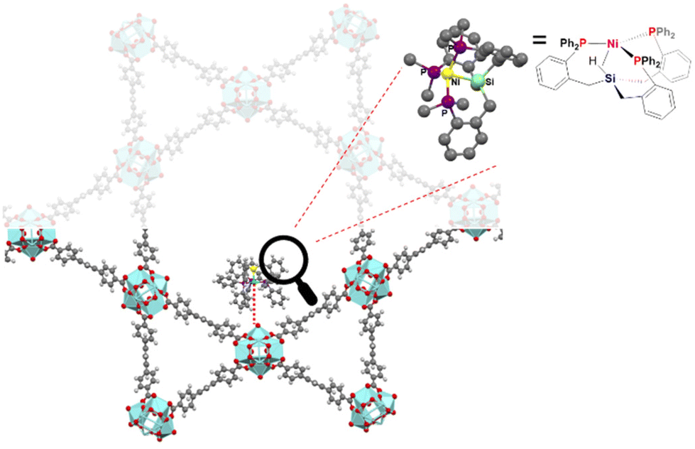 | ||
| Fig. 1 Structure representation of NU-1000 post-synthetically grafted with a nickel phosphinosilyl complex. Atom label: light blue: Zr, yellow: Ni, dark grey: carbon, light grey; hydrogen, purple: P and green: Si. | ||
[NiSi]@NU-1000 (Scheme S1†) was synthesised reported21 and obtained as a yellow powder; its crystalline structure was corroborated by PXRD (Fig. S6†). The presence of the [NiSi] complex was ascertained by a variety of techniques, including FT-IR and 1H NMR of the digested sample (Fig. S12 and S13†). XANES and SEM analyses are consistent with Ni in oxidation state of +2 and with the homogeneous distribution of the Ni complex through the material (Fig. S7 and S8†).
[NiSi]@NU-1000 displays moderate thermal stability up to 300 °C (Fig. S10†). The Ni and Zr contents were verified by ICP-MS as 0.6 wt% and 5.4 wt% respectively, corresponding to a Ni![[thin space (1/6-em)]](https://www.rsc.org/images/entities/char_2009.gif) :Zr molar ratio of ca. 1:6, very close to the ideal one Ni atom per Zr6 cluster unit of the NU-1000 material.21 The BET surface area decreased from 1970 m2 g−1 in the as-synthesized NU-1000 to 1354 m2 g−1 in [NiSi]@NU-1000 in accordance with grafting of the organometallic complex onto the MOF (Fig. S6†).21 We propose that upon grafting, the structure of the Ni silylphosphine complex is maintained, and the Ni centre binds to the zirconium node through a terminal hydroxyl group.21 Solid-state UV-Vis of [NiSi]@NU-1000 (Fig. S11†) exhibits a maximum adsorption peak at 420 nm.
:Zr molar ratio of ca. 1:6, very close to the ideal one Ni atom per Zr6 cluster unit of the NU-1000 material.21 The BET surface area decreased from 1970 m2 g−1 in the as-synthesized NU-1000 to 1354 m2 g−1 in [NiSi]@NU-1000 in accordance with grafting of the organometallic complex onto the MOF (Fig. S6†).21 We propose that upon grafting, the structure of the Ni silylphosphine complex is maintained, and the Ni centre binds to the zirconium node through a terminal hydroxyl group.21 Solid-state UV-Vis of [NiSi]@NU-1000 (Fig. S11†) exhibits a maximum adsorption peak at 420 nm.
Prior to adsorption experiments, [NiSi]@NU-1000 was activated at 120 °C under vacuum for 24 h to release the pores. Fig. 2 shows the SO2 adsorption–desorption isotherm. First, rapid capture from 0 to 0.01 bar of 0.52 mmol g−1 is observed, followed by a linear capture with a total amount of 2.48 mmol g−1 at 0.5 bar. This performance has been observed for mesoporous materials because the saturation of the material is not achieved at the experimental conditions.19,20 The isotherm shows a desorption step with a slight hysteresis, indicating that SO2 was completely released from the sample, and a relatively low SO2-MOF interaction can be inferred.22 Moreover, to explore its potential application in SO2 detection, the adsorption in the low-pressure range was analysed. It is important to highlight that adsorption in the low-pressure range can be associated with low SO2 concentrations (ppm levels).23 At low pressures (<0.01 bar), the [NiSi]@NU-1000 material shows a higher adsorption uptake in comparison with the unmodified NU-1000 material (Fig. S14†). This could be due to the higher affinity of the organometallic moiety for SO2 coordination.19 At higher pressures, the presence of the organometallic restricts access to the pores, resulting in a decrease in the adsorption capacity compared to the unmodified material (Fig. S14†). The [NiSi]@NU-1000 outperforms the SO2 uptake at 0.002 bar (0.40 mmol g−1) of NU-1000, [Ir]@NU-1000,20 [RuGa]@NU-1000,19 DUT-67(Zr),24 Zr-DMTDC, UiO-66, and MFM-133,25 all of which contain Zr-based SBUs and have surface areas above 1000 m2 g−1 (Table S2†). Nonetheless, [NiSi]@NU-1000 falls short of the best-performing MOF, Mg-gallate, with an SO2 uptake of 4.65 mmol g−1 at 0.002 bar.26
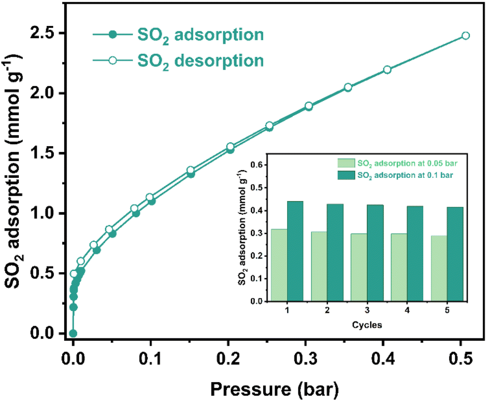 | ||
| Fig. 2 Experimental SO2 adsorption–desorption isotherm collected for a fully activated [NiSi]@NU-1000 sample (filled green diamonds = adsorption; open green diamonds = desorption) at 298 K and up to 0.5 bar. Inset: adsorption–desorption cycles for SO2 in [NiSi]@NU-1000 at 0.05–0.1 bar and 298 K. The reactivation step was carried out only by applying vacuum (1.7 × 10⁻³ torr) for 45 minutes at room temperature (298 K). | ||
Cycling SO2 experiments were conducted at 0.05–0.1 bar and 298 K (Fig. 2, inset). Each regeneration process was performed under vacuum (1.7 × 10⁻³ Torr) for 45 minutes and 298 K. It was observed that even after five adsorption–desorption experiments, the SO2 uptake was maintained at 0.05 and 0.1 bar. This confirms complete SO2 release during the desorption cycles. PXRD measurements corroborated the retention of the crystallinity (Fig. S15†) and the material's stability after the SO2 adsorption and cyclability test. Energy-dispersive X-ray spectroscopy (EDX) analyses showed no evidence of changes in the amount of Ni before and after the SO2 experiments, highlighting the stability of the isolated metal complex within the MOF framework (Table S3†). Moreover, the presence of sulphur within the MOF was corroborated, revealing that the calculated amount is two times more than expected only from the incorporated metal complex and supporting that the uptake of SO2 takes place in different parts of the MOF framework, and not exclusively in the Ni centre. Also, the stability of [NiSi]@NU-1000 under wet SO2 was tested. An activated sample was exposed for 3 h to humid SO2 (exposure at 60% relative humidity, RH), generated in a home-designed setup (Fig. S20†). It was observed by PXRD that the crystalline structure was maintained (Fig. S15†).
The host–guest interaction between SO2 and [NiSi]@NU-1000 was elucidated by calculating the isosteric enthalpy of adsorption (ΔHads) for SO2 at low coverage for a fully activated sample using the virial method (Fig. S19†).27 The calculated ΔHads at low coverage was −42.7 kJ mol−1; such value corresponds to a physisorption, in line with the reversible adsorption/desorption cycles and easy desorption step, only with vacuum. The decrease in the enthalpy of adsorption of [NiSi]@NU-1000 compared to the pristine NU-1000 (−50.8 kJ mol−1),20 could be attributed to both the high metal content (1:1, Ni per Zr6 cluster) and the steric bulk of the silicon-substituted triphosphine ligand in the [NiSi] complex. These values of enthalpies of adsorption are in agreement with the formation of hydrogen bonds between SO2 and the bare [Zr6(μ3-OH)8(OH)8] cluster as well as the establishment of electrostatic interactions between the SO2 and the grafted [NiSi] complex (Fig. S25†). The pristine NU-1000 material only exhibits hydrogen bond interactions with the SO2. In contrast, the [NiSi]@NU-1000 material additionally has lower energy SO2 electrostatic interactions with the [NiSi] complex, accounting for the decrease in the overall enthalpy of adsorption. This weak interaction was confirmed by the FTIR spectrum of an SO2-loaded sample, which showed some weak bands at 1336 and 1144 cm−1 (Fig. S18†); these correspond to adsorbed SO2, which is slightly redshifted compared to free SO2 (1362 and 1151 cm−1).41 In comparison, the previously studied [Ir]@NU-1000 material, an organometallic iridium bis(silyl)phosphine complex grafted onto NU-1000 in a molar ratio 1:11 (Ir:Zr6), exhibited a higher enthalpy of adsorption towards SO2 (−89.8 kJ mol−1), where chemisorption occurred upon SO2 bonding with the metal center.20
Several properties of MOF materials have been widely employed for the detection of SO2 gas. SO2 detection was conducted in Ni3BTC2 based on the change in its electrochemical behavior.28 Also, the detection of SO2 can be achieved using the change in the magnetic properties related to the spin-crossover transition.29 However, SO2 detection using the change in the luminescence properties is an attractive technique for the simplicity of the experiment. Indeed, modifications in the fluorescence performance of several MOF materials including MOF-303, DNA-Tb-MOF and M2(dobpdc) (M = Ni2+ and Mg2+) were investigated using this approach.9,30–32
It has been reported that NU-1000 displays photoluminescence properties based on the highly conjugated TBAPy linker, which remains unchanged after coordination with Zr4+.33 NU-100 has been evaluated to detect pesticides, explosive compounds, and metabolites in aqueous matrices by measuring the “turn-off” effect of these analytes.34–37 Also, the Ni complex incorporation in NU-1000 displays similar photoluminescence properties. Then, the pore confinement effect improves the detection effectiveness in this material at low pressures. Based on this and the SO2 adsorption properties of NU-1000 and [NiSi]@NU-1000, the materials were evaluated as fluorescent SO2 detectors for low pressure.
The fluorescence emission at different excitation wavelengths indicates that the appropriate excitation wavelength for [NiSi]@NU-1000 is 350 nm (Fig. S21†). The solid-state emission spectra of [NiSi]@NU-1000 are shown in Fig. 3a. The activated [NiSi]@NU-1000 sample displays a peak at 497 nm. Then, an activated [NiSi]@NU-1000 sample was exposed to 0.1 bar of SO2. Interestingly, a “turn-off” effect is observed with a 3.65-decreased fold in emission intensity compared to the activated sample. In order to corroborate the stability in the emission of [NiSi]@NU-1000, a cycling test was performed (Fig. S22†).
 | ||
| Fig. 3 (a) Solid-state emission spectra of activated [NiSi]@NU-1000 (purple line), and after exposure to 0.1 bar of SO2 (green line), CO2 (orange line), and air (blue line). The excitation wavelength was set at 350 nm, (b) time-resolved photoluminescence decay spectra of activated [NiSi]@NU-1000, and after exposure to 0.1 bar of SO2 measured at 335.6 nm excitation and at 500 nm emission. | ||
Five cycle experiments were conducted, observing that the emission in both cases was maintained. The SO2-exposed [NiSi]@NU-1000 shows a constant “turn-off” effect with an average 3.65 ± 0.03-fold decrease in emission intensity. Furthermore, the fluorescence of [NiSi]@NU-1000 upon exposure to air (which contains ∼78%) and CO2 was investigated to determine the selectivity (Fig. 3a). By comparison, a slight change in the fluorescence intensity is observed. However, higher selectivity is clearly observed for SO2 molecules. This can be attributed to the non-polar character of the CO2 and N2 molecules, which makes their interaction with the adsorption sites of the material unfavourable, whereas SO2, being a polar molecule, can establish specific interactions with the material, resulting in a quenching effect on the fluorescence.
This high selectivity of [NiSi]@NU-1000 for SO2 can be related to improved host–guest interactions due to the grafting of the organometallic species. As a comparison, the fluorescence performances for NU-1000 and [NiSi]@NU-1000 before and after SO2 were tested (Fig. S23†). A slight shift in the emission maxima (5 nm) for the grafted material was observed compared to the pristine material, indicating the photoluminescent properties are mainly due to the pyrene core linker. A turn-off phenomenon was observed for both materials. It is evident, however, that grafted [NiSi]@NU-1000 considerably improves the fluorescence response by turning off the response by almost 75%.
It has been reported that in NU-1000 materials, hydrogen bonding and π–π interactions could generate a quenching effect.36 One possible explanation is that the presence of the [NiSi] complex within NU-1000 reversibly bonding SO2 brings about a higher amount of SO2 molecules into the MOF pore walls, triggering SO2 packing and increasing π-SO2 interactions in the vicinity of the pyrene linkers thus turning off the fluorescence response. The proposed hydrogen-bond interactions of SO2 with [NiSi]@NU-1000 are illustrated in Fig. S24.†
Furthermore, a time-resolved photoluminescence (TRPL) experiment was performed using a 340 nm picosecond-pulsed LED as the excitation source (Fig. 3b) to investigate the possible SO2 detection. TRPL experiments were conducted on an activated [NiSi]@NU-1000 and an SO2-exposed sample. It was observed that the average decay lifetimes (Table S4†) slightly decreased from 1.91 to 1.37 ns from activated [NiSi]@NU-1000 and an SO2-exposed, respectively (a decrease of 28% in the fluorescence lifetime). This decrease can be explained by analysing the individual lifetime components (τn) and their contributions (an). Table S3† shows that, in all cases, τ1 is the shortest lifetime component, which can be associated with fast non-radiative relaxation processes, which occur in orders of less than nanoseconds,38 or very effective interactions. After SO2 adsorption, the relative contribution (a1) of this short component increases slightly, from 0.24 to 0.32, indicating that fast relaxation processes become slightly more relevant, coinciding with the observed decrease in fluorescence intensity. On the other hand, τ3, the longest lifetime component, is associated with slower phenomena, such as radiative mechanisms that occur on the order of nanoseconds.39 This component decreases both in its value, from 5.4 to 4.3 ns, as its relative contribution, from 0.21 to 0.17, after SO2 adsorption. This suggests that the interaction with SO2 induces non-radiative relaxation processes in species with longer lifetimes. This pathway could be associated with an electron transfer phenomenon related to the direct interaction between SO2 and the surface of the grafted [NiSi] complex, which deactivates the excited states in a non-radiative manner.14,40
To further support this analysis, we performed TRPL experiments for the NU-1000 material, comparing it with its modified analogue, [NiSi]@NU-1000. The data (Table S4†) reveal that, in NU-1000, the fluorescence lifetime decreases from 2.23 to 1.75 ns after SO2 adsorption, corresponding to a reduction of 21.5%. In comparison, [NiSi]@NU-1000 shows a more pronounced decrease of 28%. Similarly, the relative contributions of the lifetime components show more pronounced changes in [NiSi]@NU-1000. This indicates that the [NiSi] complex intensifies the non-radiative dissipation pathways, enhancing the sensitivity of the material to SO2 adsorption. Regarding the possible influence of absorption on fluorescence quenching, we collected solid-state UV-Vis diffuse reflectance spectra for the activated, SO2-exposed, and desorbed [NiSi]@NU-1000 samples (Fig. S11†). The spectra show that the absorption region of the material, as well as the relative intensity at the excitation wavelength (∼340 nm), do not change significantly after SO2 adsorption. This suggests that the decrease in fluorescence intensity is not related to a reduction in absorption at the excitation wavelength but to non-radiative processes associated with direct electronic interactions between SO2 and the active sites of the material.
This study investigated the SO2 adsorption and detection properties of a nickel silylphosphine organometallic complex post-synthetic grafted to a Zr-based MOF ([NiSi]@NU-1000). The material shows high stability to wet and dry SO2. Furthermore, high cyclability at the low-pressure range was achieved with a facile SO2 regeneration at room temperature. Fluorescence studies display a remarkable change in the intensity from the emission spectra after the SO2 interactions with the material and high selectivity over CO2 and air, and an evident SO2 quenching effect is observed even at low concentrations (0.1 bar of SO2). Finally, time-resolved photoluminescence experiments suggest that the turn-off effect is associated with relatively strong host–guest SO2 interactions.
Data availability
All data is available in the main text and in the ESI.†Conflicts of interest
There are no conflicts to declare.Acknowledgements
J. L. O. and V. B. L.-C. thank CONAHCYT for the Ph.D. fellowships (1003953 and 1005649). R. A. P. thanks the Autonomous University of Mexico-Iztapalapa, Mexico, for the financial support. We thank U. Winnberg (Euro Health) for scientific discussions and G. Ibarra-Winnberg for scientific encouragement. I. A. I. thanks PAPIIT UNAM (IN201123), México, for financial support. A. M. thanks to LANCAD-UNAM-DGTIC-141 for computer facilities. V. M. P. thanks the Division of Chemistry of the National Science Foundation, NSF, for financial support through project 2102689 and Dr Zamora-Moreno for technical assistance.References
- J. A. Bernstein, N. Alexis, C. Barnes, I. L. Bernstein, A. Nel, D. Peden, D. Diaz-Sanchez, S. M. Tarlo, P. B. Williams and J. A. Bernstein, J. Allergy Clin. Immunol., 2004, 114, 1116–1123 CrossRef PubMed.
- J. Roberge, H. Delgado-Granados and P. J. Wallace, Geology, 2009, 37, 107–110 CrossRef CAS.
- J. S. Pandey, R. Kumar and S. Devotta, Atmos. Environ., 2005, 39, 6868–6874 CrossRef CAS.
- R. K. Srivastava, W. Jozewicz and C. Singer, Environ. Prog., 2001, 20, 219–228 CrossRef CAS.
- J. L. Obeso, D. R. Amaro, C. V. Flores, A. Gutiérrez-Alejandre, R. A. Peralta, C. Leyva and I. A. Ibarra, Coord. Chem. Rev., 2023, 485, 215135 CrossRef CAS.
- H. Sohrabi, S. Ghasemzadeh, Z. Ghoreishi, M. R. Majidi, Y. Yoon, N. Dizge and A. Khataee, Mater. Chem. Phys., 2023, 299, 127512 CrossRef CAS.
- A. V. Leontiev and D. M. Rudkevich, J. Am. Chem. Soc., 2005, 127, 14126–14127 CrossRef CAS PubMed.
- G. Zhang, Z. Wang and X. Zhang, Mol. Phys., 2022, 120, e2018517 CrossRef.
- V. B. López-Cervantes, D. W. Kim, J. L. Obeso, E. Martínez-Ahumada, Y. A. Amador-Sánchez, E. Sánchez-González, C. Leyva, C. S. Hong, I. A. Ibarra and D. Solis-Ibarra, Nanoscale, 2023, 15, 12471–12475 RSC.
- F. Wu, J. Ye, Y. Cao, Z. Wang, T. Miao and Q. Shi, Luminescence, 2020, 35, 440–446 CrossRef PubMed.
- S. L. James, Chem. Soc. Rev., 2003, 32, 276–288 RSC.
- T. Wu, X. Gao, F. Ge and H. Zheng, CrystEngComm, 2022, 24, 7881–7901 RSC.
- X. Dou, K. Sun, H. Chen, Y. Jiang, L. Wu, J. Mei, Z. Ding and J. Xie, Antibiotics, 2021, 10, 358 CrossRef CAS PubMed.
- J.-X. Wang, J. Yin, O. Shekhah, O. M. Bakr, M. Eddaoudi and O. F. Mohammed, ACS Appl. Mater. Interfaces, 2022, 14, 9970–9986 CrossRef CAS PubMed.
- D. Mahato, S. Fajal, P. Samanta, W. Mandal and S. K. Ghosh, ChemPlusChem, 2022, 87, e202100426 CrossRef CAS PubMed.
- J. Li and A. Y. Rogachev, Phys. Chem. Chem. Phys., 2015, 17, 1987–2000 RSC.
- W. A. Schenk, Dalton Trans., 2011, 40, 1209–1219 RSC.
- A. V. Marchenko, A. N. Vedernikov, J. C. Huffman and K. G. Caulton, New J. Chem., 2003, 27, 680–683 RSC.
- J. García Ponce, M. L. Díaz-Ramírez, S. Gorla, C. Navarathna, G. Sanchez-Lecuona, B. Donnadieu, I. A. Ibarra and V. Montiel-Palma, CrystEngComm, 2021, 23, 7479–7484 RSC.
- S. Gorla, M. L. Díaz-Ramírez, N. S. Abeynayake, D. M. Kaphan, D. R. Williams, V. Martis, H. A. Lara-García, B. Donnadieu, N. Lopez, I. A. Ibarra and V. Montiel-Palma, ACS Appl. Mater. Interfaces, 2020, 12, 41758–41764 CrossRef CAS PubMed.
- L. J. Barrios-Vargas, N. S. Abeynayake, C. Secrist, N. Le, C. E. Webster, B. Donnadieu, D. M. Kaphan, A. D. Roy, I. A. Ibarra and V. Montiel-Palma, Dalton Trans., 2023, 52, 8883–8892 RSC.
- E. Martínez-Ahumada, A. López-Olvera, V. Jancik, J. E. Sánchez-Bautista, E. González-Zamora, V. Martis, D. R. Williams and I. A. Ibarra, Organometallics, 2020, 39, 883–915 CrossRef.
- S. Xing, J. Liang, P. Brandt, F. Schäfer, A. Nuhnen, T. Heinen, I. Boldog, J. Möllmer, M. Lange, O. Weingart and C. Janiak, Angew. Chem., Int. Ed., 2021, 60, 17998–18005 CrossRef CAS PubMed.
- P. Brandt, S. H. Xing, L. Liang, G. Kurt, A. Nuhnen, O. Weingart and C. Janiak, ACS Appl. Mater. Interfaces, 2021, 13, 29137–29149 CrossRef CAS PubMed.
- J. Li, G. L. Smith, Y. Chen, Y. Ma, M. Kippax-Jones, M. Fan, W. Lu, M. D. Frogley, G. Cinque, S. J. Day, S. P. Thompson, Y. Cheng, L. L. Daemen, A. J. Ramirez-Cuesta, M. Schröder and S. Yang, Angew. Chem., Int. Ed., 2022, 61, e2022072 Search PubMed.
- F. Chen, D. Lai, L. Guo, J. Wang, P. Zhang, K. Wu, Z. Zhang, Q. Yang, Y. Yang, B. Chen, Q. Ren and Z. Bao, J. Am. Chem. Soc., 2021, 143, 9040–9047 CrossRef CAS PubMed.
- A. Nuhnen and C. Janiak, Dalton Trans., 2020, 49, 10295–10307 RSC.
- N. Ingle, S. Mane, P. Sayyad, G. Bodkhe, T. Al-Gahouari, M. Mahadik, S. Shirsat and M. D. Shirsat, Front. Mater., 2020, 7, 93 CrossRef.
- C. H. Pham and F. Paesani, Inorg. Chem., 2018, 57, 9839–9843 CrossRef CAS PubMed.
- Y. Xie, H. Ma, F. L. He, J. Chen, Y. Ji, S. Han and D. Zhu, Analyst, 2020, 145, 4772–4776 RSC.
- E. Martínez-Ahumada, D. W. Kim, M. Wahiduzzaman, P. Carmona-Monroy, A. López-Olvera, D. R. Williams, V. Martis, H. A. Lara-García, S. López-Morales, D. Solis-Ibarra, G. Maurin, I. A. Ibarra and C. S. Hong, J. Mater. Chem. A, 2022, 10, 18636–18643 RSC.
- J. L. Obeso, E. Martínez-Ahumada, A. López-Olvera, J. Ortiz-Landeros, H. A. Lara-García, J. Balmaseda, S. López-Morales, E. Sánchez-González, D. Solis-Ibarra, C. Leyva and I. A. Ibarra, ACS Appl. Energy Mater., 2023, 6, 9084–9091 CrossRef CAS.
- P. Deria, J. Yu, T. Smith and R. P. Balaraman, J. Am. Chem. Soc., 2017, 139, 5973–5983 CrossRef CAS PubMed.
- Y. Zhou, Q. Yang, J. Cuan, Y. Wang, N. Gan, Y. Cao and T. Li, Analyst, 2018, 143, 3628–3634 RSC.
- W. Hao, G. Huang, G. Jiang, S. A. Dauda and F. Pi, Food Biosci., 2023, 55, 102967 CrossRef CAS.
- H. Li, Q. Chen, Z. Zhang, Z. Wang, Z. Gong and M. Fan, Dyes Pigm., 2023, 210, 111035 CrossRef CAS.
- F. Gabriel, A. Roussey, S. Sousa Nobre and A. Carella, J. Mater. Chem. C, 2024, 12, 11378–11385 RSC.
- B. Valeur and M. N. Berberan-Santos, Molecular Fluorescence, Wiley, 2012 Search PubMed.
- U. Noomnarm and R. M. Clegg, Photosynth. Res., 2009, 101, 181–194 CrossRef CAS PubMed.
- P. Chandrasekhar, A. Mukhopadhyay, G. Savitha and J. N. Moorthy, Chem. Sci., 2016, 7, 3085–3091 RSC.
- A. L. Goodman, P. Li, C. R. Usher and V. H. Grassian, J. Phys. Chem. A, 2001, 105, 6109–6120 CrossRef CAS.
Footnotes |
| † Electronic supplementary information (ESI) available: Instrumental techniques and characterization. See DOI: https://doi.org/10.1039/d3dt03985d |
| ‡ These authors contributed equally to this manuscript. |
| This journal is © The Royal Society of Chemistry 2025 |