
 Open Access Article
 This Open Access Article is licensed under a Creative Commons Attribution-Non Commercial 3.0 Unported Licence
Bioinspired micro-nano photonic materials
Chengben
Liu†
ab,
Xiaoyu
Hou†
ace,
Zixin
Zhu†
ae and
Mingzhu
Li
 *ad
*ad
aLaboratory of bio-inspired smart interface science, Technical Institute of Physics and Chemistry, Chinese Academy of Sciences, Beijing 100190, P. R. China. E-mail: mingzhu@mail.ipc.ac.cn
bSchool of Chemistry, Key Laboratory of Bio-inspired Smart Interfacial Science and Technology of Ministry of Education, Beihang University, Beijing 100190, P. R. China
cKey Laboratory of Green Printing, Institute of Chemistry, Chinese Academy of Sciences, Beijing 100190, P. R. China
dSchool of Future Technology, University of Chinese Academy of Sciences, Beijing 100049, P. R. China
eUniversity of Chinese Academy of Sciences, Beijing 100049, P. R. China
First published on 27th August 2025
Abstract
The development of micro- and nano-scale photonic materials represents a cornerstone of modern science and technology. Nature, as a master architect, has served us with the most compelling and time-tested innovations and bio-designs. From the vibrant iridescent wings of butterflies to the anti-reflective eyes of moths, biological systems have long perfected the art of light manipulation through intricate micro- and nanoscale architectures. By decoding these biological mechanisms, harnessing nature-inspired design and interdisciplinary innovation, researchers unlock pathways to create materials with tailored optical properties, durability, and environmental adaptability. In the evolving landscape of photonic materials science, bioinspired micro-nano photonic materials emerge as a transformative frontier, bridging the ingenuity of biological evolution with cutting-edge photonic technologies. In this review, we provide an overview of research efforts on bioinspired micro-nano photonic materials, which offer multifunctional, adaptive, and sustainable solutions for next generation environmentally sustainable photonic materials and ultra-compact, energy-efficient photonic devices. We summarize the typical foundational principles of biological systems where nanostructures have evolved over millennia to master light. Furthermore, we highlight recent advances in bioinspired micro-nano photonic materials which have led to tremendous progress in eco-friendly structural color display, visual chroma sensor, high-security information encryption, energy-efficient functional optoelectrical devices, and so on. Finally, we discuss the challenges and prospects of bioinspired micro-nano photonic materials, including nature-inspired design, fabrication sustainability, and interdisciplinary synergy, for applications in telecom, energy, and biomedicine in the future.
 Chengben Liu | Chengben Liu received his bachelor's (2019) and Master's (2022) degree from China University of Petroleum (East China). He is now pursuing his PhD degree in the School of Chemistry, Beihang University. His current research focuses on the design, fabrication, and applications of bioinspired optoelectronic devices. |
 Xiaoyu Hou | Xiaoyu Hou received her bachelor's degree (2018) from Beijing Institute of Technology (BIT) and subsequently obtained her Master's degree (2021) and PhD (2025) from Institute of Chemistry, Chinese Academy of Sciences (ICCAS). Her research interest mainly focuses on the design and fabrication of bioinspired structural color materials. |
 Zixin Zhu | Zixin Zhu received her bachelor's degree from Beijing University of Chemical Technology. She is now pursuing a combined master's and doctoral degree at the University of Chinese Academy of Sciences (UCAS). Her research focuses on multifunctional micro-nano optical structure design and fabrication. |
 Mingzhu Li | Mingzhu Li is a full professor at Technical Institute of Physics and Chemistry, CAS, where her interests lie in the design, fabrication, and application of bioinspired micro-nano optical structures. She received her PhD degree from Institute of Chemistry, Chinese Academy of Sciences (ICCAS), and joined the Key Laboratory of Green Printing, ICCAS in 2008. In 2023, she moved to Technical Institute of Physics and Chemistry, CAS. |
1. Introduction
In the 21st century, micro-nano photonic materials promise to overcome the “bottleneck” of electronic interconnects, ushering in an era of photonic-driven computing with enhanced speed and sustainable optical materials with energy efficiency.1 Here, micro-nano photonic materials refer to engineered systems featuring microscale or nanoscale structures that manipulate light via scattering, interference, diffraction, or resonance. Traditional photonic systems rely on bulk materials, often constrained by efficiency, scalability, and static functionality. As technology trends toward miniaturization, integrating photonic functionalities into microelectronic systems becomes critical.2 The micro-nano photonic materials transcend the limitations of conventional optics by harnessing multiscale light manipulation.3,4 These materials are not merely incremental advancements but foundational tools for a sustainable, interconnected future. The development of micro-nano photonic materials is propelled by urgent needs: sustaining digital growth, combating climate change, democratizing healthcare, and ensuring national security. Developing micro-nano photonic materials has emerged as a scientific and technological imperative.Nature has served us with the most compelling and time-tested innovations and bio-designs.5 Gaining wisdom from nature's genius forms the foundation of human survival and serves as the source of knowledge. Biological systems achieve extraordinary control of light–matter interactions through multiscale structural organization.6–8 For instance, the iridescent wings of Morpho butterflies achieve vibrant structural color through a hierarchical architecture of alternating chitin–air layers.9,10 The feathers of peacocks employ ordered melanin granules embedded in keratin matrices, leveraging coherent scattering to produce iridescence.11 Beyond aesthetics, natural systems optimize energy utilization: chloroplasts in plant leaves organize thylakoid membranes into stacked grana to enhance light absorption and electron transport efficiency.12–14 Corals employing porous, branching architectures redistribute weak light energy to their algal symbionts by lots of diffuse scattering.15–17 Natural organisms employ a limited palette of biological materials such as chitin, keratin, and melanin to create hierarchical architectures with tailored optical functionalities.6,18,19 The fascinating optical properties of biological systems are due to their exquisite micro-/nano structures refined through eons of evolution.20–27
By decoding these biological mechanisms, researchers unlock pathways to create materials with tailored optical properties, durability, and environmental adaptability. Bioinspired micro-nano photonic materials stand out as a transformative frontier, merging biological wisdom with advanced nanofabrication to create devices capable of precise light manipulation. Bioinspired designs enable on-chip photonics,28–30 optical metamaterials,31,32 and high-performance optoelectronics,33 fostering advancements in eco-friendly colorful materials,34,35 reflective display,36,37 secure communication,38–40 energy-efficient lighting,41,42 efficient renewable energy43–45 and so on. For instance, artificial structural color is more durable than pigments, astonishingly reducing reliance on dyes that fade or are toxic.46,47 Anti-reflective coating mimicking the moth's eyes leads to efficient solar panels.48 Passive radiative cooling coating inspired by the bio-white color offers a zero-energy solution for managing thermal radiation in buildings.49,50 Photonic crystals (PhCs) following the opal lattice structure can control light propagation and emission, leading to applications in telecommunications, sensors, or solar cells (SCs).51–53 With the rapid development of materials science and manufacturing processes, researchers combine nature's subtle bionic strategies with the ever-expanding library of advanced materials to create a new paradigm of function-oriented micro- and nano-photonic materials design. This cross-fertilization not only breaks through the boundaries of traditional material properties but also gives rise to innovative systems that combine bionic intelligence with engineering practicality. Therefore, it is important to summarize and review the development of this field. In this review, we will explore how humans learn from nature by following a three-step process: find and observe natural phenomena, analyze and extract physical mechanisms, and design and construct artificial bionic materials (Fig. 1). The review is divided into four sections. In the current chapter, we introduce the importance of bionics and bioinspired micro-/nano-photonic materials. Following this brief introduction, we summarize classic natural inspirations and categorize them into three aspects briefly, including reflective structural color: from disordered to ordered, light absorption, and light transmission. In each part, we present some classic examples to render basic mechanisms underlying the unique optical properties found in nature and biology. By utilizing these natural mechanisms and versatile man-made materials, researchers have devoted significant efforts to creating many biomimetic materials with superior optical functions. We focus on the representative applications of reflective display, information security and encryption, optical sensors, and light and thermal management. In Section 3, we summarize the state-of-the-art advances in artificial optical materials and structures in those application fields. Finally, we provide an outlook on the future development of bioinspired micro- and nanophotonic materials, emphasizing their potential to drive innovation in diverse fields.
 | ||
| Fig. 1 Overview of the steps of biomimicry in micro-nano photonic materials, including investigating natural photonic issues, decoding underlying optical mechanisms, and bioinspired materials and applications. Part “reflective structural color” (from left to right) reproduced from ref. 20 with permission from Wiley-VCH, copyright 2016; ref. 21 with permission from National Academy of Sciences, USA, copyright 2012; and ref. 11 with permission from National Academy of Sciences, USA, copyright 2003. Part “light absorption” (from left to right) reproduced from ref. 22 with permission from Springer Nature, copyright 2018; ref. 23 with permission from Athena International Publishing B.V., copyright 2023; and ref. 24 with permission from Wiley-VCH, copyright 2010. Part “light transmission” (from left to right) reproduced from ref. 25 with permission from OSA, copyright 2013; ref. 26 with permission from Nature Publishing Group, copyright 2003; and ref. 27 with permission from OSA, copyright 2018. | ||
2. Natural inspirations and optical mechanisms
Bionic optics, a compelling field, is booming at an unprecedented rate. It is not just a simple imitation of nature, but also an in-depth exploration and creative application of the wisdom of nature. Biomimetics is the transfer of nature's superior design to technical applications.54–57 How to do this? The logical and reasonable way is “Inverse design”. Inverse design and fabrication of bioinspired micro-nano materials constitutes an interdisciplinary approach that leverages nature-inspired structural principles and advanced computational modeling to inversely engineer multiscale architectures with tailored functionalities, enabling breakthroughs in advanced materials, energy systems, and sustainable technologies. Then it is most significant to figure out the underlying optical mechanisms. The final step is to fabricate artificial materials.This chapter focuses on the paradigm of bioinspired optical design, emphasizing two critical steps: identifying biological models exhibiting target functionalities and deciphering the underlying physical mechanisms. Only by rigorously understanding these mechanisms can researchers translate biological principles into functional artificial structures. For instance, the anti-reflective moth-eye motif has been replicated in silicon SCs, boosting efficiency via nanoimprinting. Crucially, mechanism-based design avoids blind mimicry. In exploring biological structures, scientists have found that the optical function of many organisms is not attributed to a single structure, but the result of multiple structures and mechanisms working together and cooperating. This complex synergistic effect for the study of bionic optics has brought great challenges, but is also pregnant with unlimited opportunities. Thus, we try to abstract universal principles that transcend specific biological contexts in the next content. According to the interaction between light and micro-nano structures, we categorize some classical bioinspiration into three groups, namely light reflection, light absorption, and light transmission.
2.1 Reflective structural color: from disordered to ordered
Coloration represents a milestone of evolutionary adaptation, driving critical survival strategies across life. In nature, colors are intricately linked to ecological survival, enabling camouflage against predators, sexual signaling for mate selection, aposematism to deter threats, and thermoregulation through light reflection. These functions are achieved through three principal mechanisms: pigment-based coloration, bioluminescence, and structural coloration. While pigments and bioluminescence have evolved to address specific needs, structural colors—a product of nanoscale architecture—stand out as a paradigm of evolutionary optimization, offering unparalleled durability and optical versatility.Structural coloration arises from the interaction of light with subwavelength-scale architectures. These nanostructures manipulate light via fundamental optical phenomena, including interference, diffraction, and coherent scattering.58 The evolutionary advantage of structural colors lies in their inherent robustness and energy efficiency. Unlike pigments, which degrade under UV exposure or oxidative stress, structural colors remain vibrant as long as their nano-architectures remain intact—a feature critical for long-term signaling in harsh environments. Furthermore, structural systems can be easily tunable. Organisms such as cephalopods and chameleons have evolved dynamic structural colors by reconfiguring the spacing of reflective platelets or chromatophores, enabling real-time camouflage or communication. This tunability is governed by precise control of structural parameters, including lattice periodicity, layer thickness, and refractive index contrast, which collectively determine the spectral and angular response of reflected light.
 | ||
| Fig. 2 The physical mechanisms and natural examples for structural coloration from disordered to ordered. (a) Disordered structures achieve wavelength-independent all-directional reflection, resulting in a white color. Pieris rapae (left), Cyphochilus beetle (center), and Pacific cleaner shrimp (Lysmata amboinensis, right), the corresponding SEM images of their disordered structures, and the extracted optical structure models. Reproduced from ref. 20 with permission from Wiley-VCH, copyright 2016; ref. 61 with permission from Wiley-VCH GmbH, copyright 2015; ref. 62 with permission from Springer Nature, copyright 2023. (b) Quasi-ordered structures exhibit weak angle-independent coloration from coherent scattering. The feathers of the Scarlet macaw (Ara macao, left) and Cotinga maynana (right) all exhibit non-iridescent blue color, and the corresponding SEM images and 2D Fourier transform spectra are given. Reproduced from ref. 21 with permission from National Academy of Sciences, USA, copyright 2012; ref. 66 with permission from Royal Society of Chemistry, copyright 2009. (c) Ordered structures, that is, PhC, enable iridescent color leveraging constructive and destructive interference. 1D PhC structure of jewel beetle (Chrysochroa fulgidissima, left), 2D PhC structure of peacock (Pavo muticus, center),3D PhC structure of natural opals (right). SEM images and photonic band structures are provided. Reproduced from ref. 74 with permission from John Wiley & Sons, Ltd, copyright 2020; ref. 11 with permission from National Academy of Sciences, USA, copyright 2003. | ||
Sometimes brilliant structure designs help broaden the selection of materials, meaning achieving whiteness will not be limited to high refractive index. Normally, when the refractive index is lower, increasing material thickness must be done to achieve whiteness. However, in nature, another ingenious structure for well-optimized whiteness was uncovered in the scales of the beetle genus Cyphochilus.59,60 The Cyphochilus beetle broke the rule since it possesses a fabulous white color with thin scales ca. 7 μm in thickness. Meanwhile, their scales are composed of chitin with a refractive index of around 1.55–1.56 and have a disordered network of fibers orienting vertically to the direction of the incident light. Lorenzo et al. proved that anisotropic light transport in such a structure boosts scattering strength, facilitating the achievement of a high white reflectance (>0.8) across the visible wavelength range.61 Achieving intense whiteness in aqueous environments presents unique challenges for the relative refractive index of nrel = nmaterial/nwater is lower than that in air (nrel = nmaterial/nair). Nature provides novel problem-solving strategies inspired by biological systems. The Pacific cleaner shrimp (L. amboinensis) reveals the important role of birefringence in obtaining whiteness.62 Cryo-electron microscopy unveiled densely packed nanospheres within submicron-thick chromatophores of the shrimp's white markings. These particles consist of radially aligned isoxanthopterin molecules, creating optical anisotropy with distinct refractive indices (nradial = 1.4 vs. ntangential = 1.96). Numerical simulation demonstrates that a polydispersity of 10% could suppress structural color while maintaining broadband reflectance. Crucially, the particles' anisotropic refractive index distribution reduces near-field interactions and spatial crowding effects that plague conventional isotropic scatterers. This dual strategy enables higher visible-light reflectance compared to isotropic counterparts across diverse packing densities. Such bioinspired structural principles are revolutionizing radiative cooling technologies. By decoupling optical performance from intrinsic material properties, these whiteness models exemplify nature's strategy to overcome physical limitations through topology optimization. This paradigm shift informs next-generation photonic materials for sustainable cooling, energy-efficient architecture, and low-carbon manufacturing, underscoring the transformative potential of bioinspired disorder engineering in addressing global thermal management challenges.
Recent synthetic advances have successfully replicated quasi-ordered photonic architectures. For example, photonic glasses based on colloidal particles or bottlebrush block copolymers can mimic the short-range order found in nature while offering tunable optical responses.67,68 These artificial analogues benefit from scalable fabrication techniques, such as colloidal self-assembly and thermal annealing, and can produce non-iridescent structural color across a broad spectrum. Moreover, the development of optical modeling approaches—such as Monte Carlo simulations that incorporate both multiple scattering and absorption effects—has enabled predictive control over color properties in disordered media.69,70 These developments establish a unified framework for engineering angle-independent structural colors across both biological and synthetic systems.
Quantifying the relationship between structural order and optical output remains challenging due to the nuanced interplay of spatial correlations. Fourier analysis has emerged as a critical tool, decomposing electron microscopy images into reciprocal space representations.71 The resulting power spectra reveal characteristic ring patterns—sharp rings indicate high periodicity, while diffuse halos signify disorder. Intermediate states exhibit both features, confirming the coexistence of short-range order and long-range disorder in quasi-ordered systems. The structure factor S(q) serves as a fundamental metric for characterizing the degree of spatial order in these systems. It quantifies how incident waves are scattered by the material's internal structure, with its spectral features directly reflecting the system's correlation properties.72,73 These structural correlations enable weak but significant interference effects, as quantified by the structure factor, which modulates the phase relationships between scattered waves. In quasi-ordered structures, S(q) plays a central role in determining the resulting structural colors by governing both the intensity of coherent scattering and the wavelength selectivity of reflected light. Although these structures lack perfect periodicity, they typically exhibit statistically preferred length scales, which manifest as broadened peaks in the structure factor. The position of these peaks determines the dominant reflected wavelength—and thus the hue of the structural color—while their sharpness influences the spectral purity: sharper peaks indicate stronger wavelength selectivity and result in more saturated colors. Therefore, S(q) not only reveals the degree of spatial order but also directly dictates the spectral characteristics of the optical response. Despite lacking perfect crystalline order, quasi-ordered architectures achieve functional optical performance through evolutionary optimization. Their design principles—balancing manufacturability with optical efficiency—inspire biomimetic materials for applications requiring angle-independent colors, such as anti-glare coatings and durable textile dyes. By decoding how biological systems navigate the order–disorder continuum, researchers gain insights into nature's strategies for engineering light at subwavelength scales.
1D PhC consists of alternating planar layers of high- and low-refractive-index materials. Light waves reflecting at each interface interfere coherently, with the phase difference determined by layer thickness and refractive indices. According to Bragg's law, constructive interference occurs when:
2neffd![[thin space (1/6-em)]](https://www.rsc.org/images/entities/char_2009.gif) sinθ = mλ sinθ = mλ |
2D PhC features periodic structures in two dimensions, such as hexagonal arrays of cylindrical rods or holes. Light propagates in-plane, with photonic bandgaps emerging from periodic variations in the transverse direction. For example, the iridescent eye spots on peacock (Pavo cristatus) feathers derive their brilliance from 2D PhC lattices within barbules.11 Each barbule contains melanin rods embedded in a keratin matrix, arranged in a square lattice with a periodicity of ∼140 nm. This lattice selectively reinforces green and blue wavelengths (450–500 nm) via Bragg scattering while suppressing others, creating angle-dependent metallic hues: incident light undergoes interference at melanin–keratin interfaces, with only wavelengths satisfying the Bragg condition being strongly reflected, while others are suppressed by destructive interference, resulting in angle-dependent iridescence.
3D PhC exhibits periodicity in all spatial dimensions, akin to atomic lattices in crystals. Natural opals exemplify this class, comprising close-packed silica spheres in a face-centered cubic (FCC) arrangement. Incident light undergoes Bragg diffraction across multiple crystallographic planes, generating iridescent “play-of-color” that shifts with viewing angle. The photonic bandgap in 3D situations can be calculated by the following formula:

| λ = 2np |
 | ||
| Fig. 3 Biological examples of manipulating light's polarization state and corresponding schematic diagram of the optical principle. (a) Photographs of the beetle Cataglyphis gloriosa under left and right circular polarizers. SEM image showing chiral helical nanostructure. Reproduced from ref. 79 with permission from American Association for the Advancement of Science, copyright 2009. (b) Photograph of Ornithoptera croesus lydius and optical images of scales at 45° and 0/90° azimuth under crossed polarizers. TEM image exhibiting a composite structure comprising a grating and a multilayer. Reproduced from ref. 80 with permission from Royal Society of Chemistry, copyright 2014. (c) The green color of the Papilio blumei butterfly results from color blending. Optical micrographs observed without (left) and with (right) cross-polarizers. SEM (top) showing the concavities of scales and SEM (bottom) of a biomimetic sample showing a conformal multilayer stack. Reproduced from ref. 81 with permission from Nature Publishing Group, copyright 2010. | ||
Another mechanism for achieving polarization-sensitive color was demonstrated by Zhang and coworkers through a sophisticated dual-architecture design within the orange scales in the butterfly Ornithoptera croesus lydius.80 As illustrated in Fig. 3b, the scale integrates two photonic components: surface-level tapered triangular gratings and subsurface multilayer reflectors (alternating chitin/air layers). This hierarchical configuration generates coloration through synergistic light–matter interactions: the upper grating produces zero-order diffraction while the underlying multilayer induces first-order interference, collectively enhancing orange wavelength reflectance. Polarization sensitivity emerges from azimuth-dependent optical transformations. Under crossed polarizer–analyzer configurations, reflected light intensity exhibits angular modulation, reaching minimum intensity when scale ridges align parallel/perpendicular to the incident polarization axis, yet recovering maximum reflectance at ±45° orientations. Through simulations, the team elucidated the polarization conversion mechanism. The tapered grating geometry functions as a quarter-wave plate, inducing phase retardation that converts p-polarized incident light into s-polarized output when illuminated at 45° incidence.
The optical complexity of biological systems often arises from the integration of hierarchical structures, enabling multifunctional performance beyond singular mechanisms. A striking example is the Southeast Asian butterfly Papilio palinurus, which achieves simultaneous polarization conversion and color blending through a meticulously organized multiscale photonic architecture (Fig. 3c).81 In this case, the wing scales feature concave microstructures (5–10 μm diameter) arranged in ordered arrays, each comprising a dual-level design. A multilayer stack of alternating chitin and air films generates yellow reflectance via Bragg interference at near-normal incidence. The curved sidewalls of each concavity impose oblique illumination angles on the underlying multilayers, inducing a blue shift through angularly tuned constructive interference. This geometric tuning creates spatially separated color domains—yellow from cavity bases and blue from sidewalls—which blend perceptually into a vibrant green. Moreover, the concave geometry also introduces polarization conversion. Light incident on cavity edges undergoes double reflection, imparting a geometric phase shift that converts linearly polarized incident light to cross-polarized states. This polarization conversion is experimentally validated by placing wing scales between crossed polarizers: only retroreflected edge segments retain detectable intensity, while normally reflected central light is extinguished. Such dual functionality—spectral blending and polarization modulation—stems from the hierarchical coupling of micro- and nanoscale features. This integrative strategy inspires novel optical devices, such as polarization-encoded security tags.
In summary, structural colors epitomize nature's mastery of optics, merging evolutionary fitness with nanoscale engineering. By decoding their underlying principles—from photon bandgap engineering to multilayer interference—researchers can harness them to advance photonic materials, ecological monitoring tools, and sustainable industrial applications.
2.2 Light absorption
The efficient capture of solar energy has emerged as a critical scientific frontier, driven by escalating global energy demands and the urgent need to transition from fossil fuels to renewable resources. As the largest accessible renewable energy flux, solar radiation offers a sustainable pathway to meet these challenges, as long as we overcome limitations in photon management, energy conversion, and storage. Traditional photovoltaic and photocatalytic technologies often struggle with inefficiencies stemming from spectral mismatch, reflection losses, and charge recombination. Herein lies the promise of biomimicry: over billions of years, evolutionary pressures have refined biological systems to optimize light harvesting under extreme environmental constraints, from sun-baked deserts to aphotic ocean depths. These organisms achieve remarkable efficiencies through nanoscale architectures that defy conventional engineering paradigms, offering scientists enormous brilliant ideas to design incredible materials.82Biological light-harvesting architectures span a wide range of length scales—from nanometers to microns—each exploiting distinct physical principles to optimize solar energy capture. At the nanoscale, structures interact with light primarily via near-field effects, enabling sub-diffraction light confinement and enhanced absorption. In the subwavelength regime, periodic or quasi-periodic features produce interference-based phenomena including diffraction, Bragg scattering, and photonic bandgap effects. At the micron scale, geometric optics governs light behavior—via refraction, lensing, and multiple scattering—allowing for extended photon path lengths within absorbing tissues. Notably, many biological systems combine multiple hierarchical features to achieve broadband absorption, where micron-scale light focusing enhances delivery to subwavelength structures, which in turn channel photons toward nanoscale absorbers. This integrated multiscale strategy not only improves light-trapping efficiency across the visible spectrum (400–700 nm) but also offers critical design insights for bioinspired optical materials.
Biological systems have evolved sophisticated strategies to maximize photon capture, primarily through two complementary approaches: boosting multiple scattering and enhancing light concentration via structural engineering. Among natural light-absorbing materials, ultra-black surfaces with little reflectance are observed across diverse taxa, including araneids, avians, and lepidopterans.83 These organisms provide critical insights for developing robust, lightweight absorbers with multifunctional capabilities. Zhang et al. revealed that the synergistic effect of inverse-V ridges and subwavelength nanoholes was responsible for ultra-black coloration. Experimental and computational analyses of Troides aeacus black scales demonstrated that inverse-V ridges enhance light penetration depth, directing photons into subsurface nanohole layers where near-total absorption occurs.84 Recently, Alexander and colleagues systematically studied the nanostructure and physical mechanisms of ultra-black wings in butterflies (Fig. 4a).85 It has been proven that expanded trabeculae and steep ridges in the scales can decrease reflectance by 16-fold and increase multiple scattering. These biological principles mirror those governing synthetic ultra-black materials, emphasizing two cardinal rules: maximization of photon trapping through multiscale roughness, and enhancement of effective absorption cross-sections via hierarchical porosity. Different from butterflies, the super black bird of paradise feathers provide another structural absorption mechanism with highly modified microstructures (Fig. 4b).22 McCoy et al. observed vertically tilted barbule arrays creating deep, curved cavities leading to more light absorption via multiple scattering. Ultrablack-inspired architectures now guide innovations in aerospace coatings that reduce radar cross-sections, optical sensors that improve signal-to-noise ratios, and space technologies that minimize stray light in telescopes.
 | ||
| Fig. 4 Classic biological instances with light absorption function, the corresponding SEM images of their structures, and the strategy of designing intricate structures for highly efficient light absorption. (a) Trogonoptera brookiana. Reproduced from ref. 85 with permission from Springer Nature, copyright 2020. (b) Wahnes’ Parotia Parotia wahnesi. Reproduced from ref. 22 with permission from Springer Nature, copyright 2018. (c) Green leaves. Reproduced from ref. 24 with permission from Wiley-VCH, copyright 2010. (d) Arachnoidiscus ehrenbergii. Reproduced from ref. 23 with permission from Athena International Publishing B.V., copyright 2023. | ||
Beyond animal photonic systems, terrestrial plants have evolved hierarchical architectures optimized for solar energy harvesting through 3.8 billion years of photosynthetic evolution. Natural leaves achieve quantum efficiencies exceeding 90% in the light-harvesting process, a feat enabled by chloroplast nanostructures (Fig. 4c).24,86,87 The 3D arranged lamellar membrane systems within chloroplasts demonstrate remarkable photon management strategies through their extended interfacial architectures. These biomolecular assemblies achieve enhanced photon capture efficiency through synergistic mechanisms: simultaneous light absorption at pigment–protein complexes, recurrent internal reflection across adjacent membranes, and diffusive scattering within confined luminal spaces. Such nanoscale light confinement extends optical path lengths compared to planar configurations. Inspired by this hierarchical structure, many high-performance photocatalysts have been developed, aiming to efficiently utilize solar energy.88,89
In terms of light concentration, among the organisms that developed complex photonic architectures, particular interest has recently been focused on diatoms.42,90 Diatoms, unicellular microalgae responsible for 20–25% of global photosynthetic oxygen production, thrive in aquatic environments through exquisitely engineered silica exoskeletons (frustules). The diatom's silica frustule (cell wall) contains periodic or quasi-periodic distributions of nanopores ranging from some tens of nanometers to one micron.91,92 These nanoporous architectures, particularly in species like Arachnoidiscus ehrenbergii, can selectively reflect/scatter specific photons to enhance light harvesting efficiency (Fig. 4d).23 SEM reveals heterovalvar frustules comprising two morphologically distinct valves: a planar central region surrounded by radially aligned slits and concentric rings of progressively shrinking pores. The valve's internal costa system radiates from a central flange, creating wavelength-dependent optical phenomena. Under visible light illumination, interference between diffracted waves from individual pores generates coherent hotspots at the frustule center. In contrast, UV wavelengths experience minimal concentration due to destructive phase matching, inherently shielding photosynthetic machinery from harmful radiation. This spectral selectivity combining visible light focusing with UV rejection shows evolutionary optimization for both energy capture and photoprotection. Parallel to diatoms, Ruben et al. found terrestrial plants like Rosa ‘El Toro’ employ epidermal micro-nanostructures to synergize anti-reflection and light concentration.93 It's because the micropapillae act as microlenses, focusing incident light into the petal's interior, which indirectly prolongs the average optical path length and finally boosts light absorption.
2.3 Light transmission
Improving light transmission efficiency—the enhanced ability to propagate photons with minimal energy loss—is a cornerstone of optical technologies, governing performance in applications ranging from illumination, display, telecommunications, and energy systems. High transmission efficiency ensures energy conservation, signal fidelity, and spatial resolution, particularly in low-light environments or miniaturized devices. In nature, organisms have evolved intricate photonic architectures to optimize light transmission, balancing energy expenditure with functional demands. These biological solutions often surpass synthetic counterparts in efficiency and multifunctionality. Below, we explore three paradigms of natural light transmission optimization and their physical mechanisms.Bioluminescence is a large part of light transmission and refers to the phenomenon in which an organism converts chemical energy into light energy through chemical reactions. This phenomenon is widespread among marine organisms, plants, and insects, playing diverse critical roles for both the organisms themselves and their ecological surroundings—including communication and information exchange, predation and defense mechanisms, as well as physiological regulation and energy metabolism. However, reflections at the interface prevent the organism from emitting light internally, reducing the organism's adaptability to the environment and the probability of survival. Through natural evolution and selection, many organisms have specialized structures that aid in light extraction and enable them to survive in harsh competition. There are many great instances. Here, fireflies are our main characters (Fig. 5a).25 Fireflies achieve remarkable bioluminescence efficiency through hierarchical nanostructures on their lantern cuticles. The lantern surface is textured with asymmetric, tilted microribs that function as a diffraction grating. These microstructures reduce total internal reflection (TIR) at the cuticle-air interface by redirecting trapped photons outward, minimizing backscattering. This design enables fireflies to broadcast mating signals over long distances with minimal metabolic cost, as photons generated by luciferase reactions are channeled directionally rather than isotropically. Inspired by firefly lanterns, researchers have engineered biomimetic light-emitting diodes (LEDs) with nanostructured encapsulation layers.94,95 Such designs are pivotal for energy-efficient displays, surgical lighting, and wearable optoelectronics.42
 | ||
| Fig. 5 Examples of light transmission structures in the natural world. (a) Schematic illustration demonstrating reduced total internal reflection and increased emission. The abdominal segments of Photuris fireflies with complex optical structures can improve light extraction. SEM images of misfit scales. Reproduced from ref. 25 with permission from OSA, copyright 2013. (b) Diagrams of the gradient index mechanism for antireflection (enhancing transmission) of the wings of bee hawkmoth and moth eye. Photographs of Cephonodes hylas (bee hawkmoth) and moth, and the corresponding SEM characterization of nanostructure. Reproduced from ref. 96 with permission from Zoological Society of Japan, copyright 1997; ref. 26 with permission from Nature Publishing Group, copyright 2003. (c) Schematic diagram of light transmission fiber structure. Spider silk’ photograph and SEM image are given. Reproduced from ref. 27 with permission from OSA, copyright 2018. Heart cockle (Corculum cardissa) cross-sectional SEM of shell window showing fibrous prisms acting like fiber bundles (red line refers to fiber orientation). Reproduced from ref. 99 with permission from Springer Nature, copyright 2024. | ||
Besides, decreasing light reflection from the interface is an ingenious way to increase transmission efficiency. Meanwhile, it is especially conducive for visual imaging and escaping from enemies. Several mechanisms have been observed to achieve anti-reflective structures in the eyes and wings of insects (Fig. 5b). For instance, transparent wings of Cephonodes hylas (bee hawkmoth) utilize nanopillar arrays to minimize surface reflections, enabling near-invisible camouflage during flight.96 The main mechanism is that nanopillar arrays gradually change the average refractive index, smoothing the impedance mismatch of the interface. Moths employ similar subwavelength nanostructures to maximize light transmission in dim environments. Their compound eyes feature hexagonal arrays of conical nipples that create a graded refractive index transition from air to the corneal chitin. This gradient suppresses Fresnel reflections across broad angles and wavelengths, achieving extreme transmittance. For moths, enhanced light transmission improves nocturnal vision sensitivity, while hawkmoth wings reduce predator detection by eliminating glare.26 Nowadays, bioinspired anti-reflective structures are revolutionizing photovoltaics and optical sensors. These coatings also benefit camera lenses, laser optics, and augmented reality displays by eliminating ghost imaging.
From a different aspect of thinking, a highly efficient transmission means that light can propagate longer. In nature, fiber-optic systems achieve low-loss light transmission through cylindrical waveguiding (Fig. 5c). Remarkably, studies have provided many interesting results about using natural spider silk as an optical fiber.27,97,98 Similarly, the heart cockle (Corculum cardissa) embeds lucent calcium carbonate fibers within its shell, channeling sunlight to photosynthetic symbionts, sustaining photosynthesis in sediment-buried habitats.99 Cockle fibers enable mutualistic energy harvesting.
Nature's mastery of light transmission stresses the synergy between evolutionary adaptation and physical law. By decoding biological designs from firefly gratings to spider waveguides, researchers are transcending traditional optical engineering, achieving unprecedented efficiency in illumination, imaging, and energy harvesting.
3. Applications of bioinspired optical materials
3.1 Reflectivity display
Micro/nano photonic structures play a significant role in the field of display technology for their ability to generate structural colors and dynamically modulate light.100,101 Inspired by natural photonic systems such as the Morpho butterfly wings, artificial structural color pixels based on PhCs and plasmonic nanostructures can achieve brilliant color display without relying on traditional pigments.102–104 These structures exploit geometry-dependent optical effects—such as the photonic bandgap in PhCs and localized surface plasmon resonance in metallic nanostructures—to selectively filter or reflect specific wavelengths, thereby producing various structural colors. This color generation mechanism not only eliminates the need for high-power backlighting but also ensures stable and vibrant color reproduction under bright ambient lighting conditions, making it an ideal solution for energy-efficient applications including electronic paper, smart labels, and outdoor signage. Furthermore, their superior performance characteristics including wide color gamut, high visibility under daylight conditions, and environmentally friendly position them as promising candidates for next-generation display technologies.105,106 Beyond static color, some biological organisms, such as chameleons107–109 and cuttlefish,110 achieve dynamic color changes through real-time modulation of nanostructures in their skin. Inspired by this, researchers have developed light-, thermal-, and ion-responsive materials by integrating stimuli-responsive photonic structures, enabling full-color tuning, thermal sensing, and synchronized electro-optical output. These biomimetic systems bridge the gap between static and adaptive photonic displays, opening new opportunities for intelligent visual technologies. | ||
| Fig. 6 Structural color imaging based on PhCs or microdomes. (a) Fabrication of a colorful image based on an inverse opal PhC structure. (b) Representative SEM images of inverse opal PhC with different colors. (c) Schematic of the interaction between the inverse opal structure and light. (d) Pixelated image based on inverse opal PhC structure. Reproduced from ref. 113 with permission from John Wiley and Sons, copyright 2019. (e) Schematic of direct ink writing blue, red, and green lines based on cholesteric liquid crystal structure. Inset is the cross-sectional SEM image of the printed green line. (f) Schematic of the interaction between cholesteric liquid crystals (CLCs) and light. (g) Temple of Heaven pattern based on CLCs. Reproduced from ref. 114 with permission from Elsevier, copyright 2024. (h) Schematic illustration of the droplet-by-droplet printing to create microdomes with different diameters. (i) SEM image of printed microdome structure with different diameters. (j) Schematic of the interaction between microdome and light. (k) The large-area color image based on microdomes. Reproduced from ref. 116 with permission from American Association for the Advancement of Science, copyright 2021. | ||
Unlike traditional structural colors that depend on periodic arrangements of photonic structure units, a novel coloration mechanism based on TIR and interference was introduced in 2019 by Zarzar's group.115 This approach eliminates the need for periodic nanostructures, enabling brilliant colors from individual microscale concave interfaces. Furthermore, Song's group developed an inkjet-printing strategy to rapidly fabricate dome-shaped microstructures (microdomes) for generating structural colors based on TIR and interference (Fig. 6h–k).116 By controlling droplet volume and substrate wettability, they achieved full-color printing with a single transparent ink, covering 72% of the standard Red Green Blue (sRGB) color gamut. The pixelated microdomes allowed grayscale tuning (0.152–0.803) and high-resolution imaging (up to 842 dpi), producing photorealistic portraits and functional patterns like QR codes. Notably, the optical Janus effect-color on one side, and transparency on the other-expanded applications in static displays (e.g., anti-counterfeiting labels) without requiring dynamic stimuli.
Moreover, dielectric- and metal-based metasurfaces/metamaterials have emerged as important platforms for reflective displays due to their high resolution.117–120 For instance, Yang et al. demonstrated an all-dielectric Si metasurface integrated with a refractive index matching layer, achieving record color gamuts exceeding 180% sRGB (Fig. 7a and b).121 By leveraging the interplay between electric and magnetic dipole resonances, this design significantly suppressed broadband background reflection and narrowed full width at half maximum (FWHM), enabling high-brightness, high-purity structural colors. Moreover, Sahu et al. demonstrated a single-step thermal evaporation process to fabricate liquid gallium nanoparticles embedded in PDMS, where capillary interactions between uncrosslinked oligomers and Ga vapor drove the self-organization of plasmonic nanostructures (Fig. 7c and d).122 By tuning the PDMS oligomer content and deposition parameters, the Ga nanodroplets formed multi-layered configurations with controlled size distributions, enabling various structural colors via localized surface plasmon resonances. These resonances spanned a broad CIE gamut, with chromaticity modulated by the interplay of inter- and intra-layer gap plasmons. The technique achieved scalable patterning of structural colors over large areas through substrate-selective deposition. Encapsulation with PDMS superstrates further ensured environmental stability against humidity, temperature, and mechanical wear, making the platform viable for outdoor signage and durable decorative surfaces. Moreover, Geng et al. utilized femtosecond lasers to induce oxidation in TiAlN–TiN hybrid films, generating structural colors with 90% sRGB gamut and over 10000 dpi resolution (Fig. 7e and f).123 The double-resonance absorption mechanism, arising from non-trivial phase shifts at the oxide-TiAlN interface and TiAlN–TiN interface, enabled high-throughput inkless printing on rough and flexible substrates. The laser-oxidized films demonstrated exceptional fastness against UV, corrosion, and abrasion, surpassing traditional plasmonic colorants in durability. This non-contact, single-step fabrication circumvents the cost and complexity of e-beam lithography, paving the way for its industrial adoption in decorative and security printing.
 | ||
| Fig. 7 Structural color imaging based on metasurface or metamaterials. (a) and (b) Full-color image printing with Si metasurface. Reproduced from ref. 121 with permission from Springer Nature, copyright 2020. (c) and (d) Image based on Ga-deposited nanoparticles. Reproduced from ref. 122 with permission from Springer Nature, copyright 2024. (e) and (f) Large-scale structural color patterns printed on TiAlN–TiN hybrid films on a polished 2-in. Si wafer. Reproduced from ref. 123 with permission from Springer Nature, copyright 2023. | ||
000-cycle durability, demonstrating robust potential for mechanically adaptive displays. Moreover, tuning the refractive index of the building blocks or the surrounding matrix in photonic structures provides an alternative strategy for dynamic structural color displays. This modulation typically involves physicochemical mechanisms such as thermally driven phase transitions, electric field-mediated liquid crystal reorientation, or the introduction of new substances. Franklin et al. presented a tunable color system by coupling high birefringence liquid crystals with plasmonic surfaces (Fig. 8d).133 Voltage-induced reorientation of liquid crystal molecules alters the refractive index surrounding the plasmonic surface, leading to full-color coverage across the visible spectrum.
 | ||
| Fig. 8 The applications of micro/nano photonic materials in dynamic display. (a) Dynamic display based on the change of PhC period. Reproduced from ref. 127 with permission from Wiley-VCH Verlag GmbH & Co. KGaA, Weinheim, copyright 2017. (b) Dynamic display based on the change of microdome size. Reproduced from ref. 38 with permission from Wiley-VCH GmbH, copyright 2024. (c) Dynamic display based on the change of grating orientation. Reproduced from ref. 132 with permission from Science China Press, copyright 2023. (d) Dynamic display based on the change of refractive index. Reproduced from ref. 133 with permission from Macmillan Publishers Limited, copyright 2015. | ||
3.2 Information encryption
Micro/nano photonic materials have emerged as powerful platforms for advanced information encryption due to their unique ability to manipulate light–matter interactions at subwavelength scales.134–137 Unlike conventional encryption methods relying on digital algorithms, photonic encryption leverages the multidimensional degrees of freedom (DoFs) of light—such as wavelength, polarization, amplitude, phase, and propagation direction—to achieve high-capacity and high-level security features. Bioinspired designs further enhance these systems by mimicking natural structural coloration and optical phenomena observed in butterflies, beetles, and other organisms. It offers a versatile toolkit for information security, spanning static multiplexing of light's DoFs and dynamic stimuli-responsive encryption. This section categorizes photonic encryption strategies into two ways: (1) static systems where information is multiplexed by controlling optical DoFs without structural changes, and (2) dynamic systems combining tunable structural colors or fluorescence with external stimuli (e.g., strain, humidity). | ||
| Fig. 9 Multiple DoFs of light as input for multi-channel information encryption and storage. | ||
 | ||
| Fig. 10 Multichannel information encryption based on multidimensional regulation of light by optical structure. (a)–(c) Multidimensional encrypted meta-optics storage based on order-decoupled metasurface. Reproduced from ref. 149 with permission from Wiley-VCH GmbH, copyright 2025. (d)–(f) Multichannel image switching based on polarization-sensitive PhC composite film. Reproduced from ref. 150 with permission from Wiley-VCH GmbH, copyright 2022. (g) and (h) Contact lenses integrated with chiral-structural-color microdomes for identity authentication. Reproduced from ref. 151 with permission from the National Academy of Sciences, USA, copyright 2025. | ||
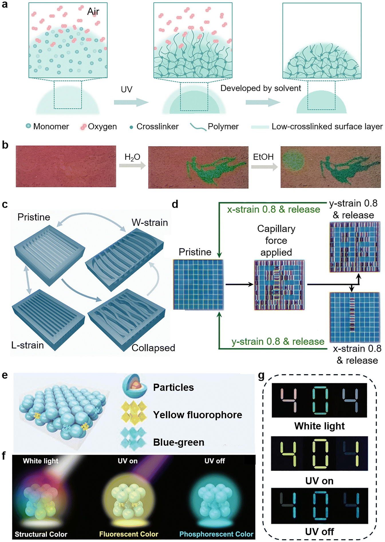 | ||
| Fig. 11 Multichannel information encryption based on responsive materials. (a) and (b) Solvent responsive information encryption based on Gradient-crosslinked hydrogel microdome pattern. Reproduced from ref. 155 with permission from Science China Press, copyright 2023. (c) and (d) Schematic diagram of PDMS trenches with multiple sets of encoding information. With directional strains, the width and length of trenches can be changed, and capillary force collapsed the structure. Under appropriate conditions, hidden information will be revealed. Reproduced from ref. 156 with permission from Wiley-VCH GmbH, copyright 2021. (e)–(g) Tri-state PhC film for multilevel information encryption enabled by both physical and chemical structures. Reproduced from ref. 157 with permission from Wiley-VCH GmbH, copyright 2023. | ||
3.3 Optical sensors
Natural biological optical systems rely on the precise design of microstructures rather than chemical dyes, offering profound insights for the development of artificial optical sensors. Biomimetic micro- and nano-optical structures for sensing have long been a hot topic of research. Sensors with fine structural design rely on sub-wavelength scale optical field localization and light–matter interactions to respond to external stimuli (including physical (light, heat, humidity, etc.), chemical (pH, VOC gas, etc.), and biological) in a label-free, real-time, highly sensitive, and visual way. People are fascinated with the convenience of naked eye sensors. In this chapter, we will focus on visualizing and miniaturizing bionic optical sensors.PhCs as a typical photonic structure are always the most famous carriers for visual sensing. The use of PhCs to detect external stimuli with the naked eye is a research hotspot. The sensing mechanisms can be broadly categorized into two aspects: one is the variation of structural parameters and the other is the manipulation of the optical field. PhCs are composed of periodically arranged materials, and their optical properties are highly dependent on structural parameters (lattice spacing, porosity, refractive index contrast, etc.). When external stimuli (e.g., temperature, humidity, pressure, chemical substances) lead to deformation, expansion, or reorganization of the PhC structural units, their photonic band gap positions are shifted, which in turn causes structural color changes visible to the naked eye. By establishing a quantitative relationship between the structure color and the parameter to be measured, visual sensing can be realized. Inspired by this iridescent color-changing principle, there exists a vast body of remarkable literature. For example, Zhao et al. proposed a novel structural color microneedle patches (EMNs) composed of PhCs and microneedle arrays to in situ detect multiple wound biomarkers (Fig. 12a).158 The EMNs were divided into different regions for the detection of distinct small molecules. By selecting specific stimulative response materials, the presence of target molecules can create responsive volume change, resulting in a change in structural color. As a demonstration, they chose the wound as the inspection object (Fig. 12b). Notably, for pH detection, the bacterial infection group exhibited a red-shifted color compared to the other two groups. For glucose detection, the diabetes group showed a redder color than the other two groups.
 | ||
| Fig. 12 Bioinspired visual optical sensors based on different working principles. (a) and (b) Schematic illustration of the sensing mechanism of small molecule detection of the EMNs. Target molecules cause changes in EMNs' volume, leading to a vivid visual color change. In vivo detection of EMNs for pH and glucose detection. Reproduced from ref. 158 with permission from Wiley-VCH GmbH, copyright 2023. (c) Schematic of the setup of coaxial 3D printing and illustration of the CLC tensile sensor change in the pitch length under uniaxial tension and the corresponding optical images. Reproduced from ref. 160 with permission from Wiley-VCH GmbH, copyright 2025. (d) Schematic illustration of the thermally responsive color variation property of objects based on cholesteric cellulose liquid crystal. Optical images of an object showing color variations under different temperatures. Reproduced from ref. 161 with permission from National Academy of Sciences, USA, copyright 2022. (e) and (f) Color contrasts of bacteria in the BCC-coated glass slide and bare glass can be improved 10.1-fold. Near-field analysis of BCCs indicates strong spatial confinement of light. Reproduced from ref. 162 with permission from American Chemical Society, copyright 2025. (g) Schematic of colorimetric sensor array relying on dye-cPhC. Detecting acetaldehyde at 1 ppm (limit of detection) and 100 ppm (permissible exposure limit) with pronounced color changes. Reproduced from ref. 163 with permission from Wiley-VCH GmbH, copyright 2024. | ||
Beyond common colloidal PhC structure colors, CLCs, as a unique type of PhC, form helical superstructures through the self-assembly of chiral supramolecules, which is popular in fabricating sensors.159 These structures can selectively reflect circularly polarized light at specific wavelengths, exhibiting dynamically tunable structural colors. With significant optical responsiveness to external stimuli such as temperature, light, and electric fields, CLCs demonstrate rapid response speeds, excellent reversibility, and outstanding processing flexibility. These characteristics have established them as a “star performer” widely employed in smart sensing and dynamic optical devices, making them a prominent material in cutting-edge photonic applications. Yang et al. successfully fabricated cholesteric liquid crystal elastomers via direct ink writing with prominent mechanochromic properties (Fig. 12c).160 They attempted to utilize two inks for coaxial printing: CLC ink as the core material and transparent silicone ink as the shell material. Using this method, the core can be supported by the silicone shell, meanwhile enabling the cholesteric phase to rapidly self-assemble. The mechanochromic properties of the sample were characterized. As can be seen, the sample showed a blue shift in color when tensile stress was applied. Because lateral compression accompanying uniaxial tension reversibly compresses the pitch, resulting in the blue shift. The mechanochromic sensitivity of the printed sample is higher than that of bare CLC. It should be noted that the blue shift in color can be reversible for at least 100 cycles under 20% strain. Combining parameter-sensitive structures with stimulative responsive materials, researchers have designed various types of sensors. Another similar but classic example was done by Zhao's group.161 They achieved a printable structural color material relying on cholesteric cellulose liquid crystals mixing with gelatin and a thermal-responsive hydrogel (Fig. 12d). Both of these filling substances have brilliant thermal responsiveness. Thus, the helical pitch could be modulated under the control of environmental temperature leading to color tunability. It exhibited a pronounced color change from green to red in the range of 20–40 °C.
PhCs have many intriguing optical functions, including the aforementioned tunable structural color and the regulation of optical fields. There are enormous papers about the latter in the sensing field. In general, slow light effect or increasing local optical density enhances light–matter interactions and the modulation of spontaneous radiation efficiency (Purcell effect) can extremely boost the detection signal when the fluorescence emission peak overlaps with the PhC bandgap edge. Based on these basic mechanisms, some novel ideas are emerging to improve sensing performance. In a more recent study, inspired by the Brazilian opal, binary colloidal crystals (BCCs) for high-contrast imaging were produced by Song et al. (Fig. 12e).162 It is a unique structure unlike traditional PhCs, which consist of two distinct nanoparticles. Compared to bare glass, the BCCs can improve color contrasts by an order of magnitude (Fig. 12f). As the figures show, the same undyed bacteria is difficult to identify with a commercial optical microscope because of the weak scattering signals. But it can be seen easily in cyan colors using the red BCCs substrate. Near-field analysis of BCCs gives us the reason for high-contrast Imaging. The scattering signals are remarkably amplified by taking advantage of the strong resonance-induced spatial confinement effect.
In addition, unlike the common principle of modulating the rate of spontaneous radiation to enhance the fluorescent signal, Yang and coworkers mentioned a new type of design combining absorption dyes and PhCs to construct a dye-cPhC colorimetric sensor array (Fig. 12g).163 It has been demonstrated that spectral overlap between the dye's absorption peak and the bandgap of cPhCs significantly provides a more noticeable VOC sensing signal. Therefore, cPhCs with broad and multiple reflective peaks were produced through spin-coating to improve the likelihood of overlapping with dyes. As a preliminary validation, they constructed a 5 × 8 array, a total of 40 spots to test the detection performance for acetaldehyde, acetone, and acetic acid. Taking acetaldehyde as an instance, the color change was observed at 1 ppm and became more and more pronounced with increasing concentration. The sensor array can distinguish acetaldehyde, acetone, and acetic acid with enhanced sensitivity.
In conclusion, bionic micro-nano optical structures provide a revolutionary solution for optical sensing technology with high performance and miniaturization. With the cross-fertilization of nanotechnology, smart materials, and artificial intelligence, this field is gradually moving towards major application scenarios such as clinical diagnosis, becoming the core driving force of next-generation optical sensing technologies.
3.4 Thermal management
Engineered micro-nano photonic structures are revolutionizing radiative cooling technologies, establishing themselves as indispensable platforms for next-generation thermal management.164–166 By decoding biological mechanisms of organisms from extreme environments, these materials achieve sub-ambient cooling through multiscale structural hierarchies that synergistically manipulate photon transport and phonon scattering.167,168 Their engineered spectral selectivity enables simultaneous high solar reflectance (>95%) and mid-infrared emissivity (>90%), while adaptive interfacial architectures allow real-time thermal modulation in response to environmental stimuli.169 Such breakthroughs are redefining sustainability paradigms across scales—from zero-energy building climatization to spacecraft thermal shields—by converting waste heat into radiative power flows through the atmospheric transparency window (8–13 μm), ultimately bridging photonic innovation with global carbon neutrality imperatives.170In harsh environments, organisms have evolved sophisticated optical strategies to manage thermal loads. A quintessential example is the Saharan silver ant (Cataglyphis bombycina), which survives desert surface temperatures exceeding 60 °C through a dual-functional hair structure (Fig. 13a–e).171 These triangular, micrometer-scale hairs enhance solar reflectance (67% in visible and near-infrared (NIR) wavelengths) via Mie scattering and total internal reflection while simultaneously boosting mid-infrared emissivity for radiative heat dissipation. This biological solution enables the ants to maintain body temperatures below 53.6 °C during peak solar exposure. Such mechanisms highlight nature's ability to decouple optical properties across spectral bands-a principle that has inspired the design of artificial radiative coolers.
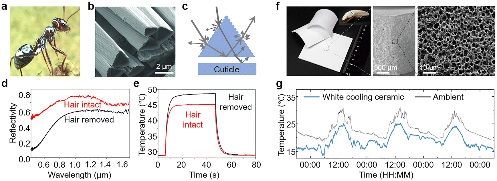 | ||
| Fig. 13 Passive radiative cooling based on optical structure. (a)–(e) Structure and cooling mechanism of silver ant hair. Reproduced from ref. 171 with permission from AAAS, copyright 2015. (f) and (g) Cooling ceramics with a hierarchically porous structure. Reproduced from ref. 173 with permission from AAAS, copyright 2023. | ||
Building on these biological insights, researchers have developed inorganic materials that replicate natural thermal regulation strategies.172 Lin et al. engineered a hierarchically porous ceramic inspired by the Cyphochilus beetle's ultra-white scales (Fig. 13f and g).173 The ceramic with a multidispersed pore system achieves a near-perfect solar reflectivity of 99.6% and high mid-infrared emissivity (96.5%). The cooling ceramic enables an average subambient temperature reduction of 4.3 °C during the daytime. However, while such high-reflectance materials excel in cooling efficiency, their monotonous white appearance limits aesthetic integration in urban and architectural contexts.
To address this limitation, color layers like pigment color,174,175 photoluminescence,176–178 and structural color179–182 have been introduced in the radiative coolers. For example, Li et al. demonstrated a pink-colored radiative cooler employing a seven-layer Si/SiO2/TiO2 stack. The color comes from Fabry–Pérot resonator (Fig. 14a and b).183 By designing photonic structures with tailored absorption in the visible range and high reflectance in the NIR, they achieved pink-colored surfaces that were 22.6 °C cooler than conventional paints under identical solar exposure.
 | ||
| Fig. 14 Colorful passive radiative cooling coating. (a) and (b) Colored radiative coolers based on multilayer photonic structures. Reproduced from ref. 183 with permission from Springer Nature, copyright 2018. (c) and (d) Bioinspired colorful daytime radiative cooling film based on a hierarchically porous nested structure. Reproduced from ref. 185 with permission from Elsevier, copyright 2024. | ||
However, the introduction of absorption-based colorants, such as pigments/dyes, photoluminescent materials, and resonant absorption structures, in radiative coolers inevitably results in absorption across the solar spectrum, which reduces the cooling performance.184 To solve these problems, Hou et al. developed a bioinspired radiative cooling film mimicking the hierarchical structure of scarab beetle elytra (Fig. 14c and d).185 Their design integrates close-packed colloidal PhC hemispheres within a porous polymethyl methacrylate (PMMA) substrate, achieving 93.4% solar reflectance and 92.3% atmospheric window emissivity. This colorful cooling film can realize subambient cooling of ∼10.2 °C at night and ∼7.2 °C at midday. The simple nesting of a colorful coating in surface pits can preserve its subambient cooling performance and provide an appealing aesthetic option. This approach bridges the gap between aesthetics and performance, offering a scalable solution for applications requiring both visual appeal and thermal efficiency.
In addition, PhC films have emerged as promising candidates for smart window applications in thermal management. Inspired by reptiles such as geckos that combine structural and pigmentary colors for adaptive camouflage, researchers have integrated thermochromic dyes and electrochromic materials into colloidal PhCs, achieving dual responsiveness to temperature and voltage with adjustable color intensity.186 More recently, Li et al. developed a smart window system by embedding PhCs into a thermoresponsive hydrogel matrix, enabling multicolor switching and enhanced NIR shielding under varying thermal conditions.187
3.5 Light management in optoelectronic devices
Light management refers to the process of engineering the interaction between photons and optoelectronic materials/devices at the micro- and nanoscale.188 This technology plays a crucial role in enhancing the performance of optoelectronic devices such as SCs, photodetectors (PDs), and LEDs.189,190 Research has demonstrated a strong correlation between device efficiency and structural design, where bioinspired micro-nano structures offer innovative solutions for light-field manipulation. In the fields of SCs and PDs, bioinspired micro/nano structures—including unitary and hierarchical architectures—significantly improve energy conversion efficiency and photoresponse performance through advanced light-trapping mechanisms such as reduced reflection, enhanced scattering, and enhanced optical path length.45 Additionally, in photodetectors, the incorporation of polarization-sensitive structures, such as grating structures, not only enhances photoelectric responses but also endows the PDs with polarization-sensitive capabilities.191 Notably, groundbreaking advances in bioinspired vision chips, incorporating structures mimicking human eyes and compound eyes, are reshaping multiple technological domains, including medical diagnostics, artificial intelligence, and autonomous driving.192,193 For LEDs, bioinspired strategies such as moth-eye antireflective surfaces, PhCs, scattering structures, and optical microcavities effectively address the limitations of total internal reflection, thereby enhancing the light out-coupling efficiency (OCE).194,195 This section reviews representative applications of bioinspired photonic structures in optoelectronic devices, focusing on three main functional domains: (1) light management in SCs and PDs (Section 3.5.1), (2) bioinspired visual sensing systems (Section 3.5.2), and (3) light management in LEDs (Section 3.5.3). These topics share similar underlying photonic principles discussed above, and their device architectures and performance objectives differ significantly. | ||
| Fig. 15 Light management with single and composite structures in SCs and PDs. Example of light management through (a) single structure and (b) composite structure. (c) Device architecture and light management of the device with grating structure. (g) Influence of grating structure on device performance. Reproduced from ref. 197 with permission from Wiley-VCH Verlag GmbH & Co. KGaA, copyright 2018. (d) Device architecture and light management of the device with moth-eye structure. (h) Influence of moth-eye structure on device performance. Reproduced from ref. 206 with permission from Wiley-VCH Verlag GmbH & Co. KGaA, copyright 2016. (e) Device architecture and light management of moiré perovskite PDs. (i) Polarization response of moiré perovskite PDs. Reproduced from ref. 208 with permission from Wiley-VCH GmbH, copyright 2021. (f) Device architecture and light management of bioinspired perovskite PDs. (j) Polarization-sensitive light detection of bioinspired perovskite PDs. Reproduced from ref. 209 with permission from Wiley-VCH Verlag GmbH &Co. KGaA, copyright 2019. | ||
3.5.1.1 Single structure. Researchers have integrated various types of single micro-nano structures into SCs and PDs, including gratings,196–200 nanopillars,201 nanorods,202,203 nanocones,189,204 and moth-eye structures (Fig. 15a).205,206 As a light-absorbing layer in SCs and PDs, the introduction of photonic structures can reduce the mismatch of refractive indices at interfaces, thus enhancing the light-trapping and efficiency of devices. Wang et al. innovatively employed commercial optical discs (CDs/DVDs) as templates to construct large-area grating structures on perovskite active layers via nanoimprinting technology.197 They demonstrated that the grating structures can simultaneously enhance both light-harvesting capability and charge collection efficiency in perovskite SCs, providing a novel approach for improving device performance (Fig. 15c). The precisely patterned diffraction gratings (with periods of 0.75 μm for DVD and 1.5 μm for CD structures) effectively reduced optical reflection while prolonging the photon propagation path through multi-order diffraction effects, resulting in significantly improved light-trapping capability. Ultimately, the DVD-derived grating structure boosted the PCE by 18% (from 16.71% to 19.71%) through enhanced photon management and carrier collection optimization (Fig. 15g). Kang et al. introduced moth-eye nanostructures into TiO2 ETL to enhance the light-harvesting efficiency of perovskite SCs (Fig. 15d).206 These moth-eye structures effectively confine incident photons within the perovskite active layer through the unique light-management characteristics. The moth-eye structure with a conical shape possesses gradient refractive index properties that efficiently guide incident light into the device while enhancing light absorption in the perovskite film. The moth-eye structure demonstrates superior light-harvesting efficiency across the entire visible spectrum. Ultimately, the perovskite SCs incorporating moth-eye TiO2 ETL achieved a 10.7% improvement in PCE over their planar counterparts (Fig. 15h). Besides, Wei et al. fabricated a moth-eye structure on the [6,6]-phenyl-C61-butyric acid methyl ester (PCBM) ETLs via a PDMS template nanoimprinting route.205 In this configuration, the moth-eye PCBM ETL simultaneously functions as a back electrode, while its PhC characteristics act as a reflective mirror to suppress light leakage, thereby effectively trapping photons within the perovskite active layer for enhanced absorption. They confirmed that this photon structure achieves broadband light absorption enhancement across the entire visible wavelength range. Furthermore, the incorporation of micro-nano structures enables spectrally selective response. Cao et al. developed bioinspired perovskite photodetectors with NIR-I selective photodetection and imaging capabilities by engineering precisely designed optical microcavities.207 These biomimetic microcavities consist of alternately stacked lithium fluoride (LiF) and N,N′-bis(naphthalen-1-yl)-N,N′-bis(phenyl)benzidine (NPB) layers, with each layer's thickness precisely controlled to quarter-wavelength dimensions of the target spectrum. This NIR-I selective detection technology based on optical microcavity architecture demonstrates significant application potential in biological imaging and optical communications.
3.5.1.2 Complex structure. Usually, hierarchical light-trapping structures demonstrate superior incident light utilization efficiency compared to single photonic structures.45 Researchers have further proposed composite structures combining multiple nanostructures to synergistically improve light extraction efficiency while enabling polarization-selective functionalities.43,208–210 Song et al. developed Moiré perovskite photodetectors with a stacked dual shallow grating structure using grating patterning (Fig. 15e).208 The dual-grating architecture synergistically enhances light-harvesting capability and polarization sensitivity through combined diffraction effects, waveguide modes, and anti-reflection characteristics. Beyond enhancing light trapping, grating structures have demonstrated polarization-sensitive responses (Fig. 15i). Besides, Sun et al. integrated a moiré-grating structure into perovskite single crystals, achieving a polarization ratio of 9.1 in the fabricated photodetector.211 Inspired by the multilevel structures of butterfly wing scales, Zhan et al. ingeniously constructed a 1D nano-grating bonded porous 2D PhC perovskite photodetector (Fig. 15f).209 This design innovatively integrates multilevel optical structures with perovskite materials. This hierarchically designed composite structure ingeniously combines the diffraction properties of gratings with the photonic bandgap characteristics of PhCs, significantly enhancing the light-trapping capability of the perovskite active layer while simultaneously improving the device's photoresponse performance and enabling polarization-sensitive detection (Fig. 15j). Beyond light harvesting, biological photonic architectures have also inspired the design of advanced vision systems. In the following section, we shift our focus from energy collection to sensory perception, examining how nature's visual systems guide the development of artificial visual technologies.
 | ||
| Fig. 16 Biomimetic neuronal photo-vision. (a) The human eye's anatomical structure primarily comprises the cornea, pupil, lens, vitreous body, and retina. Within the retina, photoreceptor cells (specifically cones and rods) play crucial roles in detecting light intensity and facilitating visual adaptation functions. Reproduced from ref. 213 with permission from Springer Nature, copyright 2023. (b) Structure of the compound eye (top), and structure of the ommatidium (down). Reproduced from ref. 192 with permission from Springer Nature, copyright 2024. (c) Exploded view of biomimetic electrochemical eye. (d) Side view of the biomimetic electrochemical eye. PDMS, polydimethylsiloxane. Reproduced from ref. 217 with permission from Springer Nature, copyright 2020. (e) Exploded view illustration of an artificial compound eye. Reproduced from ref. 192 with permission from Springer Nature, copyright 2024. (f) Macro photograph of a robber fly's eye. (g) Illustration exploded view of the pinhole compound eye system. Reproduced from ref. 221 with permission from AAAS, copyright 2024. | ||
3.5.2.1 Bionic eye light-sensing vision. The development of bionic human eyes offers hope for visually impaired individuals. The core innovation in bionic human eyes lies in mimicking the structural and functional complexity of biological retinas (Fig. 16a). Typical bionic vision systems for the human eye generally comprise single-lens optical structure coupled with a curved image sensor. In 2020, Gu et al. successfully developed a hemispherical perovskite nanowire array retinal system (Fig. 16c and d) with the potential for high-resolution imaging comparable to the human eye.217 They integrated high-density nanoscale photodetectors on a curved substrate. Notably, each perovskite nanowire demonstrated detectable strong photoresponse, while the porous aluminum membrane structure effectively isolated individual nanowires, significantly suppressing optical crosstalk. However, the device's resolution was limited by backside wire bonding processes. Subsequently, inspired by tunable optical structures like the crystalline lens and iris, the team further developed a bioinspired ocular device with adjustable optical components.218 Bionic human eyes hold immense potential in the medical field, promising to restore near-natural vision for the visually impaired and enhance their quality of life. In AI and robotics, they can endow robots with more human-like vision, enabling precise object and environment recognition and advancing human–robot interaction. Insect compound eyes are intelligent imaging systems that are small-sized, multi-apertured, and wide field-of-view (Fig. 16b).219 Insect compound eyes show high sensitivity to moving targets and strong differentiation ability regarding light intensity, wavelength, and color.220 Hence, researching bionic insect compound eyes is highly significant (Fig. 16e). In 2024, Prof. Fan's team designed a pinhole compound eye system inspired by predatory flies (Fig. 16f and g).221 This system features a 3D-printed hexagonal pinhole array and a hemispherical perovskite nanowire detector, achieving a 140° FOV in monocular mode and 220° in binocular configurations. It enables precise target localization and real-time tracking of quadruped robots via drones.
3.5.2.2 Bionic eye polarization vision. Polarization detection plays a crucial role in advanced optical sensing, offering capabilities beyond conventional intensity- or wavelength-based imaging. In remote sensing, polarization imaging can enhance detail visibility and help identify targets difficult to detect with traditional imaging. In environmental monitoring, polarization information can be used to analyze atmospheric aerosols and suspended particles in water. In information encryption and transmission, the properties of polarized light can ensure high-security data transmission. Additionally, these technologies can play significant roles in biomedical imaging and robotic vision. Many biological systems, such as mantis shrimp,222–224 dung beetle225 and some insect species,226 naturally perceive polarized light, using it for navigation, communication, and prey detection. These organisms have evolved sophisticated structures—like the microvillar arrays in compound eyes—to efficiently decode polarization information from their environment.227 For instance, desert ants (Cataglyphis) can forage hundreds of meters from their nest and return without getting lost. This is attributed to their polarization-sensitive compound eyes. Their photoreceptor cells have two sets of perpendicular microvilli, making them sensitive to the polarization direction of incident light (Fig. 17a–c). Inspired by desert ant eyes, Fang et al. used a nanoimprinting-assisted one-step solution-crystallization molding method to create a perovskite single-crystal thin film with 6400 four-directional grating arrays (Fig. 17d–f).226
 | ||
| Fig. 17 Bioinspired polarization cameras. (a) SEM image of the head of Cataglyphis. (b) SEM image of the Cataglyphis compound eyes. (c) TEM image of the cross section of one small eye of the Cataglyphis compound eyes. There are two sets of photoreceptor microvilli perpendicular to each other. (d) Device architecture of bioinspired single-shot on-chip pol-PD. (e) Optical image of the four-directional grating arrays capped perovskite single-crystal thin film (80 by 80 pixels). (f) SEM image of a polarization unit. (g) Optical imaging of the sample. (h) Polarization imaging of sample in haze. Reproduced from ref. 226 with permission from AAAS, copyright 2024. (i) Optical image of the mantis shrimp. (j) SEM image of the compound eye of the mantis shrimp. (k) Diagrammatic sagittal section through the eye, showing the internal structure of dorsal and ventral hemispheres (DH and VH) and the six rows of the mid-band. (l) Device architecture of a full-Stokes polarization camera beside a U.S. quarter dollar coin (lens not attached). (m) Image of full Stokes polarimetric CMOS imaging sensor (n) Top: 3D Conceptual illustration of chip integrated full Stokes CMOS polarimetric imaging sensor. Here P1–P4 denotes the LP filters with transmission axes at 0°, 90°, 45°, 135° respectively. P5, P5′, and P6, P6′ denote chiral metasurface filters transmitting RCP and LCP, respectively. Here, P5 and P5′, P6 and P6′ are identical in dimensions respectively. Bottom: 2D cross-section of the chip-integrated polarimetric imaging sensor. (o) Optical imaging of the plastic ruler. (p) Polarization imaging of the plastic ruler. Reproduced from ref. 229 with permission from Springer Nature, copyright 2023. | ||
Based on high-performance four-directional grating arrays capped perovskite single-crystal thin film, they designed a single-shot, on-chip integrated polarization photodetector without extra polarizing elements, achieving high-sensitivity polarization imaging. This on-chip polarization detector precisely measures known angle of polarization (AoP) and degree of polarization (DoP), with accuracies of ±1.2° and ±0.1%, respectively. As a proof-of-concept, the polarization photodetector has been applied in many fields including visual dehazing (Fig. 17g and h), polymer stress visualization, and detection of cancerous areas in tissues. This design offers a new approach for high-sensitivity, miniaturized, and on-chip integrated polarization imaging systems. The mantis shrimp has one of the most complex visual systems on Earth, with 16 photoreceptors to capture light of 300–720 nm wavelength and sense polarized light. Its eyes comprise numerous optical units. The middle part has six specialized ommatidia rows. Each ommatidium has different functions, including ultraviolet, linearly polarized, and circularly polarized light receptors (Fig. 17i–k). In 1999, N. J. Marshall proved mantis shrimps have linear polarization vision.224 Their ommatidia in rows 5 and 6 have microvilli in parallel and perpendicular arrangements, forming orthogonal polarization-sensitive channels. This structure enhances polarization signal amplitude and contrast. Subsequently, researchers demonstrated that mantis shrimp can detect circularly polarized light.222 The R8 photoreceptor, acting like a quarter-wave plate with microvilli at a precise 45°, provides the structural basis for this detection. Bionic polarization detection technologies inspired by mantis shrimp compound eyes have broad application prospects in various fields. In 2021, Michael Kudenov's team developed a mantis shrimp (stomatopod)-inspired multispectral and polarization-sensitive detector.228 It consists of six semi-transparent polarization-sensitive organic photovoltaic detectors and polymer retarders. The design can sense 15 spectral channels over a 350-nanometer bandwidth. They also experimentally demonstrated a detector that simultaneously registered four spectral channels and three polarization channels. This system is the first to fully mimic the mantis shrimp's visual DoFs, offering a new way to enhance hyperspectral and polarization imaging by enabling simultaneous multispectral and polarization sensing along the same optical axis. Yu Yao's team at Arizona State University developed a metasurface-based full-Stokes polarization imaging sensor integrated on a chip (Fig. 17l–n).229 They designed high performance micro-scale polarization filters, including broadband linear polarization filters and dual-color (green and red) chiral metasurfaces. Integrated into an imaging sensor, it achieved high precision polarization detection with an average polarization measurement error of less than 2% for red and green light.
Despite remarkable advancements, state-of-the-art PeLEDs remain constrained by suboptimal EQE, primarily limited by low light OCE.194 Approximately 80% of generated photons are trapped within the device stack through non-ideal optical coupling, leading to energy dissipation via lossy pathways (Fig. 18a and b).194,195 The restricted photon extraction originates from these mechanisms: waveguide modes caused by the refractive index mismatch between perovskite layers (∼2.5) and charge transport materials (∼1.7–1.8); total internal reflection at substrate/air interfaces due to their refractive index contrast, confining photon emission to a narrow escape cone while trapping most photons in waveguide and substrate modes; and the evanescent SP mode at the metal/dielectric interface arises from the resonance between collective electron oscillations in the metal and electromagnetic waves. Critically, unextracted photons in these modes are converted to thermal energy, accelerating perovskite degradation and degrading device performance, ultimately suppressing EQE. Stranks et al. provided the EQE calculation formula for PeLEDs as follows:235
| EQE = fbalance × fe–h × ηradiative × foutcoupling |
 | ||
| Fig. 18 Light loss analysis and enhancement of OCE in LEDs. (a) Schematic representation of the generation, propagation, emission, and losses of light in a typical planar PeLED structure. CTL, charge-transport layer. (b) Optical power distribution versus the thickness of emissive layer in a MAPbI3 PeLED. Reproduced from ref. 195 with permission from Springer Nature, copyright 2023. (c) SEM image of an AAM-based device. Br-Pero refers to CH3NH3PbBr3. F8 refers to poly(9,9-di-n-octylfluorenyl-2,7-diyl), and PEDOT:PSS refers to poly(3,4-ethylenedioxythiophene) polystyrene sulfonate. (d) Device schematic. AAM channels are filled with TiO2. (e) EQEs of the thin film and AAM samples. Inset figures in a are the optical photos for different devices driving at 3.5V. Reproduced from ref. 239 with permission from Springer Nature, copyright 2019. (f) Scanning tunnelling electron microscope image of a PeLED with submicrometre structures. PEIE stands for polyethylenimine ethoxylated. (g) Light trapped in devices with a continuous emitting layer, which can be extracted by the submicrometre structure. (h) EQE and ECE plotted against current density. Reproduced from ref. 244 with permission from Springer Nature, copyright 2018. (i) The device architecture of a top-emission PeLED, where a Fabry–Perot microcavity forms between two metal electrodes. The bottom Au electrode is thick and fully reflective, while the top Au film is thin and semi-transparent, allowing a small fraction of light to exit the device. (j) Emission intensity versus microcavity length. (k) The impact of varying the thickness of the charge injection/transport and top Au layers on the EQE. Reproduced from ref. 253 with permission from Springer Nature, copyright 2020. | ||
3.5.3.1 PhC structures. The emitted light is confined within waveguide and substrate modes due to the significant disparity in the refractive index between the interfaces. PhCs can enhance the OCE of LEDs by suppressing guided-mode emission, boosting spontaneous emission via the Purcell effect, and facilitating light extraction across the entire surface through leaky-mode coupling.191,237,238 Zhang et al. have pioneered the fabrication of PeLEDs on anodic alumina membrane (AAM) substrates, which are configured with hexagonal nanodome arrays and TiO2 nanowire arrays (Fig. 18c and d).239 The hexagonal nanodome arrays act as the barrier layer and the light outcoupler, and the TiO2 nanowires function as optical antennas to enhance light extraction. The AAM has a thickness of ∼3 μm, a pore diameter of ∼120 nm, and two pitches of 500 and 1000 nm. All functional layers of the LEDs have conformal nanodome structures derived from the porous alumina membrane, with TiO2 nanowires embedded in the pores. The higher refractive index of TiO2 (n = 2.6) than alumina (n = 1.7) enables the TiO2 nanowires to serve as optical antennas. Devices based on AAM with a 1000 nm pitch achieved an EQE of 17.5%, nearly double that of planar control devices (Fig. 18e). The photonic substrate in these PeLEDs facilitates a two-step light extraction process: light is coupled from the nanodome coupler to the nanowire antenna, which converts the guided modes into leaky modes for extraction as radiative light. This synergistic effect markedly improves the light extraction efficiency of PeLEDs. Shen et al. developed bioinspired PeLEDs by introducing a moth-eye nanostructured ZnO layer at the interface between the front electrode and the perovskite active layer using nanoimprinting.240 The patterned ZnO served as a hole-injection layer and as an out-coupling structure to extract waveguide-mode photons. The maximum EQE of the moth-eye PeLEDs was enhanced to 20.3%, and an EQE of 28.2% was achieved with a half-ball lens. Moreover, the moth-eye PeLEDs exhibited angle-independent light emission. These findings demonstrate the nanostructure's effectiveness in extracting confined waveguide-mode photons. Furthermore, Fu et al. demonstrated that photonic structures integrated into functional layers can simultaneously suppress substrate modes, extract waveguide-mode light, and achieve full-area directional polarized emission through controlled light redirection.237 Besides, using the PhCs is an effective approach to improve the OCE of OLEDs and conventional semiconductor LEDs.238 For example, Do et al. demonstrated 2D SiO2/SiNx PhCs between ITO and glass substrate to achieve a significant OCE improvement. Wiesmann et al. comprehensively discussed PhC LEDs in 2009, and more theoretical details can be found in their report.241
3.5.3.2 Scattering structures. To extract more confined light within LEDs, it's worthwhile to adopt internal scattering structures to compensate for light losses. Modifying the active layer's morphology can enhance the light scattering in the active layer, randomize the waveguided photon paths, and boost the OCE.233,242,243 For instance, light scattering in suitably sized crystallites offers advantages similar to those of deliberately patterned or textured structures.242 Cao et al. introduced sub-micrometer-scale structures to formamidinium lead iodide-based LEDs, leading to an enhanced light outcoupling efficiency greater than 30% (Fig. 18f and g).244 Submicron-scale structures spontaneously formed through a simple low-temperature solvent engineering approach. The resulting perovskite films (doped with 5-aminovaleric acid) exhibited a discontinuous morphology with randomly distributed planar flakes and roughly rectangular shapes, featuring grain sizes ranging from 100 to 500 nm. By leveraging structural conformality, photon structures with random concave–convex features were constructed on both the high-refractive-index perovskite layer and low-refractive-index organic functional layers. The continuous photonic cycling within these structures enables efficient extraction of light trapped in waveguide modes, thereby enhancing the OCE (Fig. 18g). This structural optimization ultimately enables the device to achieve a maximum EQE of 20.7% and a peak energy-conversion efficiency of 12% (Fig. 18h). Subsequently, Various passivation strategies have been used to reduce defect densities in discrete perovskite films, and the EQE based on the discrete perovskite films has been raised to 32.0%.242,245–247 In OLEDs, nanoparticles patterned scattering structures have been incorporated between the substrate and the OLED structure to enhance light extraction.248,249 These nanoparticles aid in extracting light from confined optical modes.
3.5.3.3 Optical microcavity structures. Optical microcavities can significantly enhance light outcoupling efficiency by manipulating photonic mode distribution.250 A typical microcavity consists of a pair of parallel mirrors, including metallic reflectors (e.g., Ag, semi-transparent SnO2, or NiO), oxide-based stacks (e.g., SiO2–TiO2), or dielectric distributed Bragg reflectors (DBRs).251 When the emissive layer is embedded in a cavity with a thickness matching an integer multiple of the optical wavelength (e.g., λ/2 or λ), the optical mode density near the emission wavelength is selectively amplified, thereby boosting spontaneous emission via the Purcell effect. Furthermore, phase-matching conditions from multiple-beam interference between the two mirrors enable directional control of light propagation, leading to pronounced enhancement of spontaneous emission intensity in the forward direction. Additionally, the microcavities could enhance radiative recombination rates, and reduce the electroluminescence spectral widths. By adjusting the thickness of the charge transport layers to change the cavity length, a narrowing and blue shift of the electroluminescence full width at half maximum were observed.252 Optical microcavities offer a universal approach to boost LED performance via photonic confinement and interference, requiring synergistic design of resonance matching, mirror optimization, and process compatibility. Miao et al. showed that high-efficiency PeLEDs with a high EQE of 20.2% and an ultrahigh radiant exitance up to 114.9 mW cm−2 could be achieved by employing the microcavity effect to enhance light extraction (Fig. 18i–k).253
4. Challenges & future
In this review, we systematically overview the recent advances in bioinspired micro-nano photonic materials, mainly involving many classical natural examples, the underlying mechanisms of interaction between light and biological structures and materials, the various biomimetic materials constructed for applications in several fields. Living organisms have developed and refined materials, structures, and processes to selectively absorb, reflect, scatter, transmit, guide, and harvest light. Inspired by nature, researchers have achieved a broad range of photonic structures with relatively simple materials and under mild processing conditions.Despite great advances and breakthroughs that have been made in recent years, three persistent challenges dominate the field: the constrained biological design exploration, the performance-directed design of multiscale ordered structures, and the scalability-reliability paradox in manufacturing. Addressing these challenges requires a paradigm shift in research methodologies, manufacturing technologies, and cross-disciplinary collaboration frameworks.
Firstly, while existing research has focused on well-characterized biological models (e.g., butterfly wings, peacock feathers), over 80% of nature's photonic diversity remains unexplored. Underexplored organisms—particularly extremophiles and microorganisms—harbor unique nanostructural solutions to light management under extreme conditions. Research in bioinspired micro-nano optics encompasses not only the study of structure–property relationships in biological optical materials, but also the investigation of material formation processes (e.g., biomineralization pathways, self-assembly mechanisms), which fundamentally shape the design, fabrication, characterization, and application of biomimetic materials.
Secondly, the true potential of bioinspired photonics lies in replicating nature's ability to integrate multiple optical functions within unified architectures. Biological systems achieve this through evolutionary-optimized hierarchical designs that balance competing physical requirements—a capability artificial materials currently lack. Challenges stem from incompatible material interfaces, cross-scale interference effects, and limited dynamic control mechanisms. Critical research needs span from atomic-scale material purity to macroscale system integration, bridging quantum and continuum models to achieve desired multifunctional properties (e.g., energy-efficient photonics, mechanical robustness, or self-healing capability). AI-driven inverse design is emerging as a powerful strategy to address this complexity. AI-driven inverse design refers to the use of machine learning models—such as deep neural networks or generative algorithms—to automatically generate structures that meet desired optical or functional targets. These tools invert the traditional trial-and-error process by predicting optimal architectures directly from performance requirements. While most existing models are trained on simulation datasets, biological systems adapt through continuous environmental feedback. A bioinspired AI design framework could incorporate real-time experimental inputs, such as optical performance data or environmental responses, into a closed-loop learning cycle. This approach would emulate evolutionary processes in nature and enable adaptive, context-aware design strategies beyond purely synthetic optimization.
Thirdly, the development of bioinspired photonic materials has reached a critical juncture, where breakthroughs in fundamental understanding must now translate into scalable technological solutions. Current fabrication approaches face intrinsic limitations in reconciling nanoscale precision with industrial viability. Top-down lithography techniques, while achieving atomic-scale resolution, remain energy-intensive and cost-prohibitive for large-area applications. Conversely, biomimetic self-assembly methods struggle with structural reproducibility and material compatibility. Emerging hybrid strategies combining programmable molecular assembly with advanced additive manufacturing show promise in bridging this gap. In addition, recent advances in fabrication techniques such as roll-to-roll printing, inkjet-assisted self-assembly, and template-free nanofabrication now allow high-throughput production of photonic structures over large areas while retaining optical precision. These scalable fabrication techniques have facilitated various real-world applications, including photonic textiles with durable structural colors,254 architectural coatings,255 anti-counterfeiting films,256 and thermochromic printable materials for rewritable full-color displays.257,258 Key priorities moving forward include developing self-healing materials, establishing closed-loop manufacturing cycles, and formulating standardized sustainability metrics. To support this goal, preliminary sustainability metrics have been proposed in recent studies, including fabrication energy consumption,259 environmental toxicity of raw materials,260 recyclability or biodegradability of structural components,261 and operational lifetime under environmental stressors.259 Although a unified framework remains under development, incorporating these metrics into material design and reporting standards is essential to guide the eco-responsible advancement of scalable photonic systems. Progress in these areas could substantially reduce production costs while meeting industrial performance requirements.
In summary, the ultimate goal extends beyond mimicking nature—it lies in creating adaptive photonic materials that surpass biological constraints through rational design. Potential applications span holographic encrypted information storage, intelligent optical sensors, and ultra-efficient solar energy systems. Achieving these milestones demands a highly convergent interdisciplinary collaboration across biology, optics, materials science, and artificial intelligence. By integrating biological wisdom with cutting-edge technologies, this convergence unlocks materials with unprecedented functionalities—nature's brilliance, amplified by human ingenuity. Bioinspired photonics stands as a key technology for addressing 21st-century challenges in information, energy, and environmental stewardship.
Conflicts of interest
There are no conflicts to declare.Data availability
No primary research results, software or code have been included and no new data were generated or analysed as part of this review.Acknowledgements
This work was financial supported by the National Science Fund for Distinguished Young Scholars (22225502), the National Natural Science Foundation of China (22435004, 22073107, and 52321006), and Bureau of International Cooperation, Chinese Academy of Sciences (027GJHZ2022044MI).References
- S. Hua, E. Divita, S. Yu, B. Peng, C. Roques-Carmes, Z. Su, Z. Chen, Y. Bai, J. Zou, Y. Zhu, Y. Xu, C. K. Lu, Y. Di, H. Chen, L. Jiang, L. Wang, L. Ou, C. Zhang, J. Chen, W. Zhang, H. Zhu, W. Kuang, L. Wang, H. Meng, M. Steinman and Y. Shen, Nature, 2025, 640, 361–367 CrossRef CAS PubMed.
- K. Liao, Y. Lian, M. Yu, Z. Du, T. Dai, Y. Wang, H. Yan, S. Wang, C. Lu, C. T. Chan, R. Zhu, D. Di, X. Hu and Q. Gong, Nat. Photonics, 2025, 19, 358–368 CrossRef CAS.
- S. Dou, H. Xu, J. Zhao, K. Zhang, N. Li, Y. Lin, L. Pan and Y. Li, Adv. Mater., 2021, 33, e2000697 CrossRef PubMed.
- M. Kolle and S. Lee, Adv. Mater., 2018, 30, 1702669 CrossRef PubMed.
- S. An, B. Shi, M. Jiang, B. Fu, C. Song, P. Tao, W. Shang and T. Deng, Chem. Rev., 2023, 123, 7081–7118 CrossRef CAS PubMed.
- L. P. Biró and J. P. Vigneron, Laser Photonics Rev., 2010, 5, 27–51 CrossRef.
- C. Chen, Y. Kuang, S. Zhu, I. Burgert, T. Keplinger, A. Gong, T. Li, L. Berglund, S. J. Eichhorn and L. Hu, Nat. Rev. Mater., 2020, 5, 642–666 CrossRef CAS.
- D. Nepal, S. Kang, K. M. Adstedt, K. Kanhaiya, M. R. Bockstaller, L. C. Brinson, M. J. Buehler, P. V. Coveney, K. Dayal, J. A. El-Awady, L. C. Henderson, D. L. Kaplan, S. Keten, N. A. Kotov, G. C. Schatz, S. Vignolini, F. Vollrath, Y. Wang, B. I. Yakobson, V. V. Tsukruk and H. Heinz, Nat. Mater., 2023, 22, 18–35 CrossRef CAS PubMed.
- P. Vukusic, J. R. Sambles and C. R. Lawrence, Nature, 2000, 404, 457 CrossRef CAS PubMed.
- S. Kinoshita, S. Yoshioka and K. Kawagoe, Proc. R. Soc. London, Ser. B, 2002, 269, 1417–1421 Search PubMed.
- J. Zi, X. Yu, Y. Li, X. Hu, C. Xu, X. Wang, X. Liu and R. Fu, Proc. Natl. Acad. Sci. U. S. A., 2003, 100, 12576–12578 CrossRef CAS PubMed.
- M. Jacobs, M. Lopez-Garcia, O. P. Phrathep, T. Lawson, R. Oulton and H. M. Whitney, Nat. Plants, 2016, 2, 16162 CrossRef CAS PubMed.
- X. Li, T. Fan, H. Zhou, S. K. Chow, W. Zhang, D. Zhang, Q. Guo and H. Ogawa, Adv. Funct. Mater., 2008, 19, 45–56 CrossRef.
- K. Liu, C. Yuan, Q. Zou, Z. Xie and X. Yan, Angew. Chem., Int. Ed., 2017, 56, 7876–7880 CrossRef CAS PubMed.
- S. Enriquez, E. R. Mendez, O. Hoegh-Guldberg and R. Iglesias-Prieto, Proc. R. Soc. London, Ser. B, 2017, 284, 20161667 CrossRef PubMed.
- D. Wangpraseurt, S. You, F. Azam, G. Jacucci, O. Gaidarenko, M. Hildebrand, M. Kuhl, A. G. Smith, M. P. Davey, A. Smith, D. D. Deheyn, S. Chen and S. Vignolini, Nat. Commun., 2020, 11, 1748 CrossRef CAS PubMed.
- D. Wangpraseurt, S. Jacques, N. Lyndby, J. B. Holm, C. F. Pages and M. Kuhl, J. R. Soc., Interface, 2019, 16, 20180567 CrossRef CAS PubMed.
- R. Xiong, J. Luan, S. Kang, C. Ye, S. Singamaneni and V. V. Tsukruk, Chem. Soc. Rev., 2020, 49, 983–1031 RSC.
- S. Tadepalli, J. M. Slocik, M. K. Gupta, R. R. Naik and S. Singamaneni, Chem. Rev., 2017, 117, 12705–12763 CrossRef CAS PubMed.
- B. D. Wilts, B. Wijnen, H. L. Leertouwer, U. Steiner and D. G. Stavenga, Adv. Opt. Mater., 2017, 5, 1600879 CrossRef.
- H. Yin, B. Dong, X. Liu, T. Zhan, L. Shi, J. Zi and E. Yablonovitch, Proc. Natl. Acad. Sci. U. S. A., 2012, 109, 10798–10801 CrossRef CAS PubMed.
- D. E. McCoy, T. Feo, T. A. Harvey and R. O. Prum, Nat. Commun., 2018, 9, 1 CrossRef CAS PubMed.
- A. Rogato and T. De Edoardo, Growth Form, 2023, 4, 3–10 CrossRef.
- H. Zhou, X. Li, T. Fan, F. E. Osterloh, J. Ding, E. M. Sabio, D. Zhang and Q. Guo, Adv. Mater., 2010, 22, 951–956 CrossRef CAS PubMed.
- A. Bay, P. Cloetens, H. Suhonen and J. P. Vigneron, Opt. Express, 2013, 21, 764–780 CrossRef PubMed.
- P. Vukusic and J. R. Sambles, Nature, 2003, 424, 852–855 CrossRef CAS PubMed.
- K. Hey Tow, D. M. Chow, F. Vollrath, I. Dicaire, T. Gheysens and L. Thevenaz, J. Lightwave Technol., 2018, 36, 1138–1144 Search PubMed.
- Z. Hayran and F. Monticone, Nat. Photonics, 2025, 19, 126–128 CrossRef CAS.
- C. Huang, Y. Luo, Y. Zhao, X. Ma, Z. Yan, Z. Liu, C. Sheng, S. Zhu and H. Liu, Nat. Photonics, 2024, 18, 471–477 CrossRef CAS.
- Y. Yang, Y. Yamagami, X. Yu, P. Pitchappa, J. Webber, B. Zhang, M. Fujita, T. Nagatsuma and R. Singh, Nat. Photonics, 2020, 14, 446–451 CrossRef CAS.
- Y. Li, S. Kita, P. Muñoz, O. Reshef, D. I. Vulis, M. Yin, M. Lončar and E. Mazur, Nat. Photonics, 2015, 9, 738–742 CrossRef CAS.
- H. Qin, Z. Su, Z. Zhang, W. Lv, Z. Yang, W. Chen, X. Gao, H. Wei, Y. Shi, B. Li, J. Zhou, R. Fleury, C. W. Qiu and Q. Song, Nature, 2025, 639, 602–608 CrossRef CAS PubMed.
- S. T. Ha, Q. Li, J. K. W. Yang, H. V. Demir, M. L. Brongersma and A. I. Kuznetsov, Science, 2024, 386, eadm7442 CrossRef CAS.
- F. Chen, Y. Huang, R. Li, S. Zhang, B. Wang, W. Zhang, X. Wu, Q. Jiang, F. Wang and R. Zhang, Chem. Commun., 2021, 57, 13448–13464 RSC.
- S. Zhou, C. Zhang, Z. Fu, Q. Zhu, Z. Zhou, J. Gong, N. Zhu, X. Wang, X. Wei, L. Xia and W. Xu, Nat. Commun., 2024, 15, 1979 CrossRef CAS PubMed.
- K. Li, T. Li, T. Zhang, H. Li, A. Li, Z. Li, X. Lai, X. Hou, Y. Wang, L. Shi, M. Li and Y. Song, Sci. Adv., 2021, 7, eabh1992 CrossRef CAS PubMed.
- Y. Liu, X. Hou, Y. Song and M. Li, Interdiscip. Mater, 2024, 3, 54–73 CAS.
- R. Li, K. Li, X. Deng, C. Jiang, A. Li, L. Xue, R. Yuan, Q. Liu, Z. Zhang, H. Li and Y. Song, Adv. Funct. Mater., 2024, 34, 2404706 CrossRef CAS.
- T. Wang, Y. Wang, Y. Fu, Z. Chen, C. Jiang, Y. E. Ji and Y. Lu, Adv. Sci., 2024, 11, e2400442 CrossRef PubMed.
- X. Lai, J. Peng, Q. Cheng, A. P. Tomsia, G. Zhao, L. Liu, G. Zou, Y. Song, L. Jiang and M. Li, Angew. Chem., Int. Ed., 2021, 60, 14307–14312 CrossRef CAS PubMed.
- K. Kang, I. Byeon, Y. G. Kim, J. R. Choi and D. Kim, Laser Photonics Rev, 2024, 18, 2400547 CrossRef CAS.
- S. Cui, K. Sun, Z. Liao, Q. Zhou, L. Jin, C. Jin, J. Hu, K. S. Wen, S. Liu and S. Zhou, Sci. Bull., 2024, 69, 2080–2088 CrossRef CAS PubMed.
- Y. Wang, Y. Lan, Q. Song, F. Vogelbacher, T. Xu, Y. Zhan, M. Li, W. E. I. Sha and Y. Song, Adv. Mater., 2021, 33, e2008091 CrossRef PubMed.
- C. Jeffryes, J. Campbell, H. Y. Li, J. Jiao and G. Rorrer, Energy Environ. Sci., 2011, 4, 3930–3941 RSC.
- Y. Zhan, Q. Cheng, Y. Song and M. Li, Adv. Funct. Mater., 2022, 32, 2200385 CrossRef CAS.
- Y. Fu, C. A. Tippets, E. U. Donev and R. Lopez, Wiley Interdiscip. Rev.: Nanomed. Nanobiotechnol., 2016, 8, 758–775 Search PubMed.
- X. Hou, F. Li, Y. Song and M. Li, J. Phys. Chem. Lett., 2022, 13, 2885–2900 CrossRef CAS PubMed.
- H. K. Raut, V. A. Ganesh, A. S. Nair and S. Ramakrishna, Energy Environ. Sci., 2011, 4, 3779 RSC.
- X. K. Zhao, Y. Q. Xiong, W. L. Wang, W. Zhang and D. Zhang, NPG Asia Mater., 2023, 15, 20 CrossRef.
- X. Ling, M. I. Osotsi, W. Zhang, Y. Wu, Q. J. Jin and D. Zhang, J. Bionic Eng., 2023, 20, 873–899 CrossRef.
- W. Zhang, Y. Hu, P. Feng, Z. Li, H. Zhang, B. Zhang, D. Xu, J. Qi, H. Wang, L. Xu, Z. Li, M. Xia, J. Li, R. Chai and L. Tian, Adv. Sci., 2024, 11, e2403173 CrossRef PubMed.
- G. Isapour and M. Lattuada, Adv. Mater., 2018, 30, e1707069 CrossRef PubMed.
- J. Y. Sun, B. Bhushan and J. Tong, RSC Adv., 2013, 3, 14862–14889 RSC.
- P. Tao, W. Shang, C. Song, Q. Shen, F. Zhang, Z. Luo, N. Yi, D. Zhang and T. Deng, Adv. Mater., 2015, 27, 428–463 CrossRef CAS PubMed.
- X. Cheng, Z. Shen and Y. Zhang, Nat. Sci. Rev., 2024, 11, nwad314 CrossRef PubMed.
- A. Sandak and K. Butina Ogorelec, Front. Mater., 2023, 10, 1283163 CrossRef.
- Q. Shen, Z. Luo, S. Ma, P. Tao, C. Song, J. Wu, W. Shang and T. Deng, Adv. Mater., 2018, 30, e1707632 CrossRef PubMed.
- B. Datta, E. F. Spero, F. J. Martin-Martinez and C. Ortiz, Adv. Mater., 2022, 34, e2100939 CrossRef PubMed.
- B. D. Wilts, X. Sheng, M. Holler, A. Diaz, M. Guizar-Sicairos, J. Raabe, R. Hoppe, S. H. Liu, R. Langford, O. D. Onelli, D. Chen, S. Torquato, U. Steiner, C. G. Schroer, S. Vignolini and A. Sepe, Adv. Mater., 2018, 30, e1702057 CrossRef.
- S. H. Lee, S. M. Han and S. E. Han, APL Photonics, 2020, 5, 056103 CrossRef.
- L. Cortese, L. Pattelli, F. Utel, S. Vignolini, M. Burresi and D. S. Wiersma, Adv. Opt. Mater., 2015, 3, 1337–1341 CrossRef CAS.
- T. Lemcoff, L. Alus, J. S. Haataja, A. Wagner, G. Zhang, M. J. Pavan, V. J. Yallapragada, S. Vignolini, D. Oron, L. Schertel and B. A. Palmer, Nat. Photonics, 2023, 17, 485–493 CrossRef CAS PubMed.
- R. Middleton, S. A. Tunstad, A. Knapp, S. Winters, S. McCallum and H. Whitney, Sci. Adv., 2024, 10, eadk4219 CrossRef CAS PubMed.
- B. Q. Dong, T. R. Zhan, X. H. Liu, L. P. Jiang, F. Liu, X. H. Hu and J. Zi, Phys. Rev. E: Stat., Nonlinear, Soft Matter Phys., 2011, 84, 011915 CrossRef CAS PubMed.
- G. Jacucci, S. Vignolini and L. Schertel, Proc. Natl. Acad. Sci. U. S. A., 2020, 117, 23345–23349 CrossRef CAS PubMed.
- E. R. Dufresne, H. Noh, V. Saranathan, S. G. J. Mochrie, H. Cao and R. O. Prum, Soft Matter, 2009, 5, 1792–1795 RSC.
- M. Xiao, Z. Hu, Z. Wang, Y. Li, A. D. Tormo, N. Le Thomas, B. Wang, N. C. Gianneschi, M. D. Shawkey and A. Dhinojwala, Sci. Adv., 2017, 3, e1701151 CrossRef PubMed.
- Z. Wang, R. Li, Y. Zhang, C. L. C. Chan, J. S. Haataja, K. Yu, R. M. Parker and S. Vignolini, Adv. Mater., 2023, 35, 2207923 CrossRef CAS PubMed.
- V. Hwang, A. B. Stephenson, S. Barkley, S. Brandt, M. Xiao, J. Aizenberg and V. N. Manoharan, Proc. Natl. Acad. Sci. U. S. A., 2021, 118, e2015551118 CrossRef CAS PubMed.
- G. Zhang and M. Xiao, Opt. Express, 2024, 32, 20432 CrossRef CAS PubMed.
- V. E. Johansen, O. D. Onelli, L. M. Steiner and S. Vignolini, Functional Surfaces in Biology III, Springer International Publishing, 2017, ch. 3, pp. 53–89 DOI:10.1007/978-3-319-74144-4_3.
- P. D. Kaplan, A. D. Dinsmore, A. G. Yodh and D. J. Pine, Phys. Rev. E: Stat. Phys., Plasmas, Fluids, Relat. Interdiscip. Top., 1994, 50, 4827–4835 CrossRef CAS PubMed.
- L. F. Rojas-Ochoa, J. M. Mendez-Alcaraz, J. J. Sáenz, P. Schurtenberger and F. Scheffold, Phys. Rev. Lett., 2004, 93, 073903 CrossRef CAS PubMed.
- J. Lee, T. Terlier, Y. J. Jang, K. B. Lee and Y. Lee, Surf. Interface Anal., 2020, 52, 656–664 CrossRef CAS.
- S. Berthier, Optical Biomimetics, Woodhead, Woodhead, 2012, pp. 79–115e DOI:10.1533/9780857097651.79.
- B. Frka-Petesic, T. G. Parton, C. Honorato-Rios, A. Narkevicius, K. Ballu, Q. Shen, Z. Lu, Y. Ogawa, J. S. Haataja, B. E. Droguet, R. M. Parker and S. Vignolini, Chem. Rev., 2023, 123, 12595–12756 CrossRef CAS PubMed.
- S. Vignolini, P. J. Rudall, A. V. Rowland, A. Reed, E. Moyroud, R. B. Faden, J. J. Baumberg, B. J. Glover and U. Steiner, Proc. Natl. Acad. Sci. U. S. A., 2012, 109, 15712–15715 CrossRef CAS PubMed.
- L. T. McDonald, E. D. Finlayson, B. D. Wilts and P. Vukusic, Interface Focus, 2017, 7, 20160129 CrossRef PubMed.
- V. Sharma, M. Crne, J. O. Park and M. Srinivasarao, Science, 2009, 325, 449–451 CrossRef CAS PubMed.
- K. Zhang, S. J. Zhou, Y. W. Tang, G. Wang, H. Zhou, T. X. Fan and D. Zhang, RSC Adv., 2014, 4, 51865–51871 RSC.
- M. Kolle, P. M. Salgard-Cunha, M. R. Scherer, F. Huang, P. Vukusic, S. Mahajan, J. J. Baumberg and U. Steiner, Nat. Nanotechnol., 2010, 5, 511–515 CrossRef CAS PubMed.
- H. Zhou, J. Xu, X. Liu, H. Zhang, D. Wang, Z. Chen, D. Zhang and T. Fan, Adv. Funct. Mater., 2017, 28, 1705309 CrossRef.
- D. E. McCoy, V. E. McCoy, N. K. Mandsberg, A. V. Shneidman, J. Aizenberg, R. O. Prum and D. Haig, Proc. R. Soc. London, Ser. B, 2019, 286, 20190589 CrossRef PubMed.
- Q. Zhao, X. Guo, T. Fan, J. Ding, D. Zhang and Q. Guo, Soft Matter, 2011, 7, 11433 RSC.
- A. L. Davis, H. F. Nijhout and S. Johnsen, Nat. Commun., 2020, 11, 1294 CrossRef CAS PubMed.
- A. S. Fiorucci and C. Fankhauser, Curr. Biol., 2017, 27, R931–R940 CrossRef CAS PubMed.
- Z. Tong, D. Yang, Z. Li, Y. Nan, F. Ding, Y. Shen and Z. Jiang, ACS Nano, 2017, 11, 1103–1112 CrossRef CAS PubMed.
- J. H. Sun, J. S. Zhang, M. W. Zhang, M. Antonietti, X. Z. Fu and X. C. Wang, Nat. Commun., 2012, 3, 1139 CrossRef.
- J. Liu, Q. Yang, W. T. Yang, M. Z. Li and Y. L. Song, J. Mater. Chem. A, 2013, 1, 7760–7766 RSC.
- X. W. Sun, Y. X. Zhang and D. Losic, J. Mater. Chem. A, 2017, 5, 8847–8859 RSC.
- J. W. Goessling, W. P. Wardley and M. Lopez-Garcia, Adv. Sci., 2020, 7, 1903726 CrossRef CAS PubMed.
- E. De Tommasi, J. Spectrosc., 2016, 2016, 1–13 CrossRef.
- R. Hünig, A. Mertens, M. Stephan, A. Schulz, B. Richter, M. Hetterich, M. Powalla, U. Lemmer, A. Colsmann and G. Gomard, Adv. Opt. Mater., 2016, 4, 1487–1493 CrossRef.
- J. J. Kim, Y. Lee, H. G. Kim, K. J. Choi, H. S. Kweon, S. Park and K. H. Jeong, Proc. Natl. Acad. Sci. U. S. A., 2012, 109, 18674–18678 CrossRef CAS PubMed.
- P. Mao, C. Liu, X. Li, M. Liu, Q. Chen, M. Han, S. A. Maier, E. H. Sargent and S. Zhang, Light: Sci. Appl., 2021, 10, 180 CrossRef CAS PubMed.
- A. Yoshida, M. Motoyama, A. Kosaku and K. Miyamoto, Zool. Sci., 1997, 14, 737–741 CrossRef.
- N. Huby, V. Vié, A. Renault, S. Beaufils, T. Lefèvre, F. Paquet-Mercier, M. Pézolet and B. Bêche, Appl. Phys. Lett., 2013, 102, 123702 CrossRef.
- J. Li, S. Li, J. Huang, A. Q. Khan, B. An, X. Zhou, Z. Liu and M. Zhu, Adv. Sci., 2022, 9, e2103965 CrossRef PubMed.
- D. E. McCoy, D. H. Burns, E. Klopfer, L. K. Herndon, B. Ogunlade, J. A. Dionne and S. Johnsen, Nat. Commun., 2024, 15, 9445 CrossRef CAS PubMed.
- M. M. Ito, A. H. Gibbons, D. Qin, D. Yamamoto, H. Jiang, D. Yamaguchi, K. Tanaka and E. Sivaniah, Nature, 2019, 570, 363–367 CrossRef CAS PubMed.
- S. U. Kim, Y. J. Lee, J. Liu, D. S. Kim, H. Wang and S. Yang, Nat. Mater., 2022, 21, 41–46 CrossRef CAS PubMed.
- A. Kristensen, J. K. W. Yang, S. I. Bozhevolnyi, S. Link, P. Nordlander, N. J. Halas and N. A. Mortensen, Nat. Rev. Mater., 2016, 2, 16088 CrossRef.
- X. Hou, F. Li, Y. Song and M. Li, J. Phys. Chem. Lett., 2022, 13, 2885–2900 CrossRef CAS PubMed.
- J. B. Kim, C. Chae, S. H. Han, S. Y. Lee and S. H. Kim, Sci. Adv., 2021, 7, eabj8780 CrossRef CAS PubMed.
- D. Y. Kim, M. J. Kim, G. Sung and J. Y. Sun, Nano Converge., 2019, 6, 21 CrossRef PubMed.
- Y. Zhao, Y. Zhao, S. Hu, J. Lv, Y. Ying, G. Gervinskas and G. Si, Materials, 2017, 10, 944 CrossRef PubMed.
- Y. Sun, Y. Wang, Y. Liu, S. Wu, S. Zhang and W. Niu, Adv. Funct. Mater., 2022, 32, 2204467 CrossRef CAS.
- Q. Shi, J. Liang, X. Wang, K. Yao, Y. Tang, C. Pan, J. Sun, R. Liu, H. Tan and T. Tang, Chem. Eng. J., 2024, 485, 149966 CrossRef CAS.
- G. H. Lee, T. M. Choi, B. Kim, S. H. Han, J. M. Lee and S.-H. Kim, ACS Nano, 2017, 11, 11350–11357 CrossRef CAS PubMed.
- G. Bogdanov, A. A. Strzelecka, N. Kaimal, S. L. Senft, S. Lee, R. T. Hanlon and A. A. Gorodetsky, Science, 2025, 388, 1389–1395 CrossRef CAS PubMed.
- Y. Wang, Q. Zhao and X. Du, Mater. Horiz., 2020, 7, 1341–1347 RSC.
- Q. Fu, W. Yu, G. Bao and J. Ge, Nat. Commun., 2022, 13, 7007 CrossRef CAS PubMed.
- W. Li, Y. Wang, M. Li, L. P. Garbarini and F. G. Omenetto, Adv. Mater., 2019, 31, 1901036 CrossRef PubMed.
- Y. Z. Yang, X. Zhang, C. Valenzuela, R. Bi, Y. H. Chen, Y. Liu, C. Zhang, W. B. Li, L. Wang and W. Feng, Matter, 2024, 7, 1–17 CrossRef.
- A. E. Goodling, S. Nagelberg, B. Kaehr, C. H. Meredith, S. I. Cheon, A. P. Saunders, M. Kolle and L. D. Zarzar, Nature, 2019, 566, 523–527 CrossRef CAS PubMed.
- K. Li, T. Li, T. Zhang, H. Li, A. Li, Z. Li, X. Lai, X. Hou, Y. Wang, L. Shi, M. Li and Y. Song, Sci. Adv., 2021, 7, eabh1992 CrossRef CAS PubMed.
- P. Cencillo-Abad, D. Franklin, P. Mastranzo-Ortega, J. Sanchez-Mondragon and D. Chanda, Sci. Adv., 2023, 9, eadf7207 CrossRef CAS PubMed.
- D. Franklin, Z. He, P. Mastranzo Ortega, A. Safaei, P. Cencillo-Abad, S. T. Wu and D. Chanda, Proc. Natl. Acad. Sci. U. S. A., 2020, 117, 13350–13358 CrossRef CAS PubMed.
- S. Sun, W. Yang, C. Zhang, J. Jing, Y. Gao, X. Yu, Q. Song and S. Xiao, ACS Nano, 2018, 12, 2151–2159 CrossRef CAS PubMed.
- X. Liu, Z. Huang and J. Zang, Nano Lett., 2020, 20, 8739–8744 CrossRef CAS PubMed.
- W. Yang, S. Xiao, Q. Song, Y. Liu, Y. Wu, S. Wang, J. Yu, J. Han and D.-P. Tsai, Nat. Commun., 2020, 11, 1864 CrossRef CAS PubMed.
- R. R. Sahu, A. S. Ramasamy, S. Bhonsle, M. Vailshery, A. S, H. Kumar and T. Das Gupta, Nat. Nanotechnol., 2024, 19, 766–774 CrossRef CAS PubMed.
- J. Geng, L. Xu, W. Yan, L. Shi and M. Qiu, Nat. Commun., 2023, 14, 565 CrossRef CAS PubMed.
- F. Neubrech, X. Duan and N. Liu, Sci. Adv., 2020, 6, eabc2709 CrossRef CAS PubMed.
- Z. Li and Y. Yin, Adv. Mater., 2019, 31, e1807061 CrossRef PubMed.
- C. Li, X. Zhou, K. Wang, K. Li, M. Li and Y. Song, Chem. Commun., 2019, 12, 47–53 Search PubMed.
- K. Chen, Q. Fu, S. Ye and J. Ge, Adv. Funct. Mater., 2017, 27, 1702825 CrossRef.
- W. Yu, Y. Zhao, W. Sheng and J. Ge, Adv. Funct. Mater., 2023, 33, 2304474 CrossRef CAS.
- Q. Fu, Y. Zhao, W. Yu and J. Ge, Chem. Eng. J., 2025, 505, 159050 CrossRef CAS.
- W. Yu, Y. Zhao and J. Ge, J. Colloid Interface Sci., 2024, 659, 603–610 CrossRef CAS PubMed.
- B. H. Miller, H. Liu and M. Kolle, Nat. Mater., 2022, 21, 1014 CrossRef CAS PubMed.
- X. Hou, F. Vogelbacher, X. Lai, K. Li, Y. Song and M. Li, Sci. Bull., 2023, 68, 276–283 CrossRef PubMed.
- D. Franklin, Y. Chen, A. Vazquez-Guardado, S. Modak, J. Boroumand, D. Xu, S. T. Wu and D. Chanda, Nat. Commun., 2015, 6, 7337 CrossRef CAS PubMed.
- H. Zheng, W. Li, W. Li, X. Wang, Z. Tang, S. X. Zhang and Y. Xu, Adv. Mater., 2018, 30, e1705948 CrossRef PubMed.
- Y. J. Hongkyu Eoh, C. Park, C. Eun Lee, T. Hyun Park, H. Sol Kang, S. Jeon, D. Yeol Ryu, J. Huh and C. Park, Adv. Funct. Mater., 2021, 32, 2103697 Search PubMed.
- Q. Zhou, J. G. Park, J. Bae, D. Ha, J. Park, K. Song and T. Kim, Adv. Mater., 2020, 32, e2001467 CrossRef PubMed.
- G. Qu, W. Yang, Q. Song, Y. Liu, C. W. Qiu, J. Han, D. P. Tsai and S. Xiao, Nat. Commun., 2020, 11, 5484 CrossRef CAS PubMed.
- D. Wen, F. Yue, G. Li, G. Zheng, K. Chan, S. Chen, M. Chen, K. F. Li, P. W. H. Wong, K. W. Cheah, E. Yue Bun Pun, S. Zhang and X. Chen, Nat. Commun., 2015, 6, 8241 CrossRef PubMed.
- M. Khorasaninejad, A. Ambrosio, P. Kanhaiya and F. Capasso, Sci. Adv., 2016, 2, e1501258 CrossRef PubMed.
- P. Chen, C. Wang, D. Wei, Y. Hu, X. Xu, J. Li, D. Wu, J. Ma, S. Ji, L. Zhang, L. Xu, T. Wang, C. Xu, J. Chu, S. Zhu, M. Xiao and Y. Zhang, Light: Sci. Appl., 2021, 10, 146 CrossRef CAS PubMed.
- R. Zhao, B. Sain, Q. Wei, C. Tang, X. Li, T. Weiss, L. Huang, Y. Wang and T. Zentgraf, Light: Sci. Appl., 2018, 7, 95 CrossRef PubMed.
- I. Kim, J. Jang, G. Kim, J. Lee, T. Badloe, J. Mun and J. Rho, Nat. Commun., 2021, 12, 3614 CrossRef CAS PubMed.
- H. Ren, X. Fang, J. Jang, J. Burger, J. Rho and S. A. Maier, Nat. Nanotechnol., 2020, 15, 948–955 CrossRef CAS PubMed.
- X. Ouyang, Y. Xu, M. Xian, Z. Feng, L. Zhu, Y. Cao, S. Lan, B.-O. Guan, C.-W. Qiu, M. Gu and X. Li, Nat. Photonics, 2021, 15, 901–907 CrossRef CAS.
- I. Kim, W. S. Kim, K. Kim, M. A. Ansari, M. Q. Mehmood, T. Badloe, Y. Kim, J. Gwak, H. Lee, Y. K. Kim and J. Rho, Sci. Adv., 2021, 7, eabe9943 CrossRef CAS PubMed.
- P. Zheng, Q. Dai, Z. Li, Z. Ye, J. Xiong, H. C. Liu, G. Zheng and S. Zhang, Sci. Adv., 2021, 7, eabg0363 CrossRef PubMed.
- Z. L. Deng, Q. A. Tu, Y. Wang, Z. Q. Wang, T. Shi, Z. Feng, X. C. Qiao, G. P. Wang, S. Xiao and X. Li, Adv. Mater., 2021, 33, e2103472 CrossRef PubMed.
- F. Dong and W. Chu, Adv. Mater., 2019, 31, e1804921 CrossRef PubMed.
- Z. Zhao, Z. Wang, Y. Shi, S. Wan and Z. Li, Adv. Mater., 2025, 37, 2419322 CrossRef CAS.
- X. Lai, Q. Ren, F. Vogelbacher, W. E. I. Sha, X. Hou, X. Yao, Y. Song and M. Li, Adv. Mater., 2022, 34, 2107243 CrossRef CAS PubMed.
- X. Lai, T. Li, X. Hou, F. Vogelbacher, J. Wang, Y. Song, L. Shi and M. Li, Proc. Natl. Acad. Sci. U. S. A., 2025, 122, e2419113122 CrossRef CAS PubMed.
- W. Wang, Y. Zhou, L. Yang, X. Yang, Y. Yao, Y. Meng and B. Tang, Adv. Funct. Mater., 2022, 32, 2204744 CrossRef CAS.
- W. Hong, Z. Yuan and X. Chen, Small, 2020, 16, e1907626 CrossRef PubMed.
- T. Lee, J. Jang, H. Jeong and J. Rho, Nano Converge., 2018, 5, 1 CrossRef CAS PubMed.
- J. Xue, Y. Wang, T. Zhang, K. Li, F. Vogelbacher, Y. Zhang, X. Hou, Z. Zhu, Y. Tian, Y. Song and M. Li, Sci. China: Chem., 2023, 66, 3567–3575 CrossRef CAS.
- Q. Ruan, W. Zhang, H. Wang, J. Y. E. Chan, H. Wang, H. Liu, D. Fan, Y. Li, C. W. Qiu and J. K. W. Yang, Adv. Mater., 2021, 34, 2108128 CrossRef PubMed.
- H. Huang, H. Li, J. Yin, K. Gu, J. Guo and C. Wang, Adv. Mater., 2023, 35, 2211117 CrossRef CAS PubMed.
- M. Lu, X. Zhang, D. Xu, N. Li and Y. Zhao, Adv. Mater., 2023, 35, e2211330 CrossRef PubMed.
- K. Yao, Q. Meng, V. Bulone and Q. Zhou, Adv. Mater., 2017, 29, 1701323 CrossRef PubMed.
- A. Ng, R. Telles, K. S. Riley, J. A. Lewis, C. C. Cook, E. Lee and S. Yang, Adv. Mater., 2025, 37, e2416621 CrossRef PubMed.
- Z. Zhang, C. Wang, Q. Wang, Y. Zhao and L. Shang, Proc. Natl. Acad. Sci. U. S. A., 2022, 119, e2204113119 CrossRef CAS PubMed.
- X. Yang, Z. Zhang, H. Sun, Y. Yun, H. Xie, Z. Tan, H. Wang, Y. Yang, B. Chen, H. Teng, X. Pan, M. Yang, Y. Sun, Y. Song and M. Su, J. Am. Chem. Soc., 2025, 147, 3383–3391 CrossRef CAS PubMed.
- S. H. Nah, J. B. Kim, H. N. T. Chui, Y. Suh and S. Yang, Adv. Mater., 2024, 36, e2409297 CrossRef PubMed.
- S. Dou, H. Xu, J. Zhao, K. Zhang, N. Li, Y. Lin, L. Pan and Y. Li, Adv. Mater., 2020, 33, 2000697 CrossRef PubMed.
- J. Liu, H. Tang, C. Jiang, S. Wu, L. Ye, D. Zhao and Z. Zhou, Adv. Funct. Mater., 2022, 32, 2206962 CrossRef CAS.
- M. Lee, G. Kim, Y. Jung, K. R. Pyun, J. Lee, B. W. Kim and S. H. Ko, Light: Sci. Appl., 2023, 12, 134 CrossRef CAS PubMed.
- X. Ling, M. I. Osotsi, W. Zhang, Y. Wu, Q. Jin and D. Zhang, J. Bionic Eng., 2022, 20, 873–899 CrossRef.
- H. Zhang, Q. He, F. Zhang, Y. Duan, M. Qin and W. Feng, Adv. Mater., 2025, 37, 202503140 Search PubMed.
- S. Fan and W. Li, Nat. Photonics, 2022, 16, 182–190 CrossRef CAS.
- X. Yin, R. Yang, G. Tan and S. Fan, Science, 2020, 370, 786–791 CrossRef CAS PubMed.
- N. N. Shi, C.-C. Tsai, F. Camino, G. D. Bernard, N. Yu and R. Wehner, Science, 2015, 349, 298–301 CrossRef CAS PubMed.
- D. Zhao and H. Tang, Science, 2023, 382, 644–645 CrossRef CAS PubMed.
- K. Lin, S. Chen, Y. Zeng, T. C. Ho, Y. Zhu, X. Wang, F. Liu, B. Huang, C. Y.-H. Chao, Z. Wang and C. Y. Tso, Science, 2023, 382, 691–697 CrossRef CAS PubMed.
- Y. Chen, J. Mandal, W. Li, A. Smith-Washington, C. C. Tsai, W. Huang, S. Shrestha, N. Yu, R. P. S. Han, A. Cao and Y. Yang, Sci. Adv., 2020, 6, eaaz5413 CrossRef CAS PubMed.
- L. Cai, Y. Peng, J. Xu, C. Zhou, C. Zhou, P. Wu, D. Lin, S. Fan and Y. Cui, Joule, 2019, 3, 1478–1486 CrossRef CAS.
- X. Wang, Q. Zhang, S. Wang, C. Jin, B. Zhu, Y. Su, X. Dong, J. Liang, Z. Lu, L. Zhou, W. Li, S. Zhu and J. Zhu, Sci. Bull., 2022, 67, 1874–1881 CrossRef CAS PubMed.
- S. Son, S. Jeon, D. Chae, S. Y. Lee, Y. Liu, H. Lim, S. J. Oh and H. Lee, Nano Energy, 2021, 79, 105461 CrossRef CAS.
- Y. Yin, P. Sun, Y. Zeng, M. Yang, S. Gao, S. Wang, Z. Huang, Y. Zhang, Y. Wang and Z. Wang, Adv. Energy Mater., 2024, 14, 2402202 CrossRef CAS.
- Y. Lin, C. Qin, Z. Liang, W. Lin, J. Wang and D. Li, Adv. Opt. Mater., 2024, 12, 2401020 CrossRef CAS.
- C. Sheng, Y. An, J. Du and X. Li, ACS Photonics, 2019, 6, 2545–2552 CrossRef CAS.
- G. P. Wang, W. Zhang, L. Sun, B. Hong, X. Yan, X. Zhao, X. Shu, H. Xing and W. Wang, Optica, 2023, 10, 1059–1066 CrossRef.
- G. J. Lee, Y. J. Kim, H. M. Kim, Y. J. Yoo and Y. M. Song, Adv. Opt. Mater., 2018, 6, 1800707 CrossRef.
- W. Li, Y. Shi, Z. Chen and S. Fan, Nat. Commun., 2018, 9, 4240 CrossRef PubMed.
- W. Zhu, B. Droguet, Q. Shen, Y. Zhang, T. G. Parton, X. Shan, R. M. Parker, M. F. L. De Volder, T. Deng, S. Vignolini and T. Li, Adv. Sci., 2022, 9, 2202061 CrossRef CAS PubMed.
- X. Hou, K. Zhang, X. Lai, L. Hu, F. Vogelbacher, Y. Song, L. Jiang and M. Li, Matter, 2025, 8, 101898 CrossRef.
- H. Huang, H. Li, X. Shen, K. Gu, J. Guo and C. Wang, Chem. Eng. J., 2022, 429, 132437 CrossRef CAS.
- C. Li, A. Ke, H. Shen and X. Zhang, J. Mater. Chem. A, 2025, 13, 18833–18841 RSC.
- M. L. Brongersma, Y. Cui and S. Fan, Nat. Mater., 2014, 13, 451–460 CrossRef CAS PubMed.
- I. Massiot, A. Cattoni and S. Collin, Nat. Energy, 2020, 5, 959–972 CrossRef.
- S.-F. Leung, Q. Zhang, F. Xiu, D. Yu, J. C. Ho, D. Li and Z. Fan, J. Phys. Chem. Lett., 2014, 5, 1479–1495 CrossRef CAS PubMed.
- Y. Zhan, C. Li, Z. Che, H. C. Shum, X. Hu and H. Li, Energy Environ. Sci., 2023, 16, 4135–4163 RSC.
- Z. Long, Y. Zhou, Y. Ding, X. Qiu, S. Poddar and Z. Fan, Nat. Rev. Mater., 2024, 10, 128–146 CrossRef.
- Z. Long, Y. Ding, S. Poddar, L. Gu, Q. Zhang and Z. Fan, Mater. Today Electron., 2023, 6, 100071 CrossRef.
- Q. Zhang, D. Zhang, Y. Fu, S. Poddar, L. Shu, X. Mo and Z. Fan, Adv. Funct. Mater., 2020, 30, 2002570 CrossRef CAS.
- B. Zhao, M. Vasilopoulou, A. Fakharuddin, F. Gao, A. R. B. Mohd Yusoff, R. H. Friend and D. Di, Nat. Nanotechnol., 2023, 18, 981–992 CrossRef CAS PubMed.
- J. Zhang, J. Zhao, Y. Zhou, Y. Wang, K. S. Blankenagel, X. Wang, M. Tabassum and L. Su, Adv. Opt. Mater., 2021, 9, 2100524 CrossRef CAS.
- Y. Wang, P. Wang, X. Zhou, C. Li, H. Li, X. Hu, F. Li, X. Liu, M. Li and Y. Song, Adv. Energy Mater., 2018, 8, 1702960 CrossRef.
- K. Deng, Z. Liu, M. Wang and L. Li, Adv. Funct. Mater., 2019, 29, 1900830 CrossRef.
- H. Lee and H. J. Kim, Electron. Mater. Lett., 2022, 18, 407–414 CrossRef CAS.
- G. Hu, J. Guo, J. Jiang, L. Wang, J. Zhang, H. Chen, G. Lou, W. Wei and L. Shen, Light: Sci. Appl., 2025, 14, 61 CrossRef CAS PubMed.
- Y. Zhu, L. Shu, S. Poddar, Q. Zhang, Z. Chen, Y. Ding, Z. Long, S. Ma, B. Ren, X. Qiu and Z. Fan, Nano Lett., 2022, 22, 9586–9595 CrossRef CAS PubMed.
- J. Peng, D. Walter, Y. Ren, M. Tebyetekerwa, Y. Wu, T. Duong, Q. Lin, J. Li, T. Lu, M. A. Mahmud, O. L. C. Lem, S. Zhao, W. Liu, Y. Liu, H. Shen, L. Li, F. Kremer, H. T. Nguyen, D. Y. Choi, K. J. Weber, K. R. Catchpole and T. P. White, Science, 2021, 371, 390–395 CrossRef CAS PubMed.
- Y. Wang, M. Li, X. Zhou, P. Li, X. Hu and Y. Song, Nano Energy, 2018, 51, 556–562 CrossRef CAS.
- M. M. Tavakoli, K.-H. Tsui, Q. Zhang, J. He, Y. Yao, D. Li and Z. Fan, ACS Nano, 2015, 9, 10287–10295 CrossRef CAS PubMed.
- J. Wei, R. P. Xu, Y. Q. Li, C. Li, J. D. Chen, X. D. Zhao, Z. Z. Xie, C. S. Lee, W. J. Zhang and J. X. Tang, Adv. Energy Mater., 2017, 7, 1700492 CrossRef.
- S. M. Kang, S. Jang, J. K. Lee, J. Yoon, D. E. Yoo, J. W. Lee, M. Choi and N. G. Park, Small, 2016, 12, 2443–2449 CrossRef CAS PubMed.
- F. Cao, J. Chen, D. Yu, S. Wang, X. Xu, J. Liu, Z. Han, B. Huang, Y. Gu, K. L. Choy and H. Zeng, Adv. Mater., 2020, 32, e1905362 CrossRef PubMed.
- Q. Song, Y. Wang, F. Vogelbacher, Y. Zhan, D. Zhu, Y. Lan, W. Fang, Z. Zhang, L. Jiang, Y. Song and M. Li, Adv. Energy Mater., 2021, 11, 2100742 CrossRef CAS.
- Y. Zhan, Y. Wang, Q. Cheng, C. Li, K. Li, H. Li, J. Peng, B. Lu, Y. Wang, Y. Song, L. Jiang and M. Li, Angew. Chem., Int. Ed., 2019, 58, 16456–16462 CrossRef CAS PubMed.
- Y. Zhan, Q. Cheng, J. Peng, Y. Zhao, F. Vogelbacher, X. Lai, F. Wang, Y. Song and M. Li, Nano Energy, 2022, 98, 107254 CrossRef CAS.
- S. X. Li, H. Xia, T. Y. Liu, H. Zhu, J. C. Feng, Y. An, X. L. Zhang and H. B. Sun, Adv. Mater., 2023, 35, 2207771 CrossRef CAS PubMed.
- K.-H. Jeong, J. Kim and L. P. Lee, Science, 2006, 312, 557–561 CrossRef CAS PubMed.
- W. Liu, X. Yang, Z. Wang, Y. Li, J. Li, Q. Feng, X. Xie, W. Xin, H. Xu and Y. Liu, Light: Sci. Appl., 2023, 12, 180 CrossRef CAS PubMed.
- X. Luo, C. Chen, Z. He, M. Wang, K. Pan, X. Dong, Z. Li, B. Liu, Z. Zhang, Y. Wu, C. Ban, R. Chen, D. Zhang, K. Wang, Q. Wang, J. Li, G. Lu, J. Liu, Z. Liu and W. Huang, Nat. Commun., 2024, 15, 3086 CrossRef CAS PubMed.
- Y. Liu, Z. Ji, G. Cen, H. Sun, H. Wang, C. Zhao, Z. L. Wang and W. Mai, Light: Sci. Appl., 2023, 12, 43 CrossRef CAS PubMed.
- Z. Rao, Y. Lu, Z. Li, K. Sim, Z. Ma, J. Xiao and C. Yu, Nat. Electron., 2021, 4, 513–521 CrossRef.
- L. Gu, S. Poddar, Y. Lin, Z. Long, D. Zhang, Q. Zhang, L. Shu, X. Qiu, M. Kam, A. Javey and Z. Fan, Nature, 2020, 581, 278–282 CrossRef CAS PubMed.
- Z. Long, X. Qiu, C. L. J. Chan, Z. Sun, Z. Yuan, S. Poddar, Y. Zhang, Y. Ding, L. Gu, Y. Zhou, W. Tang, A. K. Srivastava, C. Yu, X. Zou, G. Shen and Z. Fan, Nat. Commun., 2023, 14, 1972 CrossRef CAS PubMed.
- Y. M. Song, Y. Xie, V. Malyarchuk, J. Xiao, I. Jung, K. J. Choi, Z. Liu, H. Park, C. Lu, R. H. Kim, R. Li, K. B. Crozier, Y. Huang and J. A. Rogers, Nature, 2013, 497, 95–99 CrossRef CAS PubMed.
- Y. Long, B. Dai, C. Chang, N. Upreti, L. Wei, L. Zheng, S. Zhuang, T. J. Huang and D. Zhang, Sci. Adv., 2025, 11, eadt3505 CrossRef PubMed.
- Y. Zhou, Z. Sun, Y. Ding, Z. Yuan, X. Qiu, Y. B. Cao, Z. A. Wan, Z. Long, S. Poddar, S. Kumar, W. Ye, C. L. J. Chan, D. Zhang, B. Ren, Q. Zhang, H.-S. Kwok, M. G. Li and Z. Fan, Sci. Robot., 2024, 9, eadi8666 CrossRef PubMed.
- I. M. Daly, M. J. How, J. C. Partridge, S. E. Temple, N. J. Marshall, T. W. Cronin and N. W. Roberts, Nat. Commun., 2016, 7, 12140 CrossRef CAS PubMed.
- J. Marshall, T. W. Cronin and S. Kleinlogel, Arthropod Struct. Dev., 2007, 36, 420–448 CrossRef PubMed.
- H. H. Thoen, M. J. How, T.-H. Chiou and J. Marshall, Science, 2014, 343, 411–413 CrossRef CAS PubMed.
- J. Pan, S. Chen, S. Chen, J. Wang, M. Su, R. Jia, J. Jie and X. Zhang, Adv. Mater., 2025, 37, e2415530 CrossRef PubMed.
- W. Fang, C. Liu, Z. Zhu, C. Wu, Q. Cheng, Q. Song, Y. Wang, X. Lai, Y. Song, L. Jiang and M. Li, Sci. Adv., 2024, 10, eadr5375 CrossRef CAS PubMed.
- G. Horváth, Polarized Light and Polarization Vision in Animal Sciences, Springer Berlin, Heidelberg, 2014 Search PubMed.
- A. Altaqui, P. Sen, H. Schrickx, J. Rech, J.-W. Lee, M. Escuti, W. You, B. J. Kim, R. Kolbas, B. T. O’Connor and M. Kudenov, Sci. Adv., 2021, 7, eabe3196 CrossRef CAS PubMed.
- J. Zuo, J. Bai, S. Choi, A. Basiri, X. Chen, C. Wang and Y. Yao, Light: Sci. Appl., 2023, 12, 218 CrossRef CAS PubMed.
- B. R. Sutherland and E. H. Sargent, Nat. Photonics, 2016, 10, 295–302 CrossRef CAS.
- T.-H. Han, K. Y. Jang, Y. Dong, R. H. Friend, E. H. Sargent and T.-W. Lee, Nat. Rev. Mater., 2022, 7, 757–777 CrossRef.
- Y. R. Do, Y.-C. Kim, Y.-W. Song and Y.-H. Lee, J. Appl. Phys., 2004, 96, 7629–7636 CrossRef CAS.
- K. Lin, J. Xing, L. N. Quan, F. P. G. de Arquer, X. Gong, J. Lu, L. Xie, W. Zhao, D. Zhang, C. Yan, W. Li, X. Liu, Y. Lu, J. Kirman, E. H. Sargent, Q. Xiong and Z. Wei, Nature, 2018, 562, 245–248 CrossRef CAS PubMed.
- M. Zhang, X. Ma, J. L. Esguerra, H. Yu, O. Hjelm, J. Li and F. Gao, Nat. Sustain., 2025, 8, 315–324 CrossRef.
- S. D. Stranks, R. L. Z. Hoye, D. Di, R. H. Friend and F. Deschler, Adv. Mater., 2019, 31, e1803336 CrossRef PubMed.
- C. Cho, B. Zhao, G. D. Tainter, J.-Y. Lee, R. H. Friend, D. Di, F. Deschler and N. C. Greenham, Nat. Commun., 2020, 11, 611 CrossRef CAS PubMed.
- X. Fu, Y. Mehta, Y. A. Chen, L. Lei, L. Zhu, N. Barange, Q. Dong, S. Yin, J. Mendes, S. He, R. Gogusetti, C. H. Chang and F. So, Adv. Mater., 2021, 33, e2006801 CrossRef PubMed.
- B. J. Matterson, J. M. Lupton, A. F. Safonov, M. G. Salt, W. L. Barnes and I. D. W. Samuel, Adv. Mater., 2001, 13, 123–127 CrossRef CAS.
- Q. Zhang, M. M. Tavakoli, L. Gu, D. Zhang, L. Tang, Y. Gao, J. Guo, Y. Lin, S. F. Leung, S. Poddar, Y. Fu and Z. Fan, Nat. Commun., 2019, 10, 727 CrossRef PubMed.
- Y. Shen, L. P. Cheng, Y. Q. Li, W. Li, J. D. Chen, S. T. Lee and J. X. Tang, Adv. Mater., 2019, 31, e1901517 CrossRef PubMed.
- C. Wiesmann, K. Bergenek, N. Linder and U. T. Schwarz, Laser Photonics Rev., 2009, 3, 262–286 CrossRef CAS.
- M. Li, Y. Yang, Z. Kuang, C. Hao, S. Wang, F. Lu, Z. Liu, J. Liu, L. Zeng, Y. Cai, Y. Mao, J. Guo, H. Tian, G. Xing, Y. Cao, C. Ma, N. Wang, Q. Peng, L. Zhu, W. Huang and J. Wang, Nature, 2024, 630, 631–635 CrossRef CAS PubMed.
- W. Xu, Q. Hu, S. Bai, C. Bao, Y. Miao, Z. Yuan, T. Borzda, A. J. Barker, E. Tyukalova, Z. Hu, M. Kawecki, H. Wang, Z. Yan, X. Liu, X. Shi, K. Uvdal, M. Fahlman, W. Zhang, M. Duchamp, J.-M. Liu, A. Petrozza, J. Wang, L.-M. Liu, W. Huang and F. Gao, Nat. Photonics, 2019, 13, 418–424 CrossRef CAS.
- Y. Cao, N. Wang, H. Tian, J. Guo, Y. Wei, H. Chen, Y. Miao, W. Zou, K. Pan, Y. He, H. Cao, Y. Ke, M. Xu, Y. Wang, M. Yang, K. Du, Z. Fu, D. Kong, D. Dai, Y. Jin, G. Li, H. Li, Q. Peng, J. Wang and W. Huang, Nature, 2018, 562, 249–253 CrossRef CAS PubMed.
- Z. Liu, W. Qiu, X. Peng, G. Sun, X. Liu, D. Liu, Z. Li, F. He, C. Shen, Q. Gu, F. Ma, H. L. Yip, L. Hou, Z. Qi and S. J. Su, Adv. Mater., 2021, 33, e2103268 CrossRef PubMed.
- Y. Chen, R. Wang, G. Kusch, B. Xu, C. Hao, C. Xue, L. Cheng, L. Zhu, J. Wang, H. Li, R. A. Oliver, N. Wang, W. Huang and J. Wang, Nat. Commun., 2025, 16, 3254 CrossRef CAS PubMed.
- Y. Sun, L. Ge, L. Dai, C. Cho, J. Ferrer Orri, K. Ji, S. J. Zelewski, Y. Liu, A. J. Mirabelli, Y. Zhang, J. Y. Huang, Y. Wang, K. Gong, M. C. Lai, L. Zhang, D. Yang, J. Lin, E. M. Tennyson, C. Ducati, S. D. Stranks, L. S. Cui and N. C. Greenham, Nature, 2023, 615, 830–835 CrossRef CAS PubMed.
- S. A. Choulis, M. K. Mathai and V.-E. Choong, Appl. Phys. Lett., 2006, 88, 213503 CrossRef.
- J. Y. Kim, C. S. Choi, W. H. Kim, D. Y. Kim, D. H. Kim and K. C. Choi, Opt. Express, 2013, 21, 5424–5431 CrossRef CAS PubMed.
- E. F. Schubert, N. E. J. Hunt, M. Micovic, R. J. Malik, D. L. Sivco, A. Y. Cho and G. J. Zydzik, Science, 1994, 265, 943–945 CrossRef CAS PubMed.
- T.-Y. Cho, C.-L. Lin and C.-C. Wu, Appl. Phys. Lett., 2006, 88, 111106 CrossRef.
- S. Hofmann, M. Thomschke, B. Lüssem and K. Leo, Opt. Express, 2011, 19, A1250–A1264 CrossRef CAS PubMed.
- Y. Miao, L. Cheng, W. Zou, L. Gu, J. Zhang, Q. Guo, Q. Peng, M. Xu, Y. He, S. Zhang, Y. Cao, R. Li, N. Wang, W. Huang and J. Wang, Light: Sci. Appl., 2020, 9, 89 CrossRef CAS PubMed.
- Y. He, L. Liu, Q. Fu and J. Ge, Adv. Funct. Mater., 2022, 32, 2200330 CrossRef CAS.
- C. Wang, X. Lin, C. G. Schäfer, S. Hirsemann and J. Ge, Adv. Funct. Mater., 2021, 31, 2008601 CrossRef CAS.
- J. Wang, F. Pang, Q. Fu and J. Ge, Sci. China Mater., 2022, 66, 1623–1631 CrossRef.
- X. Zhang, Y. Ran, Q. Fu and J. Ge, Small, 2022, 18, 2106533 CrossRef CAS PubMed.
- X. Zhang, T. Yin and J. Ge, Adv. Mater., 2023, 36, 2309344 CrossRef PubMed.
- R. Vaz, M. F. Frasco and M. G. F. Sales, Nanoscale Adv., 2020, 2, 5106–5129 RSC.
- Y.-X. Zhang, Y. Wang, K. Zhang, D. Liu, R. Fatima, Y. Li and D.-P. Song, Adv. Mater., 2025, 37, 2501303 CrossRef CAS PubMed.
- X. Ma, B. Wu, L. Hou and P. Wu, ACS Nano, 2025, 19, 23945–23954 CrossRef CAS PubMed.
Footnote |
| † Chengben Liu, Xiaoyu Hou and Zixin Zhu contributed equally to this work. |
| This journal is © The Royal Society of Chemistry 2025 |