
 Open Access Article
 This Open Access Article is licensed under a
 Creative Commons Attribution 3.0 Unported Licence
Crystal structure and magnetoresistance of vacancy-ordered perovskite SrV0.3Fe0.7O2.8 at low temperature†
Teppei
Nagase
 a,
Takumi
Nishikubo
abc,
Yuki
Sakai
ac,
Kei
Shigematsu
abc,
Ko
Mibu
d,
Masato
Hagihala
e,
Masaki
Azuma
abc and
Takafumi
Yamamoto
*af
a,
Takumi
Nishikubo
abc,
Yuki
Sakai
ac,
Kei
Shigematsu
abc,
Ko
Mibu
d,
Masato
Hagihala
e,
Masaki
Azuma
abc and
Takafumi
Yamamoto
*af
aMaterials and Structures Laboratory, Institute of Integrated Research, Institute of Science Tokyo, Yokohama, Kanagawa 226-8501, Japan
bResearch Center for Autonomous Systems Materialogy (ASMat), Institute of Integrated Research, Institute of Science Tokyo, 4259 Nagatsuta-cho, Midori-ku, Yokohama, Kanagawa 226-8501, Japan
cKanagawa Institute of Industrial Science and Technology, 705–1 Shimoimaizumi, Ebina 243-0435, Japan
dDepartment of Physical Science and Engineering, Nagoya Institute of Technology, Nagoya 466-8555, Japan
eNeutron Science Division, Institute of Materials Structure Science, High Energy Accelerator Research Organization, Tsukuba, Japan
fDepartment of Chemistry, Graduate School of Science, Kyoto University, Kitashirakawa Oiwake-cho, Sakyoku, Kyoto, 606-8502, Japan. E-mail: yama@kuchem.kyoto-u.ac.jp
First published on 1st April 2025
Abstract
Vacancy-ordering in perovskite oxides brings a rich variety of structures and accompanying physical properties. Previously, we reported room-temperature magnetoresistance in SrV0.3Fe0.7O2.8 with ordered oxygen vacancies in the primitive perovskite (111)p plane. In this report, we characterize the structure and physical properties of SrV0.3Fe0.7O2.8 at low temperatures. This compound undergoes a structural phase transition from the rhombohedral phase to the monoclinic phase by cooling down to Ts = 200 K. The transition induces octahedral tilting, affecting its magnetization behaviour: The compound shows weak ferromagnetism with almost zero coercivity above Ts, while the coercivity prominently increases below Ts, keeping its weak ferromagnetism. We also observed an enhancement of magnetoresistance by decreasing temperature, reaching −18% at 130 K and 9 T.
Introduction
Perovskite oxides (ABO3, A and B are cations) show fascinating properties owing to their variety of composition and derived structures. Many perovskite oxides can adopt oxygen vacancies, which modify their crystal structure and electronic state, bringing various properties.1–3 When the oxygen vacancy level (δ in ABO3−δ) is commensurate with respect to the underlying lattice, the oxygen vacancies often align as typically seen in the ABO2.5 brownmillerite structure4,5 and ABO2 infinite-layer structure.6,7 Recently, several compounds with ordered oxygen vacancies in every fifth primitive perovskite (111)p plane (p represents primitive perovskite cell), the so-called “15R-structure”, have been reported.8–11 These materials show unique characteristics such as charge density wave-type ordering in SrCrO2.8 and the quasi-two-dimensional metallic state caused by the layered structure along the [111]p direction in SrVO2.2N0.6.In a previous report, we found a new 15R perovskite oxide SrV0.3Fe0.7O2.8 (Fig. 1a).9 In this compound, B-site cations are ordered along the [111]p direction accompanied by the ordered oxygen vacancies: the octahedral sites are occupied by 100% of Fe3+, and the tetrahedral site is occupied by 25% of Fe3+ and 75% of V5+, leading to a layered structure of a semiconducting magnetic octahedral layer and an insulating diluted magnetic tetrahedral layer with 25% of magnetic cation, Fe3+(d5). This compound shows weak ferromagnetic ordering with a small saturation moment (ca. 0.4 μB/Fe atom) at room temperature. In addition, this compound exhibits large negative magnetoresistance (−5% at 9 T) at room temperature by the tunnelling mechanism.
 | ||
| Fig. 1 (a) Ideal crystal structure of SrV0.3Fe0.7O2.8 at room temperature. The displacement disorders of Sr1 and O3 sites are simplified in Fig. 1a. (b) Synchrotron XRD patterns for SrVe0.3Fe0.7O2.8. The asterisk * in Fig. 1b shows the impurity Sr3V2O8. Expansions of synchrotron XRD patterns are shown in the inset figures (c) and (d). The arrows in Fig. 1c denote the peak positions originating from the low-temperature phase. The detailed peak indexes are shown in Fig. S2.† Crystal structures are visualized with VESTA 3.12 | ||
Here, we report the crystal structure and physical properties of SrV0.3Fe0.7O2.8 at low temperatures. We found that this compound undergoes a structural phase transition at 200 K, caused by octahedral tilting. We discuss the effect of the transition on the magnetic property. Furthermore, we also found enhancement of the magnetoresistance at low temperatures (−18% at 130 K and 9 T).
Experimental
SrV0.3Fe0.7O2.8 was synthesized by the standard high-temperature solid-state reaction as reported previously.9 The raw materials, SrCO3, V2O5, and Fe2O3, were mixed and ground with acetone. The obtained dry powder was pelletized and calcined at 1000 °C for 1 h in air. The calcined sample was crushed and ground. The obtained powder was pelletized again and sintered at 1150 °C for 72 h in air with two intermediate grindings.The detailed structure at low temperatures was investigated by using high-resolution synchrotron X-ray diffraction (XRD) measurements to detect the peak splitting and negligibly small peaks. The synchrotron XRD data were collected at the beamline BL02B2 (JASRI, SPring-8). The incident beam from a bending magnet was monochromatized to 0.77407 Å. The ground powder sample (particle size is about 1–10 μm; see Fig. S1†) was put into a quartz capillary of 0.2 mm in outer diameter. The sealed capillary was cooled with a low temperature N2 gas flow device and rotated during measurements to reduce the effect of potential preferential orientation. We carried out temperature dependence of neutron diffraction (ND) below room temperature on a high-resolution time-of-flight powder diffractometer SuperHRPD in the Materials and Life Science Experimental Facility (MLF) of the Japan Proton Accelerator Research Complex (J-PARC). The obtained synchrotron XRD and ND data were analyzed using Jana2006.13
Mössbauer spectra for SrV0.3Fe0.7O2.8 were taken at 78 K, 200 K, and 300 K in a conventional transmission geometry using a 57Co/Rh γ-ray source. The source velocity was calibrated by α-Fe as a reference material.
Magnetic susceptibility was measured by using a SQUID magnetometer (MPMS, Quantum Design, Inc.). Electric resistance was recorded in the range of 130–300 K by a two-probe method in PPMS with an externally equipped source (Keithley 2450 SMU, Tektronix lnc.). The magnetic field was applied using PPMS. The magnetoresistance MR is calculated using the following equation.
 | (1) |
Results and discussion
Fig. 1b–d show the temperature dependence of the synchrotron XRD pattern for SrV0.3Fe0.7O2.8. At room temperature, the pattern could be indexed on a rhombohedral cell (R![[3 with combining macron]](https://www.rsc.org/images/entities/char_0033_0304.gif) m) with a = 5.59349(1) Å and c = 34.7975(1) Å, consistent with the previous report.9 When the temperature decreases, the peaks gradually shift to higher angles. In addition, small peaks appear below 200 K (arrows in Fig. 1c). Simultaneously, the splitting of the main peaks (1 1 0 and 1 0 10) was observed (Fig. 1d), indicating the occurrence of the structural phase transition (Ts = 200 K). The peak splitting occurs gradually, indicating a second-order phase transition. Among the maximal non-isomorphic subgroups of Rm (I: R3m, R32, R1, and C2/m; IIa: Pm1, and IIb: Rm, and Rc), C2/m is a plausible space group for the low-temperature phase since it can only allow the peak splitting of the main peaks (1 1 0 and 1 0 10). We screened the superstructure by using ISODISTORT.14 Considering the peak positions of the synchrotron XRD pattern, we selected space groups C2/m or C2/c with a = 9.68 Å, b = 5.59 Å, c = 23.39 Å, and β = 97.93°, which are transformed from the original rhombohedral cell by the following matrix (Fig. 3a and b).
m) with a = 5.59349(1) Å and c = 34.7975(1) Å, consistent with the previous report.9 When the temperature decreases, the peaks gradually shift to higher angles. In addition, small peaks appear below 200 K (arrows in Fig. 1c). Simultaneously, the splitting of the main peaks (1 1 0 and 1 0 10) was observed (Fig. 1d), indicating the occurrence of the structural phase transition (Ts = 200 K). The peak splitting occurs gradually, indicating a second-order phase transition. Among the maximal non-isomorphic subgroups of Rm (I: R3m, R32, R1, and C2/m; IIa: Pm1, and IIb: Rm, and Rc), C2/m is a plausible space group for the low-temperature phase since it can only allow the peak splitting of the main peaks (1 1 0 and 1 0 10). We screened the superstructure by using ISODISTORT.14 Considering the peak positions of the synchrotron XRD pattern, we selected space groups C2/m or C2/c with a = 9.68 Å, b = 5.59 Å, c = 23.39 Å, and β = 97.93°, which are transformed from the original rhombohedral cell by the following matrix (Fig. 3a and b). | (2) |
Fig. 2 shows the temperature dependence of lattice parameters aR and cR for the high-temperature rhombohedral phase and bM, normalized aM and normalized cM
 for the low-temperature monoclinic phase. Here, subscripted M and R denote monoclinic and rhombohedral cells, respectively. The changes from aR to normalized aM and bM and from cR to normalized cM are continuous, confirming the second-order phase transition. Although cR and cM decrease monotonously, aR splits into aM and bM below Ts. aM and bM are longer and shorter, respectively, than aave by 0.1–0.2%.
 | ||
| Fig. 2 Temperature dependence of lattice parameters of SrV0.3Fe0.7O2.8. Here, aave represents the geometric mean of bM and normalized aM. | ||
There are several structures that can be considered as a member of the (111) vacancy-ordered perovskite AnBoctn−2Btet2O3n−1 (Boct and Btet represent cations in octahedral and tetrahedral sites, respectively).8–11,15,16 Glaserite-type oxides Na2BaMV2O8 (M = Mn, Fe, and Mn0.6Co0.4),17–19 which are examples of that member, show a second order phase transition with the symmetry reduction (from Pm1 to C2/c) at low temperature. The primary origin of the phase transition was reported as polyhedral tilting (Fig. 3d).17 Considering the similarities of the structure and symmetry reduction between SrV0.3Fe0.7O2.8 and Na2BaMV2O8, the primary origin of the phase transition in SrV0.3Fe0.7O2.8 is also considered as polyhedral tilting.
 | ||
| Fig. 3 (a and b) The transformation from Rm (black) to C2/c (light blue) in SrV0.3Fe0.7O2.8. (c) Octahedral tilting of SrV0.3Fe0.7O2.8. Left is the high temperature phase without polyhedral tilting and right is the low temperature phase with polyhedral tilting. (d) Octahedral tilting of Na2BaFeV2O8 at high (left) and low (right) temperatures. | ||
To prove the octahedral tilting, we performed Rietveld analysis of synchrotron X-ray and neutron powder diffraction data to precisely determine the atomic positions. We refined atomic positions of oxygen with those of cations fixed in the analysis of neutron diffraction data. The obtained neutron diffraction pattern contains some magnetic reflections. The magnetic structure of SrV0.3Fe0.7O2.8 consists of the G-type antiferromagnetic component with the propagation vector k = (0, 0, 3/2) and ferromagnetic component with k = (0, 0, 0) at room temperature.9 The result of refinement, including the magnetic structure, is shown in Fig. S4 and Table S1.† Although it is difficult to determine the precious oxygen positions due to the overlapped magnetic reflections, the combination of polyhedral rotation along the aM-axis and  -axis is observed as illustrated in Fig. S5,† which is the same to Glaserite-type oxides. Considering the symmetry reduction and the change of the lattice parameters, the rotation along the aM-axis is dominant. Therefore, this structural phase transition is caused by the polyhedral rotation along mainly the aM-axis (Fig. 3c).
In order to obtain further information about the transition, we carried out 57Fe Mössbauer spectroscopy at low temperatures (Fig. 4). In the previous report, we measured the 57Fe Mössbauer spectrum for SrV0.3Fe0.7O2.8 at 300 K, and three sets of subspectra were assigned to Fe1, Fe2, and Fe3 sites in the crystal structure.9 Increased splitting width at low temperatures suggests an increase in the magnetic moments. Notably, the observed spectra also consist of three sets of sextet peaks even well below Ts, at 78 K. This means that there is no further site separation at each Fe site, consistent with the C2/c structural model. Monotonous increase in the isomer shift (IS) and the almost constant quadrupole shift (QS) at Fe1 and Fe2 suggest that no significant deformation of the octahedra occurs (Table 1). In contrast, the increase of the QS at Fe3 at low temperatures indicates a significant change of local structure around the tetrahedra, which is also indicated by the shift of the Sr1 site from the result of the structural refinement. Overall, the 57Fe Mössbauer spectra are consistent with the results of the structural analysis. Thus, the distorted structure of SrV0.3Fe0.7O2.8 below Ts is caused by the polyhedral tilting along the aM-axis.
 | ||
| Fig. 4 57Fe Mössbauer spectra for SrV0.3Fe0.7O2.8 taken at 78 K, 200 K, and 300 K. The spectrum at 300 K is taken from a previous study.9 Black closed circles and lines represent the observed and calculated intensities. The green, blue, and pink lines represent subspectra from Fe1, Fe2, and Fe3, respectively. The red line represents the total fitted spectrum. | ||
| Site | IS (mm s−1) | QS (mm s−1) | HF (T) | |
|---|---|---|---|---|
| 300 K | Fe1 | 0.39 | 0.10 | 38.3 |
| Fe2 | 0.36 | −0.19 | 34.9 | |
| Fe3 | 0.19 | 0.35 | 29.0 | |
| 200 K | Fe1 | 0.45 | 0.10 | 47.4 |
| Fe2 | 0.42 | −0.16 | 44.1 | |
| Fe3 | 0.26 | 0.41 | 37.6 | |
| 78 K | Fe1 | 0.51 | 0.11 | 54.7 |
| Fe2 | 0.47 | −0.13 | 51.8 | |
| Fe3 | 0.29 | 0.52 | 44.3 |
We investigated the effects of structural phase transition on the physical properties. Fig. 5a shows field cool (FC) and zero field cool (ZFC) at 0.1 T (its derivative is shown in Fig. S6†). The anomaly can be observed at 200 K, corresponding to Ts. Around Ts, the shape of magnetization curves clearly changes (Fig. 5b). Although the coercivity (hysteresis) is almost zero above Ts, it prominently increases as temperature decreases below Ts. This is consistent with the discrepancy (hysteresis) between FC and ZFC becoming prominent below Ts. Namely, it can be assigned to the soft-to-hard ferrite transition.
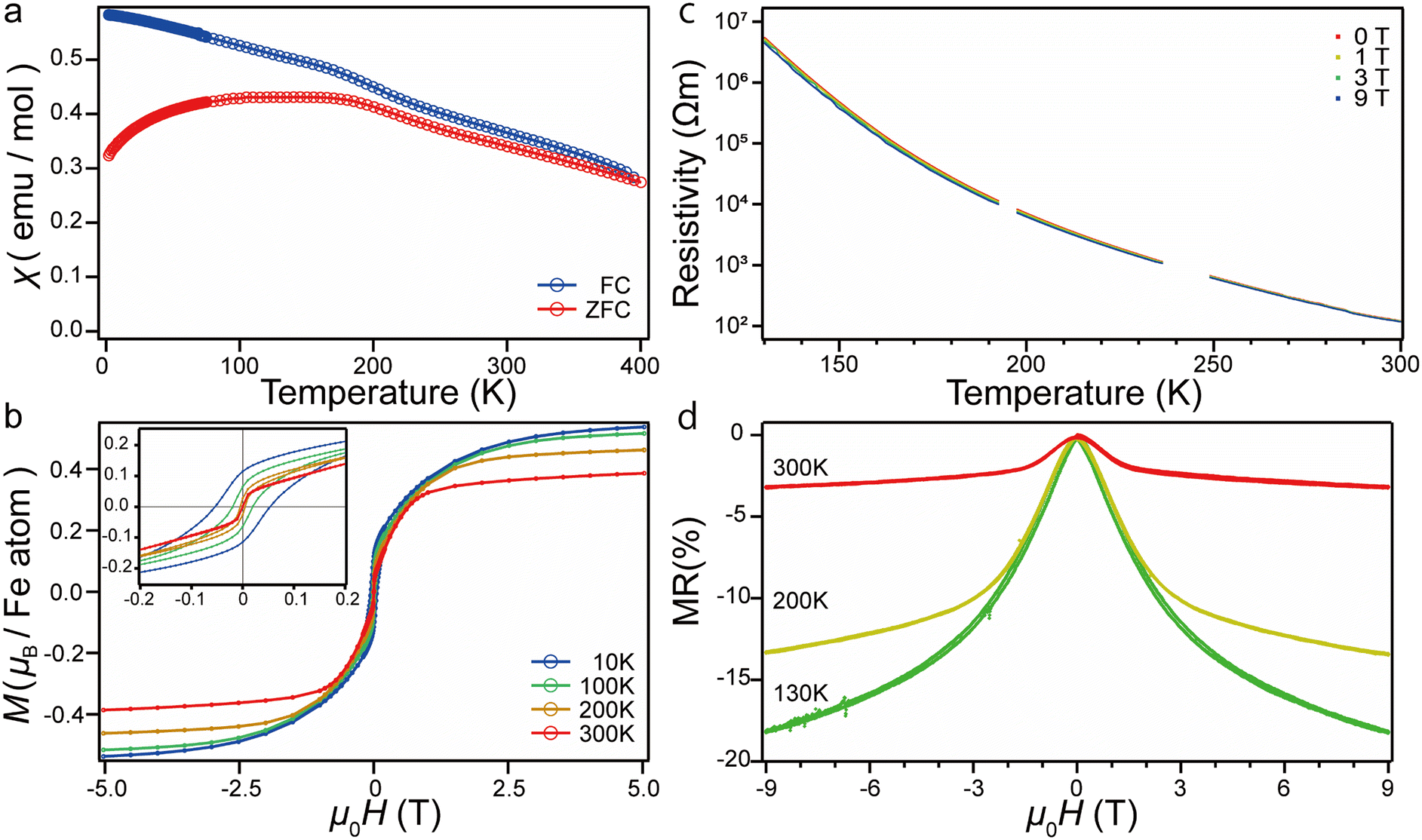 | ||
| Fig. 5 FC-ZFC curves at 0.1 T (a) and M–H curves at 10, 100, 200, and 300 K (b) for SrV0.3Fe0.7O2.8. The inset in Fig. 5b is an expansion near the zero-feild region. The M–H curve at 300 K is taken from the previous study.9 (c) Temperature dependence of resistivity. (d) Magnetoresistance at 300, 200, and 130 K. | ||
The magnetic structure analysis shows that the low temperature phase also has antiferromagnetic component in the ab plane as the high temperature phase (Fig. S7†). Thus, the magnetic phase below Ts is also weak ferromagnetism based on the G-type antiferromagnetic order. Considering the determined magnetic structure and polyhedral tilting, we deduce that the change of magnetic behaviours originates from the change in magnetic anisotropy.
Temperature dependence of resistivity at 0 T, 1 T, 3 T, and 9 T are shown in Fig. 5c. Semiconductor-like behaviour and the decrease of resistance in the magnetic field are observed in the entire measurement range. In contrast to the magnetization, no anomaly at Ts is observed in the resistivity. We converted the resistance into the MR at each temperature (Fig. 5d). At 300 K, MR is proportional to the square of magnetization, indicating the tunnelling mechanism, as reported previously.9 However, this relationship is broken below 300 K: the shape of MR becomes broader than that of magnetization (Fig. S8†). The MR increases as temperature decreases and reaches ca. −18% at 130 K and 9 T. This value is comparable to the magnetoresistance reported for other iron-base oxides (Zn0.2Ni0.8Fe2O4: ca. −20% at 150 K and 9 T (ref. 20)). Thus, SrV0.3Fe0.7O2.8 can potentially exceed these values by chemical substitution to tune its physical properties.
Conclusions
In summary, we observed the structural phase transition from the rhombohedral to monoclinic phase caused by the polyhedral tilting in SrV0.3Fe0.7O2.8 at 200 K. This phase transition affects the magnetic properties: the hysteresis becomes open and coercivity increases from almost zero as the temperature decreases. In addition, magnetoresistance increases as the temperature decreases, reaching ca. −18% at 130 K at 9 T. This material has the potential to show further large magnetoresistance by chemical substation.Data availability
The data supporting this article have been included as part of the ESI.†Author contributions
T. Nagase and T. Y. designed the research. T. Nagase performed synthesis and characterization. T. Nagase, T. Nishikubo, Y. S., and K. S. performed physical measurements and K. M. performed and analyzed Mössbauer spectroscopy. M. H. performed neutron diffraction measurements. M. A. aided in the analysis of data. All the authors discussed the results. T. Nagase and T. Y. wrote the manuscript, with comments from all the authors.Conflicts of interest
There are no conflicts to declare.Acknowledgements
This work was supported by JSPS KAKENHI Grant Number JP22H01767. This work was also supported by the Design and Engineering by the Joint Inverse Innovation for Materials Architecture (DEJI2MA), MEXT. This work was supported by JST SPRING, Grant Number JPMJSP2106. Teppei Nagase was supported by the Japan Society for the Promotion of Science for Young Scientists (JP24KJ1052). The synchrotron radiation experiments were performed at the BL02B2 of SPring-8 with the approval of the Japan Synchrotron Radiation Research Institute (JASRI) (Proposal No. 2020A1068, 2024A1801). The neutron diffraction experiment was performed at the SuperHRPD diffractometer of J-PARC, MLF, with the approval of the Institute of Materials Structure Science (Proposal No. 2019S05). The Mössbauer spectroscopic study was supported by the Advanced Research Infrastructure for Materials and Nanotechnology (ARIM) Program of MEXT, Japan.Notes and references
- J. P. Hodges, S. Short, J. D. Jorgensen, X. Xiong, B. Dabrowski, S. M. Mini and C. W. Kimball, J. Solid State Chem., 2000, 151, 190–209 CAS.
- L. Suescun, O. Chmaissem, J. Mais, B. Dabrowski and J. D. Jorgensen, J. Solid State Chem., 2007, 180, 1698–1707 CAS.
- O. H. Hansteen, H. Fjellvåg and B. C. Hauback, J. Mater. Chem., 1998, 8, 2081–2088 CAS.
- C. Greaves, A. J. Jacobson, B. C. Tofield and B. E. F. Fender, Acta Crystallogr., 1975, 31, 641–646 Search PubMed.
- J. Berggren, O. Foss, I. Roti, H. Okinaka, K. Kosuge and S. Kachi, Acta Chem. Scand., 1971, 25, 3616–3624 CAS.
- M. Azuma, Z. Hiroi, M. Takano, Y. Bando and Y. Takeda, Nature, 1992, 356, 775–776 CrossRef CAS.
- Y. Tsujimoto, C. Tassel, N. Hayashi, T. Watanabe, H. Kageyama, K. Yoshimura, M. Takano, M. Ceretti, C. Ritter and W. Paulus, Nature, 2007, 450, 1062–1065 CAS.
- T. Yamamoto, A. Chikamatsu, S. Kitagawa, N. Izumo, S. Yamashita, H. Takatsu, M. Ochi, T. Maruyama, M. Namba, W. Sun, T. Nakashima, F. Takeiri, K. Fujii, M. Yashima, Y. Sugisawa, M. Sano, Y. Hirose, D. Sekiba, C. M. Brown, T. Honda, K. Ikeda, T. Otomo, K. Kuroki, K. Ishida, T. Mori, K. Kimoto, T. Hasegawa and H. Kageyama, Nat. Commun., 2020, 11, 5923 CrossRef CAS PubMed.
- T. Nagase, T. Nishikubo, M. Fukuda, Y. Sakai, K. Shigematsu, Y. Ikeda, Y. Nambu, Q. Zhang, M. Matsuda, K. Mibu, M. Azuma and T. Yamamoto, Inorg. Chem., 2022, 61, 8987–8991 CrossRef CAS PubMed.
- A. M. Arévalo-López, F. Sher, J. Farnham, A. J. Watson and J. P. Attfield, Chem. Mater., 2013, 25, 2346–2351 CrossRef.
- A. M. Arévalo-Lõpez, J. A. Rodgers, M. S. Senn, F. Sher, J. Farnham, W. Gibbs and J. P. Attfield, Angew. Chem., Int. Ed., 2012, 51, 10791–10794 CrossRef PubMed.
- K. Momma and F. Izumi, J. Appl. Crystallogr., 2011, 44, 1272–1276 CrossRef CAS.
- V. Petrícek, M. Dušek and L. Palatinus, Z. Kristallogr., 2014, 229, 345–352 Search PubMed.
- B. J. Campbell, H. T. Stokes, D. E. Tanner and D. M. Hatch, J. Appl. Crystallogr., 2006, 39, 607–614 CrossRef CAS.
- Y. Han, X. Ye, H. Zhu, Y. Li and X. Kuang, J. Solid State Chem., 2017, 247, 20–23 CrossRef CAS.
- T. Yamamoto, Y. Otsubo, T. Nagase, T. Kosuge and M. Azuma, Inorg. Chem., 2024, 63, 4482–4486 CrossRef CAS.
- L. D. Sanjeewa, V. O. Garlea, M. A. McGuire, M. Frontzek, C. D. McMillen, K. Fulle and J. W. Kolis, Inorg. Chem., 2017, 56, 14842–14849 CrossRef CAS PubMed.
- A. Reuß, V. Ksenofontov, J. Tapp, D. Wulferding, P. Lemmens, M. Panthöfer and A. Möller, Inorg. Chem., 2018, 57, 6300–6308 CrossRef PubMed.
- L. D. Sanjeewa, V. O. Garlea, M. A. McGuire, C. D. McMillen and J. W. Kolis, Inorg. Chem., 2019, 58, 2813–2821 CrossRef CAS PubMed.
- A. K. M. A. Hossain, M. Seki, T. Kawai and H. Tabata, J. Appl. Phys., 2004, 96, 1273–1275 CrossRef.
Footnote |
| † Electronic supplementary information (ESI) available. See DOI: https://doi.org/10.1039/d5ce00028a |
| This journal is © The Royal Society of Chemistry 2025 |