
Thiadiazole-based 3D covalent organic framework for efficient anhydrous proton conduction†
Yaoyao
Pan
a,
Zhen
Shan
a,
Ziya
Liu
 a,
Jian
Su
*a and
Gen
Zhang
*ab
a,
Jian
Su
*a and
Gen
Zhang
*ab
aKey Laboratory for Soft Chemistry and Functional Materials of Ministry of Education, School of Chemistry and Chemical Engineering, Nanjing University of Science and Technology, Nanjing, Jiangsu 210094, China. E-mail: sujian@njust.edu.cn; zhanggen@njust.edu.cn
bKey Laboratory of Preclinical Study for New Drugs of Gansu Province, Lanzhou University, Lanzhou 730000, China
First published on 20th November 2024
Abstract
The design and synthesis of three-dimensional (3D) covalent organic frameworks (COFs) with exceptional stability and high proton conductivity are critical for advancing high-temperature fuel cells but remain significantly challenging. In this study, thiadiazole groups were successfully incorporated into a novel 3D COF featuring a five-fold interpenetrated diamond network through a bottom-up self-assembly strategy. The proton conduction of the thiadiazole-based 3D COF (NUST-28) under anhydrous conditions reached up to 8.40 × 10⁻² S cm−1 at 120 °C after phosphoric acid doping. Furthermore, the NUST-28 conductor demonstrated good stability at constant temperature and in cyclic experiments. This work paves the way for the design and construction of 3D COFs as platforms for fast ion transportation using reticular chemistry.
Introduction
Proton-exchange membrane fuel cells (PEMFCs) directly convert the chemical energy of hydrogen-containing fuels into electrical energy.1 Enhancing the efficiency of proton conduction is crucial for this conversion and has been a long-standing requirement in this field.2,3 However, research on proton conduction encounters challenges such as the use of environmentally harmful materials and low conductivity efficiency. To address these issues, researchers have explored new solid materials with high crystallinity,4–6 such as metal–organic frameworks (MOFs),7–9 and hydrogen-bonded organic frameworks (HOFs),10–12 which show promise as viable alternatives. The crystalline structure of these materials not only enhances the understanding of the mechanisms underlying proton transfer but also holds potential for improved proton conductivity. Nevertheless, they still face significant challenges related to stability.13–16Since the concept of covalent organic framework (COFs) was first introduced in 2005 by O. M. Yaghi,17 COFs have attracted significant attention as a rapidly developing field of chemical research due to their unique properties as crystalline organic porous polymer materials.18–25 Compared to other porous organic materials, COFs offer distinct advantages, including excellent stability, environmental friendliness, and a higher capacity for proton conductors.26–32 In 2014, R. Banerjee and colleagues first investigated the proton conductive properties of azo-based COFs.33 Recently, interest in proton-conductive COFs has grown, highlighting their significant structural and performance advantages in proton conductivity research. Most proton-conductive COFs are two-dimensional, with few examples of 3D COFs34–37 used for proton conduction. 3D COFs offer similar advantages to 2D COFs, including pre-design and modification potential, pure covalent bond construction, absence of heavy metal ions, and environmental friendliness.38 Additionally, 3D COFs enhance proton conduction due to their diverse pore directions and anisotropic structures, which facilitate efficient proton transport by eliminating the layered stacking found in 2D COFs. Their dense interspersed structure also improves the accommodation of functional groups for loaded proton conductors. Their dense interspersed structure also improves the accommodation of functional groups for loaded proton conductors.39
In this study, we synthesized a novel 3D COF featuring thiadiazole groups, labeled as NUST-28, and investigated its proton conductivity after doping with phosphoric acid (NUST-28-P). This was achieved by leveraging the hydrogen bonding between the thiadiazole and C![[double bond, length as m-dash]](https://www.rsc.org/images/entities/char_e001.gif) N groups within the framework and phosphoric acid. We capitalized on the structural advantages of the synthesized 3D COF to achieve efficient anhydrous proton conduction through optimal phosphoric acid doping. Notably, the proton conductivity of NUST-28-60% peaked at 1.46 × 10⁻² S cm−1 at 120 °C under anhydrous conditions, significantly surpassing those of most porous materials. The presence of multi-pore channels and the abundance of thiadiazole groups facilitate hydrogen bonding with phosphoric acid, promoting efficient proton transport and stable performance (Fig. 1).
N groups within the framework and phosphoric acid. We capitalized on the structural advantages of the synthesized 3D COF to achieve efficient anhydrous proton conduction through optimal phosphoric acid doping. Notably, the proton conductivity of NUST-28-60% peaked at 1.46 × 10⁻² S cm−1 at 120 °C under anhydrous conditions, significantly surpassing those of most porous materials. The presence of multi-pore channels and the abundance of thiadiazole groups facilitate hydrogen bonding with phosphoric acid, promoting efficient proton transport and stable performance (Fig. 1).
 | ||
| Fig. 1 (a) Illustration for the synthesis of NUST-28. (b) The hydrogen bonding between NUST-28 and H3PO4 (single unit cell schematic, not the actual interlaced structure). | ||
Experimental
Synthesis of 3D COF NUST-28
The precursors, 4,4′-(benzo[c][1,2,5]thiadiazole-4,7-diyl)dibenzaldehyde (BT-BA)40 and 4,4′,4″,4‴-methanetetrayltetraaniline (TAM), were dissolved in a ternary solvent mixture consisting of 1,2-dichlorobenzene, 1-butanol, and 3 M acetic acid (a volume ratio of 6![[thin space (1/6-em)]](https://www.rsc.org/images/entities/char_2009.gif) :9:2; a total volume of 1.7 mL). After sonication for 15 minutes, the reaction mixture was purged with nitrogen for an additional 15 minutes and then heated at 120 °C for six days. Following cooling to room temperature, the precipitate was washed with THF and acetone and subsequently dried under vacuum at 120 °C for 10 hours. Ultimately, NUST-28 was obtained as yellow powders.
:9:2; a total volume of 1.7 mL). After sonication for 15 minutes, the reaction mixture was purged with nitrogen for an additional 15 minutes and then heated at 120 °C for six days. Following cooling to room temperature, the precipitate was washed with THF and acetone and subsequently dried under vacuum at 120 °C for 10 hours. Ultimately, NUST-28 was obtained as yellow powders.
Characterization
The crystallinity of the product was analyzed using powder X-ray diffraction (PXRD) with a resolution of 0.02° and copper radiation over the range of 2° to 30°. The morphology of the synthesized NUST-28 and NUST-28-P was characterized using a Hitachi TM3000 scanning electron microscope (SEM, Tokyo, Japan). Solid-state nuclear magnetic resonance (NMR) spectroscopy was conducted on a Bruker ADVANCE IIIWB 400 MHz spectrometer. Fourier transform infrared (FT-IR) spectra in the range of 4000–400 cm−1 were acquired utilizing a NICOLET IS10 Fourier transform infrared spectrometer (Thermo Fisher, USA). Thermogravimetric analysis (TGA) was performed using a TGA/STGA851E thermogravimetric analyzer (METTLER TOLEDO) at a heating rate of 5 K min−1 in a nitrogen atmosphere.Electrochemical impedance spectroscopy (EIS) measurements were carried out using a SOLAR-TROM instrument.
Results and discussion
Materials synthesis and characterization
The precursor BT-BA was synthesized according to the procedure detailed in the ESI† and characterized via1H-NMR (Fig. S1†) and 13C-NMR (Fig. S2†). The precursor TAM was acquired commercially. The successful reaction between BT-BA and TAM under solvothermal conditions was confirmed through solid-state 13C-NMR (Fig. S3†) and FT-IR spectra (Fig. S4†). The FT-IR spectra indicated the elimination of amine and aldehyde groups from the raw materials and the formation of imine bonds in the resultant yellow solid powder. Specifically, the FT-IR spectrum of NUST-28 displayed the disappearance of the CO absorption peak at 1570 cm−1 and the emergence of an absorption peak at 1672 cm−1, thereby confirming the formation of the imine bond (Fig. S4†).
The unit cell parameters and crystal structure of NUST-28 were confirmed by integrating PXRD pattern measurements with structural simulations performed using Pawley refinements. Following geometric energy minimization using Materials Studio software based on the five-fold interpenetrated diamond network for NUST-28 (Fig. 2), the unit cell parameters were determined (a = 64.1404 Å, b = 67.2409 Å, c = 58.8001 Å, α = γ = 90°, β = 90.243°). The simulated PXRD patterns closely matched the experimental results (Fig. 3a).
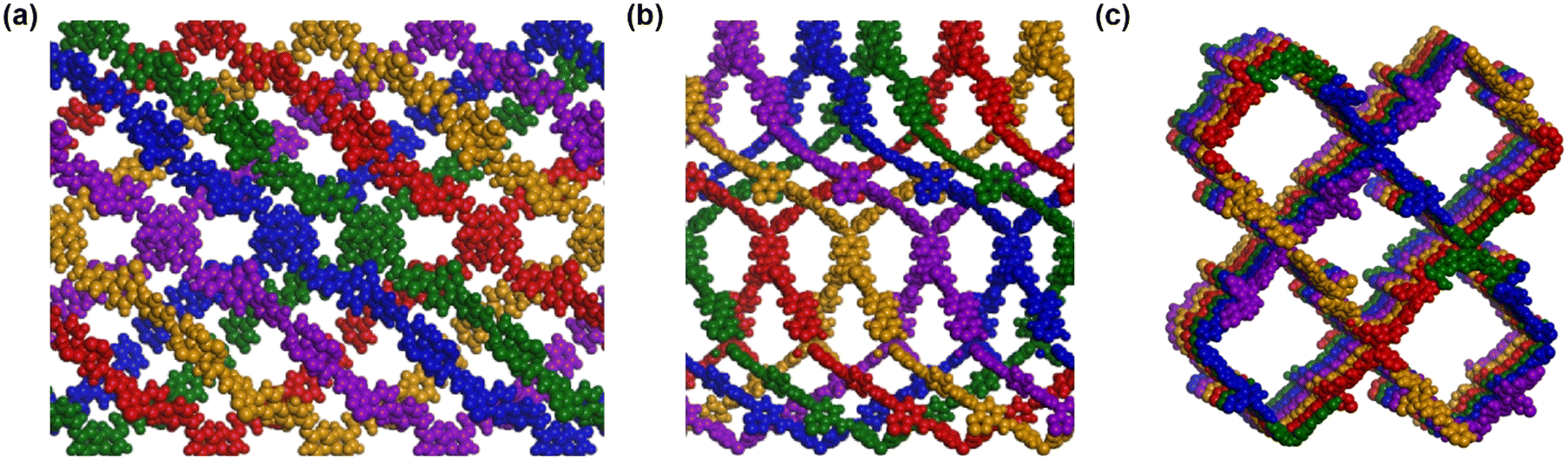 | ||
| Fig. 2 Schematic diagram of the 5-fold interpenetrated 3D COF structure from different dimensions (a) front view, (b) side view and (c) top view of 5-fold interpenetrated schematic diagram of NUST-28. | ||
 | ||
| Fig. 3 (a) PXRD patterns of NUST-28 with the experimental profile in red, simulated profile in black, the difference between the experimental and refined PXRD patterns in blue and the Bragg position in green. (b) TGA plot of pure NUST-28. (c) SEM image of NUST-28 powder. | ||
The 2θ peaks observed at 4.32°, 6.02°, 8.65°, and 13.08° for NUST-28 correspond to the (113), (124), (424), and (091) Bragg peaks of the space group P21 (no. 4), respectively. Compared to the PXRD pattern of the raw material, these peaks confirm the successful synthesis of NUST-28 (see Fig. S5†). The refinement results indicate that the unit cell parameters closely match the predicted values, with excellent agreement factors (wRp = 0.52% and Rp = 0.23% for NUST-28). Based on these findings, it can be proposed that NUST-28 possesses the anticipated architecture of a five-fold interpenetrated diamond network (Fig. 2). The structural morphology of NUST-28 was examined using scanning electron microscopy, as shown in Fig. 3c. The images reveal that NUST-28 exhibits a spindle-shaped aggregation structure with sizes ranging from 200 to 300 nm. Moreover, several spindle-shaped particles aggregate to form a hydrangea-like structure.
The stability of covalent organic frameworks (COFs) is crucial for their applications in proton conduction. The PXRD patterns of phosphoric acid-treated NUST-28 show its excellent chemical stability (Fig. S6†). Additionally, the unchanged morphology of the phosphoric acid-treated NUST-28 further indicates its structural stability (Fig. S8†). As illustrated in Fig. 3b, thermogravimetric analysis conducted in a nitrogen atmosphere demonstrates that NUST-28 is thermally stable, exhibiting no significant mass loss up to 550 °C. This stability allows for the exploration of proton conduction when doped with phosphoric acid under anhydrous conditions.
Proton conduction properties
The powder of NUST-28 was pressed into flakes under a pressure of 1 MPa, resulting in flakes with a diameter of 5 mm and a thickness of approximately 0.5 mm. These pellets were then sandwiched between two stainless steel plates. To test proton conductivity in an anhydrous environment, the COF flakes were encapsulated in a coin cell battery. This encapsulation process was performed within an anhydrous, oxygen-free glove box. Other phosphoric acid-treated NUST-28 flakes were prepared using a similar method but varied in thickness: 0.418 mm for NUST-28-10%, 0.414 mm for NUST-28-20%, 0.518 mm for NUST-28-30%, 0.671 mm for NUST-28-40%, 0.782 mm for NUST-28-50%, 0.465 mm for NUST-28-60%, 1.115 mm for NUST-28-70%, and 0.653 mm for NUST-28-80%.Electrochemical impedance spectroscopy (EIS) was employed to measure the proton conductivities of the COFs under anhydrous conditions. For all measurements, the frequency range was set from 1 Hz to 1 MHz, and the temperature was controlled using an oven. The proton conductivity (σ, S cm−1) was calculated using the following equation:
where Ea (eV) is the transport activation energy, kB is the Boltzmann constant, T (K) is the temperature, and A is a pre-exponential factor.

The impedance of the phosphoric acid-doped flakes was measured in a water-free and oxygen-free environment, and the proton conductivity was calculated according to the formula above. Fig. 4a presents the Nyquist plot of NUST-28-60% at temperatures ranging from 30 °C to 120 °C. Notably, impedance could not be measured for flakes made of pure COF, regardless of temperature, indicating that pure COFs lack proton-conducting properties and function primarily as a medium for loading phosphoric acid. The proton conductivity values for NUST-28-10% were 5.58 × 10⁻⁶ and 1.01 × 10⁻⁴ S cm−1 at 30 °C and 120 °C, respectively. NUST-28-20% exhibited improved proton conductivity, reaching 1.2 × 10⁻³ S cm−1 at 120 °C (Fig. S14†). As the proportion of phosphoric acid in the flakes increased, the proton conductivity of the COF–phosphoric acid flakes also improved (Fig. 4b and S11–S13†). The highest recorded proton conductivity was 1.46 × 10⁻² S cm−1 for NUST-28-60% and 8.40 × 10⁻² S cm−1 for NUST-28-70% at 120 °C. The phosphoric acid-doped COFs demonstrated excellent proton conduction properties compared to other doped porous organometal-free materials reported thus far. The activation energies, derived from the conductivity curves measured at varying temperatures, were found to be 0.47, 0.59, 0.65, 0.51, 0.42, 0.47, and 0.28 eV for NUST-28-10%, NUST-28-20%, NUST-28-30%, NUST-28-40%, NUST-28-50%, NUST-28-60%, and NUST-28-70%, respectively, indicating different conduction mechanisms.
 | ||
| Fig. 4 (a) Nyquist plots of NUST-28-60% from 30 to 120 °C. (b) Proton conductivities of NUST-28-30%, NUST-28-40%, NUST-28-50%, and NUST-28-60% at different temperatures. (c) Long-term proton conduction of NUST-28-60%. | ||
In addition to exhibiting high proton conductivity, COFs doped with phosphoric acid also show excellent stability. After measuring the thermostatic proton conduction in a NUST-28-60% sheet encapsulated in a coin cell, it was cooled to room temperature, and then reheated to 30 °C for further measurements. The results indicated minimal changes in proton conductivity between the first and second heating cycles (Fig. S16†), suggesting that the proton conduction properties of NUST-28-60% are relatively stable under varying temperature conditions. Additionally, we tested the proton conductivity of NUST-28-60% flakes at a constant temperature in a dry atmosphere, finding that they remained stable for at least 48 hours (Fig. 4c). Although NUST-28-70% exhibited higher proton conductivity (Fig. S17†), it was less stable, maintaining its conductivity for only 16 hours. In contrast, NUST-28-80% showed neither significantly higher proton conductivity nor stability (Fig. S18†).
The proton conductivity of NUST-28-60% in an anhydrous environment surpasses those of most published materials (Fig. 5). Other materials exhibit lower proton conductivity both at lower temperatures and at the same temperature as NUST-28-60% at 120 °C. Even at temperatures above 120 °C, some materials fail to achieve the proton conductivity of NUST-28-60% at that same temperature. While certain materials demonstrate higher proton conductivity at elevated temperatures, this is primarily due to the increased availability of active free protons. Overall, NUST-28-60% has a significant advantage over other materials in terms of both proton conductivity and stability across a range of temperatures.
 | ||
| Fig. 5 Contrast scatterplot of the proton conductivity of NUST-28-60% and other materials in a dry environment. | ||
There are two key mechanisms for proton conduction: the Grotthuss mechanism41 and the vehicle mechanism,42,43 which can be distinguished primarily by their activation energy (vehicle mechanism: Ea > 0.4 eV, Grotthuss mechanism: Ea < 0.4 eV). The activation energies vary among different COF samples. When the phosphoric acid doping level is below 70% in a dry atmosphere, the activation energy exceeds 0.40 eV, indicating that the COFs operate under the vehicle mechanism. In this mechanism, phosphoric acid molecules continuously form hydrogen bonds with nitrogen atoms in the thiadiazole group and the COF backbone, aiding proton migration. However, for NUST-28-70%, the conduction mechanism switches to Grotthuss, where protons rapidly hop along the established hydrogen-bonding network while the carrier molecules remain immobilized within the COF framework.
Notably, FT-IR (Fig. 6a) and X-ray photoelectron spectroscopy (Fig. 6b) results indicate the formation of hydrogen bonds between CN bonds and phosphoric acid molecules. For instance, the nitrogen binding energies of NUST-28 and NUST-28-40% were measured to be 396.8 eV and 399.0 eV, respectively (Fig. 6b).
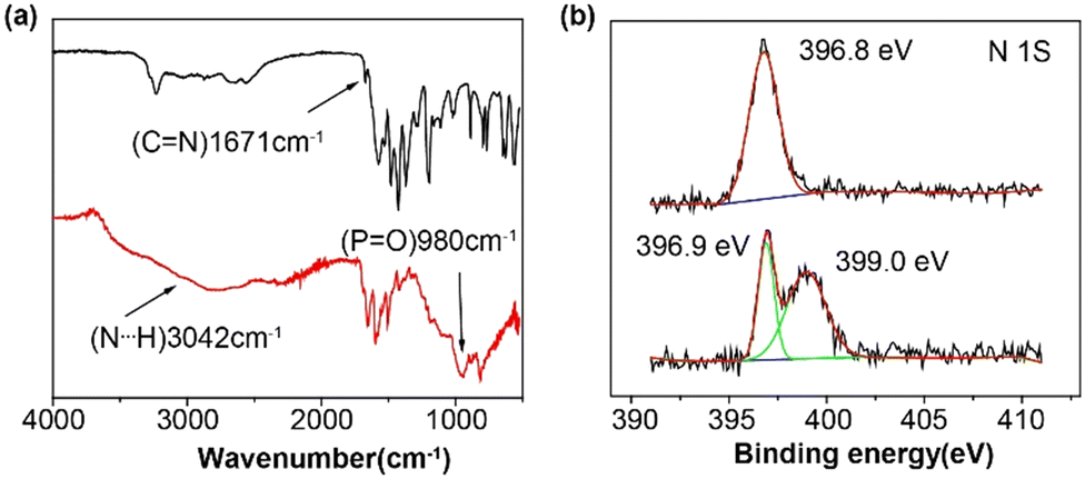 | ||
| Fig. 6 (a) FT-IR spectra of pure NUST-28 and NUST-28-60%. (b) X-ray photoelectron spectra (XPS) of NUST-28 and NUST-28-40%. | ||
Conclusions
In conclusion, we have successfully synthesized a 3D COF containing thiadiazole groups and utilized its structural advantages to achieve efficient anhydrous proton conduction through phosphoric acid doping. The proton conductivity of NUST-28-60% reached a maximum value of 1.46 × 10⁻² at 120 °C under anhydrous conditions, which significantly surpasses those of most porous materials. The presence of multi-pore channels and a high abundance of thiadiazole groups in the 3D COF facilitate hydrogen bonding with phosphoric acid. This optimal proportion of phosphoric acid enhances efficient proton transport and ensures stable performance. These findings offer valuable insights into developing 3D COFs with superior anhydrous proton conduction, as well as their potential applications in proton exchange membrane fuel cells (PEMFCs).Data availability
The data supporting this article have been included as part of the ESI.†Author contributions
The manuscript was written through contributions of all authors. All authors have given approval to the final version of the manuscript.Conflicts of interest
There are no conflicts to declare.Acknowledgements
This work was supported by the National Natural Science Foundation of China (Grant No. 22171136), the Natural Science Foundation of Jiangsu Province (BK20220079 and BK20220928), “the Fundamental Research Funds for the Central Universities” (30921011102 and 30922010301) and the startup funding from Nanjing University of Science and Technology (AE89990 and AE89991/376).Notes and references
- X. Wu, Y. L. Hong, B. Xu, Y. Nishiyama, W. Jiang, J. Zhu, G. Zhang, S. Kitagawa and S. Horike, J. Am. Chem. Soc., 2020, 142, 14357–14364 CrossRef CAS PubMed.
- G. Zhang, Y. L. Hong, Y. Nishiyama, S. Bai, S. Kitagawa and S. Horike, J. Am. Chem. Soc., 2019, 141, 1227–1234 CrossRef CAS PubMed.
- Z. Shan, M. Wu, Y. Du, B. Xu, B. He, X. Wu and G. Zhang, Chem. Mater., 2021, 33, 5058–5066 CrossRef CAS.
- J. Wu, F. Xu, S. Li, P. Ma, X. Zhang, Q. Liu, R. Fu and D. Wu, Adv. Mater., 2019, 31, e1802922 CrossRef PubMed.
- T. Zhu, B. Shi, H. Wu, X. You, X. Wang, C. Fan, Q. Peng and Z. Jiang, Ind. Eng. Chem. Res., 2021, 60, 6337–6343 CrossRef CAS.
- T. Ma, E. A. Kapustin, S. X. Yin, L. Liang, Z. Zhou and J. Niu, Science, 2018, 361, 48–52 CrossRef CAS PubMed.
- Y. Peng, M. Zhao, B. Chen, Z. Zhang, Y. Huang, F. Dai, Z. Lai, X. Cui, C. Tan and H. Zhang, Adv. Mater., 2018, 30, 1705454 CrossRef PubMed.
- D. W. Lim and H. Kitagawa, Chem. Soc. Rev., 2021, 50, 6349–6368 RSC.
- Z.-C. Guo, Z.-Q. Shi, X.-Y. Wang, Z.-F. Li and G. Li, Coord. Chem. Rev., 2020, 422, 213465 CrossRef CAS.
- S. Tao, L. Zhai, A. D. Dinga Wonanke, M. A. Addicoat, Q. Jiang and D. Jiang, Nat. Commun., 2020, 11, 1981 CrossRef CAS PubMed.
- Y. Qin, T. L. Gao, W. P. Xie, Z. Li and G. Li, ACS Appl. Mater. Interfaces, 2019, 11, 31018–31027 CrossRef CAS PubMed.
- B.-B. Hao, X.-X. Wang, C.-X. Zhang and Q. Wang, Cryst. Growth Des., 2021, 21, 3908–3915 CrossRef CAS.
- Z. Li, Y. Zhi, P. Shao, H. Xia, G. Li, X. Feng, X. Chen, Z. Shi and X. Liu, Appl. Catal., B, 2019, 245, 334–342 CrossRef CAS.
- Y. Yao, Y. Hu, H. Hu, L. Chen, M. Yu, M. Gao and S. Wang, J. Colloid Interface Sci., 2019, 554, 376–387 CrossRef CAS PubMed.
- G. Zhao, Y. Zhang, Z. Gao, H. Li, S. Liu, S. Cai, X. Yang, H. Guo and X. Sun, ACS Energy Lett., 2020, 5, 1022–1031 CrossRef CAS.
- S. Li, Y. Liu, L. Li, C. Liu, J. Li, S. Ashraf, P. Li and B. Wang, ACS Appl. Mater. Interfaces, 2020, 12, 22910–22916 CrossRef CAS PubMed.
- A. P. Cote, A. I. Benin, N. W. Ockwig, M. O'Keeffe, A. J. Matzger and O. M. Yaghi, Science, 2005, 310, 1166–1170 CrossRef CAS PubMed.
- X. Liu, J. Li, B. Gui, G. Lin, Q. Fu, S. Yin, X. Liu, J. Sun and C. Wang, J. Am. Chem. Soc., 2021, 143, 2123–2129 CrossRef CAS PubMed.
- X. He, Y. Yang, H. Wu, G. He, Z. Xu, Y. Kong, L. Cao, B. Shi, Z. Zhang, C. Tongsh, K. Jiao, K. Zhu and Z. Jiang, Adv. Mater., 2020, 32, e2001284 CrossRef PubMed.
- F. J. Uribe-Romo, C. J. Doonan, H. Furukawa, K. Oisaki and O. M. Yaghi, J. Am. Chem. Soc., 2011, 133, 11478–11481 CrossRef CAS PubMed.
- G. He, R. Zhang and Z. Jiang, Acc. Mater. Res., 2021, 2, 630–643 CrossRef CAS.
- X. Guo, T. Mao, Z. Wang, P. Cheng, Y. Chen, S. Ma and Z. Zhang, ACS Cent. Sci., 2020, 6, 787–794 CrossRef CAS PubMed.
- M. Zhang, J. Chen, S. Zhang, X. Zhou, L. He, M. V. Sheridan, M. Yuan, M. Zhang, L. Chen, X. Dai, F. Ma, J. Wang, J. Hu, G. Wu, X. Kong, R. Zhou, T. E. Albrecht-Schmitt, Z. Chai and S. Wang, J. Am. Chem. Soc., 2020, 142, 9169–9174 CrossRef CAS PubMed.
- Y. Li, W. Chen, G. Xing, D. Jiang and L. Chen, Chem. Soc. Rev., 2020, 49, 2852–2868 RSC.
- J. Y. Zeng, X. S. Wang and X. Z. Zhang, Chemistry, 2020, 26, 16568–16581 CrossRef CAS PubMed.
- M. C. Scicluna and L. Vella-Zarb, ACS Appl. Nano Mater., 2020, 3, 3097–3115 CrossRef CAS.
- Y. Liu, H. Wu, S. Wu, S. Song, Z. Guo, Y. Ren, R. Zhao, L. Yang, Y. Wu and Z. Jiang, J. Membr. Sci., 2021, 618, 118693 CrossRef CAS.
- J. Liu, N. Wang and L. Ma, Chem. – Asian J., 2020, 15, 338–351 CrossRef CAS PubMed.
- J. Wang, J. Li, M. Gao and X. Zhang, TrAC, Trends Anal. Chem., 2018, 108, 98–109 CrossRef CAS.
- L. Ma, S. Wang, X. Feng and B. Wang, Chin. Chem. Lett., 2016, 27, 1383–1394 CrossRef CAS.
- X. Liu, D. Huang, C. Lai, G. Zeng, L. Qin, H. Wang, H. Yi, B. Li, S. Liu, M. Zhang, R. Deng, Y. Fu, L. Li, W. Xue and S. Chen, Chem. Soc. Rev., 2019, 48, 5266–5302 RSC.
- C.-R. Zhang, W.-R. Cui, W. Jiang, F.-F. Li, Y.-D. Wu, R.-P. Liang and J.-D. Qiu, Environ. Sci.:Nano, 2020, 7, 842–850 RSC.
- S. Chandra, T. Kundu, S. Kandambeth, R. Babarao, Y. Marathe, S. M. Kunjir and R. Banerjee, J. Am. Chem. Soc., 2014, 136, 6570–6573 CrossRef CAS PubMed.
- X. Jiang, K. Zhang, Y. Huang, B. Xu, X. Xu, J. Zhang, Z. Liu, Y. Wang, Y. Pan, S. Bian, Q. Chen, X. Wu and G. Zhang, ACS Appl. Mater. Interfaces, 2021, 13, 15536–15541 CrossRef CAS PubMed.
- Y. X. Ma, Z. J. Li, L. Wei, S. Y. Ding, Y. B. Zhang and W. Wang, J. Am. Chem. Soc., 2017, 139, 4995–4998 CrossRef CAS PubMed.
- B. Gui, G. Lin, H. Ding, C. Gao, A. Mal and C. Wang, Acc. Chem. Res., 2020, 53, 2225–2234 CrossRef CAS PubMed.
- Z. Shan, M. Wu, D. Zhu, X. Wu, K. Zhang, R. Verduzco and G. Zhang, J. Am. Chem. Soc., 2022, 144, 5728–5733 CrossRef CAS PubMed.
- Y. Zhi, Z. Li, X. Feng, H. Xia, Y. Zhang, Z. Shi, Y. Mu and X. Liu, J. Mater. Chem. A, 2017, 5, 22933–22938 RSC.
- P. Guan, J. Qiu, Y. Zhao, H. Wang, Z. Li, Y. Shi and J. Wang, Chem. Commun., 2019, 55, 12459–12462 RSC.
- L. Pei, J. Su, H. Yang, Y. Wu, Y. Du and Y. Zhu, Microporous Mesoporous Mater., 2022, 333, 111742–111750 CrossRef CAS.
- N. Agmon, Chem. Phys. Lett., 1995, 244, 456–462 CrossRef CAS.
- J. B. Asbury, T. Steinel and M. D. Fayer, J. Phys. Chem. B, 2004, 108, 6544–6554 CrossRef CAS.
- H. Wu, L. Li, M. Tsuboi, Y. Cheng, W. Wang, E. Mamontov, S. Uchida, Z. Wang and P. Yin, J. Phys. Chem. Lett., 2018, 9, 5772–5777 CrossRef CAS PubMed.
Footnote |
| † Electronic supplementary information (ESI) available. See DOI: https://doi.org/10.1039/d4ce00995a |
| This journal is © The Royal Society of Chemistry 2025 |