
Tissue analysis by vibrational spectroscopy in Hirschsprung disease: feasibility and potential as a new intraoperative tool†
C.
Combescot
 a,
O.
Piot
*a,
V.
Untereiner
b,
A.
Durlach
cd and
F.
Laconi
e
a,
O.
Piot
*a,
V.
Untereiner
b,
A.
Durlach
cd and
F.
Laconi
e
aUniversité de Reims Champagne-Ardenne, BioSpecT UR 7506, Reims, France. E-mail: olivier.piot@univ-reims.fr
bUniversité de Reims Champagne-Ardenne, URCATech, PICT, Reims, France
cCentre Hospitalier Universitaire de Reims, Hôpital Maison Blanche, Département de biopathologie, Reims, France
dUniversité de Reims Champagne-Ardenne, Inserm UMR-S 1250, P3Cell, Reims, France
eCentre Hospitalier Universitaire de Reims, Département de chirurgie pédiatrique, Reims, France
First published on 10th February 2025
Abstract
Hirschsprung disease is a congenital anomaly characterised by an absence of innervation in the colon. The current diagnosis, which involves identifying the non-functional part of the colon through histological examination, is unsatisfactory. The objective of our study was to assess the potential of infrared spectroscopy as a label-free method to distinguish between functional and non-functional parts of the colon. Tissue samples from FFPE sections of Hirschsprung patients, taken from both functional and non-functional regions, were analysed by mid-infrared imaging. Colour-coded spectral images, reconstructed using multivariate data processing, were compared to the gold standard (hematoxylin–eosin–safran staining) to associate a specific spectral signature with each histological structure. Statistical analyses were also carried out to highlight infrared markers associated with Hirschsprung disease. The search for ganglion cells and cholinergic threads, the usual markers of the disease, was unsuccessful. However, our approach was efficient in differentiating between functional and non-functional parts of the colon by focussing on the muscularis. As such, vibrational spectroscopy can highlight biochemical differences that are not visible using standard histology. This proof-of-concept study suggests that vibrational spectroscopy is a candidate method for diagnosing Hirschsprung disease, paving the way for intraoperative application by assisting surgeons and histologists in delineating the pathological region.
Introduction
Hirschsprung disease (HD) is a congenital condition arising from disruption in the migration of neuroblasts from the neural crest during colon formation. It affects 1 in 5000 births, predominantly boys (ratio 4![[thin space (1/6-em)]](https://www.rsc.org/images/entities/char_2009.gif) :1), and is associated with more than 12 genetic mutations.1,2 In most cases, the diagnosis is clinically suspected within the first days of life, and a subsequent histological examination of rectal biopsies is the gold standard for confirming the diagnosis. Once the diagnosis is made, the treatment consists of surgical pull-through of the aganglionic tract. At that point, determining histologically the boundaries between the ganglionic/functional and aganglionic/non-functional colon segments involves confirming the presence of ganglion cells, which can be particularly challenging. Indeed, the identification of ganglionic cells necessitates an experienced pathologist with specific expertise. In addition, the tissue section examined may contain no ganglionic cells, even though the tissue is functional. In this case, it is necessary to redo the analysis to search for another part of the biopsy containing ganglionic cells. This extemporaneous examination, performed intraoperatively, takes approximately thirty minutes, during which the baby remains under general anaesthesia. Alternative techniques, such as enzymatic tests for detecting cholinergic networks, are too time-consuming for intraoperative use.
:1), and is associated with more than 12 genetic mutations.1,2 In most cases, the diagnosis is clinically suspected within the first days of life, and a subsequent histological examination of rectal biopsies is the gold standard for confirming the diagnosis. Once the diagnosis is made, the treatment consists of surgical pull-through of the aganglionic tract. At that point, determining histologically the boundaries between the ganglionic/functional and aganglionic/non-functional colon segments involves confirming the presence of ganglion cells, which can be particularly challenging. Indeed, the identification of ganglionic cells necessitates an experienced pathologist with specific expertise. In addition, the tissue section examined may contain no ganglionic cells, even though the tissue is functional. In this case, it is necessary to redo the analysis to search for another part of the biopsy containing ganglionic cells. This extemporaneous examination, performed intraoperatively, takes approximately thirty minutes, during which the baby remains under general anaesthesia. Alternative techniques, such as enzymatic tests for detecting cholinergic networks, are too time-consuming for intraoperative use.
Aware of these challenges and limitations, we believe that introducing a new diagnostic tool could be valuable in the clinical management of this disease. Vibrational spectroscopy, an optical technique corresponding to two modalities, namely, Raman scattering and infrared (IR) absorption, appears to be a promising candidate tool, as it gives access to the overall biochemical composition of a sample in a single, label-free measurement. To our knowledge, the scientific literature includes only two studies related to the use of vibrational spectroscopy in HD.3,4 These studies demonstrated the ability of Raman spectroscopy to obtain the spectral signature of ganglionic cells in a mouse model3 and to detect the human enteric nervous system.4 In our study, we focused our investigation on the mid-infrared imaging of tissue sections. This modality allows for the combination of chemical specificity with spatial information at the cellular scale without the need for extrinsic chemical treatments. The analysis relies on the non-destructive interaction between the infrared radiation and the matter, permitting the reuse of the sample for further additional analysis.5 Combined with multivariate data processing, the approach turned out to be successful in the characterisation of cancerous5–9 tissues by providing information on tumour heterogeneity, sometimes non accessible by conventional histology.
Vibrational spectroscopy is a new and promising candidate in the medical field, allowing diagnosis in a non-invasive or non-destructive way.10–13 This technique can lead to the development of new approaches, such as the daily non-invasive monitoring of glycaemia.12,13 Combined with a microscopic imaging device, this technique can provide complementary diagnostic markers to conventional histopathology, as demonstrated by studies on various diseases, such as inflammatory skin lesions,11 lung fibrosis,14 or cancerous malignancy.15
The objective of this proof-of-concept study was to assess the capacity of mid-infrared spectral imaging to identify HD tissues by distinguishing the functional part from the non-functional part of the colon. First, we evaluated the ability of IR imaging to detect the standard indicators of routine histological examination, namely, the ganglion cells at the level of Auerbach and Meissner plexus in functional tissue, and the cholinergic nets within the chorion of the mucosa of non-functional tissue. Second, our investigation focused on the muscularis of the colon; this structure showed no morphological alteration visible by standard histology.
Materials and methods
The methodology workflow is summarized in Fig. 1. | ||
| Fig. 1 Experimental approach for the acquisition and processing of a spectral image from a paraffin-embedded histological section acquired by FTIR. EMSC: extended multiplicative signal correction; KM: K-means. | ||
Sample collection and preparation
The study utilized tissue samples from colon macro-biopsies (n = 6) obtained from pull-through surgery, available in Formalin Fixed Paraffin-Embedded (FFPE) form, and from patients previously diagnosed with Hirschsprung disease (Table 1). Each patient contributed two samples, both from the functional and non-functional segments of the colon, resulting in a total of 12 samples for this proof-of-concept study.| Patient number | Age | Sex | Extension of the aganglionosis |
|---|---|---|---|
| 1 | 6 months | F | Sigmoid |
| 2 | 6 months | M | Sigmoid |
| 3 | 4 months | F | Sigmoid |
| 4 | 6 months | F | Sigmoid |
| 5 | 3 months | M | Sigmoid |
| 6 | 1 month | M | Sigmoid |
For each sample, two successive 8 μm sections made with a microtome were used. The first section was deposited on a calcium fluoride (CaF2) slide to carry out the acquisition by infrared imaging. The second adjacent section was placed on a glass slide for hematoxylin–eosin–safran (HES) staining, which marked the tissue structures. All stained tissue sections were digitalized using a VS120 digital scanner (Olympus, Tokyo, Japan).
All experiments were performed in accordance with French laws and approved by the local ethics committee (Comité d’éthique du Centre Hospitalier Universaire de Reims, France) under the approval number AC 2019-3408, after obtaining informed consent from the parents of every patient.
Analysis of spectral images acquired by infrared spectroscopy
The spectral images were recorded with a spectral resolution of 4 cm−1, 4 accumulations per pixel, over the spectral range of 4000–800 cm−1. A pixel size of 6.25 × 6.25 μm2 was chosen, offering spatial precision at the cellular scale.
In addition, polarisation-resolved infrared acquisitions were led with the same parameters by incorporating a polariser on the incident beam; images were collected with three orientations at 0°, 45° and 90° according to the sample orientation.
The spectral data were then corrected using Extended Multiplicative Signal Correction (EMSC), which simultaneously performed baseline correction, vector normalisation and digital neutralization of the paraffin interferences (S.D.1†). It also permits the removal of outlier spectra corresponding to noisy or weak intensity spectra. Normalisation uses a reference spectrum named the target. In the present study, two distinctive targets were used depending on the analysis made. Target #1 was the average of the spectra of a complete tissue (mucosa, sub-mucosa, muscularis and nerve plexus) and was used for the spectral analysis at the complete tissue level. For analysis focused on the muscularis structure, a second target, target #2, was used; it corresponded to the mean of spectra extracted from a reduced zone containing only the muscularis and parts of the nerve plexus. To determine these targets, we considered the sample from patient #1 for the quality of the section (S.D.1†). For this pre-processing step, the spectral range was reduced to the 1800–900 cm−1 fingerprint region, containing the most vibrational information in biological samples.
An inferential non-parametric univariate analysis was carried out for each feature of the spectral data using a Kruskal–Wallis test, followed by the calculation of the p-value using a χ2 test, with 95% and 99% thresholds.
Results
Definition of histological structures of interest
In HD, the functional or non-functional area of the colon is characterised respectively by the presence or absence of ganglion cells and is validated by histopathological examination of HES-stained tissue samples observed under a microscope (Fig. 2). The tissue is deemed functional if the ganglion cells are present, or non-functional otherwise. Thus, confirming the presence of ganglion cells is the gold standard for classifying a sample as healthy, ruling out a diagnosis of disease. | ||
| Fig. 2 Simplified anatomy of the colon and corresponding HES-stained slide. (A) Colon histology and structure in an HES-stained section. (B) and (C) Stained sections showing, respectively, hypertrophied nerve trunks within the mucosa and ganglion cells within the Auerbach plexus. The HES-stained slides were scanned at a magnification of ×40. | ||
Another way to validate the HD diagnosis is the presence of a cholinergic net in the mucosa and submucosa of the non-functional area.16 This second marker has two flaws, making it difficult to use as a reliable and reproducible gold standard.
The first is that the presence of a cholinergic net can only be verified in fresh or frozen tissues by treating the tissue with acetylcholinesterase (AchE). Our study relied on paraffin-embedded tissues, and it was not possible to determine the presence and quantity of cholinergic nets by AchE staining. The second is the density of these nets, which varies among patients, with some exhibiting dense nets, whereas others have nearly non-existent ones.17 However, some of the non-functional tissues presented large and visible hypertrophied nerve trunks with HES staining, which is known to be correlated with the presence of cholinergic nets. We tried to search for a spectral signature for the hypertrophied nerve trunks or for the cholinergic nets on the FFPE tissues, but we were not able to isolate any specific spectral signature (data not shown). Therefore, the question of the cholinergic net is still pending, and more tests should be carried out on fresh or frozen tissue.
Characterisation of the HD tissues using IR microimaging
This part of the analysis focused on the myenteric plexus of confirmed functional tissues because the ganglion cells were easier to find compared to the submucosal plexus. First, the tissue sections, including the mucosa, sub-mucosa and muscularis, were analysed by infrared to find a specific spectral signature of the ganglion cells.The KM analysis on spectral images did not reveal a specific cluster associated with the ganglionic cells even if the number of clusters was increased. However, the submucosa heterogeneity was highlighted by multiplying clusters in this structure but not in the plexus. Then, the analysis focused on a reduced area containing only the myenteric plexus and the surrounding muscularis (S.D 2†). Despite increasing up to fifteen clusters, we were not able to find a signal specific to the ganglion cells (S.D 2†). Although a specific spectral signature for ganglion cells was identified in both IR and Raman micro-spectroscopy in previous studies,18,19 we were not able to locate them using IR microimaging with a pixel size of 6.25 × 6.25 μm2. This could be attributed to the size of the cells averaging 20 μm. Thus, if a ganglion cell was between two pixels, its spectral signal would be mixed with that of the plexus. This hypothesis is reinforced by the fact that other teams were able to obtain an IR spectral signature of the ganglion using a pixel size of 2.6 × 2.6 μm2.18 Moreover, although two adjacent thin sections were used for infrared imaging and HES staining, it could be possible that one section contained cells whereas the other did not.
Comparison of infrared imaging of healthy and pathological tissue
Considering that the ganglion cells were undetectable with the spatial resolution of our equipment, our analysis was then focused on other tissue structures of functional and non-functional parts of the colon samples from patients with Hirschsprung disease.Although standard histology did not show any morphological differences, infrared imaging permitted highlighting differences at the level of the muscularis between the non-functional and functional segments in HD tissues. Fig. 3 shows the KM results obtained for a representative case. Tests were conducted to find the minimum number of clusters required to separate the HD and healthy muscularis segments. A difference in the cluster number was observed among the patients: for patients #1, #2, and #3, 7 clusters were found to be necessary, while patient #4 required 9 clusters, and patients #5 and #6 required 10 clusters (data not shown). These disparities can be explained by high inter-patient variability in the sub-mucosa and mucosa. These spectral variabilities in the mucosa and sub-mucosa are not attributed to the disease, as the clusters are present both in the functional and non-functional segments of the same patient.
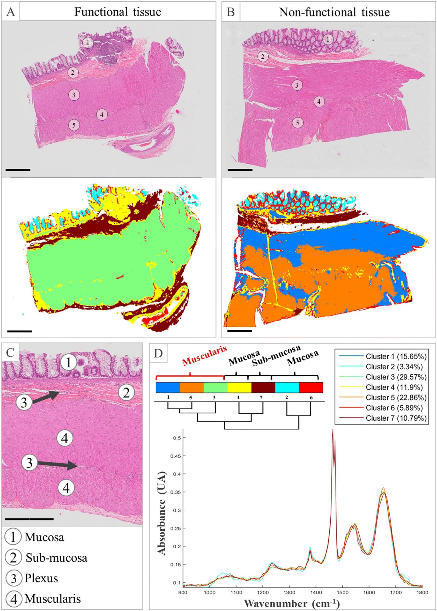 | ||
| Fig. 3 KM clustering of ganglionic and aganglionic parts of Hirschsprung patient samples. (A) and (B) HES-stained tissue and common KM with 7 clusters of functional and non-functional (ganglionic) tissue (×40), respectively. (C) Anatomy of the colon (×40). (D) Dendrogram and centroid of common KM. The scale bar represents 500 μm. The HES-stained slides were scanned at a magnification of ×40. | ||
At the methodological level, this disparity constitutes a limitation in the automation of the approach because the operator must intervene to adjust the parameters for each case. To avoid interferences originating from the mucosa and sub-mucosa, the solution is to use spectral images containing only the muscularis and the plexus of the tissue. It was then possible to discriminate for all cases between the non-functional and functional parts of the colon with a cluster number fixed to 4 (S.D 3†).
Based on these positive results, the last step of our study was to identify wavenumbers that can be considered spectral markers to discriminate between the functional and non-functional segments of the tissue. Thus, the centroids corresponding to the muscularis extracted from 4 distinctive spectral images for each patient and each condition were statistically compared through a Kruskal–Wallis test; thus, 24 spectra representatives of each of the 2 conditions were considered. For a given wavenumber, a p-value over the significance level (here 0.05 or 0.01) reflects a difference in intensity between the two conditions. The wavenumbers associated with significant differences are shown in Fig. 4. The mean spectra and standard deviations are also displayed to better visualize the position of the spectral discriminant differences. From the scientific literature, we were able to assign the majority of these most discriminative wavenumbers mainly to protein, RNA and phospholipid contents. The wavenumbers around 1720 cm−1 are associated with C![[double bond, length as m-dash]](https://www.rsc.org/images/entities/char_e001.gif) O stretching found in lipid esters.20,21 The 1540 cm−1 vibration is associated with the Amide II band, resulting from N—H bending and C—N stretching modes in proteins.20–23 The vibrations at 1428–1444 cm−1 can correspond to both CH2 bending in lipids and CH3 bending in proteins.20,21 The spectral range 1386–1398 cm−1 is associated with CH3 bending vibrations present in fatty acids and phospholipids,20 and the wavenumbers at 1320–1330 cm−1 correspond to C—O stretching vibrations of carbohydates.24 The wavenumbers between 1232 and 1250 cm−1 can contribute to the Amide III band, arising from C—N stretching and N—H bending modes in proteins and PO2− stretching vibration of RNA,20,21 while the wavenumbers between 1268 and 1280 cm−1 are assigned to PO2− stretching vibration of phospholipids.21,22 The wavenumber around 1110 cm−1 can be associated with stretching vibration of the C—O ester-groups.21 The 1028 and 1154 cm−1 wavenumbers can correspond to C—O—H and C—O—C bending vibrations of glycogen.20 The set of vibrations located between 900 and 974 cm−1 are likely to be associated with the C—N—C stretching in RNA and phospholipids, or with C—O—C deformations in carbohydtates.22,24 Since the tissues were paraffined, the vibrations associated with the lipid contents correspond to components of the cell membrane. The discriminative wavenumbers implicated suggest several biochemical changes in the tissues, most likely an alteration in the protein content and structure. But it could also be due to a difference in the molecular components of muscle cells, the absence of ganglion cells inducing a modification in the expression of muscle cells, and particularly those of the outer tunic. This hypothesis is supported by the numerous genetic mutations as described in the literature in Hirschsprung patients.2 Moreover, the heterogeneity between the patients must be considered. The most discriminative wavenumbers varied between patients, and this statistical analysis permitted to highlight the most common variations across all patients.
O stretching found in lipid esters.20,21 The 1540 cm−1 vibration is associated with the Amide II band, resulting from N—H bending and C—N stretching modes in proteins.20–23 The vibrations at 1428–1444 cm−1 can correspond to both CH2 bending in lipids and CH3 bending in proteins.20,21 The spectral range 1386–1398 cm−1 is associated with CH3 bending vibrations present in fatty acids and phospholipids,20 and the wavenumbers at 1320–1330 cm−1 correspond to C—O stretching vibrations of carbohydates.24 The wavenumbers between 1232 and 1250 cm−1 can contribute to the Amide III band, arising from C—N stretching and N—H bending modes in proteins and PO2− stretching vibration of RNA,20,21 while the wavenumbers between 1268 and 1280 cm−1 are assigned to PO2− stretching vibration of phospholipids.21,22 The wavenumber around 1110 cm−1 can be associated with stretching vibration of the C—O ester-groups.21 The 1028 and 1154 cm−1 wavenumbers can correspond to C—O—H and C—O—C bending vibrations of glycogen.20 The set of vibrations located between 900 and 974 cm−1 are likely to be associated with the C—N—C stretching in RNA and phospholipids, or with C—O—C deformations in carbohydtates.22,24 Since the tissues were paraffined, the vibrations associated with the lipid contents correspond to components of the cell membrane. The discriminative wavenumbers implicated suggest several biochemical changes in the tissues, most likely an alteration in the protein content and structure. But it could also be due to a difference in the molecular components of muscle cells, the absence of ganglion cells inducing a modification in the expression of muscle cells, and particularly those of the outer tunic. This hypothesis is supported by the numerous genetic mutations as described in the literature in Hirschsprung patients.2 Moreover, the heterogeneity between the patients must be considered. The most discriminative wavenumbers varied between patients, and this statistical analysis permitted to highlight the most common variations across all patients.
 | ||
| Fig. 4 Mean spectra and standard deviations of the functional and non-functional parts of the colon (shown in blue and red, respectively), with the statistically different wavenumbers identified from a Kruskal–Wallis test and a χ2 test. (A) The areas where the peak absorbances are significantly different are colored in yellow (95%) and green (99%). (B) The H values calculated from the rank of the Kruskal–Wallis test and the p-value are reported in the table. | ||
The discriminant vibrations identified previously can be considered spectroscopic markers of HD and permit biochemical interpretation of the spectral changes observed. In the case of the muscularis structures, it is also questionable whether there is a relationship between the spectral features and the muscle structure and/or organization. Indeed, the muscle fibres are differently oriented between the inner tunic and the outer tunic of the colon. Possibly, analysing the fibre orientation could be a supplementary indicator in HD diagnosis. Following this hypothesis, our idea was to check whether polarisation-resolved infrared imaging would enhance spectral differences within the muscularis when compared to non-polarised measurements. For this test, a healthy segment was analysed by changing the polarisation of the incident infrared beam. Previous studies conducted in our laboratory revealed the interest in polarising the infrared incident beam to probe the orientation of biological macromolecules, such as type I collagen.25 The polarisation measurements were carried out by orientating the electric field of the incident beam at a precise angle in relation to the structures of the tissue (aligned horizontally as shown in Fig. 1A and B). The polarised measurements at 0°, 45° and 90° were compared with each other and with non-polarised measurements through a common KM clustering. From this experiment, it appeared that the polarisation of the infrared incident beam had a negligible effect on the spectral signatures of the tissue structures (Fig. 5). Indeed, common KM clustering was carried out on both functional and pathological parts of an HD specimen for three conditions: without polarisation, and with polarisation angles of 0° and 90° according to the tissue orientation on the motorized plate of the instrument. Five clusters were chosen for this KM clustering. The overall spatial distribution of the clusters and the respective spectral distances between their centroids, as can be assessed by the dendrograms, were similar independently to the polarisation condition. In particular, the Amide I-to-Amide II intensity ratio, previously shown to be affected by the polarisation,25 appeared here stable. This experiment demonstrated that the tissue components of the colon are insensitive to the orientation of the electric field of the infrared radiation and that using polarisation does not enhance the spectral differences between the inner and outer tunics.
 | ||
| Fig. 5 Analyses of the orientation of the fibres on the spectral image of the functional (①) and non-functional (②) parts of the colon of the same patient with KM clustering. Each spectral image is a cropped part of the whole tissue, composed of the submucosa, the circular muscle and the longitudinal muscle. Five clusters were used for each pair with a 0° and 90° polariser and without a polariser. The scale bar represents 500 μm. | ||
Our results demonstrated that IR imaging can distinguish between functional and non-functional parts of the colon of Hirschsprung patients; however, the main study's limitation is the small number of patients (n = 6). Because Hirschsprung disease is a rare disease, we obtained samples from only 6 patients after obtaining their consent, in collaboration with local medical doctors. An expanded patient cohort relying on multicentric sampling would enhance result reliability and permit the creation of a predictive model using supervised classification methods for the implementation of an automated diagnostic tool. Additionally, there are case reports where samples containing ganglion cells are found to be non-functional after surgery, necessitating further intervention. This region, known as the transitional zone, poses significant risks when performing an anastomosis (Transition Zone Anastomosis – TZA), potentially leading to serious complications and adversely affecting patient outcomes.26,27 Exploring the spectral characteristics of the TZA seems necessary because there are no existing methods to differentiate this part from the functional area of the colon. This could offer valuable insights because our approach is not based on the presence or absence of the cells. Addressing this clinical question may aid in defining surgical excision limits. However, this exploration requires the inclusion of new patients present with this complication because none of the patients included in the present study had this complication.
Conclusions
Infrared microimaging permits the differentiation of the functional and non-functional parts by analysing the muscularis structure. Our proof of concept paves the way for developing an automated tool complementary to the standard histological technique to assist in the diagnosis of Hirschsprung disease.Author contributions
O. Piot, F. Laconi contributed to the conception, design and drafting of the paper. C. Combescot performed the acquisitions and curation of the data, computational experiments, and data analysis. F. Laconi performed the surgeries from where the samples were taken. A. Durlach confirmed the histological composition of the sample and the patient's disease and selected the tissues used for this study. V. Untereiner and O. Piot supervised the acquisition and validated the data analysis of the study. All authors reviewed the final version and approved the submission.Data availability
Data for this article are available at the SSDS of the University of Reims Champagne-Ardenne at https://ssds-cloud.univ-reims.fr/index.php/s/LsGmQZeBy6WQoYD. As per the University's policy, sharing links have a limited lifespan; after expiration, the link will be reactivated upon request. However, personal data collected from human participants, as described in [Table 1], are not available due to confidentiality reasons.Conflicts of interest
There are no conflicts to declare.Acknowledgements
The authors thank the URCATech, PICT-IBiSA platform of the University of Reims Champagne-Ardenne for spectroscopy facilities. This work has been made within the framework of the Graduate School NANO-PHOT (École Universitaire de Recherche, PIA3, contract ANR-18-EURE-0013). The authors also thank the biobank of the CHU of Reims (Département de Biopathologie) for the biopsies. The authors would also like to thank Dr Cyril GOBINET, from BioSpecT UR 7506 (Reims, France), for his help and insight with the statistical analysis.References
- R. O. Heuckeroth, Nat. Rev. Gastroenterol. Hepatol., 2018, 15, 152–167 CrossRef PubMed.
- C. S. Tang, A. Karim, Y. Zhong, P. H. Chung and P. K. Tam, Pediatr. Surg. Int., 2023, 39, 104 Search PubMed.
- M. A. Soares de Oliveira, L. Galganski, S. Stokes, C.-W. Chang, C. D. Pivetti, B. Zhang, K. E. Matsukuma, P. Saadai and J. W. Chan, Sci. Rep., 2021, 11, 1398 CrossRef CAS PubMed.
- K. Ogawa, Y. Oshima, T. Etoh, Y. Kaisyakuji, M. Tojigamori, Y. Ohno, N. Shiraishi and M. Inomata, J. Pediatr. Surg., 2021, 56, 1150–1156 CrossRef PubMed.
- K.-Y. Su and W.-L. Lee, Cancers, 2020, 12, 115 CAS.
- J. Nallala, M.-D. Diebold, C. Gobinet, O. Bouché, G. D. Sockalingum, O. Piot and M. Manfait, Analyst, 2014, 139, 4005–4015 CAS.
- G. M. Magno, G. E. Lotilla, E. R. Ambrosio, F. L. Nagera, M. I. Grino, M. R. Castro, J. A. Millan, J. A. Manzano, R. E. Tiongco and P. M. Albano, Asian Pac. J. Cancer Prev., 2024, 25, 1487–1495 CrossRef PubMed.
- L. A. Courtenay, I. Barbero-García, S. Martínez-Lastras, S. Del Pozo, M. Corral de la Calle, A. Garrido, D. Guerrero-Sevilla, D. Hernandez-Lopez and D. González-Aguilera, PLoS One, 2024, 19, e0300400 CrossRef CAS PubMed.
- M. Féré, C. Gobinet, L. H. Liu, A. Beljebbar, V. Untereiner, D. Gheldof, M. Chollat, J. Klossa, B. Chatelain and O. Piot, Anal. Bioanal. Chem., 2020, 412, 949–962 CrossRef PubMed.
- Z. Fan, J. Zhang, C. Ma, B. Cong and P. Huang, Forensic Sci., Med., Pathol., 2024 DOI:10.1007/s12024-024-00866-9.
- D. Sebiskveradze, B. Bertino, V. Gaydou, A.-S. Dugaret, M. Roquet, D. E. Zugaj, J. J. Voegel, P. Jeannesson, M. Manfait and O. Piot, J. Biophotonics, 2018, 11, e201700380 CrossRef PubMed.
- J. W. Kang, Y. S. Park, H. Chang, W. Lee, S. P. Singh, W. Choi, L. H. Galindo, R. R. Dasari, S. H. Nam, J. Park and P. T. C. So, Sci. Adv., 2020, 6, eaay5206 CrossRef CAS PubMed.
- J. Liu, J. Chu, J. Xu, Z. Zhang and S. Wang, Spectrochim. Acta, Part A, 2025, 329, 125584 CrossRef CAS PubMed.
- V. Suryadevara, S. S. Nazeer, H. Sreedhar, O. Adelaja, A. Kajdacsy-Balla, V. Natarajan and M. J. Walsh, Biomed. Opt. Express, 2020, 11, 3996–4007 CrossRef CAS PubMed.
- M. Diem, A. Mazur, K. Lenau, J. Schubert, B. Bird, M. Miljković, C. Krafft and J. Popp, J. Biophotonics, 2013, 6, 855–886 CrossRef CAS PubMed.
- R. K. Agrawal, N. Kakkar, R. K. Vasishta, V. Kumari, R. Samujh and K. L. N. Rao, Diagn. Pathol., 2015, 10, 208 CrossRef CAS PubMed.
- A. K. Braczynski, S. Gfroerer, R. Beschorner, P. N. Harter, P. Baumgarten, U. Rolle and M. Mittelbronn, BMC Pediatr., 2020, 20, 399 CrossRef CAS PubMed.
- C. Krafft, D. Codrich, G. Pelizzo and V. Sergo, J. Biophotonics, 2008, 1, 154–169 CrossRef CAS PubMed.
- M. A. Soares De Oliveira, L. Galganski, S. Stokes, C.-W. Chang, C. D. Pivetti, B. Zhang, K. E. Matsukuma, P. Saadai and J. W. Chan, Sci. Rep., 2021, 11, 1398 CrossRef CAS PubMed.
- B. Zupančič, N. Umek, C. K. Ugwoke, E. Cvetko, S. Horvat and J. Grdadolnik, Int. J. Mol. Sci., 2022, 23, 12498 CrossRef PubMed.
- B. Zupančič, C. K. Ugwoke, M. E. A. Abdelmonaem, A. Alibegović, E. Cvetko, J. Grdadolnik, A. Šerbec and N. Umek, Front. Endocrinol., 2023, 14, 1308373 CrossRef PubMed.
- O. Bozkurt, M. Severcan and F. Severcan, Analyst, 2010, 135, 3110–3119 RSC.
- S. Magalhães, I. Almeida, F. Martins, F. Camões, A. R. Soares, B. J. Goodfellow, S. Rebelo and A. Nunes, Molecules, 2021, 26, 6410 Search PubMed.
- J. Lopes, M. Correia, I. Martins, A. G. Henriques, I. Delgadillo, O. da Cruz E Silva and A. Nunes, J. Alzheimers Dis., 2016, 53(3), 801–812 Search PubMed.
- C. Eklouh-Molinier, T. Happillon, N. Bouland, C. Fichel, M.-D. Diébold, J.-F. Angiboust, M. Manfait, S. Brassart-Pasco and O. Piot, Analyst, 2015, 140, 6260–6268 RSC.
- F. Chen, X. Wei, X. Chen, L. Xiang and J. Feng, Front. Pediatr., 2021, 9, 600316 CrossRef PubMed.
- K. Taghavi, L. Goddard, S. M. Evans, A. Hobson, S. W. Beasley, S. Sankaran, A. Kukkady, J. Stevenson and M. D. Stringer, Aust. N. Z. J. Surg., 2020, 90, 1037–1040 CrossRef PubMed.
Footnote |
| † Electronic supplementary information (ESI) available. See DOI: https://doi.org/10.1039/d4an01489h |
| This journal is © The Royal Society of Chemistry 2025 |