
 Open Access Article
 This Open Access Article is licensed under a Creative Commons Attribution-Non Commercial 3.0 Unported Licence
Contactless vital sign monitoring systems: a comprehensive survey of remote health sensing for heart rate and respiration in internet of things and sleep applications
Muhammad Salman
Raheel
a,
Faisel
Tubbal
a,
Raad
Raad
 a,
Philip
Ogunbona
b,
James
Coyte
a,
Christopher
Patterson
c,
Dana
Perlman
c,
Saeid
Iranmanesh
*a,
Nidhal
Odeh
ad and
Javad
Foroughi
*aef
a,
Philip
Ogunbona
b,
James
Coyte
a,
Christopher
Patterson
c,
Dana
Perlman
c,
Saeid
Iranmanesh
*a,
Nidhal
Odeh
ad and
Javad
Foroughi
*aef
aSchool of Electrical, Computer and Telecommunications Engineering (SECTE), University of Wollongong (UOW), Australia. E-mail: saeidim@uow.edu.au; foroughi@uow.edu.au
bSchool of Computing and Information Technology (SCIT), University of Wollongong (UOW), Australia
cSchool of Nursing; University of Wollongong (UOW), Australia
dElectronics and Telecommunications, IP Australia, Australia
eSchool of Mechanical and Manufacturing Engineering, University of New South Wales, Library Rd, Kensington, NSW, 2052 Australia
fDepartment of Cardiothoracic, Transplantation and Vascular Surgery, Hannover Medical School, Carl-Neuberg-Str, 130625 Hannover, Germany
First published on 8th May 2024
Abstract
With the coronavirus pandemic, companies and governments around the world have been investing millions of dollars in the development of contactless sensor technologies that minimize the need for physical interactions between patients and healthcare providers. This has led to rapid progress in healthcare research on innovative contactless technologies, particularly for infants and elderly individuals with chronic diseases that require continuous, real-time monitoring and control. The combination of sensing technology and wireless communication has emerged as a promising research area, as patients often find it unpleasant or anxiety-provoking to wear sensor devices, and physical contact can exacerbate the spread of contagious diseases. To address these issues, research has focused on sensor-less or contactless technology to send and analyse wireless signals to remotely monitor and measure vital signs without requiring physical contact or sensor devices. Herein, we have provided a comprehensive survey and study of non-invasive/contactless vital sign monitoring systems, particularly the heart rate and the respiration rate monitoring systems to achieve accurate and reliable measurements. We have found that there is a lack of a comprehensive comparison and analysis over existing contactless vital sign monitoring systems. Therefore, we first present and classify the existing non-invasive monitoring designs based on their approaches and techniques, and then compare them based on the performance and accuracy.
 Faisel Tubbal | Faisel Tubbal (M'15, SM'20) received the B.E. degree from the college of Electronic Technology, Tripoli, Libya, in 2004, the M.S. degrees in engineering management and Telecommunication Engineering both from the University of Wollongong, Wollongong, Australia, in 2012 and 2013, respectively. He has also received a PhD in telecommunication engineering from the University of Wollongong, Australia in 2017. |
 Raad Raad | Raad Raad received a Bachelor of Engineering degree (Hons.1) in Electrical Engineering from the University of Wollongong, Australia in 1997, and a Ph.D. degree entitled Neuro-Fuzzy Logic Admission Control in Cellular Mobile Networks, in 2006. Since 2004, he has been with the School of Electrical, Computer and Telecommunications Engineering, University of Wollongong and he is the current Head of School. His current research interests include wireless communications, CubeSat, the IoT, and Antenna design in addition to advanced power systems. He is part of the ARC ITTC for Future Grids and ARC hub for Connected Health Sensors, both major research initiatives. |
 Saeid Iranmanesh | Saeid Iranmanesh holds the Ph.D. degree in electrical and telecommunication engineering from the University of Wollongong, NSW, Australia, in 2015. He is a senior member of IEEE and a Fellow member of Engineers Australia. He is currently a Senior Research Fellow within the School of Electrical, Computer and Telecommunication Engineering, University of Wollongong. His research interests include wireless networks, vehicular networks, intelligent transportation systems, and smart cities. |
 Javad Foroughi | Prof. Foroughi is a professor of biomedical engineering at the Department of Cardiothoracic, Transplantation, and Vascular Surgery, Hannover Medical School, Hannover, Germany. Additionally, he holds the position of Senior Research Fellow and Chief Investigator within the ARC Research Hub for Connected Sensors for Health at the University of New South Wales. He is also a recipient of an Australian Research Council DECRA Fellowship. Prof. Foroughi specializes in innovative research in smart materials, focusing on artificial muscles and wearable technology for emerging advanced applications, including medical devices and health monitoring. He is internationally recognized as an emerging leader in the field of smart materials and is widely acknowledged as the inventor of Torsional Carbon Nanotube Artificial Muscles (Science). |
1. Introduction
The monitoring of vital signs such as heart rate and respiration rate of patients during sleep is considered a standard procedure in healthcare.1 It is a critical health practice for monitoring wellbeing and the detection of adverse medical events (such as arrhythmias) or the detection of conditions (such as sleep apnoea). Apnoea is an interruption of breathing during sleep, where a person stops breathing for a period of 10 seconds or longer which leads to complete or partial waking of patients.2 It has been reported that approximately 4% of middle-aged adults and 3% of children under 5 years old developed such a condition.3,4 Therefore, continuous monitoring of a patient's sleep states is essential in the determination of patients' health status by healthcare. It also provides useful additional information and hence, prevents some health implications and adverse events at a very early stage.5,6To date, clinical methods of monitoring heart rate and respiration rate during sleep monitoring in hospitals require attaching sensing devices to the patient's body such as a mask or nasal cannula for breath rate monitoring and electrodes for cardiac monitoring.7 Healthcare professionals, such as a nurse, must visit and use these dedicated devices frequently to check the patient's health status and monitor vital signs. These traditional sleep monitoring systems, however, are intrusive, expensive, and inconvenient for patients, often negatively affecting sleep hygiene and quality.8 In addition, they can cause rashes and allergic reactions for patients with sensitive skin when they are used for a long time.9 Another example of a traditional sleep monitoring system is poly-sonography (PSG) which is a conventional technique performed throughout the night and includes a number of wearable sensors, connected via wires to provide data to the computer. These sensors include electroencephalography (EEG) electrodes to track and record the electrical brain activity, electrooculography (EOG) electrodes for measuring eye movement, electromyography (EMG) electrodes for recording muscle activity, electrocardiography (ECG) electrodes for measuring the electrical heart activity, pulse oximeters to monitor blood oxygen saturation and elastic belts for measuring the respiratory effort. However, there are some limitations to utilize these sensors due to the cost and complexity.10 Henceforth, the main challenge is to monitor patients' vital signs without attaching sensors or diagnostic devices to their body and at minimal cost.
To address these aforementioned challenges, non-invasive and long-term vital sign monitoring systems are ideal, as they are simple, cheap, and convenient for patients. These systems use contactless techniques, which enable monitoring of heart rate and respiration rate remotely without attaching any device to a patient's body. However, the designing of such systems requires considering different challenges. The first concern is the type of wireless transmission medium used, which is divided into ultra sonic and radio frequency (RF) signals. The RF signals are further categorized into low frequency and high frequency signals. Furthermore, strict limits are applied on the amount of power that can be directed at soft tissue using RF signals. Indeed, this has been the subject of many medical studies.11 Prolonged exposure to high-powered RF signals increases the probability of cell manipulation. Table 1 shows the allowable power levels as determined by ANSI/IEEE C95.1-2005 standard.12 To prevent harmful effects of transmitted signals on patients, the maximum allowable observed power density of the transmitted signal must not exceed 40 mW m−2 in the frequency range of 0.4–3 GHz, and 200 mW m−2 in the high frequency range of 30–300 GHz as per the ANSI/IEEE C95.12005 standard.13–18 The other challenge is the ability to monitor the vital signs of a patient from a long distance accurately, e.g., 5 m. This is important as it allows monitoring the vital signs of sleeping patients without waking them up. Moreover, the accuracy of contactless vital sign monitoring systems is essential in order to determine patients' health status.
| Design properties | Performance | Operating ranges |
|---|---|---|
| Low transmitted power at high frequencies | Less effect on human | ≤200 mW m−2 at 30–300GHz is the allowable power to human as per the ANSI/IEEE C95.1-2005 standard |
| Long effective distance (range) | Long monitoring distance (long range) | ≥5 m |
| High patient privacy and security | Very secure | High privacy and secure |
| Accuracy | The ability to provide very accurate breathing and heart rates | ≥98% |
1.1 Related review articles on contactless monitoring systems
In ref. 19, Krishnan et al. provided a short comparative study of four different contactless methods to estimate heart rate. The methods include electromagnetic (EM) wave-based radar systems comprising continuous wave (CW) or ultrawide band (UWB) radars, laser based method, image based method and using other monitoring systems such as magnetic induction usability of each different method, and the authors suggest that EM based monitoring systems can provide promising results. However, an appropriate signal processing algorithm must be adapted to retrieve the necessary signal.Similarly, in ref. 20 Vorobyov et al. provided a feasibility study of contactless respiration, heart rate and stress level monitoring systems using short range radar-based technologies operating at different frequencies. The authors reported that automotive radar solutions can be used to estimate vital signs, but they consume high power and are particularly used for longer ranges hence, they are not typically used for healthcare applications. Similarly, the 140 GHz band is used by telecom applications. Therefore, 60 GHz and 122 GHz ISM bands accommodate (frequency modulated continuous wave) FMCW and UWB radars. Furthermore, the authors estimated the results obtained using a CW reflectometer based on commercial-off-the-shelf (COTS) components and laboratory equipment operating at 75–110 GHz. The authors also discussed the challenges to obtain a reliable vital sign monitoring system and suggested that MIMO antennas and machine learning (ML) algorithms must be used to reduce interference and harmonics in estimation breathing and heart rate.
In ref. 21, Kebe et al. presented a detailed review of conventional contact-based methods to estimate cardio-pulmonary rates and how radar-based systems can replace these methods. Furthermore, the paper highlights the challenges faced by radar-based systems in the healthcare industry and their existing solutions. Finally, a proof of concept is proposed to measure vital signs using an existing CW doppler radar operating at 10 GHz. Although the radar shows promising results to estimate vital signs, issues such as random body movement (RBM) and the separation of the heart rate signal from the breathing rate signal still exist. However, the existing solutions consume more power to reduce motion artifacts and hence, require advance signal processing algorithms to reduce motion artifacts and better estimate the heart rate.
In ref. 22, Hall et al. discussed a brief review of latest developments on non-contact vital sign sensors and compared them with an intelligent and novel phased array doppler based radar designed in the lab. The existing methods include under the bed non-contact sensors, using an accelerometer, or using a thin strip of electro-sensitive polymer film material or placing a microwave radar under the bed. On the other hand, camera based methods are used to capture human body movement using RGB, monochrome or infrared cameras. Moreover, doppler based radars are designed which reduce system complexity, power consumption and increase accuracy and robustness. However, the motion artifact is not yet overcome. Furthermore, the authors claimed that there is no existing intelligent doppler system that tracks patient on bed movement for long term vital sign measurement until they designed a first intelligent phased array doppler sensor that performs automatic beam steering to track the subject for long term vital sign monitoring.
In ref. 23, the authors explored the state-of-the-art techniques in the field of contactless vital sign monitoring based on operating bands and classification tools. The research analysis validates that contactless sensing is a reliable technology to save human life and reduces the direct physical interaction between the patient and nurse. Moreover, the existing techniques are modeled around FMCW-radars, channel state information, wireless sensors, and camera-based technologies. The authors discussed how each technology operates and their corresponding applications. Moreover, the emphasis is given more on the FMCW technique due to its high usage and potential in future healthcare monitoring. Furthermore, the authors discussed the existing challenges on each technology (Table 2).
| Reference | Year | Studied based on |
|---|---|---|
| Krishnan et al.19 | 2017 | Short comparative study on four different contactless methods; EM based, laser based, image based or by using magnetic induction or capacitive coupling to estimate HR. Based on the advantages and usability, suggested that EM based systems can provide promising results. However, requires an appropriate signal processing algorithm |
| Vorobyov et al.20 | 2020 | Feasibility study of contactless RR and HR monitoring using short range radar-solutions. For example, automotive radars can be used, but they are over designed, consume more power, 140 GHz band radars are used by telecom applications and not under ISM band. 60 GHz and 122 GHz ISM bands accommodate FMCW and UWB radars. Also est. results using a CW reflectometer operating at 75–110 GHz. Discuss challenges and suggest that MIMO antennas and machine learning (ML) algorithms must be used to reduce interference and harmonics in estimation BR and HR |
| Kebe et al.21 | 2020 | Review of conventional contact-based methods to estimate cardio-pulmonary rates vs. radar-based system. Highlights challenges faced by radar systems such and existing solutions. Proof of concept using a CW doppler radar operating at 10 GHz is discussed with promising results. Issues such as RBM & HR separation from the RR rate signal still exist and existing solutions consume high power to reduce motion artifacts. Hence, advance signal processing algorithms are required for better HR estimation |
| Hall et al.22 | 2017 | Discuss latest developments on non-contact vital sign sensors such as under the bed sensors, which use accelerometer, or a thin strip of electro-sensitive polymer film material, or by placing a microwave radar under bed. Meanwhile camera-based methods capture human body movement using RGB, monochrome or infrared cameras. Similarly, doppler based radars reduce system complexity, power consumption and increase accuracy and robustness. However, the motion artifact is not yet overcome. Furthermore, the authors design a first intelligent phased array doppler sensor that performs automatic beam steering to track the subject for long term VSM |
| Bahache et al.23 | 2020 | Explore the state-of-the-art techniques in the field of contactless vital sign monitoring based on operating bands and classification tools. The existing techniques discussed use FMCW-radars, channel state information, wireless sensors, and camera-based technology. The existing challenges for each technology are also discussed and FMCW shows promising results |
1.2 Contribution of this paper
In this survey, we explore the advancements and challenges of contact-less monitoring systems for measuring vital signs such as heart rate and respiration rate. We deliberately focus on technologies that do not require physical contact, such as microwave radars and video-based monitoring. This focus reflects the growing interest and demand in healthcare settings for solutions that minimize patient discomfort and risk of infection. As a result, conventional contact-based methods like ECG, while highly relevant in clinical diagnostics, fall outside the primary scope of this review. It is important to note that our comparisons are based on findings reported in existing studies. Each study reviewed was conducted under different conditions with distinct methodologies. As such, this manuscript does not present direct empirical testing or simulation of these technologies; instead, it relies on the critical analysis of reported results to understand the capabilities and limitations of each technology as described by previous research. This approach allows us to draw broad conclusions about the state of the art in radar technology for vital sign monitoring. Compared to the aforementioned surveys, we present the most recent developments in wireless sensing techniques used in various healthcare applications. Specifically, we analyse the existing wireless sensing techniques related to contactless technologies, particularly the heart rate and the respiration rate monitoring systems to achieve accurate and reliable measurements. Also, we provide a detailed comparison between these techniques to assist in selecting the appropriate technique for a given situation. Additionally, we discuss the main challenges faced by these systems and highlight enabling technologies.2. Contactless vital sign monitoring systems
Recently, contactless heart rate and respiration rate systems have received considerable attention due to their accuracy and suitability for monitoring the vital signs of a sleeping patient without attaching sensors or devices to his/her body. In this survey, the main techniques and approaches for contactless monitoring of vital signs have been categorised according to the segment of the electromagnetic spectrum used for sensing. This categorisation allows us to identify different groups of methods namely the microwave radar, Wi-Fi, and visible spectrum.2.1 Microwave radar technique
Since 1975, microwave Doppler radars have been used to monitor vital signs such as respiration and breathing rates.24 Initially, these devices were quite bulky and expensive. However, recently the CMOS integrated versions of the radar are presented in ref. 25 for contactless cardiopulmonary monitoring, which consumes a small amount of power. The radar technique uses the Doppler effect to produce velocity data about objects (e.g., patient's body) at a distance. As shown in Fig. 1, a transmitting antenna sends a microwave signal towards the object (human's body) and then this incident signal hits the target object, i.e., patient's chest and reflects back to the receiving antenna. The movement of the body's chest leads to a change in the reflected microwave signal and hence causes the Doppler effect. Further information such as range, angular direction, radial velocity, size and shape can be obtained from the reflected signal when it is filtered, amplified, simplified and processed.26 The transmitted signal at time t in the microwave radar technique is represented by:T(t) = AT![[thin space (1/6-em)]](https://www.rsc.org/images/entities/char_2009.gif) cos(wt + ϕ(t)) cos(wt + ϕ(t)) | (1) |
 | ||
| Fig. 1 Doppler radar block diagram for monitoring vital signs. | ||
The radar approach is based on the Doppler effect whereby the velocity information of a distant object is captured in a waveform reflected from the object. In this survey, the object is the patient's body. The apparatus consists of an arrangement of microwave transmitting and receiving antennas pointing at the patient's chest such that movements of the chest resulting from respiration and heartbeats are registered on the waveform detected at the receiving antenna (Fig. 1). Apart from respiration and heart rates, other interesting parameters that could be extracted from the received waveform include range, angular direction, radial velocity, size and shape of the reflecting object.24
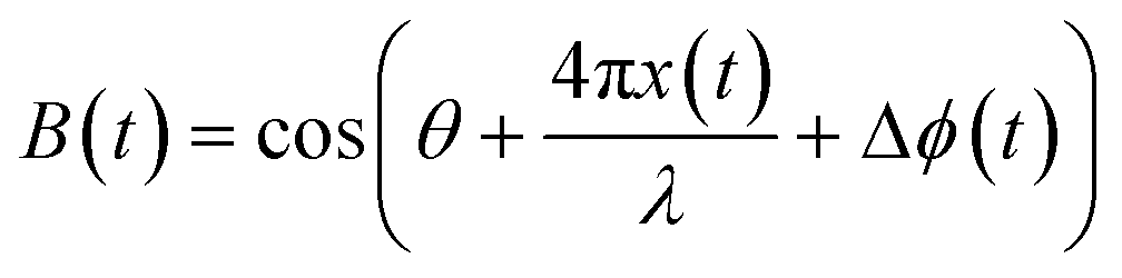
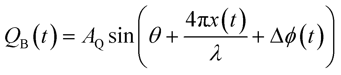
As shown in Fig. 1, dt = 2(d0 + x(t)) which is the total travel distance of the microwave signal. Therefore, the reflected signal which is influenced by the movement of the chest at a distance of d(t) in the form of a time varying displacement x(t) is represented by:
 | (2) |
 | (3) |
 is the changing phase shift and
  is the constant phase shift. Two baseband signals with 90° out of phase are generated (denoted by IB(t) and IQB(t)) and then they are processed and demodulated to obtain the heart and breathing rate measurements.
 | (4) |
 | (5) |
Huang et al.28 proposed a low cost contactless and self-calibrating I/Q Doppler radar sensing system for measuring the heartbeat and respiration rate of a sleeping patient. The main idea is to use the Doppler effect technique for motion detection of the patient's chest and hence measuring the breathing and heart rates. As shown in Fig. 2, the Doppler radar transmits a signal towards the patient's chest, then this incident signal hits the human's chest and reflects to the I/Q Doppler. The output-based band signals I(t) and Q(t) of the Doppler are then digitized using data acquisition (DAQ) and phase change. Moreover, the heart and respiration measurements are achieved by applying a demodulation process to I(t) and Q(t) signals. The proposed system consists of sensing (local oscillator and antennas), pre-processing (amplifier, down converter, and filter), modelling (phase demodulation), and information layers (vital sign analysis). One of the limitations in existing Doppler radar systems is that they require an approximation of the I/Q signals or re-calibration for the DC offset in the I/Q channels when there is a change in the environment, e.g., temperature or light.29 The authors in ref. 28 addressed this problem by using a demodulation method that does a signal model recalculation when there is any change in the temperature or light during the measurement and hence, tuning for each subject is not required. This is called a self-calibration. However, its main limitation is that the test for measurements of humans behind the wall has not been studied and presented in this work. The proposed self-calibration system achieved an accuracy of 87% for the tests that was carried out outdoor and about 92% for the tests carried out in corridors.
 | ||
| Fig. 2 I/Q Doppler radar sensing system for measuring vital signs. The Doppler radar transmits a signal towards the patient's chest, then this incident signal hits the human's chest and reflects to the I/Q Doppler. | ||
Furthermore, the antenna designs and their performance such as gain, bandwidth and beamwidth play a significant role in Doppler radar systems.30 It is important that the antenna design must provide high gain, low profile, and high signal to noise ratio (SNR). In ref. 31, Das et al. proposed a portable Doppler-based non-contact vital sign monitoring system that continuously monitors the human's heart and respiration rates. The main limitation of vital monitoring systems that operate in the X-band frequency with a penetration depth of about 3 m is that the system detects the body movement instead of the heartbeat. To address this limitation, the authors used the continuous wave (CW) Doppler radar system using 2.45 GHz helical antennas. Moreover, the movement of the human's chest causes phase modulation, which can be easily demodulated to monitor the heart and respiration rates separately. The proposed transmitter and receiver helical antennas achieve a BW of 44.6° and a high SNR. In addition, the proposed portable Doppler-based non-contact vital sign system achieves an error rate for respiration rate of ±1 breath per min for about 95% of the time while for the heart rate it is ±1 breath per min for 75% of the time.
Sadek et al.33 designed a new non-contact microwave sensor system operating at 9 GHz for heart and respiration rate estimations. The proposed system comprises two parts; the hardware part is made of a Doppler radar, made of an oscillator with 0 dBm of power emitted, an isolator, a circulator, a Schottky diode mixer and a horn antenna with a 15 dB gain and a digital multi meter (DMM), whereas the software part uses LabView for signal acquisition and two band pass Chebeychev filters are used to separate heart and respiration information. The filter used for the extraction respiration activity has a bandwidth of 0.15 and 0.4 Hz, whereas the filter used for the extraction of heart rate has a bandwidth of 0.6 to 1.5 Hz. The experimental study shows that the system is easy to use and tested to work over 50 cm from a person's chest.
Chen et al.34 presented a contactless vital sign monitoring system based on a 24 GHz continuous wave (CW) Doppler radar with the capability to separate multiple person respirations. The proposed system consists of 3 parts: radar transceivers front, USB DAQ device and LabView for processing. The radar transceivers forward electromagnetic waves on a person's chest that is reflected and received by the transceiver. Furthermore, the NI-USB DAQ device is used to convert the analog baseband signal into digital. Finally, the signal processing is performed in MATLAB and LabView using the blind source separation (BSS) algorithm. This algorithm operates on distinct individual sources from the mixture with very little information about the source signals or their mixing process. The experimental result shows the successful separation of respiration signals using Blind Source separation processing, with the estimation closer to the ground truth values. The proposed system can be used for monitoring the respiration pattern for elderly people to detect any illness.
As discussed earlier, Doppler radars transmit a continuous wave signal and receive the reflected signal with varied frequency proportional to the target's relative velocity. Meanwhile the UWB pulse radars send an electromagnetic pulse and receive the reflected echoes from the target. On the other hand, the FMCW (frequency modulated continuous wave) radar uses the phase of an estimated body point to measure the vital signs. It should be noted that the CW radar primarily includes velocity measurements through the Doppler effect; however, when incorporated into more sophisticated systems such as FMCW radar and multiple input multiple output (MIMO) configurations, it becomes possible to measure both the range and angular direction. The basic operational principle of the IR-UWB radar and FMCW radar is discussed below.
In the IR-UWB radar, a short time domain modulated electromagnetic pulse is transmitted towards the human body, where the reflected echo is received by the receiver and processed to obtain heart and breathing rates. The basic architecture of the UWB radar is discussed in Fig. 3(c). The figure shows that an analog receiver receives the echoes transmitted by an analog transmitter, sampled by the delay replica of the transmitted signal. The delay is represented by the time of flight of the pulse, which corresponds to the time taken by the pulse to travel from the transmitter to the receiver antenna. The pulse generated by a typical IR-UWB radar is represented in Fig. 3(a) for the time domain and (b) in the frequency domain.
 | ||
| Fig. 3 Overview of the UWB pulse radar, (a) time domain, (b) spectrum of the UWB signal and (c) basic architecture of the UWB radar. | ||
However, in the FMCW radar, the frequency of an output signal is varied linearly over time as shown in Fig. 4. This type of signal comprises a unity signal generated at every T time instant called chirp. Different methods are used to generate this chirp such as using a phase locked loop with frequency synthesizers or with the help of a voltage-controlled oscillator using a linear controlled voltage. The working principle of the FMCW radar is like the CW Doppler radar. A single-tone CW is forwarded by the transceiver to a persons' chest and the receiver antenna obtains the reflected wave. The obtained data is then inserted into a matrix comprising slow time and fast time data. The slow time data represents the range and the number of ramps transmitted, whereas the fast time represents the number of samples taken at each ramp and hence carries the vital sign information.
 | ||
| Fig. 4 Frequency modulated continuous wave (FMCW) signal. (a) Variation in frequency over time and (b) chirp signal. | ||
The authors35 provided a feasibility study and design of a new system on chip radar sensor for next generation wearable wireless interface to monitor human healthcare. The system comprises a UWB pulse radar sensor that exploits UWB signals at 3.1 to 10.6 GHz and a low power IEEE 802.15.4 ZigBee radio interface to provide a wireless data link with remote data acquisition and control units. The system is based on the correlation receiver topology followed by an integrator to average the received pulses to obtain an output signal that contains the heart and breathing rates. The complete working principle of the proposed radar system is shown in Fig. 5. A train of short Gaussian monocycle electromagnetic pulses at approximately 200 picoseconds is pointed towards the target. After a certain delay equal to the time of flight of pulse from the transmitter to the receiver, the system (delay and shaper block) internally generates a delayed replica of the transmitted pulse and multiplies it using a multiplier with the echo received. The multiplier output signal is then passed through the integrator, which has the maximum amplitude if the time between two signals is perfectly aligned at the input of the multiplier. The obtained signal at the output of the integrator represents the time-varying position of the heart and carries the heartrate and breathing frequencies. The simulation results agree with the theoretical analysis, which demonstrates the feasibility of using system-on-chip radars for vital sign monitoring.
 | ||
| Fig. 5 Block diagram of the fully integrated UWB radar for heart and breath monitoring. | ||
Similarly, Lena et al. used UWB radar technology to devise a measurement setup for heart and breathing rate detection using two PulsON 220RD UWB radars.36 For the measurement setup, the target is considered sitting or still at a known distance from the radar and breathing at a normal pace. Initially, the background noise subtraction is performed on the data received through the radar to remove any static reflections caused by the presence of other objects in the test area. Once the signal becomes clean, the author uses four known period estimation algorithms to estimate its period, such as Welch, multiple signal classification (MUSIC), average magnitude difference function (AMDF) and a low complexity version of maximum likelihood (ML). The performance evaluation shows that the ML algorithm outperforms the algorithm without signal processing. Furthermore, for heart and breathing rate estimation, two FIR filters at 0.8–1.2 Hz are designed with order 30 and 70 to cut everything outside the average healthy heartbeat range of an adult (80–100 bpm). However, it does not achieve the required results. Hence, a higher order is chosen, and it is identified that the 70th order filter can achieve the results except for WELCH and MUSIC, which introduces system complexity.
In another work,30 Baboli et al. proposed a detection algorithm for physiological monitoring using Pulson P210 transducers as an UWB radar. The algorithm aims to distinguish two periodic sources of motion (heart and breathing rate), with different frequencies, using wavelet and filter banks. Initially, a burst of ultra-wide band pulses is radiated to thorax and the reflected signal is stored in a matrix. The row matrices carry the sampled version of the reflected signal, where the total number of rows is equal to the total radiation time divided by the time division. On the other hand, the number of columns is proportional to the receiver sampling rate. In the next step, the static part of the received signal is removed, and the wavelet packet transformation is applied at each column matrix of the received signal. This decomposes the signal in the time domain with different scales and times, where each scale relates to a frequency band called a transform level. Afterwards, the energy of each frequency band in the last transform is calculated and stored in another matrix. The column with the highest energy level reflects the location of the desired motion. The experimental results show the algorithm robustness in the presence of environmental motion and the heart rate is detected at an accuracy of 95% whereas the respiration rate is detected at 100% accuracy when compared with a figure pressure pulse sensor and respiration effort belts.
There are some other studies in the literature that utilize the ultra-wideband array radar to monitor the respiration rate of a patient. This approach is used with an adaptive beamforming to simultaneously monitor the vital signs of multiple sleeping patients located in different ranges.37 Muragaki et al.38 proposed a new contactless monitoring system based on an array radar with the beamforming technique that can monitor the respiration rate of multiple closely positioned patients in the same range but with different lateral positions. This is important as it allows monitoring more than one patient at the same time with very small interference and small estimated displacement error, e.g., 0.13 mm, as compared to the conventional method, i.e., 2.7 mm. The proposed vital monitoring system consists of one ultra-wideband (UWB) array radar (e.g., one transmitter and four receivers) with an adaptive beamforming technique to steer the beam of the radar. Moreover, they presented a new algorithm to separate multiple targets that are located near each other and in the same range. The main limitation of existing high range resolution UWB radars is that they allow the separation of multiple targets that are located only in different ranges but not for targets positioned in the same range. Another problem is that techniques using time-frequency analysis29,39 are not able to estimate the surface displacement of each individual target's body at each measurement time. Therefore, they do not separate the received signals of multiple closely positioned patients. To overcome the aforementioned problems, it was reported that they used the Capon method40 (high resolution adaptive beamforming technique), proposed a new algorithm and modified the diagonal technique by using a diagonal loading factor of −10 dB for DOA estimation to separate multiple closely positioned targets placed in the same range but at different lateral positions.38 Moreover, the proposed UWB array radar system achieved a small root-mean-square error (estimated displacement error) less than 0.13 mm. Its main limitation, however, is that its mode of monitoring respiration rate is limited for seating or lying down positions.
A study of accurate evaluation of using an off the shelf pulse Doppler radar to estimate the respiration rate of a human body is presented by Li et al.41 For analysis purposes, the respiration data is obtained for different people sitting in front of the radar in different ranges from 1 m to 5 m with recordings at each 1 m and at different angles (18 and 36 degrees) each time. Each recording lasts for 35 seconds with each subject performing; slow, normal and fast breathing. The radar used in the study is a commercial UWB radar model Xethru by Novelda. For the signal processing part, initially, a fast Fourier transform (FFT) is applied on the raw data along with a band stop filter to remove clutter and identify the range bins that carries the subject's information. Afterwards, a window size is applied on the range bins and the spectrogram plots are obtained by applying short time fast Fourier transform (STFT). The STFT helps to detect the small frequency shifts caused by the chest displacement of a human body during the respiration from 0 to 1 Hz. The experimental result shows that the respiration rate is closer to the ground truth values which can be obtained by adjusting the relationship between Doppler bins and window sizes.
Ossberger et al.42 proposed a method to detect the respiration movement of a hidden person using an UWB pulse radar system comprising sub-nanosecond pulse generators, wideband antennas and a low noise amplifier. The authors designed a continuous wavelet transform (CWT) algorithm accompanied by a background subtraction filter to process the retrieved signal. The CWT algorithm detects sub-nanosecond pulses of different widths because of a change in the dilation factor compared to using the mother wavelet. Furthermore, the background subtraction removes unwanted signal components caused by antenna cross talk, rigging and the use of non-ideal pulse generators. The simulation result showed that the proposed system is able to detect respiration signals up to the distance of 5 meters and behind the walls. However, the heart rate is not considered, as it requires a higher amplitude signal of sub-nanosecond pulse for detection.
In another work, Venkatesh et al.43 presented the use of IR-UWB signals to detect the chest cavity motion. They proposed signal-processing algorithms suitable for the estimation of respiration and heartbeat rates, even in the absence of a direct path between the transmitter, the subject and the UWB receiver antennas. Initially, a background clutter based on a continuous time motion filter is used to remove all signal components not relevant to the motion due to respiration. Afterwards, a fast Fourier transform (FFT) is calculated based on the average energy of the motion output filter and a band pass filter is used on the spectrum to eliminate harmonics and then peak values are used to estimate the respiration rate. A similar approach is applied to obtain some preliminary results for the estimation of the heartrate. However, the proposed solution does not consider breathing and heartrate harmonics, which introduce noise factor due to false peaks in the signal.
There are several scenarios wherein patients are monitored using the UWB medical radar as would be found in heartrate, cardiac and motion activity monitoring in post-operative chambers. Immoreev and Tao44 devised an algorithm to restore and analyse the quadrature signals arising from the back and forth motion of patients' thorax and heart. An alarm signal is set off when the respiration rate crosses a given threshold. The drawback of this contribution is the lack of detailed description of the algorithm used to obtain the reported results.
Lazaro et al.45 devised a mathematical model to estimate heart and breathing rates from recorded waveforms using the IR-UWB radar. It was observed that the waveforms contain several breathing signal harmonics that could, in some cases, be stronger than the signal at heartbeat frequency. Additionally, when the harmonic is close to the frequency of heartbeat, identification is difficult. The authors have used Bessel functions and Fourier transform to obtain the breathing harmonics and the intermodulation products of breathing and heartrates. A cancellation filter was also designed to suppress the breathing harmonics. Simulation results indicated that the proposed technique achieves a high accuracy of breathing rate estimation. However, the proposed system could not handle random body movements during a one-time measurement.
Multipath impulse response of the human body and respiration noise can lead to inaccurate estimation of the respiration rate. Kang et al.46 proposed a reliable method of using IR-UWB that incorporated multipath impulse response. The estimation proceeds by using an energy detector to extract the respiration signal reflected from the human body, which was then used to reduce the multipath effect. A noise subspace technique was then used to estimate the respiration rate. The required, but unknown autocorrelation is estimated from the sample autocorrelation of the respiratory signal. Experimental results indicated that the proposed method is more reliable than conventional methods.
As stated earlier, respiration rate harmonic interference could lead to erroneous estimation of heartrates. Nguyen et al.47 proposed a harmonic path-tracking algorithm with improved performance at estimating heart and respiration rates. The algorithm first finds the peaks with power above a preselected threshold, then computes the pairwise frequency distance between these peaks, and only retains the peaks whose pairwise distance is between the human heart ranges. In addition, it calculates the peaks with equal pairwise distances from a contiguous path and the average inter-peak distance; a harmonic path (nodes bearing multiple frequency relationship) test is conducted to ascertain if a path is to be discarded. Simulation results show that the proposed method improves the heart and respiration rate estimation under harmonics interference. The proposed method did not account for possible variation in peak heights that could be due to temporal changes in chest displacement.
The wavelet transform has been used to analyse and estimate respiration rate from a 3.2 GHz bandwidth UWB signal.48 The authors selected the length of the frequency intervals and the number of decomposition levels based on the respiratory frequency range. Mean removal from the collected data was achieved by subtracting column-wise averages. The resulting mean-removed signal is processed in a packet wavelet transform. The decomposition band with the largest energy is hypothesized as identifying the location of the motion and the corresponding frequencies are used to estimate the respiration rate. When compared to the performance of a conventional contact-measuring device, the proposed algorithm provides a very close estimate. Despite the performance reported, the authors did not specify the transmission range and the computational burden might preclude use in online applications.
Sharafi et al.49 tackled the problem of estimating the respiration rate of a human located behind an obstacle. Using UWB signals, the proposed algorithm detected frequency of periodic movement by estimating energies in the frequency domain of the returned signal. Simulation results showed that the algorithm is robust to noise and has high computing speed suitable for online applications and it can approximate up to 98% of the motion frequency of the target. However, the proposed algorithm does not calculate the heartrate.
An IR-UWB-based algorithm was reported by Khan et al.,50 for the estimation of respiration and heartbeat rates. In a pre-processing phase, an averaging filter was used to remove clutter from the received signal and the breathing rate estimated from the maximum peak locations of the spectrum. The algorithm also suppressed breathing rate harmonics and intermodulation components by using a notch filter. This allowed the heartrate to be estimated from the resulting signal, selecting more than one peak in the heartrate frequency range (1–3 Hz).
In a follow up work,51 Khan et al. added a movement detection method to the algorithm developed in ref. 50 for the estimation of vital signs of non-stationary humans. The motion detection is based on cross-correlation between a signal and its shifted version. The algorithm provides stability in estimating the heartrate and breathing rate values. However, the proposed algorithm cannot estimate the vital signs during the motion and waits until the object comes to rest.
In addition to the promising applications of biometric radars in the medical field, Kocur et al.52 reported a novel signal processing procedure to estimate the breathing frequency and heart rate of static persons using a UWB radar. During the breathing rate estimation, the low signal to clutter and noise ratio (SCNR) is improved by applying an exponential averaging filter for background subtraction and a range filter and a slow time low pass filtering is applied for target echo enhancement. As the UWB radar backscatters multiple signals from a person, these backscatters are then estimated based on the SCNR to identify the backscatters with the highest SCNR based on the peak to average power ratio obtained using the Welch periodogram method, because it is likely that it carries the breathing frequency. On the other hand, the heart rate is estimated in a similar way to breathing rate, however, the ratio of power of waveform due to heart motion to clutter and noise power (HCNR) must be smaller than SCNR. Therefore, it is necessary to select those waveform components that have a high HCNR. The HCNR can be improved by applying background subtraction using the averaging filter discussed previously and a band pass filter. Moreover, the Savitzky Golay filter (GSF) is applied that uses the least fitting principle to further improve the HCNR value and a delay line canceller is used to remove the breathing harmonics components from the waveforms. Finally, the heart rate is estimated by obtaining the power spectrum using the Welch periodogram method and by looking at the peak to average power ratio. The experimental results are compared with laser rangefinders and oximeters and it shows reasonable accuracy with respect to the values obtained using the UWB radar.
For monitoring and diagnosis purposes, it is important to provide long term continuous patient monitoring under different health systems. One of such method is to provide a non-contactless vital sign monitoring system for chronic heart failure (CHF) patients. Therefore, Tran et al.53 proposed an automation estimation algorithm using a patented novel non-contact bio motion sensor by SleepMinder (SM) to monitor respiration and heart rate for CHF patients during sleep. This sensor uses the Doppler technique to transmit two short pulses at 5.8 GHz frequency, while consuming less than 1 mW of power, along the capability to measure between 0.5–3.0 meters distance. The proposed algorithm is based on three key components: signal separation and reconstruction (SSR), signal demodulation (SD) and respiration and heart rate estimation (RHE). For the SSR part, initially the detrend method is used, which subtracts the mean from the signals, to remove DC offsets. Then, the wavelet packet decomposition (WPD) is used as both detail and approximation coefficients are required to estimate the heart and respiration signals. Then, a Gram–Schmidt method is applied to correct any imbalances in these signals. In the SD part, arctangent demodulation is applied to retrieve the phase modulated signals, on which the motion scaling factor is applied to retrieve heart and respiration motions. Afterwards, a Butterworth filter of bandwidth between 0.2–0.5 GHz is applied to respiration motions to remove clutter, noises and heart motions. Similarly, another Butterworth filter with a bandwidth of 0.7–1.6 GHz is applied to heart rate motions to eliminate noises or respiration motions. Finally, the respiration rate is estimated from the spectral analysis using short time Fourier transform (STFT), and the highest peak represents the respiration rate. On the other hand, the heart rate is estimated by analysing the peaks in the time domain of the previously spectrally analysed signal for respiration rate estimation. For analysis purposes, a database of 20 CHF patients at New York Heart Association (NYHA) is taken, who underwent full PSG analysis to diagnose sleep disorder, apnea or both. The mean age of patients is 68.89 years, with a mean body mass index (BMI) of 28.83 and the mean sleep recorded is 7.78 hours. After taking the ethical approval and written consent of patients, the SM sensor is installed in line with the patient's chest at 0.5 m in the sleep laboratory and its bio motion signals are recorded along with PSG signals. The experimental result shows that across different patients' recordings, the respiration rate is estimated at 92.46% accuracy whereas the heart rate is estimated with 88.06% accuracy. Based on these results, the patented novel motion sensor can be an effective tool for long term patient monitoring in a home environment.
In this work Tariq et al.54 applied the wavelet transform method to measure the breathing and heart rates for stationary persons using the IR-UWB radar. In the proposed method, a person sits in front of the radar, where a continuous stream of pulses are transmitted towards its chest and received after reflection. This received signal is stored in a matrix; in which the rows represent the number of samples in slow time, whereas columns represent the number of samples in fast time. The aim of this method is to identify the column with the highest peak energy. Initially, an averaging filter is applied to remove any background clutter and then wavelet transform is applied to obtain wavelength coefficients. For analysis purposes, Daubechies-2 wavelet is used as a mother wavelet. Afterwards, a scalogram i.e., a graph between time and scales is made using the obtained wavelength coefficients. By observing the scales, the coefficients with the highest energy spectral density carries heart and breathing rate information. In the future, the authors aimed to implement wavelet transform to find breathing and heart rates for non-stationary objects.
Vehicle driving consumes a major part of a person's daily life, hence, it is quite important to continuously monitor the potential health issues while driving. However, the challenge arises to design a system that does not distract the driver's attention or provides discomfort. Hence, Yang et al.55 used an UWB-IR radar to design a contactless system for estimating the breathing rate of a person while driving. The authors propose two different signal processing algorithms for off-line and on-line measurements. For offline measurement, the periodic chest movement results in a dominant peak in the frequency domain. However, the proposed algorithm considers two adjacent peaks next to the highest peak and produces a narrow band pass filter that is applied on amplitude and phase data, whereas a simple peak detection algorithm is then used to estimate the breathing rate. On the other hand, for online measurement, a moving sliding window is applied on the data measured. However, in this case, instead of designing a customized filter, the estimated breathing rate is obtained by multiplying the peak with the highest magnitude in the frequency domain of a sliding window by 60 seconds. However, the size of a sliding window is an important factor to reduce percentage error. For testing purposes, the radar is moved at 16 different positions to accurately monitor the breathing rate under body motion and it is suggested that the rear-view mirror is a promising position. The experimental result is evaluated on 4 different individuals and it shows that the in a local city, the proposed system can measure breathing rate with around 1.06 estimation error per minute. In the future, the authors aimed to design a connected health system in Internet of vehicles (IoV) to improve overall safety of driving on roads.
To date, research has focused on monitoring of superficial chest motion from the front. However, in this study Schires et al.56 utilized the advantage of electromagnetic (EM) wave penetration from a body to perform back monitoring of human subjects, using body-coupled antennas and an UWB-IR radar to estimate breathing and heart rates. The radar transmits pules with 2 GHz bandwidth approximately along with 3.8 GHz carrier frequency and the body coupled antennas are designed to reduce reflections and obtain better EM wave penetration to reach the heart and other pulsating tissues from the back. For better coupling, the antenna must be within close proximity of the body. Furthermore, to enhance the SNR ratio and reduce the attenuation caused by the reflected radar signals, an optimized location for the radar is estimated to take measurements from the back of the body. Once the radar is calibrated, two methods are used to extract heart and breathing rate signals out of the radar received signal. One method works in frequency while the other method works in the time domain. Before applying any method, a simple mean clutter is used to remove the stationary background information obtained by the dashboard or steering wheel. Afterwards, in order to separate the heartbeat from respiration signal, band pass filters are used between 0.8 and 6 Hz to catch the heart frequency; and between 0.1 and 0.7 Hz to obtain respiration frequency. In the frequency domain, Fourier analysis of the fast time signals is used for phase detection. Meanwhile, the pulse carrier is at 3.8 GHz, therefore the phase variations at this frequency need to be measured. For this reason, the Fourier coefficients of each fast time frame are calculated at 3.8 GHz. After performing the DC offset compensation, the obtained signal contains the desired information and can be filtered to obtain the heart and breathing rates. On the other hand, in the time domain, the first row of the fast time matrix is correlated with all the other rows over a specific period. The resulting correlation matrix is then filtered along the slow time to obtain two matrices: with respiration and heart information. Finally, variance estimation is applied over the filtered matrices to identify the slow time signal column of the matrix that contains the highest amount of energy. The selected columns are then extracted to obtain the heart and respiration information. The experimental result showed the potential future of using this sensor to develop a smart car seat to monitor the driver's health condition.
Another radar technique that uses 60 GHz millimetre wave (mmWave) signals to measure the human's vital signs in different positions is reported by Yang et al.55 This is important for monitoring the patients using contactless devices to detect central apnea and hypopnea events for patients suffering from sleep disorders. As shown in Fig. 6, the key idea is to feed the Vubig transmitter (horn antenna) module with a 10 MHz based band sine wave signal and then at the receiver side, the 60 GHz Vubig receiver is used. This horn receiver antenna is connected to a spectrum analyser (Keysight EXA N9010A) to analyse the received signal. The main problem that authors try to address is the use of 2.45 GHz omnidirectional antennas by some of the current contactless devices which radiate in all directions including unwanted directions and hence the system becomes more complex to recover the breathing and heart rates from different reflected signals. To address this problem, the authors proposed the use of a high gain directional horn antenna to point the signal as reflected off the target human body and another directional antenna at the receiver to receive the breathing and heart rate. The use of directional antennas helps to reduce the interference and the power consumption. The proposed monitoring design can measure both breathing and heart rate even at sleeping mode. However, its main limitation is the large size and high operating frequency of the proposed horn antennas, which leads to high power consumption. Moreover, the design is implemented using the customized mmWave and not using the off-the-shelf devices. Hence, it leads to an increase in the total implementation cost. However, the authors claim that their design can be implemented using the low-cost future commercial 60GHz wireless local area network (WALAN) devices. They reported a low mean breathing rate estimation error of 0.42 Bpm (mean accuracy is 98.8%) for 8 m distance between the transmitter and the receiver. For a distance higher than 8 m, the mean estimation error is 1.07 bpm (mean accuracy is 97%). This proves that the proposed vital sign-monitoring device is very accurate and robust.
 | ||
| Fig. 6 The proposed mmWave system overview. | ||
Dai et al.57 proposed a vital sign monitoring system based on the MIMO 77 GHz FMCW radar. The proposed system utilised MIMO channel diversity properties to overcome the multipath interferences on mmWave sensors. In ref. 58, the problem of multipath was handled by separating the unwanted part of the multipath signals from the desired signal using a combination of the SISO FMCW radar and an indoor radar signal model.
Sacco et al.59 reported two different methods to monitor the breathing activity of a person and its location using a contactless frequency modulated continuous wave radar (FMCW) sensor system, made of an Infineon BGT24MTR11 integrated circuit, operating at 24 GHz ISM band with a bandwidth of 250 MHz. The system comprises two antennas, one is used to continuously transmit the signal produced by the voltage-controlled oscillator (VCO) and the other is responsible for collecting these reflected signals. For this experimental setup, a series of patch and commercial horn antennas are used. The first method uses the FMCW signal to identify the target location and the breathing rate is identified using a single tone continuous wave (CW). In this scenario, the VCO is fed up with a hybrid signal made of a triangular wave. Meanwhile the second method only uses the FMCW signal to study the breathing activity and location, based on the estimation of the magnitude and phase of the received signal. In order to estimate the performance of the proposed system, a reflecting panel is placed at different locations from the radar to stimulate the presence of human being and to identify the small movements related to respiration which is obtained using a small metallic panel driven by micrometric screw. The experimental results are obtained with an error of 6 cm in range estimation and 30 μm for breathing estimation as compared to the ones obtained using simulations over MATLAB. In the future, the authors aim to design a radar system operating at 5.8 GHz to increase phase linearity and to test the movements directly on human being.
Turppa et al.60 evaluated the performance of the FMCW radar operating at the carrier frequency of 24 GHz, with a range of 60 cm for contactless monitoring of heart and breathing rate under normal and abnormal physiological scenarios during sleep. The testing is performed on 10 subjects lying in different positions in a way to replicate real-life scenarios and then check the robustness of vital sign extraction technique which comprises of FFT based cepstral and autocorrelation analyses. Thar radar is mounted on the ceiling, facing down at 2 m. The authors compared the results using Embla titanium, a certified medical device, and achieved an absolute error of 3.6% for heart rate and 9.1% for respiration rate. However, the proposed system is mainly focused on bedridden patients or stationary subjects. Hence, the motion artefacts and real time data acquisition is still an open challenge.
Alizadeh et al.61 proposed a novel solution for contactless breathing and sleep monitoring of multiple subjects simultaneously using the multiple input multiple output (MIMO) radar. Moreover, the proposed system uses high resolution direction of arrival detection to identify more subjects in a confined space. Also, the proposed system clearly captures the sleeping postures using a support vector machine classifier. For breathing rate estimation, an optimum filter is designed to obtain a noise less breathing waveform. For testing purposes, the radar is placed in the bedroom above the bed where two subjects sleep at the same time. The overall accuracy achieved to estimate the breathing rate is approximately 99% if a person is lying on the side and approximately 94% from the back. However, the drawback of this method is that it still works for bedbound patients. Hence, the values get widely effected if the subject moves.
Mercuri et al.62 proposed an algorithm and architecture to estimate vital signs for multiple targets using a UWB radar. A three-step approach is followed to obtain multiple objects monitoring. Initially, a multiple target tracking algorithm is used to identify the targets and then motion artefacts caused by random body movement are removed and finally the vital signs are estimated. The CWT technique is used to locate the motion artefacts and then a moving average filter is used to remove them from the waveform. Afterwards, heart and breathing rate signals are separated with the help of wavelet decomposition. Finally, an FFT is applied to extract breathing and heart rates.
Lee et al.63 demonstrated the use of IR-UWB for heart and breathing rate extraction in neonatal intensive care unit along with its accuracy and reliability as compared to the traditional ECG method. The radar chip was placed on a tripod stand which was approximately 1 meter high from the floor and pointing towards the chest of the neonate. The neonate is lying in a crib or an incubator. The distance of the radar is approximately 35 cm from the chest and cradles, or the incubator is fixed to avoid any motion during the measurement. The authors obtained a sample of 34 neonates.
2.2 Wi-Fi based technique
This approach can monitor the human's breathing rate without attaching any device or sensor to the human's body using existing Wi-Fi signal with off-the-shelf mobile devices. The transmitted 2.45 GHz continuous signal c(t) from the access point (AP) is represented by:| c(t) = Accos(2πfct) | (6) |
| u(t) = Acm(t)cos(wct) | (7) |
Abdelnasser et al.66 proposed an accurate contactless system for monitoring a human's breath rate and detecting apnea event using UbiBreathe software. This software can work with any Wi Fi enabled off-the-shelf mobile devices to monitor the full breathing signal of multiple persons in parallel at real time. The authors used several models such as a breathing signal extractor (to extract the full signal), robust breathing rate extractor (to achieve high robustness of breathing rate), apnea detector (to detect apnea when human's breathing stops for more than 10 second) and a real-time visualization (to collect the output of all models and present the combined output breathing signal. Apnea alarm and/or breathing rate in a user-friendly manner using display devices such as laptops or mobile phones). The main problem of using mobile phones' camera technique for monitoring the breath rates based on the motion of the human's chest is that it consumes high power. Moreover, for effective monitoring, the mobile phone's camera requires light and hence it is not suitable for monitoring breath rates for sleeping patients such as an infant in a dark room. To address this problem, the authors proposed triggering the graphical user interface (GUI) systems on the mobile phone only during the monitoring process and while running, it uses a low beacon transmission rate of Wi Fi (IEEE 802.11 standard). This decreases the power consumption and the interference. In addition, the proposed UbiBreathe technique can measure the human's breathing rates and detect apnea during the day and night at higher accuracy. They reported a high monitoring accuracy of 99% and 96% for breathing rates and detecting apnea respectively. These results are robust for the distance of up to 8 meters (through the wall scenario) and 11 meters in free space (no obstacles). However, the proposed system does not monitor the heart rate.
Existing off-the-shelf Wi Fi devices and access points (AP) can provide low-cost breathing and heart rates monitoring systems. Liu et al.67 reported a low cost and high performance system to monitor human breathing and heart rates during sleep using the existing Wi Fi network; one AP is used as a transmitter and a single Wi Fi device such as a laptop or smartphone is used as a receiver. The main idea is to reuse the existing low-cost 802.11n Wi Fi network to track and monitor the human's vital signs during sleep without attaching sensors to the body. Moreover, they developed an algorithm based on channel state information (CSI) in both time and frequency domains to estimate the breathing and heart rates. Compared to the designs in ref. 5 and 7 that use special and complex tracking systems, reusing the existing low cost and simple 802.11n Wi Fi networks, i.e., AP and laptop is simpler and easier to use. To address the limitation of monitoring only the breathing rate such as in ref. 9 and 10 or the heart rate13,68 the authors in ref. 40 proposed a design, which is capable of monitoring both the breathing and heart rates for a distance up to 10 meters, with higher accuracy and performance using the CSI from the off-the-shelf device. Furthermore, it also uses the Omni and directional antenna to improve the system performance especially for behind the wall scenario. In addition, to distinguish between the breathing and heart rates, the authors' system technique is based on the fact that the breathing and heart rates of humans during sleep have different frequency ranges of 10–37 bpm (ref. 29 and 69) for breathing and 60–80 bpm for heart rate. For breathing rate, they reported over 90% of estimation error less than 0.4 bpm for distance of 3–7 meters between the AP ad Wi Fi device (typical case) and over 80% of an estimation error is less than 0.5 bpm for distance of 8–10 meters between the AP and Wi Fi device (challenging case). For heart rate, the system achieved 93% of estimation error less than 0.5 bpm for prone posture (body lying face down) and 80% of error estimation less than 0.2 bpm for typical posture.
Ravichandran et al.39 presented a non-invasive contactless system that continuously monitors the breathing rate of a human in any location at home under different scenarios including the line of sight, non-line of sight and behind the walls. This helps to provide early detection of abnormalities in the patient's conditions such as apnea during sleep. The key idea is to use a single transmitter–receiver pair to transmit the wireless narrow band signal, i.e., Wi Fi, towards the human's body and then receive the reflected signal. The proposed system uses an envelope detection algorithm to demodulate and extract the respiration frequency from the 2.45 GHz modulated wireless signal. The 2.45 GHz transmitter and receiver directional antennas (LP0965) are used with a total gain of 6 dBi. Moreover, the universal software radio peripheral (USRP) is used with the LP0965 directional transmitter and receiver antennas as an access point (AP). In addition, the proposed WiBreathe system has an average error of 1.54 breaths per minute in measuring the breathing rate of individual at any location at home and with different natural environments. Using a single transmitter–receiver pair with the same operating frequency leads to a continuous monitoring of the patient's respiration rate at any location within the home including behind the wall scenario. Another advantage of the proposed system is that it is not significantly affected by the change in the environment and the user's breathing pattern. However, it enables measurement of the breathing rate and not the heart rate. Another limitation of the proposed system is the poor respiratory rate estimation in the case where three of the four algorithms give an incorrect frequency estimate.
In another work Kaltiokallio et al.70 reported a non-invasive respiration rate monitoring system based on a low cost off-the-shelf (COTS) transceiver. In the proposed system, initially pre-filtering is applied that helps to increase the signal to noise ratio of received signal strength (RSS) measurements and reduces the computation requirements by down sampling the signal using a decimation factor. Then, a mean removal is applied based on the spectral estimation technique applied and to make the system environment independent. Afterwards, an algorithm that leverages the channel state information (CSI) phase difference data obtained from commodity Wi-Fi devices. The PhaseBeat architecture comprises four different modules: data extraction, data pre-processing, breathing rate estimation and heart rate estimation. The data extraction module obtains the CSI phase difference data between the two receiver antennas of a Wi-Fi device, to extract the chest movement that carries the periodic signal. The data pre-processing module first performs environment detection, which uses a threshold method to find out a person's stationary state such as siting, standing or sleeping. Secondly, the data calibration is performed by removing the direct current (DC) component along with high frequency noise, and the data is down sampled. Thirdly, the sub carrier selection is used that helps to further improve the reliability of CSI phase difference data. Finally, a discrete wavelet transform (DWT) is applied to obtain the noise free breathing signal and the reconstructed heart signal. Afterwards, the breathing rate estimation module uses peak detection to measure breathing for a single person and a root-MUSIC method for multiple persons. On the other hand, the heart rate estimation module uses an FFT based method motion interference detector that helps to detect the time intervals at which the respiration rate cannot be estimated. Finally, the respiration rate is calculated based on estimating the power spectral density of the signal, where the highest peak represents the respiration frequency. The experimental result shows that the proposed system can estimate the respiration rate with higher accuracy compared to the high-end spectrum analyser. The advantage of such a system is that it does not require any complicated hardware setup and can easily be battery powered, which opens different opportunities in future.
To measure the heart and breathing rate of one or more persons, Wang et al.71 designed PhaseBeat to measure the heart rate. For testing purposes, 5 GHz band of Wi-Fi devices is used. A desktop computer is used as an access point whereas a laptop is used as a mobile device, and both devices are equipped with Intel 5300 NIC. The access point acts in a monitor mode whereas the mobile device operates in injection mode to forward 400 packets per s using a single antenna. The distance between two adjacent antennas is kept at 2.68 cm that is half of the wavelength of the 5 GHz band. The access point extracts the CSI phase difference data between two adjacent antennas to estimate vital signs. The experimental result shows that the PhaseBeat system shows better performance as compared to the existing RSS method under different scenarios such as at different locations, orientations and during different obstacles.
2.3 Visible range technique (camera based)
This approach is a camera-based system and is mainly used for measuring the respiration rate without the need to attach any sensor or device to a human body. Bartula et al.72 proposed a cost-effective contactless system using off-the-shelf camera, i.e., monochrome camera, to measure the human's breathing rate as shown in Fig. 7. This is important as it provides a cheap non-contact monitoring system that keeps track and monitors human health during sleep at a lower cost. The proposed system is based on developing an effective image processing algorithm with the use of a low cost and simple monochrome camera for visible and/or infra-red (NIR) light imaging to process and extract the respiration signal. It achieved accuracy and precision of about 89% and 95% respectively. McDuff et al.73 provided a theoretical study of photo-plethysmography imaging methods to provide a contactless healthcare monitoring system. The technique uses colors signals generated from the camera, which are then processed to obtain the blood volume signal through which physiological data such as respiration rate, pulse rate and variations, and blood oxygen levels are collected. Similarly, Bogdan et al.74 proposed a system that processes the skin tone of a person to detect the heartrate wirelessly. The system uses principal component analysis and empirical mode decomposition to obtain the heartrate. Moreover, Chandrasekar et al.75 used an optical proximity sensor in photo-plethysmography to obtain the physiological signals. Compared to the designs in ref. 1 and 76 that use the mobile thermal imaging approach, the contactless monitoring system proposed in ref. 72 is simpler and cheaper because it uses a simple image processing algorithm and off-the-shelf monochrome camera. However, the challenges of this camera-based approach are when it is used for people with dark skin tone, under low lighting scenarios or during the movement of the subject (e.g., patient) in front of the camera. Hence, the major limitation of the camera-based approach is that the proposed design needs to be within a small distance between the camera and the human. Furthermore, the proposed design is restricted only to measure the breathing rate but not the heart rate. | ||
| Fig. 7 Overview of the acquisition system. | ||
The analysis of structured light with the use of the camera approach can provide an accurate monitoring of the respiration rate.77 Makkapati et al.78 proposed a simple contactless system and approach using the structured light-based technique and a monochromatic camera to monitor the breathing rate. As shown in Fig. 8, the main idea is to project a structured spot of light onto the chest or the abdomen region of a human, then when a person breathes, the size and shape of these spots of light vary in a periodic fashion, which is synchronised with the breathing pattern. Moreover, the parameters' changes of the shape and size are analysed, monitored using a camera and used to obtain the respiration signal. Finally, the Fourier transform is computed to obtain and monitor the respiration rate. The proposed system is computationally light, cheap, and easy to construct. Another advantage is the ability to wirelessly monitor the human's breath during sleep without attaching any sensors to its body. The proposed system achieved an accurate calculated and observed respiration rate of about 14–21 and 14–20 breaths per minute (bpm) respectively. This shows that the system achieves good matching and agreement between the measured and the ground truth breath. Compared to the designs in ref. 29, 39, 40 and 72, the proposed breathing rate system in ref. 70 is cheaper and less complex. The main limitation of the proposed contactless breathing monitoring system is that it does not measure the heart rate.
 | ||
| Fig. 8 Schematic showing structured light and camera. | ||
In another work, Abuella et al.79 discussed the idea of using visible light sensing (VLS) for wireless vital sign monitoring. The proposed system uses an off the shelf light source and a photo detector to obtain the reflected visible light signal from a person's chest. Once the raw data is collected, a simple band pass filtering is applied to estimate the heart and breathing frequency components. Afterwards, based on simple signal processing algorithms such as FFT and Hanning window, the dominant frequency is estimated. The experimental results are validated by comparing it with existing devices and it shows that the proposed system can estimate the heart rate and breathing with more than 94% accuracy.
In addition, Google glass can also be used in the clinical environment to estimate vital signs. The feasibility of utilizing Google glass and a camera-based algorithm to monitor vital signs of neonates in a clinical environment has been reported.80 The recording setup is shown in Fig. 9 which comprises two cameras (uEye, IDS imaging, UI-2220SE with 768 × 576 resolution, 20 fps) with lenses (Tamron 12VM412ASIR with manual iris, zoom and focus) mounted on top of the incubator bed to observe the infant. One camera focuses on the facial region to identify colour change for extracting the pulse rate, whereas the second camera points towards the chest region to perceive chest motion for extracting the respiration rate. These cameras are then connected to a laptop where a reference pulse rate data from Philips Intellivue MX800 Health Monitor is recorded using a serial connection. Furthermore, the authors implemented a remote photoplethysmography (rPPG) algorithm proposed in ref. 81 to generate a statistical pulse signal immune to motion noise. The rPPG algorithm carries three different stages; pixel to pixel pulse extraction, in which a region of interest is tracked using an online object tracker and a dense optical flow is used for aligning skin pixels at each adjacent frame. Moreover, the chrominance based rPPG algorithm is applied on the temporally aligned skin pairs to extract pulse durations based on RGB values of the skin pixels in parallel. Spatial pruning: after temporal normalization, the motion-induced outliers are spatially pruned along the direction of motion. Temporal filtering: the remaining pulse intervals are concatenated to derive pulse signals from multiple traces, and a band-pass filtering is applied to select a single trace with maximum variance/energy. The obtained signal is then transformed into the frequency domain to calculate the pulse rate. Furthermore, the author reported that the Google glass concept is achievable for wearable pulse rate monitoring. However, the existing limitations should be overcome such as large heat dissipation and high-power consumption. The excessive heating can cause skin damage and processor performance is degraded. Hence, the application does not meet the required performance and can only be used for around 1 min before cooling of the system is required.
 | ||
| Fig. 9 Recording setup. | ||
Similarly, thermal cameras are used to capture different levels of the emitted infrared radiation from the objects and to detect its temperature. This is important as it leads to tracking the breathing rate of a human by monitoring the changes in temperature during inhalation and exhalation in the area around the nostrils.82 Cho et al. reported1 a new thermography-based breath tracking method using the mobile thermal imaging approach. The main techniques used to achieve the breath tracking include a novel quantization for searching the optimal thermal range of interest, an enhanced visual tracking process of the nostril region, and a new technique of thermal voxel for obtaining high accuracy breathing rate.
The camera-based approaches can be integrated into various modalities, such as radio, or radar approaches in order to leverage the complementary strengths of each technology to improve accuracy and robustness under diverse conditions. Radar-camera fusion offers significant potential for enhancing contact-less vital sign monitoring; however, the complexity of integrating and optimizing these systems poses substantial challenges. Future research needs to focus not only on improving the accuracy and reliability of these systems but also on simplifying the architectures and enhancing the usability of such technologies in real-world settings. As this field evolves, it will be crucial to address these challenges to fully realize the potential of multimodal sensor fusion in healthcare and everyday environments.
2.4 Other techniques
 | ||
| Fig. 10 Block diagram of the CCECG system. | ||
In addition, Lim et al.86 reported a contactless ECG monitoring system to use over the bed during sleep. The proposed system comprises an array of 8 copper clad capacitive electrodes of 4 × 4 cm2 with entrenched electronics and a large conductive textile electrode is used as a ground plane. Furthermore, in order to estimate the heart rate, the R peak is measured from one of the 8 channels, based on the signal quality. Similarly, Eilebrecht et al.87 designed a capacitive ECG measurement system that integrates the capacitive electrodes into a pillow, allowing for use in a clinical bed or chair that is actively used.
However, earlier studies on capacitive ECG measurement do not use textile-based electrodes for both measurement and referencing. Textile based sensing is another unobtrusive approach used these days to measure electrical bio-signals using electrodes, with the help of capacitive coupling with the skin.
Consequently, Chamadiya et al. reported a flexible textile electrode based contactless ECG monitoring system for patients under different clinical fittings such as over a stretcher, in a hospital bed or on a wheelchair.88 The idea was to obtain ECG signals using contactless and conventional methods and then compare the performance by implementing signal processing algorithms based on QRS detection to obtain heart rate and power spectral density. The experimental results showed that there is a potential future to design personal, non-obstructive and permanent health monitoring systems.
3. Qualitative evaluation
In this section, we perform a qualitative evaluation based on the aforementioned techniques and approaches as discussed in Table 3 below to obtain vital signs at a higher accuracy.| Approach | Main device used | Op. frequency (GHz) | Accuracy rate | Type of vital signs | Suitability for monitoring human vital signs | References (publication year) |
|---|---|---|---|---|---|---|
| Use Doppler effect to detect chest motion. System comprises of sensing (local oscillator & antennas), pre-processing (amplifier, down converter & filter), modelling (phase demodulation), & information layers (vital sign analysis) | I/Q Doppler radar sensing system | 2.25–2.5 GHz | 87% for test carried out in outdoor & about 92% for tests in corridors | Heart & breathing rates of sleeping patient | Body movement is not considered and cannot test humans behind wall | Huang et al.28,32 (2016) |
| Propose a portable Doppler-based non-contact VSM system. Uses low pass filter of 0.66 Hz & passband from 0.9 Hz to 3 Hz. FFT response is used to extract values | Continuous wave (CW) Doppler radar system using helical antennas | 2.45 GHz | Achieves error rate for RR of ±1 breath per min for 95% of time while ±1 breath per min error for HR 75% of time | Continuously monitors the human's heart and respiration rates | Body should be stationary, requires high directivity antenna to improve SNR and robustness | Das et al.31 (2012) |
| Designed contactless VSM using Doppler radar comprises of an oscillator, an isolator, circulator, Schottky diode mixer, horn antenna plus DMM. Two passband Chebyshev filters are used at 0.15 to 0.4 Hz & 0.6 to 1.5 Hz to separate signals | Doppler radar & digital multi meter (DMM) | 9 GHz | No comparison provided, only use for shorter distance | Heart & respiration activity | Easy to use and work over a distance of 50 cm from a person's chest | Sadek et al.33 (2010) |
| Presented contactless VSM system using CW Doppler radar to separate multiple person respirations. System comprises of radar transceivers to forward EM waves, USB DAQ device to convert analog to digital and LabView for signal processing using blind source separation algorithm | Continuous wave (CW) Doppler radar | 24 GHz | Percentage error between 4% to 8% of respiration rate | Separate multiple person respirations | Monitor respiration pattern for elderly people to detect any illness or sleep monitoring heart rate is not considered | Chen et al.34 (2012) |
| Provide feasibility study & design of SOC UWB radar to monitor human healthcare. ZigBee for remote data acquisition & control. System uses correlation receiver topology, with integrator that averages received pulses to obtain an output signal with req. information | UWB pulse radar a& low power ZigBee interface | 3.1 to 10.6 GHz | No comparison provided only applicability of system is discussed | Heartrate & breathing rate | System demonstrates feasibility of using system-on-chip radars for VSM. Body movement is not considered | Zito et al.35 (2008) |
| Background clutter is removed & synchronization issues between transmitter and receiver resolved by customer-built sync algorithm. Two FIR filters at 0.8–1.2 Hz are designed to improve HR detection | Two PulsON 220RD UWB radars | 4.2 GHz | Higher order filters are required to increase system accuracy | Detect BR & HR of target sitting at known distance & breathing at a normal pace | Increases system complexity | Luigi Di Lena36 (2010) |
| Propose detection algorithm for physiological monitoring using UWB radar. Wavelet transform & filter banks are used to distinguish HR & BR | PulsON P210 transducers as an UWB radar | 4.7 GHz | Heart movement detected with 95% & breathing with 100% accuracy. With algorithm repeatability of 93% | Detects HR & BR | Robust in presence of environmental motion | Baboli et al.30 (2012) |
| Propose algorithm for contactless VSM system using beamforming technique of multiple closely positioned targets using capon method | UWB array radar with one Tx and 4 Rx | 60.5 GHz | Small interference & displacement error | Detects RR for multiple closely positioned patients | Heart rate is not considered. Only limited for seating or lying positions | Muragaki et al.38 (2017) |
| Evaluation study of RR estimation using pulse Doppler radar. For signal processing, FFT is applied on raw data and band stop filter to remove clutter and identify range bins with information. Window size is applied on range bins and spectrogram plots are obtained by applying short time fast Fourier transform (STFT) | Xethru UWB IR radar by Novelda | 10 GHz | Better results by adjusting Doppler bins and window sizes Average deviation of 4.22% | Detects RR | Heart rate is not considered Person sitting in front of radar | Li et al.41 (2019) |
| CWT algorithm to detect sub-nanosecond pulses and background clutter | UWB pulse radar | 10 GHz | Measure up to 5 m & behind walls | Detects RR | Heart rate is not considered no movement considered | Osberger et al.42 (2004) |
| Background clutter to remove irrelevant sig. FFT on avg. energy of motion output filter & band pass filter to eliminate harm | UWB pulse radar | 4 GHz | Measure HR with above 90% accuracy and RR above 95% accuracy | Detect chest cavity & Est. of BR & HR in pres. of obstacle bet. Subj. & UWB antennas | BR & HR harm. Due to false peaks are not cons. No movement considered | Venkatesh et. al43 (2005) |
| Discuss scenarios of patient monitoring and PC is used to analyse quadrature signal & alarm sets of if RR is below threshold | UWB radar | 6.2–6.6 GHz | Average deviation of 2.8% | Mon. heartrate, cardiac & mot. Activities | Noise factor is not considered Lack of detailed algorithm used | Immoreev et al.44 (2008) |
| Uses Bessel function & Fourier trans. to obtain breathing harmonic & intermodulation product of BR & HR | IR-UWB sensors | 3.1–10.6 GHz | HR and BR estimation with more than 95% accuracy | Detects HR & BR | Not deal with random body mov. | Lazaro et al.45 (2010) |
| Use energy detection to extract respiration signal & noise subspace method to estimate respiration rate | IR-UWB radar | 3.5 GHz | % relative error of around 5% | Detects RR | Heart rate is not estimated body motion is not considered | Kang et al.46 (2013) |
| Propose harmonic path algorithm. Peaks above sel. power threshold are considered & pairwise frequency & inter peak dist. is calculated harmonic test is performed to find harmonic path | IR-UWB radar | 4.2 GHz | Normalized error of 0.74 bpm whereas HR estimated correctly | Detects HR & RR | Ignore peak heights during estimation | Nguyen et al.47 (2013) |
| Design algorithm based on wavelet transformation. Selects freq. int. length based on resp. freq. & identifies trans. levels. Energy of freq. ints. is cal. to find motion & freq. spec. to calc. RR | IR-UWB radar | 3.2 GHz | Standard deviation of 19% for respiration motion for 30 samples | Detects RR | HR is not estimated & trans. Range not specified | Baboli et al.48 (2009) |
| Detect freq. of periodic mov. based on energy in freq. dom. to estimate resp. rate based on UWB signals | IR-UWB radar | 3.2 GHz | RR with around 98% accuracy | Detects RR | HR is not estimated | Sharafi et al.49 (2010) |
| Provide approximation algorithm, Avg. filter to remove clutter & est. breathing frequency based on max. pk. loc. Notch filter to remove breath. harmonic & HR est. based on sel. more than one pk. | IR-UWB radar | 3.1–10.6 GHz | RR is measured with higher accuracy whereas HR is calculated with 90% accuracy | Estimate HR & BR | Body movement is not considered | Khan et al.50 (2014) |
| Measure vital sign of non-stationary human. Use autocorrelation to match actual signal with shifted version | IR-UWB radar | 3.1–10.6 GHz | Higher accuracy except under motion | Detects HR & BR | No estimation during motion & wait until stops | Khan et al.51 (2014) |
| Proposed algorithm for VSM. For BR, low SCNR is improved by avg. filter and range filter and low pass filter. Welch periodogram method is used to est. BR. HR is estimated using avg. filter, SGF filter and delay line canceller and Welch periodogram finds highest PSD | M sequence UWB radar | 10 GHz | HR and RR is estimated with around 95% accuracy | Detects HR & BR | For static persons, less computation complexity | Kocur et al.52 (2018) |
| Estimation algorithm for VSM of CHF patients during sleep. Uses Doppler technique. 3 key components; signals separation & reconstruction (SSR) using de-trend and wavelet decomposition method, signal demodulation (SD) using arctangent demodulation plus butter worth filter. RR estimated using STFT & HR by identifying peaks in time | Bio motion sensor by SleepMinder (SM) | 5.8 GHz | RR is estimated at 92.46% accuracy whereas HR with 88.06% accuracy | Detects HR & BR | Long term patient monitoring under home environment | Tran et al.53 (2015) |
| Applies wavelet transform method. Initially avg. filter removes clutter and then wavelet transform is applied to get wavelet coefficients. The coefficients with high energy spectral density carries information | IR-UWB radar | 3.1–10.6 GHz | System applicability is considered but accuracy is not compared | Detects HR & BR | For stationary persons | Tariq et al.54 (2011) |
| Proposed two signal process. Algorithm for offline & online meas. of BR while driving. For offline, adjacent pk. next to dominant pk. are cons. And peak detection is used to estimate BR. For online, moving sliding window is used | IR-UWB radar | 10 GHz | BR with around 1.06 estimation error per minute | Detects BR | Heart rate is not considered aimed to design IoV for future | Yang et al.55 (2018) |
| Utilize adv. of EM wave penetration for back monitoring. Two method are used to extract signals one in freq. & other in time domain. Clutter is removed using mean, band pass filters are used to extract HR & RR along with var. threshold method | IR-UWB radar with body coupled antennas | 3.8–9 GHz | For static test, HR% error of around 4% for dynamic test, HR% error of around 15% | Detects HR & BR | Potential future is to design smart car seat for healthcare | Schires et al.56 (2018) |
| Use direction horn antenna to point reflected signal to receiver antenna. Data de-noising and filtering is performed to estimate BR and HR along with posture and apnea detection | Vubig transmitter and receiver (horn antennas) | 60 GHz millimetre wave signal | Using WLAN, BR estimation error of 0.42 bpm up to 8 m & error of 1.07 bpm above 8 m | Detects HR & BR | High power consumption due to high frequency of horn antennas | Yang et al.55 (2018) |
| Propose two method to detect breathing activity and location. One uses FMCW to find location and breathing activity estimated using CW. Other method uses FMCW signal to identify both | FMCW radar | 24 GHz ISM band | Location estimated with 6 cm error and 30 μm for breathing estimation | Detects breathing activity | Heart rate is not estimated future is to design radar system at 5.8 GHz & test movement | Sacco et al.59 (2018) |
| Propose Wi-sleep that utilizes Wi-Fi signals to collect fine-grained CSI around a person. When, transmitter sends packets, receiver extracts CFR matrix. Hampel identifier is used to identify & eliminate points irrelevant to chest movement. Wavelet filter approach remove noise in CFR data | TP link 802.11 AP & 802.11n commercial NIC | 2.4 GHz | Sleeping postures are detected with approximate accuracy of 86% | Detect respiration and sleeping posture | Robust under low lighting conditions & no privacy concerns | Liu et al.64 (2014) |
| Propose contactless monitoring system using UbiBreathe software, which detect multiple breathing signals in parallel. System uses breathing signal extractor (band pass filter), BR extractor and apnea detector (breath stop) | Wi-Fi (802.11) | 2.4 GHz | Accuracy of 99% & 96% for BR & detecting apnea | Detect breath rate and apnea | Robust for distance of 8 m through wall & 11 m in free space. Does not monitor HR | Abdelnasser et al.66 (2015) |
| Present low-cost system for VSM during sleep using Wi-Fi. Algorithm based on CSI in both time & frequency domains is designed for estimation | Wi-Fi 802.11n (one AP as transmitter and laptop or smart phone as receiver) | 2.4 GHz | For BR, 90% accuracy with less than 0.4 bpm est. error of 3–7 m distance b/w AP & Wi Fi device & over 80% with est. error less than 0.5 bpm for 8–10 m distance for HR, achieved 93% acc. with est. error less than 0.5 bpm for prone posture & 80% of err. est. less than 0.2 bpm for typical post | BR and HR estimation during sleep | Open new directions to obtain device free and low cost VSM | Liu et al.67 (2015) |
| Present non-invasive system to cont. monitor BR at home under different scenarios; LOS, NLOS & behind walls uses Tx–Rx pair to transmit wireless narrowband signal & envelope detection algo. to demodulate & extract breath. freq. | T x & Rx directional antennas (LP00965) | 2.45 GHz | An average error of 1.54 breaths per minute in measuring BR | BR is estimated | Helps to detect early detection of abnormalities such as apnea. HR is not measured | Ravichandran et al.39 (2015) |
| Propose non-invasive RR monitoring system. Initially, pre-filtering is applied that increase SNR of RSS measurements & reduces computation requirements by down-sampling signal. Mean removal is applied to remove clutter. Motion detector to detect intervals at which RR cannot be estimated. Use PSD to estimate RR | Low cost of-the-shelf (COTS) transceivers | 2.45 GHz | RR estimated with accuracy of 95% | RR is estimated | Does not required complicated hardware and can be battery powered | Kaltiokallio et al.70 (2014) |
| Design PhaseBeat that exploits CSI phase difference data obtained by Wi-Fi devices. System includes data extraction (obtain CSI data b/w antennas to extract chest motion), data pre-processing (detect enviro. & perform data calibration), applies discrete wavelet transform to remove noise, BR est. (peak detection & root MUSIC method) and HR est. (FFT method) | Desktop computer as an AP & laptop as mobile device (Intel 5300 NIC) | 5 GHz | RR is estimated with 98% accuracy and HR with 95% accuracy | BR and HR estimation | Better performance as compared to RSS method under different location, orientation and obstacles | Wang et al.71 (2017) |
| Propose cost effective contactless system using of-the-shelf camera using a simple image processing algo | Monochrome camera | N/A | Achieves an accuracy & precision of about 89% & 95% respectively | BR is estimated | Heart rate is not estimated Does not work on dark tone skin | Bartula et al.72 (2013) |
| Propose structured light-based technique & monochromatic camera to monitor BR. Fourier transform is used for estimation | Monochromatic camera | N/A | System shows good matching with the ground truth values | BR is estimated | Heart rate is not estimated. Cheaper and less complex | Makkapati et al.78 (2016) |
| Uses visible light sensing (VLS) for wireless VSM. Photo detector & light source detects reflected light from person's chest. Once raw data is obtained, band pass filtering is applied & simple signal processing algorithms such as FFT & Hanning window to estimate frequencies | Light source and photo detector | N/A | HR and RR estimated with 94% accuracy | RR and HR estimation | System can be used om different domains of medical facilities and residence | Abuella et al.79 (2018) |
| Provides feasibility study of utilizing Google glass & camera-based algorithm to monitor vital signs. Two cameras are mounted on top of bed. One camera focuses on facial region to identify colour change for extracting pulse rate and second camera points towards chest to extract respiration rate | Two cameras with lenses connected to laptop | N/A | System applicability is considered but accuracy is not compared | Pulse & resp. rate monitoring for neonates | Large heat dissipation & high-power consumption | Fernando et al.80 (2015) |
| Propose thermography-based breath tracking method using mobile thermal imaging approach. Main techniques include novel quantization for searching optimal thermal range of interest, enhance visual tracking process of nostril region, and thermal voxel technique for obtaining breathing rate | Thermal cameras | N/A | System applicability is considered | Breathing rate estimation | Heart rate is not estimated | Cho et al.1 (2017) |
| Method of contactless capacitive recoding of an ECG signal where electrodes are electrically insulated | Body and the electrode (TE) form a capacitor | N/A | Provides reasonable stable output for long term monitoring | ECG measurement system | Increase system complexity and needs improvement | Richardson83 (1967) |
| Proposed a capacitive coupled ECG system | High input impedance amplifiers built on MOSFETs | N/A | System applicability is considered but accuracy is not compared | ECG measurement system | Complex hardware equipment and prone to moving charges | Wolfson et al.84 (1969) |
| Introduced capacitive coupled noncontact electrode (CCNE) & designed compact sensor with capability to record ECG signals through clothing without any galvanic contact | Capacitive coupled noncontact electrode (CCNE) | N/A | Around 99% correlation in results obtained via skin contacting electrodes | ECG measurement system | Prone to motion artifacts and moving charges near electrodes | Lee85 (2004) |
| Propose contactless ECG monitoring system to use over bed during sleep. Furthermore, HR is estimated using R peak of one of 8 channels signals quality | Array of 8 copper clad capacitive electrodes of 4 × 4 cm2 & large conductive textile electrode for ground plane | N/A | System applicability is considered but accuracy is not compared | ECG monitoring and HR estimation | Not for ambulatory applications, low signal quality and high motion artifacts | Lim et al.86 (2007) |
| Design capacitive ECG measurement system to use in clinical bed or a chair | Capacitive electrodes into pillow | N/A | System applicability is considered but accuracy is not compared | ECG measurement system | High source impedance poses circuit design challenges | Eilebrecht et al.87 (2009) |
| Propose a flexible textile electrode based contactless ECG monitoring system for patients under different clinical fittings. The values were compared with signal processing algorithms based on QRS detection to obtain heart rate and power spectral density | Textile electrodes | N/A | System applicability is considered but accuracy is not compared | Obtained HR and power spectral density | Not a permanent solution | Chamadiya et al.88 (2013) |
| Designed non-invasive system for VSM using pneumatic method for a patient lying in a bed. 3 BPF filters are used to differentiate bio-signals detected by the sensor. Simple FFT is used to identify highest peak | Thin air sealed cushion placed under mattress, pressure sensor, electric fillers & signal processor | 0.1 Hz to 5 kHz (pressure sensor freq.) | 4 bio parameters are measured with SNR of 15.9 to 23.5 dB | Estimate HR, RR, snoring and body movement | Dealt with mechanical signals originating from dynamics of cardiovascular system | Watanabe et al.89 (2005) |
| Developed a smart shirt embedded with a Ballistocardiography (BCG) sensor, designed to measure a microgravity environment in an international Space Station. IR-UWB is used to communicate the measured data into the network | Ballistocardiography sensors | 2.45 GHz | System applicability is considered but accuracy is not compared | Cardiovascular parameters | Embedded into user's furniture or wearable (smart shirt) | M. Drobczyk et al.103 (2022) |
| Estimated the beat-to-beat intervals (BBI's) and heart-rate-variability using EMFit QS sensor embedded on 14 patients and 10 healthy adults' beds. The estimated parameters were then compared to an ECG reference in terms of average estimation error and temporal coverage | Ballistocardiography sensors (EMFit QS sensor) | 1 kHz (sampling frequency) | BBI estimation is comparable between healthy and patient groups in terms of average absolute estimation error | Beat-to-beat intervals, heart rate, heart rate variability | EMFit QS sensor embedded into user's beds | Hoog Antink et al.100 (2020) |
| Proposed a BCG-based cardiopulmonary health monitoring system. Piezoelectric ceramic sensor is used to transform an applied mechanical pressure into electrical charges. Signal processing tasks are performed at the edge nodes,104 while HRV, and RRV are analysed on cloud platform | Ballistocardiography sensors (piezoelectric ceramic senso) | 125 Hz. (sampling frequency) | MAE ± SDAE of 22.4 ± 31.1 ms | Heart rate variability, respiratory rate variability | BCG is collected by a piezoelectric ceramic senso placed under the pillow | J. Liu et al.101 (2021) |
| Analysed the BCG signal to extract cardiovascular related features. These features were inputted to machine learning based algorithm to classify the heart failure (i.e., compensated and decompensated) | Modified weighing scale (BC534, Tanita, Tokyo, Japan) | 125 Hz | Global mean features have the greatest impact on the accuracy | BCG signals: pre-ejection period and cardiac output | Modified weighing scale, wearable accelerometer, wearable camera, toilet seat | Aydemir, Varol et al.,102 (2020) |
| Developed a multi-person vital signs identification (VSign-ID) system which integrates multiple doppler radars and a thermal camera. The system aims track and monitor the respiration and heartbeat rates of multiple people in a public area | Doppler radars and the thermal camera | 2.4 GHz | Its accuracy of R-R intervals is 95.6% | Respiration rates and heartbeat rates | System can be used in public places with multiple people | D. M. Chian et al.,105 (2022) |
As shown in Fig. 11, the vital sign monitoring systems are classified into two broad categories: contact and contactless. The contact category mainly relies on physical contact between the monitoring probe and the patient to capture the measurements. However, this is not required for contactless-based techniques. Several techniques were adopted to implement the contactless vital sign monitoring systems such as BCG, capacitive coupling, cameras, radars and Wi-Fi technologies. Within the radar technique, different types of radar systems were used, such as continuous wave (CW), UWB, MM-wave and FMCW. The reminder of this section provides an extensive discussion and comparisons between these various techniques.
 | ||
| Fig. 11 Vital sign monitoring system classifications. | ||
As discussed earlier, unlike the contact based vital sign monitoring systems, the radar-based technique does not require any probes to be attached to the body for acquiring signals but relies on radio signals sent towards the body. Furthermore, the power emitted by radars does not exceed 12 dBm for a 2 meter distance application, which is quite less than the power transmitted by smart phones. Hence, they are considered safe. The signals generated by radars are used to categorize different types of radars, such as continuous wave radars (CW),28,31,34 frequency modulated CW (FMCW) radars59 and UWB pulsed radars.30,35–38,40–52,54–56
In a CW radar, a transceiver is used to send a continuous wave signal towards the chest of a person. The reflected waveform is then received, demodulated, and processed to obtain the breathing and heart rates as discussed in the literature. Based on the results obtained from the following different approaches,28,31,33,34 it can be easily identified that the heart and breathing rates can be obtained within percentage accuracy of around 90–95% under motionless testing conditions. However, the proposed approaches face several challenges during the detection process such as null point detection, signal deterioration at local oscillators, random body motions, and harmonic frequencies that make it difficult to separate the heart signal from the respiration signal, human detection behind the walls and multiple target heart rate detection. The null point detection and signal deterioration because of local oscillators can be reduced using a better transceiver circuit and with the help of complex demodulators that help to remove unwanted signals. Meanwhile the other issues such as harmonic frequency, random body movement and multiple target heart rate detection require better system architectures and signal processing algorithms. Therefore, the overall system complexity and power consumption increase.
However, in the case of an FMCW radar, the output signal frequency is varied linearly with respect to time. The transceiver of the FMCW radar works similarly to the CW radar to transmit continuous waveform signals which are then received and demodulated to obtain the range and vital sign information.59 In contrast to CW radars, FMCW radars can help in achieving range estimation but on the other hand consume more power than CW radars. Furthermore, issues such as random body movements, human detection behind walls and separation of breathing rate from heart rate due to harmonics frequency still exist.
On the other hand, in UWB pulse-based radars, a modulated or un-modulated pulse is transmitted towards the person within sub-nano seconds of time. The reflected waveform is then received and processed in the time domain to estimate vital signs. The impulse radio (IR) UWB radar is a common example of such type of radar operating at 3.1 to 10.6 GHz frequencies. Furthermore, the tendency of UWB pulse to penetrate through walls and concrete objects has increased its usage significant for detecting humans behind walls. In the literature, we have discussed different architectures and signal processing algorithms that utilize UWB radars to provide different vital sign estimations.30,35–38,41–52,54–56 These signal processing algorithms comprise background clutters or denoisers to remove unwanted signals, Fourier or wavelet transformation techniques and different band filters (low pass, high pass, band pass, band stop or notch) to estimate heart rate, breathing rate and multiple object detection such as in ref. 38 where the authors used the beamforming technique to detect respiration rate for multiple closely positioned patients.
As compared to CW and FMCW radars, the UWB radar consumes less power and has a stronger tendency to penetrate through walls or concrete objects, hence, making it a suitable candidate to use in biomedical application as well as for search and rescue operations. However, one of the limitations with the UWB radar is its power density which is limited to −41.3 dBm Hz−1 for indoor applications in US due to safety and interference concerns.90 Henceforth, the applications of UWB radars are limited to only short-range applications.
The performance study of different types of radars used for vital sign measurement is discussed in Table 4. As discussed earlier, the major limitation of the proposed techniques is their inability to properly estimate vital signs during random body movements. Little movements over which a subject remains stationary, such as movement caused by using a mobile phone or a laptop while sitting on a chair, are acceptable in achieving reliable estimation.91 However, it is equally important to obtain reliable estimations of vital signs irrespective of whether the subject is stationary or moving.
| Radar type | Vital sign measurement | Accuracy | Range estimation | Power utilization | Multiple subject detection | Computation complexity |
|---|---|---|---|---|---|---|
| CW | Heart & breathing rate | 90–95% | No | Medium | No | High |
| FMCW | Heart & breathing rate | 90–95% | Yes | High | Yes | High |
| UWB | Heart & breathing rate | 95–100% | Yes | Low | Yes | Low |
The existing state-of-the-art literature is largely focused on providing reliable vital sign estimations when the subject is stationary. In the case of large body motions, caused by hands or other body parts, the microwave doppler variations are suppressed due to chest or abdominal movements. In ref. 92 an autocorrelation approach was utilized by Sun et al.
For a 10 GHz Doppler radar to overcome the random body movements, however, the range for data collection was only between 0.2–0.3 m. Similarly, in ref. 93 Khan designed an algorithm to identify the random body movements by computing the signals' auto correlation width and then comparing it with the threshold to identify the movement. In the proposed method, the vital signs are only estimated when the subject is stationary whereas no estimation is performed otherwise. This helps to overcome the estimation error as vital signs are only estimated when they are mostly accurate.
However, the drawback of this approach was that the object needs to be seated for measurements. In another approach,94 a random body movement cancellation algorithm based on adaptive phase compensation is proposed that utilizes the data received from the video and radar.95 The video data helps to identify random body movements which is then fed back to the radar system as phase information and hence helps to preserve respiration signals during motion.
However, the radar-based techniques are widely used to monitor vital signs such as breathing and heart rate detection at a higher accuracy. However, they are rather expensive and require line of sight in most cases, which eventually increases directivity issues and deployment complexity. On the other hand, Wi-Fi based systems have become an effective solution for vital sign monitoring, because of their wide availability and low hardware requirements. Received signal strength (RSS) and channel state information (CSI) are important parameters that can be extracted using off-the-shelf Wi-Fi network interface cards (NIC).
Wi-Fi based techniques use existing Wi-Fi signals operating at 2.45 or 5 GHz to estimate vital signs. The signal transmitted from the AP is altered by the signal generated by the human due to breathing and a modulated signal is received at the receiver. This signal is then demodulated, down sampled, and filtered to obtain the breathing rate and further signal processing is performed to estimate the respiration rate. Based on the results obtained in the following different studies,58,96,97 it can be estimated that breathing and heart rate can be estimated at a higher accuracy, using low-cost Wi-Fi devices. However, these studies were performed under strict constrains and assumptions. For example, multiple objects are not considered, subjects are forced to lie down in between or closer to transmitter and receivers to ensure line of sight. Therefore, better Wi-Fi transceiver positioning techniques and revised signal processing algorithms are required to estimate vital signs in real life as discussed in ref. 39, 66, 67, 70 and 71. In ref. 39, vital signs are estimated at any location inside the home under different scenarios, such as LOS, NLOS and behind the walls using 2.45 GHz transmitter and receiver directional antennas and a Universal software radio peripheral is used as an AP. The experimental result shows that the proposed system has an average error of 1.54 bpm to estimate the breathing rate of a person at different locations, environment and with different breathing patterns. However, the limitation of this system is no heart rate estimation and poor respiration rate estimation if algorithms provide wrong frequency estimation. Meanwhile, in ref. 70 vital signs are estimated based on RSS measurement using a low cost off-the-shelf (COTS) transceiver. The experimental result shows that the proposed system can estimate the respiration rate with higher accuracy compared to the high-end spectrum analyzer. The system does not require any complex hardware and can be battery powered. However, this approach does not perform well if multiple bodies are located at different locations, have different orientations and in the presence of obstacles. On the other hand,71 CSI phase data obtained through commodity Wi-Fi devices were exploited. The experimental result validates that the proposed method performs better than the RSS method under the above discussed conditions.
However, to estimate vital signs for multiple persons,70 the breathing rate of multiple persons is estimated in parallel using UbiBreathe software that runs on any Wi-Fi enabled mobile device. The system triggers mobile GUI during monitoring and while running, it uses a low beacon transmission rate of Wi-Fi to reduce power consumption and interference. The experimental result shows higher accuracy of up to 99% for breathing rate up to distance of 8 meters (through walls) and 11 meters in free space. However, the system does not estimate heart rate. Similarly, ref. 67 designed an algorithm based on CSI data to estimate breathing and heart rate for distance up to 10 meters, with higher accuracy. For breathing, they reported that over 90% of estimation error is less than 0.4 bpm for distance between 3–7 meters between the AP and Wi-Fi device and 80% of estimation error is less than 0.5 bpm at distance of 8–10 meters. Meanwhile, for heart rate, the system achieved 93% estimation error less than 0.5 bpm for prone posture and 80% of estimation error less than 0.2 bpm for typical posture.
Similarly, the literature discussed different camera based non-contact vital sign monitoring systems to estimate breathing and heart rates.1,72,78–80 In ref. 72, an off the shelf low-cost monochrome camera is used to estimate breathing rate during sleep. The system achieves accuracy of about 89 to 95%. However, the major limitation of this approach was that the design requires a small distance between the camera and body to work properly. Also, it does not work effectively for people with dark skin tones, under low lighting conditions and during movement of the subject. Furthermore, the design is restricted to measuring only the breathing rate. On the other hand, the concept of using structured light along with a camera provides better breathing estimation.78 The idea is to project the structure of spot of light on the chest, and when a person breathes, the size and shape of spots vary, which represent the breathing pattern and are monitored clearly by the camera. The proposed method is comparatively cheaper and easier to make and shows good matching with ground truth values. However, it does not estimate the heart rate. On the other hand, thermal cameras are also used to detect temperature and capture different amounts of infrared radiation from the objects.1 It is important as it helps estimating the breathing rate of a human by monitoring the changes in temperature during the inhalation and exhalation in the area around the nostrils.90
In another approach,79 the concept of visible light sensing is used to monitor vital signs. The system comprises a light source and a photo detector to obtain reflected waveform from the human chest. The collected data is then processed to estimate breathing and heart rate with more than 94% accuracy. Furthermore, Google glass along with a camera-based algorithm is used in ref. 80 to estimate vital signs for neonates. The system comprises two cameras; one used to see any facial changes to identify pulse rate and other one points at the chest to extract respiration rate. However, it is investigated that the proposed system dissipates high heat and consumes more power. Therefore, the proposed scheme is not ideal to use as it requires a cooling period after every few minutes.
Ballistocardiogram (BCG) technology is also used to measure the ballistic forces generated by the heart. This is done by measuring the body motion in response to ejection of the blood into the great vessels at each cardiac cycle.98 While the process does not require a direct contact between the patient's body and the sensor, it requires a mechanical contact between the user's body and the BCG sensors integrated in the user's furniture (such as bed99–102) or in a wearable garment.89
3.1 Evaluation of commercialized radar modules for patient monitoring
In our exploration of contact-less vital sign monitoring systems, a significant focus has been placed on the radar structures utilized by numerous commercial entities, particularly the innovative use of specialized radar modules like the K-LC6 developed by RF Beam Microwave Company.106 These modules are at the forefront of radar technology, tailored specifically for healthcare applications where non-intrusive and accurate monitoring of vital signs is critical. The K-LC6, among others,107 leverages advanced microwave Doppler radar technology capable of detecting minute physiological motions associated with respiration and cardiac cycles. This capability allows for continuous, real-time health monitoring without direct contact with the patient, thereby enhancing comfort and reducing the stress associated with traditional monitoring methods.Further examination of these technologies reveals how radar modules such as the K-LC6 are integral in addressing some of the most pressing challenges in healthcare monitoring. The precision of these devices facilitates early detection of health anomalies, potentially leading to more timely interventions and improved patient outcomes. Moreover, the integration of such radar technologies into healthcare systems illustrates a broader trend towards more sophisticated, data-driven patient care paradigms. These devices not only collect valuable health data but are also increasingly capable of interfacing with AI-driven analytical tools to predict and manage health issues before they escalate. However, the deployment of these advanced radar systems in a clinical setting is not without challenges. The precision of data capture and the sensitivity of the radar to minor physiological changes, while beneficial, also demand rigorous calibration and validation to ensure accuracy and reliability. Privacy concerns are also paramount, as the continuous monitoring capabilities of these devices could potentially lead to issues surrounding consent and data protection.
4. Conclusion
Non-invasive/contactless vital sign monitoring has gained huge interest recently when encountering any epidemiological situations such as the recent outbreak of COVID-19 as it makes it difficult for medical staff to move closer to patients due to spread of this contagious infection. Moreover, it is equally important to use in situations where it is difficult to use the actual clinical equipment such as during rescue operations and over burn victims. Therefore, this paper provides a comprehensive survey and study of non-invasive/contactless vital sign monitoring systems, particularly the heart rate and the respiration rate monitoring systems to achieve accurate and reliable measurements. While our review has provided a qualitative synthesis of the existing literature on radar technologies for vital sign monitoring, the establishment of standardized experimental protocols stands out as a pivotal next step for the field. Such advancements are not only crucial for academic and clinical validation but also for the broader adoption of these technologies in practical healthcare settings. We call on the research community to prioritize this initiative, which will undoubtedly propel the field toward more reliable and universally applicable monitoring solutions. Future research also could benefit from expanding the scope to include a systematic comparison across all available monitoring technologies, including ECG and other contact-based methods. Such an approach would provide a holistic view of the strengths and limitations inherent to each technology, offering insights into their suitability for different clinical and non-clinical settings.Author contributions
Conceptualization, M. S. R., J. F. and F. T.; data curation, M. S. R.; formal analysis, M. S. R., J. F. and S. I.; funding acquisition, R. R., P. O., and D. P.; investigation, R. R. F. T., and P. O.; methodology, J. C., D. P. and F. T.; project administration, M. S. R.; supervision, J. F., R. R.; validation, N. O. and J. C.; visualization, J. C.; writing – original draft, M. S. R. J. F., and F. T.; writing – review & editing, S. I. and J. F.Conflicts of interest
The authors do not have any conflict of interest.Acknowledgements
The research project was supported by the Global Challenges Program project funding (2019–20) at University of Wollongong, Australia, under the project titled “The research and development of a prototype device for the observation of vital signs” supervised by Christopher Patterson (Faculty of Science, Medicine and Health), Raad Raad (Faculty of Engineering and Information Sciences), Philip Ogunbona (Faculty of Engineering and Information Sciences), and Dana Perlman (Faculty of Law, Humanities and Arts) at University of Wollongong, Australia.References
- Y. Cho, S. J. Julier, N. Marquardt and N. Bianchi-Berthouze, Robust tracking of respiratory rate in high-dynamic range scenes using mobile thermal imaging, Biomed. Opt. Express, 2017, 8(10), 4480–4503, DOI:10.1364/BOE.8.004480.
- E. Shahar, C. W. Whitney, S. Redline, E. T. Lee, A. B. Newman, F. Javier Nieto, G. T. O'Connor, L. L. Boland, J. E. Schwartz and J. M. Samet, Sleep-disordered breathing and cardiovascular disease: cross-sectional results of the Sleep Heart Health Study, Am. J. Respir. Crit. Care Med., 2001, 163(1), 19–25 CrossRef CAS PubMed.
- T. Young, M. Palta, J. Dempsey, J. Skatrud, S. Weber and S. Badr, The occurrence of sleep-disordered breathing among middle-aged adults, N. Engl. J. Med., 1993, 328(17), 1230–1235 CrossRef CAS PubMed.
- T. Gislason and B. Benediktsdottir, Snoring, apneic episodes, and nocturnal hypoxemia among children 6 months to 6 years old: an epidemiologic study of lower limit of prevalence, Chest, 1995, 107(4), 963–966 CrossRef CAS PubMed.
- A. Lewicke, E. Sazonov, M. J. Corwin, M. Neuman and S. Schuckers, Sleep versus wake classification from heart rate variability using computational intelligence: consideration of rejection in classification models, IEEE Trans. Biomed. Eng., 2007, 55(1), 108–118 Search PubMed.
- T. Watanabe and K. Watanabe, Noncontact method for sleep stage estimation, IEEE Trans. Biomed. Eng., 2004, 51(10), 1735–1748 CrossRef PubMed.
- M. Folke, L. Cernerud, M. Ekström and B. Hök, Critical review of non-invasive respiratory monitoring in medical care, Med. Biol. Eng. Comput, 2003, 41, 377–383 CrossRef CAS PubMed.
- L. J. Delaney, M. J. Currie, H.-C. C. Huang, V. Lopez and F. Van Haren, They can rest at home: an observational study of patients' quality of sleep in an Australian hospital, BMC Health Serv. Res., 2018, 18, 1–9 Search PubMed.
- M. S. Raheel, J. Coyte, F. Tubbal, R. Raad, P. Ogunbona, C. Patterson and D. Perlman, Breathing and heartrate monitoring system using IR-UWB radar, in 2019 13th International Conference on Signal Processing and Communication Systems (ICSPCS), IEEE, 2019, pp. 1–5 Search PubMed.
- C. A. Kushida, M. R. Littner, T. Morgenthaler, C. A. Alessi, D. Bailey, J. Coleman Jr., L. Friedman, M. Hirshkowitz, S. Kapen and M. Kramer, Practice parameters for the indications for polysomnography and related procedures: an update for 2005, Sleep, 2005, 28(4), 499–523 CrossRef PubMed.
- L. Gherardini, G. Ciuti, S. Tognarelli and C. Cinti, Searching for the perfect wave: the effect of radiofrequency electromagnetic fields on cells, Int. J. Mol. Sci., 2014, 15(4), 5366–5387 CrossRef PubMed.
- IEEE Standards Coordinating Committee, IEEE standard for safety levels with respect to human exposure to radio frequency electromagnetic fields, 3kHz to 300GHz, IEEE, 1992, vol. C95, pp. 1–1991 Search PubMed.
- Z. Yang, P. H. Pathak, Y. Zeng, X. Liran and P. Mohapatra, Vital sign and sleep monitoring using millimeter wave, ACM Trans. Sens. Netw., 2017, 13(2), 1–32 CrossRef.
- S. Liu, P. I. Theoharis, R. Raad, F. Tubbal, A. Theoharis, S. Iranmanesh, S. Abulgasem, M. U. A. Khan and L. Matekovits, A survey on CubeSat missions and their antenna designs, Electronics, 2022, 11(13), 2021 CrossRef.
- S. Abulgasem, F. Tubbal, R. Raad, P. I. Theoharis, S. Lu and S. Iranmanesh, Antenna designs for CubeSats: A review, IEEE Access, 2021, 9, 45289–45324 Search PubMed.
- M. El Bakkali, F. Tubbal, R. Raad, P. I. Theoharis, M. S. Raheel, N. E. A. El Idrissi, S. Iranmanesh and S. Abuelgasem, A Novel High Gain CPW-Feed Circular Slot Antenna with MSS for 3U CubeSats, in 2020 14th International Conference on Telecommunication Systems, Services, and Applications (TSSA), IEEE, 2020, pp. 1–5 Search PubMed.
- F. Tubbal, R. Raad, P. I. Theoharis, S. Iranmanesh, S. Abulgase and M. U. A. Khan, Dual band slot antenna with F-shaped slits for C-band and X-band applications, in 2019 13th International Conference on Signal Processing and Communication Systems(ICSPCS), IEEE, 2019, pp. 1–4 Search PubMed.
- H. Shi, H. Wang, R. Raad, S. Iranmanesh and L. Meng, Redundant Reader Elimination in large-scale IoT City Networks, in 2019 IEEE International Conference on Computational Intelligence and Virtual Environments for Measurement Systems and Applications(CIVEMSA), IEEE, 2019, pp. 1–6 Search PubMed.
- R. Krishnan, A SURVEY OF CONTACTLESS, J. Curr. Res., 2017, 9(03), 48344–48348 Search PubMed.
- A. Vorobyov, E. Daskalaki, E. Le Roux, J. Farserotu and P. Dallemagne, Contactless Vital Signs Sensing: a survey, preliminary results and challenges, in 2020 XXXIIIrd General Assembly and Scientific Symposium of the International Union of Radio Science, IEEE, 2020, pp. 1–4 Search PubMed.
- M. Kebe, R. Gadhafi, B. Mohammad, M. Sanduleanu, H. Saleh and M. Al-Qutayri, Human vital signs detection methods and potential using radars: A review, Sensors, 2020, 20(5), 1454 CrossRef PubMed.
- T. Hall, D. Y. Lie, T. Q. Nguyen, J. C. Mayeda, P. E. Lie, J. Lopez and R. E. Banister, Non-contact sensor for long-term continuous vital signs monitoring: A review on intelligent phased-array Doppler sensor design, Sensors, 2017, 17(11), 2632 CrossRef PubMed.
- M. Bahache, J. P. Lemayian, W. Wang and J. M. Hamamreh, An inclusive survey of contactless wireless sensing: A technology used for remotely monitoring vital signs has the potential to combating covid-19, RS Open Journal on Innovative Communication Technologies, 2020, 1(2) DOI:10.46470/03d8ffbd.5b3676f3.
- J. C. Lin, Microwave sensing of physiological movement and volume change: A review, Bioelectromagnetics, 1992, 13(6), 557–565 CrossRef CAS PubMed.
- A. D. Droitcour, O. Boric-Lubecke, V. M. Lubecke, J. Lin and G. T. Kovacs, Range correlation and I/Q performance benefits in single-chip silicon Doppler radars for noncontact cardiopulmonary monitoring, IEEE Trans. Microwave Theory Tech., 2004, 52(3), 838–848 CrossRef.
- M. Skolnik, Radar Handbook, McGraw-Hill Education, Electronics electrical engineering, 3rd edn, 2008 Search PubMed.
- A. D. Droitcour, O. Boric-Lubecke, V. M. Lubecke, J. Lin and G. T. A. Kovacs, Range correlation and I/Q performance benefits in single-chip silicon Doppler radars for noncontact cardiopulmonary monitoring, IEEE Trans. Microwave Theory Tech., 2004, 52(3), 838–848, DOI:10.1109/TMTT.2004.823552.
- M.-C. Huang, J. J. Liu, W. Xu, C. Gu, C. Li and M. Sarrafzadeh, A self-calibrating radar sensor system for measuring vital signs, IEEE Trans. Biomed. Circuits Syst., 2015, 10(2), 352–363 Search PubMed.
- Y. Ren, C. Wang, J. Yang and Y. Chen, Fine-grained sleep monitoring: Hearing your breathing with smartphones, in 2015 IEEE Conference on Computer Communications(INFOCOM), IEEE, 2015, pp. 1194–1202 Search PubMed.
- M. Baboli, O. Boric-Lubecke and V. Lubecke, A new algorithm for detection of heart and respiration rate with UWB signals, in 2012 annual international conference of the IEEE engineering in medicine and biology society, IEEE, 2012, pp. 3947–3950 Search PubMed.
- V. Das, A. Boothby, R. Hwang, T. Nguyen, J. Lopez and D. Y. Lie, Antenna evaluation of a non-contact vital signs sensor for continuous heart and respiration rate monitoring, in 2012 IEEE Topical Conference on Biomedical Wireless Technologies, Networks, and Sensing Systems(BioWireleSS), IEEE, 2012, pp. 13–16 Search PubMed.
- M.-C. Huang, J. J. Liu and W. Xu, A Self-Calibrating Radar Sensor System for Measuring Vital Signs, IEEE Trans. Biomed. Circuits Syst., 2016, 10(2), 352–363 Search PubMed.
- S. Sadek, L. Ghattas and L. Fawaz, A wireless microwave sensor for remote monitoring of heart and respiration activity, in 2010 10th Mediterranean Microwave Symposium, IEEE, 2010, pp. 374–376 Search PubMed.
- Y. Chen, H. Tan, B. Hu and Y. Li, Doppler radar based non-contact multi-person respiration signals separation, in Proceedings of 2012 IEEE-EMBS international conference on biomedical and health informatics, IEEE, 2012, pp. 799–802 Search PubMed.
- D. Zito, D. Pepe, B. Neri, F. Zito, D. De Rossi and A. Lanatà, Feasibility study and design of a wearable system-on-a-chip pulse radar for contactless cardiopulmonary monitoring, Int. J. Telemed. Appl., 2008, 2008, 328597 Search PubMed.
- L. Di Lena, Vital signs monitoring using Ultra Wide Band pulse radar, 2010 Search PubMed.
- T. Sakamoto1a, R. Imasaka, H. Taki, T. Sato, M. Yoshioka, K. Inoue, T. Fukuda and H. Sakai, Accurate heartbeat monitoring using ultra-wideband radar, IEICE Electron. Express, 2015, 12(3), 1–7 Search PubMed.
- M. Muragaki, S. Okumura, K. Maehara, T. Sakamoto, M. Yoshioka, K. Inoue, T. Fukuda, H. Sakai and T. Sato, Noncontact respiration monitoring of multiple closely positioned patients using ultra-wideband array radar with adaptive beamforming technique, in 2017 IEEE International Conference on Acoustics, Speech and Signal Processing(ICASSP), 2017, pp. 1118–1122, DOI:10.1109/ICASSP.2017.7952330.
- R. Ravichandran, E. Saba, K.-Y. Chen, M. Goel, S. Gupta and S. N. Patel, WiBreathe: Estimating respiration rate using wireless signals in natural settings in the home, in 2015 IEEE International Conference on Pervasive Computing and Communications(PerCom), IEEE, 2015, pp. 131–139 Search PubMed.
- H. Bo, Q. Fu, L. Xu, F. Lure and Y. Dou, Design and implementation of a 2.45 GHz RF sensor for non-contacting monitoring vital signs, in 2016 Computing in Cardiology Conference(CinC), IEEE, 2016, pp. 1113–1116 Search PubMed.
- X. Li, S. Li, H. Li and F. Fioranelli, Accuracy Evaluation on the Respiration Rate Estimation using Off-the-shelf Pulse-Doppler Radar, in 2019 IEEE MTT-S International Microwave Biomedical Conference(IMBioC), IEEE, 2019, vol. 1, pp. 1–4 Search PubMed.
- G. Ossberger, T. Buchegger, E. Schimback, A. Stelzer and R. Weigel, Non-invasive respiratory movement detection and monitoring of hidden humans using ultra wideband pulse radar, in 2004 International Workshop on Ultra Wideband Systems Joint with Conference on Ultra Wideband Systems and Technologies. Joint UWBST & IWUWBS 2004(IEEE Cat. No. 04EX812), IEEE, 2004, pp. 395–399 Search PubMed.
- S. Venkatesh, C. R. Anderson, N. V. Rivera and R. M. Buehrer, Implementation and analysis of respiration-rate estimation using impulse-based UWB, in MILCOM 2005–2005 IEEE Military Communications Conference, IEEE, 2005, pp. 3314–3320 Search PubMed.
- I. Immoreev and T.-H. Tao, UWB radar for patient monitoring, IEEE Aerosp. Electron. Syst. Mag., 2008, 23(11), 11–18 Search PubMed.
- A. Lazaro, D. Girbau and R. Villarino, Analysis of vital signs monitoring using an IR-UWB radar, Prog. Electromagn. Res., 2010, 100, 265–284 CrossRef.
- J.-M. Kang, D.-W. Lim, J.-H. Lee, C. In, H.-M. Kim, S.-C. Woo and C. Kim, Reliable estimation of respiration rate using UWB impulse radar, in 2013 Asia-Pacific Microwave Conference Proceedings(APMC), IEEE, 2013, pp. 997–999 Search PubMed.
- V. Nguyen, A. Q. Javaid and M. A. Weitnauer, Harmonic Path(HAPA) algorithm for non-contact vital signs monitoring with IR-UWB radar, in 2013 IEEE Biomedical Circuits and Systems Conference(BioCAS), IEEE, 2013, pp. 146–149 Search PubMed.
- M. Baboli, S. A. Ghorashi, N. Saniei and A. Ahmadian, A new wavelet based algorithm for estimating respiratory motion rate using UWB radar, in 2009 International Conference on Biomedical and Pharmaceutical Engineering, IEEE, 2009, pp. 1–3 Search PubMed.
- A. Sharafi, M. Baboli and M. Eshghi, A new algorithm for detection motion rate based on energy in frequency domain using UWB signals, in 2010 4th International Conference on Bioinformatics and Biomedical Engineering, IEEE, 2010, pp. 1–4 Search PubMed.
- F. Khan, J. W. Choi and S. H. Cho, Design issues in vital sign monitoring through IR UWB radar, in The 18th IEEE International Symposium on Consumer Electronics(ISCE 2014), IEEE, 2014, pp. 1–2 Search PubMed.
- F. Khan, J. W. Choi and S. H. Cho, Vital sign monitoring of a non-stationary human through IR-UWB radar, in 2014 4th IEEE International Conference on Network Infrastructure and Digital Content, IEEE, 2014, pp. 511–514 Search PubMed.
- D. Kocur, M. Svecova and J. Demcak, Estimation of breathing frequency and heart rate by biometric UWB radar, in 2018 IEEE International Conference on Systems, Man, and Cybernetics(SMC), IEEE, 2018, pp. 2570–2576 Search PubMed.
- V. P. Tran and A. A. Al-Jumaily, Non-contact dual pulse Doppler system based respiratory and heart rates estimation for CHF patients, in 2015 37th Annual International Conference of the IEEE Engineering in Medicine and Biology Society(EMBC), IEEE, 2015, pp. 4202–4205 Search PubMed.
- A. Tariq and H. Ghafouri-Shiraz, Noncontact heart rate monitoring using Doppler radar and continuous wavelet transform, Microw. Opt. Technol. Lett., 2011, 53(8), 1793–1797 CrossRef.
- Z. Yang, M. Bocca, V. Jain and P. Mohapatra, Contactless breathing rate monitoring in vehicle using UWB radar, in Proceedings of the 7th international workshop on real-world embedded wireless systems and networks, 2018, pp. 13–18.
- E. Schires, P. Georgiou and T. S. Lande, Vital sign monitoring through the back using an UWB impulse radar with body coupled antennas, IEEE Trans. Biomed. Circuits Syst., 2018, 12(2), 292–302 Search PubMed.
- T. K. V. Dai, K. Oleksak, T. Kvelashvili, F. Foroughian, C. Bauder, P. Theilmann, A. E. Fathy and O. Kilic, Enhancement of remote vital sign monitoring detection accuracy using multiple-input multiple-output 77 GHz FMCW radar, IEEE J. Electromagn. RF Microw. Med. Biol., 2021, 6(1), 111–122 Search PubMed.
- M. Mercuri, Y. Lu, S. Polito, F. Wieringa, Y.-H. Liu, A.-J. van der Veen, C. Van Hoof and T. Torfs, Enabling robust radar-based localization and vital signs monitoring in multipath propagation environments, IEEE Trans. Biomed. Eng., 2021, 68(11), 3228–3240 Search PubMed.
- G. Sacco, E. Pittella, E. Piuzzi and S. Pisa, A radar system for indoor human localization and breath monitoring, in 2018 IEEE International Symposium on Medical Measurements and Applications(MeMeA), IEEE, 2018, pp. 1–6 Search PubMed.
- E. Turppa, J. M. Kortelainen, O. Antropov and T. Kiuru, Vital sign monitoring using FMCW radar in various sleeping scenarios, Sensors, 2020, 20(22), 6505 CrossRef PubMed.
- M. Alizadeh, G. Shaker and S. Safavi-Naeini, Remote health monitoring system for bedbound patients, in 2020 IEEE 20th International Conference on Bioinformatics and Bioengineering(BIBE), IEEE, 2020, pp. 801–806 Search PubMed.
- M. Mercuri, I. R. Lorato, Y.-H. Liu, F. Wieringa, C. V. Hoof and T. Torfs, Vital-sign monitoring and spatial tracking of multiple people using a contactless radar-based sensor, Nat. Electron., 2019, 2(6), 252–262 CrossRef.
- W. H. Lee, Y. Lee, J. Y. Na, S. H. Kim, H. J. Lee, Y.-H. Lim, S. H. Cho, S. H. Cho and H.-K. Park, Feasibility of non-contact cardiorespiratory monitoring using impulse-radio ultra-wideband radar in the neonatal intensive care unit, PLoS One, 2020, 15(12), e0243939 CrossRef CAS PubMed.
- X. Liu, J. Cao, S. Tang and J. Wen, Wi-sleep: Contactless sleep monitoring via wifi signals, in 2014 IEEE Real-Time Systems Symposium, IEEE, 2014, pp. 346–355 Search PubMed.
- L. Davies and U. Gather, The identification of multiple outliers, J. Am. Stat. Assoc., 1993, 88(423), 782–792 CrossRef.
- H. Abdelnasser, K. A. Harras and M. Youssef, UbiBreathe: A ubiquitous non-invasive WiFi-based breathing estimator, in Proceedings of the 16th ACM international symposium on mobile ad hoc networking and computing, 2015, pp. 277–286 Search PubMed.
- J. Liu, Y. Wang, Y. Chen, J. Yang, X. Chen and J. Cheng, Tracking vital signs during sleep leveraging off-the-shelf wifi, in Proceedings of the 16th ACM international symposium on mobile ad hoc networking and computing, 2015, pp. 267–276 Search PubMed.
- J. Anita McCutcheon and G. Rees Doyle, Clinical procedures for safer patient care, BCcampus, 2015 Search PubMed.
- M. Baboli, A. Singh, N. Hafner and V. Lubecke, Parametric study of antennas for long range Doppler radar heart rate detection, in 2012 Annual International Conference of the IEEE Engineering in Medicine and Biology Society, IEEE, 2012, pp. 3764–3767 Search PubMed.
- O. Kaltiokallio, H. Yiğitler, R. Jäntti and N. Patwari, Non-invasive respiration rate monitoring using a single COTS TX-RX pair, in IPSN-14 proceedings of the 13th international symposium on information processing in sensor networks, IEEE, 2014, pp. 59–69 Search PubMed.
- X. Wang, C. Yang and S. Mao, PhaseBeat: Exploiting CSI phase data for vital sign monitoring with commodity WiFi devices, in 2017 IEEE 37th International Conference on Distributed Computing Systems(ICDCS), IEEE, 2017, pp. 1230–1239 Search PubMed.
- M. Bartula, T. Tigges and J. Muehlsteff, Camera-based system for contactless monitoring of respiration, in 2013 35th Annual International Conference of the IEEE Engineering in Medicine and Biology Society(EMBC), IEEE, 2013, pp. 2672–2675 Search PubMed.
- D. J. McDuff, J. R. Estepp, A. M. Piasecki and E. B. Blackford, A survey of remote optical photoplethysmographic imaging methods, in 2015 37th annual international conference of the IEEE engineering in medicine and biology society(EMBC), IEEE, 2015, pp. 6398–6404 Search PubMed.
- G. Bogdan, V. Radu, F. Octavian, B. Alin, M. Constantin and C. Cristian, Remote assessment of heart rate by skin color processing, in 2015 IEEE international black sea conference on communications and networking(BlackSeaCom), IEEE, 2015, pp. 112–116 Search PubMed.
- D. Chandrasekar, B. Arnetz, P. Levy and A. S. Basu, Plug-and-play, single-chip photoplethysmography, in 2012 annual international conference of the IEEE engineering in medicine and biology society, IEEE, 2012, pp. 3243–3246 Search PubMed.
- M. Kumar, A. Veeraraghavan and A. Sabharwal, DistancePPG: Robust non-contact vital signs monitoring using a camera, Biomed. Opt. Express, 2015, 6(5), 1565–1588 CrossRef PubMed.
- F. Meyer, Contrast feature extraction, Biology and Medicine, Quantitative Analysis of Micro-structures in Material Sciences, 1978.
- V. V. Makkapati and S. S. Rambhatla, Camera based estimation of respiration rate by analyzing shape and size variation of structured light, in 2016 IEEE International Conference on Acoustics, Speech and Signal Processing(ICASSP), IEEE, 2016, pp. 2219–2223 Search PubMed.
- H. Abuella and S. Ekin, Non-contact vital signs monitoring through visible light sensing, IEEE Sens. J., 2019, 20(7), 3859–3870 Search PubMed.
- S. Fernando, W. Wang, I. Kirenko, G. de Haan, S. B. Oetomo and H. Corporaal, Feasibility of contactless pulse rate monitoring of neonates using google glass, in MOBIHEALTH 2015, ICST, 2015, pp. 198–201 Search PubMed.
- W. Wang, S. Stuijk and G. De Haan, Exploiting spatial redundancy of image sensor for motion robust rPPG, IEEE Trans. Biomed. Eng., 2014, 62(2), 415–425 Search PubMed.
- J. Fei and I. Pavlidis, Thermistor at a distance: unobtrusive measurement of breathing, IEEE Trans. Biomed. Eng., 2009, 57(4), 988–998 Search PubMed.
- P. Richardson, The insulated electrode: A pasteless electrocardiographic technique, in 20th Annual conference on engineering in medicine and biology, Wellesley Press, Wellesley, MA, USA, 1967, vol. 9, p. 15.7 Search PubMed.
- R. Wolfson and M. Neuman, Miniature Si-SiO2 insulated electrodes based on semiconductor technology, in Proc. 22nd ACEMB, 1969 Search PubMed.
- J. M. Lee, F. Pearce, A. D. Hibbs, R. Matthews and C. Morrissette, Evaluation of a capacitively-coupled, non-contact(through clothing) electrode or ECG monitoring and life signs detection for the objective force warfighter, in Symposium on Combat Casualty Care in Ground Based Tactical Situations: Trauma Technology and Emergency Medical Procedures, Etats-Unis, St. Pete Beach, 2004 Search PubMed.
- Y. G. Lim, K. K. Kim and K. S. Park, ECG recording on a bed during sleep without direct skin-contact, IEEE Trans. Biomed. Eng., 2007, 54(4), 718–725 Search PubMed.
- B. Eilebrecht, M. Czaplik, M. Walter, T. Wartzek, R. Rossaint and S. Leonhardt, Implementation of a capacitive ECG measurement system in clinical practice: An interim report, in World Congress on Medical Physics and Biomedical Engineering, September 7–12, 2009, Munich, Germany: Vol. 25/7 Diagnostic and Therapeutic Instrumentation, Clinical Engineering, Springer, 2009, pp. 370–372 Search PubMed.
- B. Chamadiya, K. Mankodiya, M. Wagner and U. G. Hofmann, Textile-based, contactless ECG monitoring for non-ICU clinical settings, J. Ambient Intell. Humaniz. Comput., 2013, 4, 791–800 CrossRef.
- K. Watanabe, T. Watanabe, H. Watanabe, H. Ando, T. Ishikawa and K. Kobayashi, Noninvasive measurement of heartbeat, respiration, snoring and body movements of a subject in bed via a pneumatic method, IEEE Trans. Biomed. Eng., 2005, 52(12), 2100–2107 CrossRef PubMed.
- B. Schleicher and H. Schumacher, Impulse generator targeting the European UWB mask, in 2010 Topical Meeting on Silicon Monolithic Integrated Circuits in RF Systems(SiRF), IEEE, 2010, pp. 21–24 Search PubMed.
- F. Adib, H. Mao, Z. Kabelac, D. Katabi and R. C. Miller, Smart homes that monitor breathing and heart rate, in Proceedings of the 33rd annual ACM conference on human factors in computing systems, 2015, pp. 837–846 Search PubMed.
- G. Sun and T. Matsui, Rapid and stable measurement of respiratory rate from Doppler radar signals using time domain autocorrelation model, in 2015 37th Annual International Conference of the IEEE Engineering in Medicine and Biology Society(EMBC), IEEE, 2015, pp. 5985–5988 Search PubMed.
- F. Khan and S. H. Cho, A detailed algorithm for vital sign monitoring of a stationary/non-stationary human through IR-UWB radar, Sensors, 2017, 17(2), 290 CrossRef PubMed.
- C. Gu, G. Wang, Y. Li, T. Inoue and C. Li, A hybrid radar-camera sensing system with phase compensation for random body movement cancellation in Doppler vital sign detection, IEEE Trans. Microwave Theory Tech., 2013, 61(12), 4678–4688 Search PubMed.
- M. S. Raheel, S. Iranmanesh and R. Raad, A novel energy-efficient video streaming method for decentralized mobile ad-hoc networks, Pervasive Mob. Comput., 2017, 40, 301–323 CrossRef.
- P. Hillyard, A. Luong, A. S. Abrar, N. Patwari, K. Sundar, R. Farney, J. Burch, C. Porucznik and S. H. Pollard, Experience: Cross-technology radio respiratory monitoring performance study, in Proceedings of the 24th Annual International Conference on Mobile Computing and Networking, 2018, pp. 487–496 Search PubMed.
- H. Wang, D. Zhang, J. Ma, Y. Wang, Y. Wang, D. Wu, T. Gu and B. Xie, Human respiration detection with commodity WiFi devices: Do user location and body orientation matter?, in Proceedings of the 2016 ACM international joint conference on pervasive and ubiquitous computing, 2016, pp. 25–36 Search PubMed.
- L. Giovangrandi, O. T. Inan, R. M. Wiard, M. Etemadi and G. T. Kovacs, Ballistocardiography—a method worth revisiting, in 2011 annual international conference of the IEEE engineering in medicine and biology society, IEEE, 2011, pp. 4279–4282 Search PubMed.
- E. Pinheiro, O. Postolache and P. S. Girão, Ballistocardiogram: Model and sensing systems, in 2016 10th International Conference on Sensing Technology(ICST), IEEE, 2016, pp. 1–6 Search PubMed.
- C. H. Antink, Y. Mai, R. Aalto, C. Brüser, S. Leonhardt, N. Oksala and A. Vehkaoja, Ballistocardiography can estimate beat-to-beat heart rate accurately at night in patients after vascular intervention, IEEE J. Biomed. Health Inform., 2020, 24(8), 2230–2237 Search PubMed.
- J. Liu, F. Miao, L. Yin, Z. Pang and Y. Li, A noncontact ballistocardiography-based IoMT system for cardiopulmonary health monitoring of discharged COVID-19 patients, IEEE Internet Things J., 2021, 8(21), 15807–15817 Search PubMed.
- V. B. Aydemir, S. Nagesh, M. M. H. Shandhi, J. Fan, L. Klein, M. Etemadi, J. A. Heller, O. T. Inan and J. M. Rehg, Classification of decompensated heart failure from clinical and home ballistocardiography, IEEE Trans. Biomed. Eng., 2019, 67(5), 1303–1313 Search PubMed.
- M. Drobczyk, A. Lübken, C. Strowik, U.-V. Albrecht, J. Rust, J. Beringer and U. Kulau, A Wireless Communication Network With a Ballistocardiography Experiment on the ISS: Scenario, Components and Preflight Demonstration, IEEE J. Radio Freq. Identif., 2022, 6, 258–268 Search PubMed.
- F. S. Abkenar, P. Ramezani, S. Iranmanesh, S. Murali, D. Chulerttiyawong, X. Wan, A. Jamalipour and R. Raad, A Survey on Mobility of Edge Computing Networks in IoT: State-of-the-Art, Architectures, and Challenges, IEEE Commun. Surv. Tutor., 2022, 24(4), 2329–2365 Search PubMed.
- D.-M. Chian, C.-K. Wen, C.-J. Wang, M.-H. Hsu and F.-K. Wang, Vital signs identification system with Doppler radars and thermal camera, IEEE Trans. Biomed. Circuits Syst., 2022, 16(1), 153–167 Search PubMed.
- F. Pérez-Martínez, S. Martínez-Cordero, J. Calvo-Gallego and F. J. Romero-Paisano, Arc-Scanning Synthetic Aperture Radar for Accurate Location of Targets, J. Sens., 2024, 2024, 1308712 Search PubMed.
- S. D. Uddin, M. S. Hossain and S. M. Islam, Heart Rate Variability-Based Obstructive Sleep Apnea Events Classification Using Microwave Doppler Radar, IEEE J. Electromagn. RF Microw. Med. Biol., 2023, 7(4), 416–424 Search PubMed.
| This journal is © The Royal Society of Chemistry 2024 |