
 Open Access Article
 This Open Access Article is licensed under a
 Creative Commons Attribution 3.0 Unported Licence
MA’AT analysis of the O-glycosidic linkages of oligosaccharides using nonconventional NMR J-couplings: MA’AT and MD models of phi†
Reagan J. Meredithab,
Wenhui Zhangc,
Mi-Kyung Yoonac,
Xiaosong Hu d,
Ian Carmichaele and
Anthony S. Serianni*a
d,
Ian Carmichaele and
Anthony S. Serianni*a
aDepartment of Chemistry and Biochemistry, University of Notre Dame, Notre Dame, IN 46556, USA. E-mail: aseriann@nd.edu
bTexas Biomedical Research Institute, San Antonio, TX 78227, USA
cOmicron Biochemicals, Inc., South Bend, IN 46617, USA
dDepartment of Chemistry, Wuhan University of Technology, Wuhan 430070, China
eRadiation Laboratory, University of Notre Dame, Notre Dame, IN 46556, USA
First published on 23rd September 2024
Abstract
MA’AT analysis (Meredith et al., J. Chem. Inf. Model. 2022, 62, 3135–3141) is a new NMR-based method to treat ensembles of redundant NMR spin-coupling constants (J-couplings) to obtain experiment-based probability distributions of molecular torsion angles in solution. Work reported to date on modeling the conformations of O-glycosidic linkages of oligosaccharides using three conventional J-coupling constraints (2JCOC, 3JCOCH, 3JCOCC) has shown that the method gives mean torsion angles and circular standard deviations (CSDs) for psi in very good agreement with those obtained by MD simulation. On the other hand, CSDs for phi determined by MA’AT analysis have consistently been much larger than those determined by MD, calling into question either the reliability of MA’AT analysis or MD to accurately predict this behavior. Prior work has shown that this discrepancy does not stem from the limitations of DFT-based J-coupling equation parameterization where secondary conformational dependencies can introduce uncertainties. The present work re-visits this problem by incorporating a new nonconventional J-coupling constraint into MA’AT analyses of phi, namely, a geminal (two-bond) 2JCCH J-value that exhibits a strong primary dependence on phi. The latter property pertains explicitly to linkages contributed by GlcNAc pyranosyl rings and pyranosyl rings devoid of substituents at C2 (i.e., deoxy residues) where known secondary contributions to 2JCCH magnitude caused by C–O bond rotation involving the coupled carbon are negligible or absent. The results show that when 2JCCH values are added to the analysis, phi CSDs reduce considerably, bringing them into better alignment with those obtained by MD simulation. The cause of the discrepancy when only three conventional J-couplings are used to treat phi appears to be associated with the two-bond 2JCOC, which has properties that make it less effective than the non-conventional 2JCCH as a discriminator of different conformational models of phi.
1 Introduction
Oligosaccharide linkage conformation has been investigated recently using a new NMR method, MA’AT analysis, that provides mean values of linkage torsion angles and information on their librational properties in solution.1–3 For linkages involving two C–O bonds, two torsion angles define linkage conformation, denoted phi (ϕ) and psi (ψ) (see methyl β-lactoside (1), Scheme 1A). The behaviors of these angles in solution are typically evaluated by NMR spectroscopy using inter-residue 3JCOCH and 3JCCOC scalar coupling constants,4,5 inter-residue 1H–1H NOEs/ROEs (steady-state and transient),6–8 residual dipolar couplings9,10 and/or nuclear spin-relaxation.11 However, until the development of MA’AT analysis, experimental NMR parameters alone could not provide probability distributions of ϕ and ψ comparable to those determined by molecular dynamics (MD) simulation. Consequently, the resulting conformational assignments have been biased strongly by the MD models. MA’AT analysis provides experiment-based conformational models that rival those obtained by MD simulation, thus reducing or eliminating the dominant contribution that MD has made to solution models of linkage conformation. | ||
| Scheme 1 (A) Structure of methyl β-D-galactopyranosyl-(1→4)-β-D-glucopyranoside (methyl β-lactoside (1)), showing the O-glycosidic linkage torsion angles, ϕ and ψ. (B) Expansion of the βGal ring of 1 showing ϕ and θ, and the lone-pair orbitals on O2 whose orientation relative to the C2′–C1′–H1′ coupling pathway affects 2JC2′,H1′. (C) The same structure as in (B) but replacing the hydroxyl group at C2 with an N-acetyl side-chain. Torsion angle θ is more constrained in 3 than in 2, with the H2′–C2′–N2′–H torsion angle approximating 180° based on MA’AT analysis.3,30 (D)–(G) Structures of methyl 2-acetamido-2-deoxy-β-D-[1-13C]glucopyranosyl-(1→4)-2-acetamido-2-deoxy-β-D-[4-13C]glucopyranoside (41′,4), methyl 2-acetamido-2-deoxy-β-D-[1-13C] and [2-13C]glucopyranosyl-(1→4)-β-D-mannopyranoside (51′/2′), methyl 2-acetamido-2-deoxy-β-D-[1-13C]glucopyranosyl-(1→2)-α-D-[2-13C]mannopyranoside (61′,2), and methyl 2-deoxy-β-D-[1-13C]arabino-texopyranosyl-(1→4)-β-D-[4-13C]glucopyranoside (71′,4), showing the θ and ϕ torsion angles. In 1 and 4–7, anomeric carbons are labeled as either 1 or 1′. Superscripts on the compound numbers identify the carbons labeled with 13C. | ||
Over the past few years, we have reported very good agreement between MD and MA’AT analysis with regard to mean values of psi (ψ) and their circular standard deviations (CSDs) for a range of O-glycosidic linkages, with the CSDs revealing the librational behavior of the angle in solution.1–3 In contrast, while mean values for phi (ϕ) determined by MA’AT analysis were in good agreement with those obtained by MD simulation, MA’AT analysis has consistently produced significantly larger CSDs than MD, implying greater librational motion in solution. In an effort to reconcile these differences, phi-dependent J-coupling equations were parameterized that take into account secondary effects from psi by constraining the parameterization to the preferred psi values determined by MA’AT analysis (psi-constrained phi-dependent equations).12 Use of the latter equations had little effect on MA’AT-determined mean values and CSDs of phi, leading to the conclusion that secondary effects from psi on equation parameterization are not responsible for the CSD discrepancy. We also performed MD simulations using both the GLYCAM06 and CHARMM force fields and compared mean values of phi and their CSDs obtained from both methods. Both force fields gave similar models of ϕ, suggesting that they are not at fault.
Recent work has brought to light the opportunity of using non-conventional J-couplings in MA’AT analyses of phi (ϕ) and psi (ψ).13 A potential nonconventional constraint for ϕ is 2JC2′,H1′ (ref. 14) (Scheme 1B and C). As described previously, 2JCCH values involving vicinal diol (HO–C–C–OH) fragments in saccharides are particularly sensitive to rotation about the C–O bond involving the carbon bearing the coupled hydrogen (e.g., 2JC2′,H1′ is very sensitive to rotation about ϕ in an O-glycosidic linkage such as that in 1; the primary effect).14 However, its use as a phi constraint is complicated by the fact that rotation about the C–O bond involving the coupled carbon also contributes to the 2JCCH value (a secondary effect), although more modestly14 (e.g., rotation about the C2′–O2′ bond affects 2JC2′,H1′ in 1; Scheme 1A and 1B). The major impediment to using 2JC2′,H1′ values for ϕ modeling by MA’AT analysis is uncertainty about the conformational properties of the C2′–O2′ bond bearing the coupled C2′ carbon, which leads to unacceptable uncertainties in equation parameterization.
The above-noted problem with C2′–O2′ secondary effects complicating the use of 2JC2′,H1′ values as phi constraints in MA’AT analysis is eliminated when the hydroxyl group at C2′ is replaced by a N-acetyl side-chain or by a hydrogen atom (deoxygenation). For most, although probably not all, N-acetyl side-chains, conformation about the C2′–N2′ bond is highly conserved to the geometry shown in Scheme 1C, as shown by recent MA’AT analyses of these side-chains in mono- and disaccharides.15 This being the case, the secondary effects of C2′–N2′ bond rotation on the parameterization of equations relating 2JC2′,H1′ to ϕ can be accurately accounted for, thereby enabling its use in MA’AT analysis. We describe in this report the first application of 2JC2′,H1′ to MA’AT analyses of ϕ in three 13C-labeled βGlcNAc-containing disaccharides, β-[1-13C]GlcNAc-(1→4)-β-[4-13C]GlcNAcOCH3 (41′,4), β-[1,2-13C2]GlcNAc-(1→4)-βManOCH3 (51′,2′), and β-[1-13C]GlcNAc-(1→2)-α-[2-13C]ManOCH3 (61′,2), and in a deoxy disaccharide, 2dβ-[1-13C]Glc-(1→4)-β-[4-13C]GlcOCH3 (71′,4) (superscripts on compound numbers denote the 13C-labeled carbons) in which secondary contributions to 2JC2′,H1′ are eliminated (Scheme 1D–G). We show that the inclusion of 2JC2′,H1′ values significantly improves the agreement between MA’AT-determined CSDs of ϕ and corresponding values obtained by MD simulation. We argue that the poor agreement between MA’AT- and MD-derived CSDs of ϕ that has been documented previously1–3 is caused by the particular properties of geminal 2JCOC values used in conventional MA’AT analyses of ϕ.
2 Methods
2.1 Experimental
Long-range nJCH couplings across O-glycosidic linkages in unlabeled compounds were obtained from 2D 13C–1H J-HMBC spectra18 using scaling factors of 10–50 and a two-fold low-pass J-filter to suppress 1JCH values. Trans-O-glycosidic 3JCOCH values were obtained from 2D NMR spectra of unlabeled compounds when signal overlap prevented 3JCOCH measurement directly from signal splittings in 1D 1H spectra of 13C-labeled compounds. For measurements of trans-O-glycosidic 3JCOCH values from 1D 1H NMR spectra, attention was paid to potential non-first-order effects that might affect their determination directly from signal splittings. If necessary, 1H NMR spectra were collected at different spectrometer frequencies (500–800 MHz) and/or spectral simulation (Daisy in Bruker TopSpin 3.6.4) was used to obtain accurate 3JCOCH values.
Non-first-order effects were minimal in 1D 13C{1H} NMR spectra of 13C-labeled compounds, allowing trans-O-glycosidic 2JCOC and 3JCOCC values to be measured directly from the observed signal splittings.
2.2 Calculations
![[thin space (1/6-em)]](https://www.rsc.org/images/entities/char_2009.gif) 500 steps of conjugate-gradient minimization). Each system was subsequently heated to 300 K over a period of 50 ps, followed by equilibration at 300 K for a further 0.5 ns using the nPT condition, with the Berendsen thermostat35 for temperature control. All covalent bonds involving hydrogen atoms were constrained using the SHAKE algorithm,36 allowing a simulation time step of 2 fs throughout the simulation. After equilibration, production simulations were carried out with the GPU implementation37 of the PMEMD.MPI module, and trajectory frames were collected every 1 ps for a total of 1 μs. One to four non-bonded interactions were not scaled,38 and a non-bonded cut-off of 8 Å was applied to van der Waals interactions, with long-range electrostatics treated with the particle mesh Ewald approximation. The output of each MD simulation was imported into Prism39 for visualization.
500 steps of conjugate-gradient minimization). Each system was subsequently heated to 300 K over a period of 50 ps, followed by equilibration at 300 K for a further 0.5 ns using the nPT condition, with the Berendsen thermostat35 for temperature control. All covalent bonds involving hydrogen atoms were constrained using the SHAKE algorithm,36 allowing a simulation time step of 2 fs throughout the simulation. After equilibration, production simulations were carried out with the GPU implementation37 of the PMEMD.MPI module, and trajectory frames were collected every 1 ps for a total of 1 μs. One to four non-bonded interactions were not scaled,38 and a non-bonded cut-off of 8 Å was applied to van der Waals interactions, with long-range electrostatics treated with the particle mesh Ewald approximation. The output of each MD simulation was imported into Prism39 for visualization.3 Results and discussion
3.1 Structural dependencies of ϕ-dependent J-couplings in 4–7
Several J-couplings can be used to evaluate the O-glycosidic linkage torsion angles, ϕ and ψ, in oligosaccharides (Scheme 1).1–3 For ϕ, prior work has focused on three trans-O-glycosidic J-couplings: a 2JCOC, a 3JCOCH, and a 3JCOCC. For example, in 4, these J-couplings are 2JC1′,C4, 3JH1′,C4 and 3JC2′,C2 (Scheme 2). These J-values are defined as conventional J-couplings given their application in this manner over the past 40 years.40 Prior work on the development of MA’AT analysis to evaluate ϕ in O-glycosidic linkages have used these three conventional J-values. These treatments gave mean values of ϕ in close agreement with MD simulation but the calculated CSDs from MA’AT analysis have been consistently larger than those determined by MD (Fig. S5, ESI†). This discrepancy is observed regardless of the force-field used in the simulation (Fig. S5, ESI†).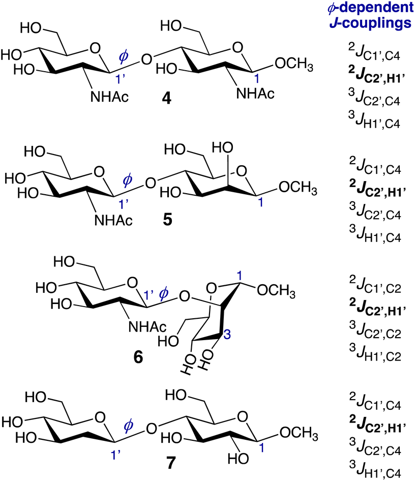 | ||
| Scheme 2 ϕ-Dependent conventional and nonconventional (in bold) J-couplings in disaccharides 4–7. In this work, the four J-couplings in each compound were used in three different combinations in MA'AT analysis of ϕ (Groups I–III). See Tables S1–S4 (ESI†) and the text for more discussion. | ||
The general dependencies of the three conventional J-values on ϕ in O-glycosidic linkages have been reported previously,4,5,41,42 and are shown in Fig. S1–S4 (ESI†) for 4–7. The ϕ-dependencies of 2JC2′,H1′ are less appreciated and are shown in Fig. 1. The calculated two-bond (geminal) J-values have positive signs and range from ∼0 Hz to ∼+7 Hz. The overall shapes of the curves are similar (bimodal), with coupling maxima found at ϕ = ∼120° and ∼300°, and minima at ∼0° and ∼180°. In these calculations, the C2′–N1′ bonds in 4–6 were constrained in geometries expected in aqueous solution based on prior MA’AT analyses (C1′–C2′–N1′–CO torsion angle near +120°),15 leaving rotation about ϕ as the only significant determinant of 2JC2′,H1′. Disaccharide 7 lacks substitution at C2′, and 2JC2′,H1′ depends mainly on ϕ. In the three perfectly-stagged geometries about ϕ (60°, 180° and 300°), 2JC2′,H1′ is most positive at 300° in which one lone-pair orbital on O1′ is anti to the C1′–C2′ bond and the other is anti to the C1′–H1′ bond (assuming ideal sp3 character of O1′). In the three eclipsed geometries (0°, 120° and 240°), that in which two O1′ lone-pair orbitals eclipse the C1′–C2′ and C1′–H1′ bonds (120°) produces a second maximum. 2JC2′,H1′ values at ϕ values of 0°, 60°, 180°, and 240° have similar small magnitudes relative to values at 120° and 300°. The dynamic ranges of the four 2JC2′,H1′ values are ∼4 Hz, making them suitable for use in MA’AT analysis.
 | ||
| Fig. 1 Plots showing the dependence of the calculated 2JC2′,H1′ value in disaccharides 4–7 on ϕ. (A) 4. (B) 5. (C) 6. (D) 7. For all four two-bond C2′–C1′–H1′ coupling pathways, the calculated geminal 2JCCH is positive. In each plot, curves corresponding to the trimmed (blue) and constrained (red) eqn [S1]–[S32] (ESI†) are shown. | ||
3.2 MA’AT analysis of ϕ in 4–7 using different combinations of ϕ-dependent J-couplings
Three different combinations of the ϕ-dependent J-couplings (Scheme 2) were used in MA’AT analyses of ϕ in 4–7. The J-couplings in each group were as follows: Group I, 2JCOC, 3JCOCC, 3JCOCH; Group II, 2JCCH, 3JCOCC, 3JCOCH; Group III, 2JCOC, 2JCCH, 3JCOCC, 3JCOCH. MA’AT analyses were conducted using the trimmed and constrained equations (see Calculations, Section 2.2.3) and the experimental J-couplings (Table 1), and the complete MA’AT results are provided in Tables S1–S4 (ESI).† Since the results do not depend significantly on the equations used in the analyses, only results using the constrained equations are shown in Table 2. Overlays of the MA’AT and MD models of ϕ are shown in Fig. 2 for the constrained equations; those for the trimmed equations are shown in Fig. S6 (ESI).†| J-coupling group/MD | ϕ mean (°) | ϕ CSDb (°) | RMSDc (Hz) |
|---|---|---|---|
| a The ϕ sensitive J-couplings in Groups I–III for 4–7 are identified in Tables S1–S4, ESI. The constrained equations were used in these analyses.b CSD = circular standard deviation.c RMSD = room mean squared deviation.d GLYCAM06; see text for description of MD simulations. | |||
| Methyl βGlcNAc-(1→4)-βGlcNAc (4) | |||
| Group I | 34.7 | 26.4 | 0.18 |
| Group II | 37.3 | 18.6 | 0.31 |
| Group III | 36.6 | 19.3 | 0.32 |
| MDd | 39.5 | 12.8 | |
|
|||
| Methyl βGlcNAc-(1→4)-βMan (5) | |||
| Group I | 33.1 | 22.9 | 0.23 |
| Group II | 34.4 | 14.5 | 0.51 |
| Group III | 33.4 | 15.9 | 0.48 |
| MD | 40.2 | 12.1 | |
|
|||
| Methyl βGlcNAc-(1→2)-αMan (6) | |||
| Group I | 33.5 | 34.5 | 0.06 |
| Group II | 36.6 | 23.7 | 0.55 |
| Group III | 35.9 | 24.4 | 0.49 |
| MD | 39.7 | 15.0 | |
|
|||
| Methyl 2dβGlc-(1→4)-βGlc (7) | |||
| Group I | 35.1 | 33.5 | 0.18 |
| Group II | 38.8 | 13.9 | 0.77 |
| Group III | 37.4 | 16.8 | 0.71 |
| MD | 40.9 | 20.8 | |
 | ||
| Fig. 2 Population distributions of ϕ in 4–7 determined by MA’AT analysis using Groups I (red), II (green) and III (black) ϕ-dependent J-couplings, superimposed on the distributions determined by MD simulation (purple hatched). (A) 4. (B) 5. (C) 6. (D) 7. MA’AT analyses were conducted using constrained eqn [S5]–[S8], [S13]–[S16], [S21]–[S24] and [S29]–[S32] (ESI).† | ||
The use of different combinations of ϕ-dependent J-couplings in 4–7 has a minor effect on MA’AT-determined mean values, varying by at most ∼3.5° (Fig. 3A). These values are uniformly smaller than those determined by MD simulation (differences <7°). The difference is smaller when geminal 2JC2′,H1′ values are included in the analyses (Groups II and III).
 | ||
| Fig. 3 (A) MA’AT-determined mean values (A) and CSDs (B) for ϕ in 4–7 determined using Groups I (red), II (blue) and III (green) ϕ-dependent J-couplings, compared to the mean value and CSDs obtained from MD simulation (black). Constrained eqn [S5]–[S8], [S13]–[S16], [S21]–[S24] and [S29]–[S32] (ESI†) were used in the MA’AT analyses. | ||
Significant effects are observed in the CSDs of ϕ when 2JC2′,H1′ values are included in MA’AT analyses, with reductions of ∼9° found for 4–6 and ∼17° for 7 (Fig. 3B). The reduced CSDs are in better agreement with those obtained by MD, although in 4 and 6 the CSDs determined by MD are still ∼6° and ∼9° smaller, respectively (Table 2 and Fig. 3B). For 7, the MA’AT-determined CSD is smaller than that determined MD by ∼5°.
RMSDs increased by ∼0.4 Hz when 2JC2′,H1′ was included in MA’AT analyses, although their values remain relatively small, indicating good fits of the data.
The MA’AT and MD results, summarized in Fig. 3, show that inclusion of 2JC2′,H1′ in MA’AT analyses of ϕ does not affect mean values appreciably but significantly reduces CSDs, bringing them into closer, albeit not quantitative, agreement with MD. The latter finding provides new experimental evidence that geminal 2JCOC values are the likely cause of aberrant CSDs of ϕ determined by MA’AT analysis when conventional (Group I) ϕ-dependent J-couplings are used to model this torsion angle in O-glycosidic linkages.
4 Conclusions
In an earlier report on the use of MA’AT analysis to model the phi (ϕ) and psi (ψ) torsion angles of O-glycosidic linkages,1 circular standard deviations (CSDs) for ϕ (but not ψ) obtained from studies of twelve disaccharides containing β-(1→4) linkages were found to be consistently and significantly larger than those determined by MD simulation using the GYCAM06 force field. Subsequent MA’AT modeling of mannose-containing di- and oligosaccharides,2 and more recently Gal-(1→3)-Gal disaccharides,43 revealed similar discrepancies. Thus, taken at face value, MA’AT analysis was indicating greater librational averaging about the mean value of ϕ in aqueous solution than predicted by MD simulation. Prior work has also shown3,12,30 that MA’AT analysis gives reliable information on librational motion (dynamics) about mean molecular torsion angles, embodied in CSD values, although of the two parameters provided by the method (mean molecular torsion angles and their CSDs), CSDs are more prone to error when insufficient numbers of J-couplings with satisfactory dynamic ranges are available and/or when equation parameterization cannot be done reliably. In pursuit of a resolution of the CSD discrepancy, a study was undertaken12 wherein parameterized equations for the three conventional J-values used in MA’AT analyses of ϕ (2JCOC, 3JCOCC and 3JCOCH; Group I in this study) were obtained by reducing contributions made by their secondary dependencies on ψ, the expectation being that these secondary dependencies, which are greater for geminal 2JCOC values than for vicinal 3JCOCC and 3JCOCH values, were the source of the discrepancy. However, these studies revealed that CSDs for ϕ determined with and without ψ constraints on equation parameterization were nearly identical, eliminating secondary effects on the three conventional ϕ-dependent J-couplings as the cause of the discrepancy but leaving the source of the discrepancy unidentified.Recent work13 aimed at improving MA’AT analysis of O-glycosidic linkages and other conformational elements in saccharides by incorporating nonconventional J-couplings in the analysis provided an impetus to re-examine this discrepancy from a different vantage point. Earlier work had shown14 that 2JCCH values in saccharides are not only useful to determine pyranosyl and furanosyl ring configuration,44–46 but also exhibit conformational dependencies that might be exploited in MA’AT analysis. These nonconventional J-couplings include not only 2JCCH values but also 1JCH, 1JCC, and 2JCCC values.13 Impediments to their use in MA’AT analyses of ϕ and ψ stem from weak understandings of the conformational properties of exocyclic hydroxyl groups in aqueous solutions of saccharides which partly determine their magnitudes and sometimes their signs. Two-bond 13C–1H spin-coupling between C2 and H1 in an aldopyranosyl ring that contributes its anomeric carbon to an O-glycosidic linkage has particular relevance to ϕ modeling. The magnitude of this 2JC2′,H1′ value depends on rotation about the C1′–O1′ bond (ϕ) (primary dependence) and on rotation about the C2′–O2′ bond (secondary dependence).14 Even though the latter dependence is smaller than the former, it is still sufficiently strong that it cannot be ignored when parameterizing equations for use in MA’AT analysis. In the absence of quantitative knowledge of the conformational properties of the C2′–O2′ bond in aqueous solution, and absent the ability to capture this dependence accurately in parameterized equations, 2JC2′,H1′ values cannot be applied with confidence in MA’AT analyses of ϕ. We have been working on applying MA’AT analysis to better understand exocyclic hydroxyl group conformations in aqueous solutions of saccharides, but results are presently incomplete.
The above-noted limitation associated with the use of nonconventional 2JC2′,H1′ values in MA’AT analyses of ϕ is circumvented when the residue participating in the O-glycosidic linkage bears an N-acetyl functional group at C2′, or no substituent at C2′, instead of a hydroxyl group. Unlike hydroxyl groups, exocyclic N-acetyl groups are highly conformationally constrained in most cases, and MA’AT analysis has recently modeled this behavior as uni-modal using six redundant J-couplings sensitive to rotation about the C2′–N′ bond.15 In general, rotation about this bond is conserved in most C2 N-acetylated aldopyranosyl rings, as demonstrated by MD in the βGlcNAc rings of 4–6 (Fig. S7, ESI†), allowing equation parameterization for 2JC2′,H1′ in which the only significant conformational dependency is on ϕ. In deoxy structures like 7, secondary effects from C2′–O2′ bond rotation are absent, making parameterization of 2JC2′,H1′ straightforward.
The results of this study confirm the value of adding a nonconventional J-coupling to MA’AT analyses of ϕ. While the addition of this constraint does not alter calculated mean values of ϕ appreciably, its inclusion results in a significant reduction in CSDs, bringing their values into closer alignment with MD simulation. As shown in the Group III calculations, even when all four redundant J-couplings are used, a significant reduction in the CSD is observed. For 7, complications arising from C2′–O2′ bond rotation are absent, making 2JC2′,H1′ a valuable constraint for ϕ. On the other hand, the approach described here cannot be applied at present to O-glycosidic linkages in which the donor residue bears an hydroxyl group at C2′. While it is anticipated that the values of MA’AT-determined CSDs of ϕ may also decline upon inclusion of 2JC2′,H1′ values in MA’AT analysis, additional work is needed to better understand the conformational behavior of C2′–O2′ bonds to enable their use to test this prediction.
Of interest is why the inclusion of 2JC2′,H1′ values in MA’AT analyses of ϕ improves the modeling of this torsion angle, as inferred from the better agreement with MD results. An inspection of the plots in Fig. 4 for 4 provides insight into this question and reveals aspects of MA’AT analysis worth illuminating. Secondary effects of ψ on 2JC1′,C4 are substantial even when ψ constraints are applied to the dataset (constrained data), leading to inherent uncertainty in equation parameterization (Fig. 4A). In contrast, secondary affects from ψ are smaller for 2JC2′,H1′ (Fig. 4B). More importantly, the plot of the dependence of 2JC1′,C4 on ϕ contains a region between ϕ = 0–180° where the J-value is relatively constant (−2–−3°), that is, in the region that includes the MA’AT-determined mean value of ϕ for 4 (35–37°; Table 2). In contrast, the same region for 2JC2′,H1′ shows values ranging from ∼1–5 Hz. While the behavior of 2JC1′,C4 is sufficient when combined with those of the two vicinal ϕ-dependent J-couplings (3JC2′,C4 and 3JH1′,C4) to compute a reproducible mean value of ϕ, the flat region of the 2JC1′,C4 curve renders it less discriminating than that for 2JC2′,H1′, leading to an artificially broadened population distribution for ϕ. This fact emphasizes the importance of not only the dynamic range of the J-coupling across the full 360° torsion angle itinerary, but also the shape of the dependency in the region where the mean value of the molecular torsion angle resides. The sinusoidal behavior of 2JC2′,H1′ and its more reliable parameterization due in part to smaller secondary effects from ψ increase its discriminatory power in MA’AT analysis relative to that of 2JC1′,C4.
 | ||
| Fig. 4 Plots of the dependencies of 2JC1′,C4 (A) and 2JC2′,H1′ (B) in 4 on the H1′–C1′–O1′–C4 torsion angle ϕ. Blue circles, trimmed data; red squares, constrained data. The solid lines are plots of the trimmed (blue) and constrained (red) equations for each J-coupling. Point scatter at discrete values of ϕ is caused by the secondary dependence of the J-coupling on ψ. | ||
Prior reports have noted that the width of the population distribution of a molecular torsion angle determined by MA’AT analysis will be affected by the dynamic ranges of the redundant J-couplings, the nature (shape) of their dependencies on the torsion angle, the quality of the parameterized equations used to fit the J-couplings, and the accuracy of the experimental J-couplings.3,30 Errors in one or more of these contributing factors will increase the width of the distribution (i.e., increase the calculated CSD). In this regard the population distribution can be compared to peak width in NMR, wherein linewidth in the absence of chemical exchange is determined by the intrinsic T2 of the nucleus (the true linewidth) and by field inhomogeneity  . A CSD value determined by MA’AT analysis is expected to always be equal to or greater than the true CSD (i.e., the MA’AT-determined CSDs are upper limits). This being the case, the smaller MA’AT-determined CSD found for ϕ in 7 (Groups II and III, Table 2) compared to that determined by MD suggests that the latter may be incorrect, that is, librational averaging about ϕ in 7 is probably smaller than that indicated by MD simulation.
Data availability
The data supporting this article have been included as part of the ESI.† The current version of the MA’AT application can be accessed online (https://rmeredit.shinyapps.io/maat24/) (last accessed: 08/21/2024) and a User's Manual is available on the software's webpage.Conflicts of interest
There are no conflicts of interest to declare.Acknowledgements
Financial support was provided by the National Science Foundation (CHE 1707660 and CHE 2002625 to A. S.) and by Omicron Biochemicals, Inc. The Notre Dame Radiation Laboratory is supported by the Department of Energy Office of Science, Office of Basic Energy Sciences under Award Number DE-FC02-04ER15533. This is document number NDRL 5437. A. S. thanks Kevin Dorst and Göran Widmalm (Stockholm University) for providing the MD data shown in Fig. S5 (ESI).†References
- W. Zhang, T. Turney, R. Meredith, Q. Pan, L. Sernau, X. Wang, X. Hu, R. J. Woods, I. Carmichael and A. S. Serianni, Conformational populations of β-(1→4) O-glycosidic linkages using redundant NMR J-couplings and circular statistics, J. Phys. Chem. B, 2017, 121, 3042–3058 CrossRef CAS.
- W. Zhang, R. Meredith, Q. Pan, X. Wang, R. J. Woods, I. Carmichael and A. S. Serianni, Use of circular statistics to model αMan-(1→2)-αMan and αMan-(1→3)-α/βMan O-glycosidic linkage conformation in 13C-labeled disaccharides and high-mannose oligosaccharides, Biochemistry, 2019, 58, 546–560 CrossRef CAS PubMed.
- R. J. Meredith, I. Carmichael, R. J. Woods and A. S. Serianni, MA’AT analysis: Probability distributions of molecular torsion angles in solution from NMR spectroscopy, Acc. Chem. Res., 2023, 56, 2313–2328 CrossRef CAS PubMed.
- I. Tvaroska, H. Hricovini and E. Petrakova, An attempt to derive a new Karplus-type equation of vicinal proton-carbon coupling constants for C–O–C–H segments of bonded atoms, Carbohydr. Res., 1989, 189, 359–362 CrossRef CAS.
- B. Mulloy, T. A. Frenkiel and D. B. Davies, Long-range carbon-proton coupling constants: Application to conformational studies of oligosaccharides, Carbohydr. Res., 1988, 184, 39–46 CrossRef CAS PubMed.
- T. Peters and T. Weimar, Assessing glycosidic linkage flexibility: Conformational analysis of the repeating trisaccharide unit of Aeromonas salmonicida, J. Biomol. NMR, 1994, 4, 97–116 CrossRef CAS PubMed.
- T. Weimar, B. Meyer and T. Peters, Conformational analysis of α-D-Fuc-(1→4)-β-D-GlcNAc-OMe. One-dimensional transient NOE experiments and metropolis Monte Carlo simulations, J. Biomol. NMR, 1993, 3, 399–414 CrossRef CAS PubMed.
- P. C. Kline, A. S. Serianni, S.-G. Huang, M. H. Hayes and R. Barker, 1H-1H internuclear distance measurements in carbohydrates: Proton transient nuclear Overhauser enhancement and spin-lattice relaxation in (13C)- and (2H)-substituted compounds, Can. J. Chem., 1990, 68, 2171–2182 CrossRef CAS.
- M. Erdélyi, E. d'Auvergne, A. Navarro-Vázquez, A. Leonov and C. Griesinger, Dynamics of the glycosidic bond: Conformational space of lactose, Chem.–Eur. J., 2011, 17, 9368–9376 CrossRef.
- N. G. A. Bell, G. Rigg, S. Masters, J. Bella and D. Uhrín, Detecting low-level flexibility using residual dipolar couplings: A study of the conformation of cellobiose, Phys. Chem. Chem. Phys., 2013, 15, 18223–18234 RSC.
- A. Almond, J. Bunkenborg, T. Franch, C. H. Gotfredsen and J. Ø. Duus, Comparison of aqueous molecular dynamics with NMR relaxation and residual dipolar couplings favors internal motion in a mannose oligosaccharide, J. Am. Chem. Soc., 2001, 123, 4792–4802 CrossRef CAS PubMed.
- R. J. Meredith, R. J. Woods, I. Carmichael and A. S. Serianni, Reconciling MA’AT and molecular dynamics models of linkage conformation in oligosaccharides, Phys. Chem. Chem. Phys., 2020, 22, 14454–14457 RSC.
- R. J. Meredith, I. Carmichael and A. S. Serianni, Nonconventional NMR spin-coupling constants in oligosaccharide conformational modeling: Structural dependencies determined from density functional theory calculations, ACS Omega, 2022, 7, 23950–23966 CrossRef CAS PubMed.
- T. E. Klepach, I. Carmichael and A. S. Serianni, Geminal 2JCCH spin-spin coupling constants as probes of the ϕ glycosidic torsion angle in oligosaccharides, J. Am. Chem. Soc., 2005, 127, 9781–9793 CrossRef CAS PubMed.
- R. J. Meredith, T. Tetrault, M.-K. Yoon, W. Zhang, I. Carmichael and A. S. Serianni, N-Acetyl side-chain conformation in saccharides: Solution models obtained from MA’AT analysis, J. Org. Chem., 2022, 87, 8368–8379 CrossRef CAS PubMed.
- M. von Kienlin, C. T. W. Moonen, A. van der Toorn and P. C. M. van Zijl, Rapid recording of solvent-suppressed 2D COSY spectra with inherent quadrature detection using pulsed field gradients, J. Magn. Reson., 1991, 93, 423–429 CAS.
- G. W. Vuister, R. Boelens, R. Kaptein, R. E. Hurd, B. John and P. C. M. van Zijl, Gradient-enhanced HMQC and HSQC spectroscopy. Applications to 15N-labeled Mnt repressor, J. Am. Chem. Soc., 1991, 113, 9688–9690 CrossRef CAS.
- A. Meissner and O. W. Sørensen, Measurement of J(H,H) and long-range J(X,H) coupling constants in small molecules. Broadband XLOC and J-HMBC, Magn. Reson. Chem., 2001, 39, 49–52 CrossRef CAS.
- M. J. Frisch, et al., Gaussian16. Revision B.01, Gaussian Inc., Wallingford, CT, 2016, https://gaussian.com/gaussian16/ Search PubMed.
- A. D. Becke, Density-functional thermochemistry. III. The role of exact exchange, J. Chem. Phys., 1993, 98, 5648–5652 CrossRef CAS.
- A. D. Becke, New mixing of Hartree-Fock and local density-functional theories, J. Chem. Phys., 1993, 98, 1372–1377 CrossRef CAS.
- W. J. Hehre, R. Ditchfield and J. A. Pople, Self-consistent molecular orbital methods. XII. Further extensions of Gaussian-type basis sets for use in molecular orbital studies of organic molecules, J. Chem. Phys., 1972, 56, 2257–2261 CrossRef CAS.
- E. Cancès, B. Mennucci and J. Tomasi, A new integral equation formalism for the polarizable continuum model: Theoretical background and applications to isotropic and anisotropic dielectrics, J. Chem. Phys., 1997, 107, 3032–3041 CrossRef.
- R. Cammi, B. Mennucci and J. Tomasi, Fast evaluation of geometries and properties of excited molecules in solution: A Tamm-Dancoff model with application to 4-dimethyl-aminobenzonitrile, J. Phys. Chem. A, 2000, 104, 5631–5637 CrossRef CAS.
- V. Sychrovský, J. Gräfenstein and D. Cremer, Nuclear magnetic resonance spin−spin coupling constants from coupled perturbed density functional theory, J. Chem. Phys., 2000, 113, 3530–3547 CrossRef.
- T. Helgaker, M. Watson and N. C. Handy, Analytical calculation of nuclear magnetic resonance indirect spin−spin coupling constants at the generalized gradient approximation and hybrid levels of density-functional theory, J. Chem. Phys., 2000, 113, 9402–9409 CrossRef CAS.
- V. Barone, J. E. Peralta, R. H. Contreras and J. P. Snyder, DFT calculation of NMR JFF spin spin coupling constants in fluorinated pyridines, J. Phys. Chem. A, 2002, 106, 5607–5612 CrossRef CAS.
- T. Klepach, H. Zhao, X. Hu, W. Zhang, R. Stenutz, M. J. Hadad, I. Carmichael and A. S. Serianni, Informing saccharide structural NMR studies with density functional theory calculations, in Glycoinformatics: Methods in Molecular Biology, ed. Lütteke, T. and Frank, M., Springer, New York, 2015, pp. 289–331 Search PubMed.
- R. Stenutz, I. Carmichael, G. Widmalm and A. S. Serianni, Hydroxymethyl group conformation in saccharides: Structural dependencies of 2JHH, 3JHH and 1JCH spin-spin coupling constants, J. Org. Chem., 2002, 67, 949–958 CrossRef CAS PubMed.
- R. J. Meredith, L. Sernau and A. S. Serianni, MA’AT: A web-based application to determine rotamer population distributions in solution from nuclear magnetic resonance spin-coupling constants, J. Chem. Inf. Model., 2022, 62, 3135–3141 CrossRef CAS.
- Complex Carbohydrate Research Center (CCRC) and University of Georgia, http://www.glycam.org.
- K. N. Kirschner, A. B. Yongye, S. M. Tschampel, J. González-Outeiriño, C. R. Daniels, B. L. Foley and R. J. Woods, GLYCAM06: A generalizable biomolecular force field. Carbohydrates, J. Comput. Chem., 2008, 29, 622–655 CrossRef CAS PubMed.
- W. L. Jorgensen, J. Chandrasekhar, J. D. Madura, R. W. Impey and M. L. Klein, Comparison of simple potential functions for simulating liquid water, J. Chem. Phys., 1983, 79, 926–935 CrossRef CAS.
- D. A. Case, V. Babin, J. T. Berryman, R. M. Betz, Q. Cai, D. S. Cerutti, T. E. I. Cheatham, T. A. Darden, R. E. Duke, H. Gohlke, et al., AMBER 14, University of California, San Francisco, 2014 Search PubMed.
- H. J. C. Berendsen, J. P. M. Postma, W. F. van Gunsteren, A. DiNola and J. R. Haak, Molecular dynamics with coupling to an external bath, J. Chem. Phys., 1984, 81, 3684–3690 CrossRef CAS.
- W. F. van Gunsteren and H. J. C. Berendsen, Algorithms for macromolecular dynamics and constraint dynamics, Mol. Phys., 1977, 34, 1311–1327 CrossRef CAS.
- A. W. Götz, M. J. Williamson, D. Xu, D. Poole, S. Le Grand and R. C. Walker, Routine microsecond molecular dynamics simulations with AMBER on GPUs. 1. Generalized Born, J. Chem. Theory Comput., 2012, 8, 1542–1555 CrossRef PubMed.
- K. N. Kirschner and R. J. Woods, Solvent interactions determine carbohydrate conformation, Proc. Natl. Acad. Sci. U.S.A., 2001, 98, 10541–10545 CrossRef CAS.
- Prism 8 for Mac OS X; GraphPad Software, Version 8.4.2 (464), 2020 Search PubMed.
- M. L. Hayes, A. S. Serianni and R. Barker, Methyl β-lactoside: 600-MHz 1H- and 75-MHz 13C-N.M.R. Studies of 2H- and 13C-enriched compounds, Carbohydr. Res., 1982, 100, 87–101 CrossRef CAS.
- B. Bose, S. Zhao, R. Stenutz, F. Cloran, P. B. Bondo, G. Bondo, B. Hertz, I. Carmichael and A. S. Serianni, Three-bond C−O−C−C spin-coupling constants in carbohydrates: Development of a Karplus relationship, J. Am. Chem. Soc., 1998, 120, 11158–11173 CrossRef CAS.
- F. Cloran, I. Carmichael and A. S. Serianni, 2JCOC spin-spin coupling constants across glycosidic linkages exhibit a valence bond-angle dependence, J. Am. Chem. Soc., 2000, 122, 396–397 CrossRef CAS.
- R. Meredith, Y. Zhu, M.-K. Yoon, T. Tetrault, J. Lin, W. Zhang, M. McGurn, E. Cook, R. Popp, P. Shit, I. Carmichael and A. S. Serianni, Methyl α-D-galactopyranosyl-(1→3)-β-D-galactopyranoside and methyl β-D-galactopyranosyl-(1→3)-β-D-galactopyranoside: Glycosidic linkage conformation determined from MA’AT analysis, Magn. Reson. Chem., 2024, 62, 544–555 CrossRef CAS.
- K. Bock and C. Pedersen, Two- and three-bond 13C-1H couplings in some carbohydrates, Acta Chem. Scand., 1977, B31, 354–358 CrossRef CAS.
- J. A. Schwarcz and A. S. Perlin, Orientational dependence of vicinal and geminal 13C-1H coupling, Can. J. Chem., 1972, 50, 3667–3676 CrossRef CAS.
- J. A. Schwarcz, N. Cyr and A. S. Perlin, Orientation effects and the sign of two-bond 13C-1H coupling, Can. J. Chem., 1975, 53, 1872–1875 CrossRef CAS.
Footnote |
| † Electronic supplementary information (ESI) available: MA’AT statistics for ϕ in 4–7 using different combinations of J-couplings; plots of DFT-calculated ϕ-dependent J-couplings in 4–7 as a function of ϕ; comparison of MD histograms of ϕ and ψ in methyl β-lactoside obtained from aqueous 1 μs simulations using GLYCAM06 and CHARMM; population distributions of ϕ in 4–7 determined by MA’AT analysis (Groups I–III) superimposed on distributions obtained from MD; MD histograms of torsion angles in the N-acetyl side-chains of 4–6; torsion angle constraints in DFT calculations of 4c–7c; DFT-parameterized spin-coupling equations for 4–7; partial 1D 13C{1H} NMR spectrum of 51′; partial 1D 13C{1H} NMR spectrum of 52′; partial 2D J-HMBC spectrum of 5 in (3JC4,H1′′ determination); partial 2D HSQC-HECADE spectrum of 5 (2JC2′,H1′ determination); partial 2D HSQC-HECADE spectrum of 7 (2JC2′,H1′ determination); partial 2D HSQC-HECADE spectrum of 4 (2JC2′,H1′ determination); preparation of disaccharide 41′,4; preparation of disaccharides 51′ and 52′; preparation of disaccharide 61′,2; preparation of disaccharide 71′,4; representative Cartesian coordinates for DFT structures 4c–7c; brief discussion of the functional and basis sets used in DFT calculations; complete ref. 19 and 34. See DOI: https://doi.org/10.1039/d4ra06062h |
| This journal is © The Royal Society of Chemistry 2024 |