
 Open Access Article
 This Open Access Article is licensed under a Creative Commons Attribution-Non Commercial 3.0 Unported Licence
Macauba oil carried by polymeric micelles reduces migration and proliferation of triple-negative breast cancer cells
Davi T.
Aleixo
ab,
Ana C. M.
Gualberto
a,
Ana B. C. dos S.
Valle
a,
Luan C.
da Silva
a,
Kézia C. B.
Ferreira
b,
Ari S. de O.
Lemos
a,
Rodrigo L.
Fabri
a,
Guilherme D.
Tavares
b,
Maurílio de S.
Cazarim
b,
Jacy
Gameiro
a and
Frederico
Pittella
 *ab
*ab
aPrograma de Pós-Graduação em Ciências Biológicas, Universidade Federal de Juiz de Fora, Juiz de Fora 36036-900, Minas Gerais, Brazil. E-mail: frederico.pittella@ufjf.br
bPrograma de Pós-Graduação em Ciências Farmacêuticas, Universidade Federal de Juiz de Fora, Juiz de Fora 36036-900, Minas Gerais, Brazil
First published on 23rd July 2024
Abstract
Triple-negative breast cancer (TNBC) accounts for about 10–15% of all breast cancer cases, often affecting younger women and those with a BRCA1 mutation. It is more aggressive and has a higher recurrence risk within the first few years after diagnosis. Due to its aggressive nature, limited treatment options, and drug resistance, alternative strategies are urgently needed. In this context, Macauba (Acrocomia aculeata) is a South American palm with antioxidant-rich fruits containing fatty acids, carotenoids, and phenolic compounds, which can remove reactive oxygen species (ROS) and protect cells. Our study focused on creating Macauba pulp oil-loaded polymeric micelles (PM-MO) and assessing their impact on triple-negative breast cancer cells in terms of cytotoxicity, antiproliferation, and antimigration. Before formulating PM-MO, we conducted chemical characterization and testing of Macauba oil. Further, PM-MO presented hydrodynamic diameters of 105 nm, polydispersity index (PdI) of 0.12 and Zeta potential of −17.5 mV. PM-MO showed enhanced cytotoxicity against triple negative breast cancer cells after 48h and 72h, while no toxicity was observed on non-tumor cells. The clonogenicity assay showed a reduction in the formation of cell colonies ranging from 97% to 81.9% at the highest concentration of PM-MO. Treatment with PM-MO reduced breast cancer cell migration in vitro, indicating potential as an anti-metastatic agent. We conclude that the method used to produce PM-MO yielded well-sized nanoparticles with uniform distribution. Results from cell viability, proliferation, and migration tests highlight its potential for future in vivo trials against triple-negative breast cancer.
1. Introduction
The latest cancer data indicate that female breast cancer (BC) has overtaken lung cancer to become the most diagnosed type in the world, with 2.26 million new cases. It is an increase of about 8% from 2018 numbers, highlighting the importance of the development of treatments for this type of disease.1,2 Triple-negative breast cancer (TNBC) is a subtype of breast cancer that lacks expression of three important receptors commonly found in other types of breast cancer: the estrogen receptor (ER), the progesterone receptor (PR), and the human epidermal growth factor receptor 2 (HER2). This indicates that TNBC does not respond to hormonal therapies designed to target estrogen and progesterone receptors, nor does it respond to medications aimed at the HER2 protein. Moreover, many conventional drugs used in current therapies present serious side effects, the risk of tumor recurrence and the resistance to these drugs.3 Thus, the search for antitumor agents that circumvent the collateral problems continue to be extremely relevant in the health field.4 Among the viable options, plant species with reported biological activity offer promising alternative sources of new molecules.4,5Acrocomia aculeata (Jacq.) Lodd. ex Mart., popularly known as “Bocaiuva” or “Macauba”, is a native plant species widely distributed throughout Brazil and the American continent, belonging to the Arecaceae family.6 Macauba oil represents a valuable natural resource with diverse applications in nutrition, cosmetics, bioenergy, and sustainable development. Due to its moisturizing, emollient, and anti-inflammatory properties, Macauba oil is commonly used in skincare and hair care products. In addition, it has potential as a renewable energy source due to its high oil content and favourable fatty acid composition. In fact, its fruits contain rich amounts of short-chain saturated fatty acids, mainly lauric acid and oleic acid, making it suitable for consumption as a dietary supplement or as an ingredient in food products. Furthermore, bioactive compounds such as β-carotene and α-tocopherol, provide antioxidant protection in biological systems – an important activity in the regulation of cellular metabolism.7–9
Therefore, Macauba oil has been reported for the treatment and control of various pathologies, including type 1 and 2 diabetes,10,11 and as a neuroprotector in cases of chronic stress observed in rats.12 It has also been used to prevent adipocyte hypertrophy in mice, and exhibit anti-inflammatory, antimutagenic and antioxidant properties.13–15 Additionally, there are studies exploring the use of Macauba as a chemopreventive agent to control the adverse effects of chemotherapeutic drugs such as cyclophosphamide in cancer treatment.16,17 Unfortunately, the pharmaceutical application of Acrocomia Aculeata pulp oil faces limitations due to its physicochemical properties, including limited solubility in water. This requires a formulation that enables the exploration of its therapeutic potential.18 In this context, the association with polymeric micelles emerges as a viable solution to overcome these challenges.
Polymeric micelles have the ability to encapsulate hydrophobic compounds in their core while maintaining a hydrophilic outer coating. This improves the aqueous stability of oily content and provide robust protection against oxidation and degradation of bioactive components such as unsaturated fatty acids, tocopherols, and carotenoids, thereby preserving the therapeutic and nutritional constituents during storage and administration.19–22 Furthermore, polymeric micelles offer the possibility of precise and targeted release of active compounds, distinguishing them from other nanoparticle systems.23,24 Their superior solubilization capacity, enhanced stability, and versatility in surface functionalization enable tailored targeting strategies in breast cancer.22,25,26 These approaches facilitate controlled and sustained release directly into tumor cells, thereby boosting therapeutic efficacy while minimizing systemic side effects.27–29 Promising outcomes have been reported with agents like fatty acids,26,30 resveratrol31 and essential oils.32
These advancements represent a compelling strategy to enhance breast cancer treatment outcomes, with several formulations advancing towards FDA approval.28,33 Thus, the present study aims to develop polymeric micelles containing Macauba pulp oil to evaluate their anticancer activity against triple negative breast cancer cells.
2. Materials and methods
2.1 Reagents and material
The Macauba pulp oil was purchased from Mundo dos Óleos, Brasília (CNPJ: 33.460.511/0001-45). Pluronic® F-127, Sodium Chloride and RPMI medium were obtained from Sigma-Aldrich (USA). Dulbecco's Modified Eagle's medium (DMEM), fetal bovine serum (FBS) and trypsin were purchased from Gibco (USA). Trypan blue and dimethyl diphenyltetrazolium bromide (MTT) purchased from Invitrogen (USA). The DMSO was acquired from Dinâmica (Brazil). Monobasic sodium phosphate, dibasic sodium phosphate, and paraformaldehyde were obtained from Synth (Brazil). SDS was obtained from Bio-rad (USA). Violet crystal was purchased from Cromoline (Brazil). Ethyl acetate, methanol and Rutin were purchased from Vetec (Brazil). Folin–Ciocalteu was purchased from MP Biomedicals (USA). 2,2-Diphenyl-1-picrylhydrazyl (DPPH) and beta-carotene were acquired from Merck (Germany), while sodium carbonate was purchased from Panreac (USA).2.2 Determination of the fatty acid profile
For the qualitative characterization of the Macauba pulp oil, the gas chromatography methodology was employed using the HP7820A Gas Chromatograph (GC) equipment from Agilent, USA. The GC was equipped with a flame ionization detector and a SUPELCOWAX-10 column (30 m × 0.2 mm × 0.2 μm) from Sigma-Aldrich. The temperature gradient used was as follows: the column temperature started at 120 °C, then increased by approximately 2.5 °C per minute until reaching 240 °C; Injector temperature was set at 240 °C with a 1/10 split ratio, and the detector temperature was set at 260 °C. Peak identification was performed through comparative analysis with Supelco37 Fame mix methylated fatty acid standards (Supelco cat no 47![[thin space (1/6-em)]](https://www.rsc.org/images/entities/char_2009.gif) 885-U).
885-U).
2.3 Determination of total carotenoids
About 1 mL of Macauba pulp oil was dissolved in ethyl acetate (100 μg mL−1), followed by the addition of a 0.5% NaCl solution and centrifugation at 1500 rpm for 10 minutes. Subsequently, 250 μL of the supernatant were collected and solubilized in methanol at concentrations ranging from 7.5 to 350 μg mL−1. From this solution, an aliquot was taken for reading in a spectrophotometer at 460 nm using the Spectramax 190 (Molecular Devices, California, USA). The total carotenoid content in the sample was expressed in miligrams equivalent to micrograms per milliliter (mg equivalent of mg per 100 g of oil). A curve of a beta carotenoid was used as a control for comparison.2.4 Determination of phenolic content
The determination of the phenolic content was performed by means of spectroscopy in the visible region using the Folin–Ciocalteu method with minor modifications.34,35 Tannic acid was used as standard. A calibration curve was prepared using concentrations ranging from 7.5 to 350 μg mL−1 of a methanolic tannic acid solution (500 μg mL−1). For the assay, 120 μL of Folin Ciocalteau 20% reagent, 30 μL of the sample, and 100 μL of 4% sodium carbonate were added to 96-well microplates. After 30 min of incubation in the dark, the absorbance was measured in a spectrophotometer at 770 nm. The same procedure was carried out for the solution at 500 μg mL−1 of Macauba pulp oil in methanol.All determinations were performed in triplicate and the mean ± standard deviation was used to calculate the phenol content. The total phenol content was expressed in mg g−1 of plant extract, in tannic acid equivalents (TAE).
2.5 Free radical scavenging potential by DPPH
The antioxidant activity of Macauba pulp oil was performed using the DPPH (2,2-diphenyl-1-picrylhydrazyl) radical reduction method.36 For this assay, 1000 μg of the oil was solubilized in 1 ml of methanol, followed by homogenization. Serial dilutions were prepared from this mixture to reach concentrations ranging from 49 to 250 μg mL−1. The calculation of the radical reduction was determined by adding DPPH solubilized in methanol (20 μg mL−1).The dilutions were stored away from light and at room temperature for 30 minutes, and the scavenging potential was quantified as the concentration of the samples capable of inhibiting 50% of the DPPH absorbance (estimated IC50) using the Spectramax 190 spectrophotometer (Molecular Devices, California, USA) in the visible radiation region of 517 nm. A solution of DPPH and Rutin (flavonoid) was also prepared and tested at the same concentrations as a positive control group. To quantify the result, the antioxidant activity was calculated using the equation:
 | (1) |
2.6 Preparation of polymeric micelles
The polymeric micelles containing Macauba pulp oil (PM-MO) were prepared through the sonication method. Initially, Pluronic® F-127 was weighed and solubilized in ultrapure water (0.7% w/v). Subsequently, the solution was added to the Macauba pulp oil at a concentration of 1.0% w/v. This mixture was then subject to ultrasonic irradiation (315 W) using an ultrasonic homogenizer (Ultrasonics) for 3 minutes, repeated 3 times in sequence. Similarly, a control polymeric micelle without oil was prepared using the same procedure.2.7 Characterization of polymeric micelles
:400) in purified water to a suitable dispersion intensity. The Z-average data were evaluated through the distribution by intensity. Mean diameter (based on the Stokes–Einstein equation) and PdI were given as the average of three individual measurements.
:400 v/v) and subjected to an electric field with an established potential of approximately 150 mV, using a specific cuvette (DTS1060).
000 rpm for 20 minutes). The filtrated was collected and measured in a Nanodrop Lite spectrophotometer (Thermo Scientific) at a wavelength of 450 nm, referring to oleic acid. From the calculated concentration after filtration, it was possible to determine the percentage of free Macaba pulp oil and the percentage of encapsulation (%) through the formula: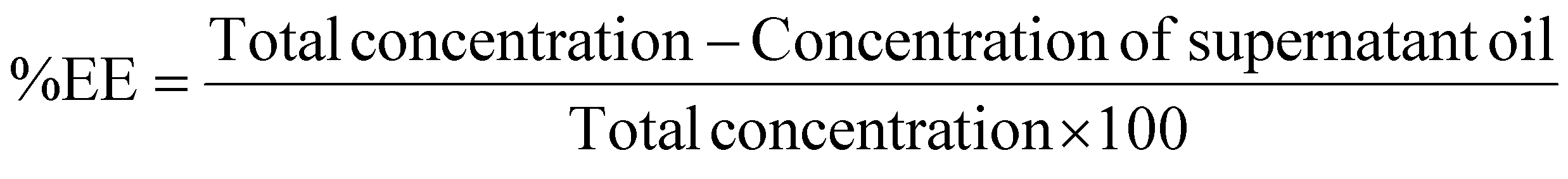 | (2) |
2.8 Transmission electron microscopy (TEM)
The morphology of the nanoparticles was determined using transmission electron microscopy (TEM) with a JEM-1011 TEM (Jeol LTD, Tokyo, Japan) operated at an accelerating voltage of 80 kV. A sample of the nanoparticle solution was deposited on Parlodion® 200-mesh nickel grids (CF200-Ni, EMS) coated with amorphous carbon and dried for 24 hours at room temperature. For the morphology analysis, the microscope was operated in bright field mode with up to 150000× magnification.
2.9 Cell culture
The triple negative breast cancer cell line MDA-MB-231 was gently donated by Prof. Tania Pasa, at Federal University of Santa Catarina, Brazil. The fibroblast cell line L929 was purchased from the Cell Bank of Rio de Janeiro (BCRJ), Brazil. Both cell lines were maintained in Dulbecco's Eagle Medium Mod (DMEM) supplemented with 4500 mg L−1 glucose, 1500 mg L−1 glucose, 1500 mg L−1 sodium bicarbonate, 10% inactivated Fetal Bovine Serum (FBS), and 1% penicillin–streptomycin. The cells were incubated at 37 °C with 5% CO2.2.10 MTT assay
Both cell lines were plated in 96-well plates at a density of 5 × 10³ per well and incubated overnight at 37 °C in 5% CO2. Afterwards, the medium was removed, and different concentrations of free Macauba oil (F-MO, 48.25–386 μg mL−1) was used to evaluate the cytotoxicity in 24, 48 and 72 hours. Further, the wells were washed and 500 μg mL−1 of MTT {[3-(4,5-dimethylthiazol-492yl)-2,5-diphenyl tetrazolium bromide]} reagent in medium was added. After 4 hours, the solution was removed and 100 μL per well of DMSO (dimethylsulfoxide) was added to dissolve the MTT-formazan crystals. The absorbance was measured in a Spectramax 190 spectrophotometer (Molecular Devices, California, USA) at a wavelength of 540nm. The control group (without treatment) containing only culture medium was considered as 100% viable cells. Polymeric micelles without oil and DMSO/Tween80 groups (90% DMSO + 10% Tween 80) were used as controls. Each experiment was performed in triplicate.2.11 Antiproliferative activity by Clonogenic assay
In colony formation assay, MDA-MB-231 cells were cultured in 6-well plate, in density of 300 cells per well and incubated at 37 °C in 5% CO2. After 24 hours, the medium was removed, the wells washed with PBS and the treatments with PM-MO, PM mock and F-MO were added at concentrations ranging from 386 μg mL−1 to 96.5 μg mL−1. The plates were incubated for 14 days at 37 °C and 5% CO2 for the formation of clones. Later, cells in the plates were fixed in paraformaldehyde and stained with crystal violet. The colony counts were calculated using CFU Scope (Medixgraph, California, EUA).2.12 Migration assay
For the migration assays, cells were seeded into 24-well plates using medium without FBS and incubated at 37 °C in 5% CO2. After reaching 90% of monolayer confluence, the cells were scratched with a 200 μL sterile pipette tip. The cancer cells were subsequently treated with the same concentration used in clonogenic assay of PM-MO and F-MO, and photographed with an inverted microscope at different time points (0, 24 and 48hr). Further, the area of scratch was calculated daily, using the ImageJ program (National Institute of Health, Maryland, USA).2.13 Statistical analysis
The results were expressed as the mean ± standard error of the mean (SEM). The analysis of variance (ANOVA) test was used to access differences between groups, after which post hoc tests with the Bonferroni correction were used for comparison between individual groups. A value of p < 0.05 was considered significant.3. Results
3.1. Quantification of fatty acids, phenolic and carotenoids compounds and antioxidant activity of Macauba pulp oil
The initial phase of this research aimed to identify and quantify the fatty acids present in Macauba pulp oil. Table 1 displays the results of the gas chromatography (GC) analysis of the fatty acid profile of Macauba pulp oil. Approximately 78.8% of the fatty acids are unsaturated, while saturated fatty acids account for around 17.5%. Among the unsaturated fatty acids, oleic acid (C18:1) predominates, comprising 45.5% of the total composition. Following oleic acid, linoleic acid constitutes 26.8%, and palmitic acid contributes with 12%. These results align with the data found in the literature for Acrocomia aculeata, considering regional, seasonal and temporal variations.21,34,37| Compounds | Percentage (%) |
|---|---|
| Unsaturated | 17.5 |
| Monounsaturated | 41.7 |
| Polyunsaturated | 37.1 |
| Others | 3.7 |
| Individual fatty acids | |
| C12:0 (lauric acid) | 1.2 |
| C14:0 (myristic acid) | 0.4 |
| C16:0 (palmitic acid) | 12.0 |
| C18:1 (oleic acid) | 45.1 |
| C18:2 (linoleic acid) | 26.8 |
| Others | 7.3 |
Phenolic compounds and carotenoids are bioactive compounds with significant antioxidant activity. In this work, we quantified both compounds in Macauba pulp oil (Table 2). In general, the amount of phenolic compound was 632 ± 41/100 g equivalent in tannic acid, while the carotenoid content was 2.81 ± 0.18 mg/100 g of oil.
| Contents and activity of Macauba pulp oil | Sample (1 mL = 0.856g) |
|---|---|
| a TAE = tannic acid equivalent. b βCE = β-carotene equivalent. Positive control for antioxidant activity: Rutin (IC50 = 3371 μg mL−1) | |
| β-carotene (βCEb per 100 g) | 2.81 ± 0.18 |
| Phenolic content (mg TAEa per g) | 632 ± 41 |
| Antioxidant activity IC50 (μg mL−1) | 141.8 |
The presence of these bioactive molecules are good indicators of a possible antioxidant activity in natural samples. In fact, the Acrocomia aculeata oil sample presented a statistically significant value of 141.8 μg mL−1 of IC50, compared to the flavonoid Rutin, used as positive control, that presented an IC50 value of 3371 μg mL−1. Following the determination of its composition, the oil underwent a sophisticated nanoencapsulation procedure.
3.2. Characterization of polymeric micelles containing Macauba oil (PM-MO)
Macroscopically, PM-MO suspension presented an opaque whitish color, with high fluidity and homogeneity. The micelles are a monodisperse suspension with an averaged hydrodynamic diameter (HDM) of 105 nm ± 0.7 and PdI of 0.1 ± 0.03 (Fig. 1A). The Zeta potential of the polymeric micelles was −17.20 mV ± 0.35. The blank polymeric micelles (B-PM) showed values of 394.2 nm ± 205.8 for HDM, 0.54 ± 0.17 for PdI and Zeta potential of −0.17 mV ± 0.15. The encapsulation efficiency of the Macauba pulp oil complexed in the polymeric micelles was approximately 80.51 ± 13.45%. | ||
| Fig. 1 Characterization and schematic representation of PM-MO. (A) PM-MO size distribution by DLS. PM-MO stability stored at 4 °C (B) and at 25 °C (C). (D) Schematic illustration. (E) Images of PM-MO and (F) transmission electron microscopy of PM-MO distribution (D). Hydrodynamic diameters (circles) and a polydispersity index (PdI) (black squares) determined by DLS. The particle size scale is located on the left Y axis and the PdI is located on the right Y axis. | ||
Stability was evaluated over a period of time from the moment PM-MO was prepared until 185 days (6 months), with measurements taken on days 1, 2, 3, 10, 17, 24, 31, 62, 93, 124, 155, and 185. The micelles were stored at 4 °C (Fig. 1B) and 25 °C (Fig. 1C), and the diameter and PdI remained stable throughout the evaluation period. Fig. 1D shows a schematic illustration of the Macauba Oil Polymeric Micelle, while Fig. 1E shows a representative transmission electron microscopy (TEM) image of PM-MO.
3.3. Cell viability assay
Fig. 2 illustrates the cytotoxic effects of PM-MO, F-MO, and PM mock on the MDA-MB-231 cancer cell line and L929 epithelial cell line at various concentrations after 48 hours (Fig. 2A) and 72 hours (Fig. 2B). We observed that prolonged incubation periods and high doses of nanoemulsions significantly reduce cell survival (p-value <0.05). No significant inhibition was noted at 24 hours in any treatment group. However, noteworthy cytotoxicity was observed at concentrations of 0.193 and 386 μg mL−1, where a decrease in cell viability of 25.04% and 20.74%, respectively, was recorded at 48 hours, and 38.52% and 25.87%, respectively, at 72 hours. Interestingly, free Macauba oil (F-MO) did not exhibit cytotoxicity at any of the tested concentrations, suggesting that the micelles are modulating the oil's activity. | ||
| Fig. 2 Viability of triple-negative breast cancer cells (MDA-MB-231) at (A) 48 hours and (B) 72 hours, and fibroblast lines (L929) viability at (C) 48 hours and (D) 72 hours after treatment with PM-MO, F-MO, and controls. The D + T80 (DMSO plus Tween 80), used to solubilize F-MO, served as the control group. Results are expressed as mean ± standard error of the mean (SEM); (a) significant reduction compared to the free group of p < 0.05; *p < 0.05 (ANOVA followed by Bonferroni); Data are shown as the average of at least 3 independent experiments. | ||
The cytotoxicity on normal L929 epithelial cells was also assessed to evaluate whether the cytotoxic effect was selective for cancer cells (Fig. 2C and D). Surprisingly, no reduction in cell viability was observed in any of the groups at all treatment times, indicating treatment safety without harmful effects of PM-MO. Interestingly, after 72 hours, an increase in viability was observed in the free oil (F-MO) groups, and in the group treated with PM-MO.
3.4. Clonogenic assay
Triple negative breast cancer cells (MDA-MB-231) were treated with PM-MO at concentrations of 386 μg mL−1 and 96.5 μg mL−1 and incubated for a period of 14 days at 37 °C. Fig. 3A shows the wells with formed colonies that were macroscopically visible. The total count of colonies is shown in Fig. 3B. The PM-MO group presented a 97% reduction at the concentration of 386 μg mL−1, resulting in a colony count of 7.3 ± 2.5. The group treated with the concentration of 96.5 μg ml−1 had a still significant but smaller reduction compared to the control, with the colony count of 170.3 ± 20.0 (30% reduction compared to the control group). These results indicate an antiproliferative activity of the polymeric micelle associated with Macauba pulp oil, against triple negative breast cancer cells. | ||
| Fig. 3 Colony formation in breast cancer cells (MDA-MB-231) after the treatment with PM-MO and controls. (A) Colony count of the various treatments. Results expressed as mean ± standard error of the mean (SEM); (B) photographs of the wells treated with PM-MO and controls after 14 days. (a) Significant reduction compared to the F-MO group of p < 0.05; *p < 0.05 (ANOVA followed by Bonferroni). | ||
3.5. Migration assay
To determine the effectiveness of PM-MO in inhibiting the migration process of MDA-MB-231 cells, the two concentrations previously used in the clonogenicity assay were selected. Fig. 4A shows the evolution of the scratch in a delimited area. The treatment of MDA-MB-231 cells with PM-MO visibly prevented the closure of the scratched area, indicating that it prevented the migration of the cancer cells. This was confirmed by the measurement of the scratched area relative to the first day as shown in Fig. 4B. At 48 hours, the cell-free area of the control group was 23.1% ± 12.7, while the PM-MO treatment groups were 65% ± 2.4 for 386 μg mL−1, and 61.9% ± 4.7 for 96.5 μg mL−1. The group treated with free oil showed a free area of 38.8% ± 4.1 after 48 hours, but with no significant difference compared to the control group. | ||
| Fig. 4 Cell migratory assay in MDA-MB-231 cells after the treatment with PM-MO. (A) Microscopical images of the same determined scratched areas at 0, 24 and 48 hours. (B) Measurement of the cell-free area relative to the first day in percentage, of the treated groups, in different time points. Results expressed as mean ± standard error of the mean (SEM); *p < 0.05 (ANOVA followed by Bonferroni). | ||
4. Discussion
Natural products emerge as a promising resource for the formulation of therapeutic alternatives due to their abundance of bioactive compounds possessing significant therapeutic potential.35,38 Considerable efforts have been made to isolate novel compounds from these products, aiming to identify viable options, including antitumor agents, ranging from plant species to microorganisms.38 Previous studies suggest that around 25% of all drugs employed in cancer treatments have origins in natural products.34–38 In this study, our objective was to develop a polymeric micelle for incorporating and administering Macauba pulp oil. Within this group, A. aculeata has important bioactive compounds, such as oleic acid, β-carotene and tocopherols. Tables 1 and 2 describe the presence of bioactive compounds in Macauba pulp oil.In general, literature reports indicate that Macaúba pulp oil typically contains approximately 80–45% oleic acid, 25–10% palmitic acid, and 20–5% linoleic acid, although variations may occur based on geographic location, climate conditions, and sample age. Our findings are consistent with these trends, underscoring the robustness of the compositional profile observed in previous studies.39–42
Phenolic compounds and carotenoids, known for their potent antioxidant properties, were quantified in Macauba pulp. These compounds play crucial roles in regulating reactive oxygen species (ROS) levels, which are closely linked to tumor cell proliferation.38,39,42 However, the intricate physicochemical nature of Macauba oil presents challenges for its use as an antitumor agent in biological settings.
The physicochemical properties of Macauba oil pose challenges for its use as an antitumor agent in the body. Nanocarriers with particle sizes ranging from 50 to 300 nm have been shown to preferentially accumulate in tumors, enhancing cancer treatment efficacy.43,44 In light of this, we recognized the necessity of encapsulating Macauba oil within nanocarriers to improve its physicochemical properties and enhance tumor targeting. To our knowledge, only one study has reported the use of Acrocomia aculeata oil with nanoparticles, where Dario and colleagues (2018) employed a Solid Lipid Nanoparticles (SLN) system. The morphology of PM-MO (Fig. 1F) was observed using transmission electron microscopy (TEM). The TEM results revealed that the nanoparticle cores exhibited variable, mostly rounded shapes, with a homogeneous distribution and a dark contrast. Given the distinctive features of polymeric micelles, our system demonstrated satisfactory results in terms of size, polydispersity index (PdI), and zeta potential, making it suitable for biomedical applications.18
The high concentration of fatty acids in Acrocomia aculeata oil played a pivotal role in the generation of the negative Zeta potential of PM-MO, primarily attributed to its major constituent, oleic acid.21,43 The incorporation of the oil with Pluronic® F-127, an amphiphilic triblock copolymer consisting of polyethylene glycol – propylene oxide – polyethylene glycol, favored the micellization process, and allowed a high value of encapsulation efficiency (80.51%).
Cell culture investigations have significantly advanced our understanding of how formulation composition impacts cytotoxicity. As shown in Fig. 2, our findings indicate that at high concentrations (96.5 and 386 μg mL−1) of PM-MO, significant cytotoxic activity was observed in triple-negative breast cancer cells (MDA-MB-231) after 48 hours (Fig. 2A), compared to free oil and controls. This effect was even more pronounced after 72 hours of treatment (Fig. 2B), with the highest concentration reducing cell viability by up to 55%. Similar studies on other vegetable oils associated with nanostructures have reported antioxidant activity in cancer cells, suggesting that the successful combination of vegetable lipids and nanoparticles disrupts the antioxidant physiology of cancer cells. This disruption may be related to shared constituents.41,45,46
Two hypotheses arise from the cell viability assays: firstly, that the use of the oil encapsulated by polymeric micelle emerges as selective option for inducing cytotoxicity in cancer cells without causing damages to non-cancer cell lines, as we demonstrate in Fig. 2A and B; secondly, free Acrocomia aculeata oil is not able to demonstrate cytotoxic activity in cancer cells, requiring nanocarriers for the cytotoxic effect to occur. Moreover, free Macauba oil (F-MO) may increase the viability of fibroblasts (L929), suggesting its potential use in other therapeutic approaches such as wound healing.
Achieving selective killing of cancer cells with minimal side effects on normal cells remains one of the major challenges in effective cancer treatment. There are several theories that attempt to explain the cytotoxic selectivity of compounds across different cell types. One of the primary theories suggests that this selectivity arises from variations in physiological and metabolic properties between healthy and cancerous cell lines. For instance, cancer cells exhibit a more active metabolism compared to healthy cells, leading to an increased demand for energy to support their survival and rapid proliferation.47,48 In addition, cancer cells often possess more flexible and permeable cell membranes, potentially enhancing the absorption of cytotoxic compounds.49
Regarding MDA-MB-231 cell line, a subtype of triple-negative breast cancer cells, the absence of estrogen, progesterone, and HER2 receptors renders them less responsive to treatments specifically designed for these receptors.50 This could also elucidate why Macauba oil exhibits minimal impact on the cell viability of MDA-MB-231, as these cells lack known receptors for the oil.
Nonetheless, when nanoencapsulated within PM-MO, it is possible that the encapsulation has augmented the oil`s cytotoxicity by redirecting it towards other cellular signaling pathways, which may not be directly related to hormonal receptors. This redirection could account for the observed cytotoxic activity against this specific cancer cell line. In fact, the subcellular compartmentation of scavengers plays a key role in eukaryotic cells, serving as a critical factor for the proper distribution of metabolism and various essential biological functions.51
Among the constituents of Macauba oil, oleic acid has been reported as capable of reducing cell viability, inducing apoptosis and inhibiting the proliferation of cancer cells through different pathways, such as MAPK signaling and in synergy with other compounds, such as paclitaxel.52,53 There are studies that highlight the cytotoxic action through apoptosis and necrosis for phenolic and carotenoid compounds.54–57 However, this activity does not seem to be possible without the micellization of Macauba oil.
In addition, nanoencapsulation in PM-MO may have increased the protection of the oil`s biomolecules from enzymatic degradation and prolonged its presence in active form inside cells, thus increasing antioxidant activity in cellular compartments and consequently enhancing cytotoxicity.
On the other hand, the increase in viability at higher concentrations of free oil in healthy L929 fibroblast lines may be related to the protection against oxidative and inflammatory damage, as well as the stimulation of cell metabolism and cell proliferation generated by bioactive compounds or fatty acids present in Macauba. Another theory that may explain this difference comes from the lower energy demand of this lineage compared to cancerous cells, in addition to the fact that healthy cells have stiffer and less permeable membranes, which may limit the absorption of cytotoxic compounds.58 It is worth noting that the exact mechanisms of cytotoxic selectivity are not fully understood and may vary depending on the substance and cell lines involved.
Cell cycle progression is essential for cell proliferation. In this context, certain anticancer molecules aim to disrupt this cycle, preventing cancer cells from completing cell division and thereby inhibiting tumor growth.40,59 Consistent with the findings of the cytotoxicity assay, the results of the clonogenic assay (Fig. 3) demonstrate that PM-MO inhibits the in vitro growth of MDA-MB-231 cells at both tested concentrations. To date, no studies have reported the inhibition of breast cancer colony formation by macaúba oil encapsulated in polymeric micelles. Therefore, experiments evaluating the impact of PM-MO on the cell cycle appear to be a promising next step to further investigate the antiproliferative activity observed in our study.
One of the most challenging issues faced by patients diagnosed with triple-negative breast cancer (TNBC) is the high risk of metastasis. In this context, the anti-migration assay emerges as a valuable tool to evaluate formulations for their potential to inhibit the metastatic mechanisms of TNBC cells.60–62 The tested concentrations of PM-MO (386 μg mL−1 and 96.5 μg mL−1) led to a significant inhibition of MDA-MB-231 cell migration (Fig. 4). At 48 hours, the difference between the treatment and control groups was approximately 40%. While a reduction in migration was observed with the treatment using free oil, it did not exhibit a significant difference compared to the control, suggesting that the anti-migratory effect was specifically related to the encapsulation of A. aculeata oil within the micelles. By delivering the Macaúba oil components into cancer cells, the formulation may stimulate a downstream inhibition of molecular pathways related to migration, ultimately blocking cell migration as observed in this assay. These results indicate an anti-migration activity of the polymeric micelle associated with Macaúba pulp oil against TNBC cells. To the best of our knowledge, this represents the first reported evidence of such activity for Acrocomia aculeata oil.
The in vitro results indicate that polymeric micelles are effective in solubilizing and encapsulating Macauba oil, significantly enhancing its stability in aqueous media. This feature is crucial for potential intravenous administration, where solubility and stability are vital for the bioavailability and therapeutic efficacy of the bioactive compounds present in the oil. Additionally, polymeric micelles may protect the oil content and provide controlled release at the tumor site after dilution in the bloodstream. As intravenous administration of nanoparticles can lead to nonspecific uptake by Kupffer cells in the liver, the stealthy properties of the hydrophilic outer layer comprised of poly(ethylene-glycol) may impair the release of the oil content in the bloodstream while improving the circulation half-life.63,64 Further studies are needed to clarify such aspects and ensure that the oil is released exclusively in the tumor microenvironment, promoting a higher concentration of the therapeutic agent at the desired site while reducing interaction with healthy cells.
5. Conclusion
Macauba oil emerges as an important source of biomolecules, with distinct activities. In this study, the formation of a polymeric micelle loaded with Acrocomia aculeata pulp oil showed promising cytotoxic, antiproliferative and antimigratory activity against triple negative breast cancer cell lines. It was also possible to observe a low cytotoxicity in non-cancer cells. We observed an improved antioxidant effect, and a selective cytotoxicity of Macauba oil incorporated in polymeric micelles against triple-negative tumor cells, which was not observed for the free oil. Further in vitro studies may be of interest to fully elucidate the molecular pathways and the effects of this system for a safe and efficient alternative to the treatment of breast cancer.Author contributions
Davi T. Aleixo: conceptualization, methodology, validation, formal analysis, investigation, data curation, writing – original draft, visualization. Ana C. M. Gualberto: conceptualization, methodology, validation, formal analysis, data curation, writing – original draft. Ana B. C. dos S. Valle: methodology, data curation. Luan C. da Silva: methodology, formal analysis, data curation. Kézia C.B. Ferreira: methodology, validation, formal analysis. Ari S. de O. Lemos: methodology, formal analysis. Rodrigo L. Fabri: methodology, resources, funding acquisition. Guilherme D. Tavares: resources, funding acquisition. Maurílio de S. Cazarim: methodology, resources. Jacy Gameiro: methodology, validation, formal analysis, resources, data curation, writing – original draft, writing – review & editing, funding acquisition. Frederico Pittella; methodology, validation, formal analysis, investigation, resources, data curation, writing – original draft, writing – review & editing, visualization, supervision, project administration, funding acquisition. All authors have read and agreed to the published version of the manuscript.Data availability
Data for this article, including colloidal stability, antioxidant activity, viability assay, TEM, clonogenic and migration assays are available at Open Science Framework (OSF) at https://osf.io/gpcxw/?view_only=f6247d8a6e6e4b7086b89d9a5a9a708d.Conflicts of interest
The authors declare that they have no known competing financial interests or personal relationships that could have appeared to influence the work reported in this paper.Acknowledgements
This work was partially supported by CNPq, under the number CNPq 311195/2022-9, and FAPEMIG under the number RED-00053-21. The authors are thankful to FAPEMIG, CNPq, and CAPES (Coordenação de Aperfeiçoamento de Pessoal de Nível Superior) for fellowships. In addition, authors would like to thank the Laboratório Multiusuário de Bioprodutos e Bioprocessos (CentralBio) from UFJF, for providing access to some of the equipment used in this research.References
- W. Cao, H. D. Chen, Y. W. Yu, N. Li and W. Q. Chen, Changing profiles of cancer burden worldwide and in China: a secondary analysis of the global cancer statistics 2020, China Med. J., 2021, 134(07), 783–791 CrossRef PubMed.
- J. Ferlay, M. Colombet, I. Soerjomataram, D. M. Parkin, M. Piñeros and A. Znaor, et al., Cancer statistics for the year 2020: An overview, Int. J. Cancer, 2021, 149(4), 778–789 CrossRef CAS PubMed.
- M. Nikolaou, A. Pavlopoulou, A. G. Georgakilas and E. Kyrodimos, The challenge of drug resistance in cancer treatment: a current overview, Clin. Exp. Metastasis, 2018, 35(4), 309–318 CrossRef CAS PubMed.
- Y. Zhang, H. Li, J. Zhang, C. Zhao, S. Lu and J. Qiao, et al., The combinatory effects of natural products and chemotherapy drugs and their mechanisms in breast cancer treatment, Phytochem. Rev., 2020, 19(5), 1179–1197 CrossRef CAS.
- S. Mitra and R. Dash, Natural Products for the Management and Prevention of Breast Cancer, J. Evidence-Based Complementary Altern. Med., 2018, 2018, e8324696 Search PubMed.
- A. Henderson, G. Galeano and R. Bernal, Field Guide to the Palms of the Americas, Princeton University Press, 2019, p. 505 Search PubMed.
- J. C. del Río, A. B. Evaristo, G. Marques, P. Martín-Ramos, J. Martín-Gil and A. Gutiérrez, Chemical composition and thermal behavior of the pulp and kernel oils from macauba palm (Acrocomia aculeata) fruit, Ind. Crops Prod., 2016, 84, 294–304 CrossRef.
- V. M. Lieb, R. Schex, P. Esquivel, V. M. Jiménez, H. G. Schmarr and R. Carle, et al., Fatty acids and triacylglycerols in the mesocarp and kernel oils of maturing Costa Rican Acrocomia aculeata fruits, NFS J., 2019, 14–15, 6–13 CrossRef.
- P. Prates-Valério, J. M. F. Celayeta and E. C. Cren, Quality Parameters of Mechanically Extracted Edible Macauba Oils (Acrocomia aculeata) for Potential Food and Alternative Industrial Feedstock Application, Eur. J. Lipid Sci. Technol., 2019, 121(5), 1800329 CrossRef.
- P. V. B. da Silva, M. M. Ramiro, E. K. K. Iriguchi, W. A. Corrêa, J. Lowe and C. A. L. Cardoso, et al., Antidiabetic, cytotoxic and antioxidant activities of oil extracted from Acrocomia aculeata pulp, Nat. Prod. Res., 2019, 33(16), 2413–2416 CrossRef CAS PubMed.
- A. Nunes, D. F. Buccini, J. A. S. Jaques, L. C. Portugal, R. C. A. Guimarães and S. P. Favaro, et al., Effect of Acrocomia aculeata Kernel Oil on Adiposity in Type 2 Diabetic Rats, Plant Foods Hum. Nutr., 2018, 73(1), 61–67 CrossRef CAS PubMed.
- A. C. Jacobowski, E. B. Parisotto, L. R. Aydos, R. S. de Souza, S. Viveros and A. L. Colín-Gonzalez, et al., Neuroprotective Effects of Acrocomia aculeata Pulp Oil Microcapsules on Rats Subjected to Chronic Stress, J. Med. Food, 2021, 24(10), 1068–1075 CrossRef CAS PubMed.
- G. L. A. Costa, D. F. Buccini, A. L. A. Arruda, S. P. Favaro and S. E. Moreno, Phytochemical profile, anti-inflammatory, antimutagenic and antioxidant properties of Acrocomia aculeata (Jacq.) Lodd. pulp oil, Food Sci. Technol., 2020, 40, 963–971 CrossRef.
- M. R. Fernandes, K. S. Rezende, A. C. Inada, K. d. C. Freitas, W. d. O. Filiú and L. F. Cavalheiro, et al., High-Fat Diet with Lyophilized Acrocomia aculeata Pulp Increases High-Density Lipoprotein-Cholesterol Levels and Inhibits Adipocyte Hypertrophy in Mice, J. Med. Food, 2021, 24(8), 841–851 CrossRef CAS PubMed.
- C. H. Lescano, R. D. Iwamoto, E. J. Sanjinez-Argandoña and C. A. L. Kassuya, Diuretic and Anti-Inflammatory Activities of the Microencapsulated Acrocomia aculeata (Arecaceae) Oil on Wistar Rats, J. Med. Food, 2015, 18(6), 656–662 CrossRef CAS PubMed.
- A. C. Arena, B. C. Jorge, M. C. Silva, A. L. de Barros, A. a. H. Fernandes and R. H. Nóbrega, et al., Acrocomia aculeata oil: Beneficial effects on cyclophosphamide-induced reproductive toxicity in male rats, Andrologia, 2018, 50(6), e13028 CrossRef CAS PubMed.
- M. F. Magosso, P. C. Carvalho, B. U. C. Shneider, L. R. Pessatto, J. R. Pesarini and P. V. B. Silva, et al., Acrocomia aculeata prevents toxicogenetic damage caused by the antitumor agent cyclophosphamide, GMR, Genet. Mol. Res., 2016, 15(2), 1–14 CAS.
- M. F. Dario, F. F. Oliveira, D. S. S. Marins, A. R. Baby, M. V. R. Velasco and R. Löbenberg, et al., Synergistic photoprotective activity of nanocarrier containing oil of Acrocomia aculeata (Jacq.) Lodd. Ex. Martius—Arecaceae, Ind. Crops Prod., 2018, 112, 305–312 CrossRef CAS.
- M. Almeida, M. Magalhães, F. Veiga and A. Figueiras, Poloxamers, poloxamines and polymeric micelles: Definition, structure and therapeutic applications in cancer, J. Polym. Res., 2017, 25(1), 31 CrossRef.
- E. V. Batrakova and A. V. Kabanov, Pluronic Block Copolymers, J. Controlled Release, 2008, 130(2), 98–106 CrossRef CAS PubMed.
- Y. Masayuki, M. Mizue, Y. Noriko, O. Teruo, S. Yasuhisa and K. Kazunori, et al., Polymer micelles as novel drug carrier: Adriamycin-conjugated poly(ethylene glycol)-poly(aspartic acid) block copolymer, J. Controlled Release, 1990, 11(1), 269–278 CrossRef.
- R. Gupta, J. Shea, C. Scaife, A. Shurlygina and N. Rapoport, Polymeric micelles and nanoemulsions as drug carriers: Therapeutic efficacy, toxicity, and drug resistance, J. Controlled Release, 2015, 212, 70–77 CrossRef CAS PubMed.
- N. Rapoport, R. Gupta, Y. S. Kim and B. E. O'Neill, Polymeric micelles and nanoemulsions as tumor-targeted drug carriers: Insight through intravital imaging, J. Controlled Release, 2015, 206, 153–160 CrossRef CAS PubMed.
- N. G. Majumder, N. Das and S. K. Das, Polymeric Micelles for Anticancer Drug Delivery, Ther. Delivery, 2020, 11(10), 613–635 CrossRef CAS PubMed.
- W. Weisany, S. Yousefi, N. A. R. Tahir, N. Golestanehzadeh, D. J. McClements and B. Adhikari, et al., Targeted delivery and controlled released of essential oils using nanoencapsulation: A review, Adv. Colloid Interface Sci., 2022, 303, 102655 CrossRef CAS PubMed.
- E. Güç, G. Gündüz and U. Gündüz, Fatty acid based hyperbranched polymeric nanoparticles for hydrophobic drug delivery, Drug Dev. Ind. Pharm., 2010, 36(10), 1139–1148 CrossRef PubMed.
- S. K. Hari, A. Gauba, N. Shrivastava, R. M. Tripathi, S. K. Jain and A. K. Pandey, Polymeric micelles and cancer therapy: an ingenious multimodal tumor-targeted drug delivery system, Drug Delivery Transl. Res., 2023, 13(1), 135–163 CrossRef PubMed.
- A. Chaudhuri, K. Ramesh, D. N. Kumar, D. Dehari, S. Singh and D. Kumar, et al., Polymeric micelles: A novel drug delivery system for the treatment of breast cancer, J. Drug Delivery Sci. Technol., 2022, 77, 103886 CrossRef CAS.
- V. Junnuthula, P. Kolimi, D. Nyavanandi, S. Sampathi, L. K. Vora and S. Dyawanapelly, Polymeric Micelles for Breast Cancer Therapy: Recent Updates, Clinical Translation and Regulatory Considerations, Pharmaceutics, 2022, 14(9), 1860 CrossRef CAS PubMed.
- I. D. Zlotnikov, D. A. Streltsov, N. G. Belogurova and E. V. Kudryashova, Chitosan or Cyclodextrin Grafted with Oleic Acid Self-Assemble into Stabilized Polymeric Micelles with Potential of Drug Carriers, Life, 2023, 13(2), 446 CrossRef CAS PubMed.
- Y. Gregoriou, G. Gregoriou, V. Yilmaz, K. Kapnisis, M. Prokopi and A. Anayiotos, et al., Resveratrol loaded polymeric micelles for theranostic targeting of breast cancer cells, Nanotheranostics, 2021, 5(1), 113–124 CrossRef PubMed.
- S. B. Adeyemi, A. M. Akere, J. I. Orege, O. Ejeromeghene, O. B. Orege and J. O. Akolade, Polymeric nanoparticles for enhanced delivery and improved bioactivity of essential oils, Heliyon, 2023, 9(6), 1–18 CrossRef PubMed.
- D. Bobo, K. J. Robinson, J. Islam, K. J. Thurecht and S. R. Corrie, Nanoparticle-Based Medicines: A Review of FDA-Approved Materials and Clinical Trials to Date, Pharm. Res., 2016, 33(10), 2373–2387 CrossRef CAS PubMed.
- M. Huang, J. J. Lu and J. Ding, Natural Products in Cancer Therapy: Past, Present and Future, Nat. Prod. Bioprospect., 2021, 11(1), 5–13 CrossRef PubMed.
- D. J. Newman and G. M. Cragg, Natural Products as Sources of New Drugs over the Nearly Four Decades from 01/1981 to 09/2019, J. Nat. Prod., 2020, 83(3), 770–803 CrossRef CAS PubMed.
- P. T. Schumacker, Reactive oxygen species in cancer cells: Live by the sword, die by the sword, Cancer Cell, 2006, 10(3), 175–176 CrossRef CAS PubMed.
- F. P. do Amaral, F. Broetto, C. B. Batistella and S. M. A. Jorge, Extração e Caracterização Qualitativa do Óleo da Polpa e Amendoas de Frutos de Macaúba [Acrocomia aculeata (Jacq) Lodd. ex Mart] Coletada Na Região De Botucatu – SP, Energ. NA Agric., 2011, 26(1), 12–20 CrossRef.
- J. Wang and J. Yi, Cancer cell killing via ROS: To increase or decrease, that is the question, Cancer Biol. Ther., 2008, 7(12), 1875–1884 CrossRef CAS PubMed.
- D. Kashyap, H. S. Tuli, K. Sak, V. K. Garg, N. Goel and S. Punia, et al., Role of Reactive Oxygen Species in Cancer Progression, Curr. Pharmacol. Rep., 2019, 5(2), 79–86 CrossRef CAS.
- G. K. Jayaprakasha, K. N. Chidambara Murthy, F. Pellati and B. S. Patil, BetaSweet carrot extracts have antioxidant activity and in vitro antiproliferative effects against breast cancer cells, J. Funct. Foods, 2019, 62, 103552 CrossRef CAS.
- B. Kühn, C. Brat, J. Fettel, N. Hellmuth, I. V. Maucher and U. Bulut, et al., Anti-inflammatory nitro-fatty acids suppress tumor growth by triggering mitochondrial dysfunction and activation of the intrinsic apoptotic pathway in colorectal cancer cells, Biochem. Pharmacol., 2018, 155, 48–60 CrossRef PubMed.
- R. J. S. do Nascimento, S. Couri, R. Antoniassi and S. P. Freitas, Fatty acids composition of açaí pulp oil obtained by enzymatic technology and hexane, Rev. Bras. Frutic., 2008, 30, 498–502 CrossRef.
- L. Liu, K. Xu, H. Wang, P. K. J. Tan, W. Fan and S. S. Venkatraman, et al., Self-assembled cationic peptide nanoparticles as an efficient antimicrobial agent, Nat. Nanotechnol., 2009, 4(7), 457–463 CrossRef CAS PubMed.
- R. Sun, J. Xiang, Q. Zhou, Y. Piao, J. Tang and S. Shao, et al., The tumor EPR effect for cancer drug delivery: Current status, limitations, and alternatives, Adv. Drug Delivery Rev., 2022, 191, 114614 CrossRef CAS PubMed.
- P. Fazelifar, M. H. Tabrizi and A. Rafiee, The Arachis hypogaea Essential Oil Nanoemulsion as an Efficient Safe Apoptosis Inducer in Human Lung Cancer Cells (A549), Nutr. Cancer, 2021, 73(6), 1059–1067 CrossRef CAS PubMed.
- R. Niranjana, R. Gayathri, S. N. Mol, T. Sugawara, T. Hirata and K. Miyashita, et al., Carotenoids modulate the hallmarks of cancer cells, J. Funct. Foods, 2015, 18, 968–985 CrossRef CAS.
- K. Hönigova, J. Navratil, B. Peltanova, H. H. Polanska, M. Raudenska and M. Masarik, Metabolic tricks of cancer cells, Biochim. Biophys. Acta, Rev. Cancer, 2022, 1877(3), 188705 CrossRef PubMed.
- R. A. Cairns, I. S. Harris and T. W. Mak, Regulation of cancer cell metabolism, Nat. Rev. Cancer, 2011, 11(2), 85–95 CrossRef CAS PubMed.
- J. Yin, W. Ren, X. Huang, J. Deng, T. Li and Y. Yin, Potential Mechanisms Connecting Purine Metabolism and Cancer Therapy, Front. Immunol., 2018, 9, 1–8 CrossRef PubMed.
- R. Beňačka, D. Szabóová, Z. Guľašová, Z. Hertelyová and J. Radoňák, Classic and New Markers in Diagnostics and Classification of Breast Cancer, Cancers, 2022, 14(21), 5444 CrossRef PubMed.
- G. Noctor and C. H. Foyer, Intracellular Redox Compartmentation and ROS-Related Communication in Regulation and Signaling, Plant Physiol., 2016, 171(3), 1581–1592 CrossRef CAS PubMed.
- S. Li, T. Zhou, C. Li, Z. Dai, D. Che and Y. Yao, et al., High Metastaticgastric and Breast Cancer Cells Consume Oleic Acid in an AMPK Dependent Manner, PLoS One, 2014, 9(5), e97330 CrossRef PubMed.
- J. A. Menéndez, M. del Mar Barbacid, S. Montero, E. Sevilla, E. Escrich and M. Solanas, et al., Effects of gamma-linolenic acid and oleic acid on paclitaxel cytotoxicity in human breast cancer cells, Eur. J. Cancer, 2001, 37(3), 402–413 CrossRef PubMed.
- A. B. Granado-Serrano, M. A. Martín, M. Izquierdo-Pulido, L. Goya, L. Bravo and S. Ramos, Molecular Mechanisms of (−)-Epicatechin and Chlorogenic Acid on the Regulation of the Apoptotic and Survival/Proliferation Pathways in a Human Hepatoma Cell Line, J. Agric. Food Chem., 2007, 55(5), 2020–2027 CrossRef CAS PubMed.
- D. Hwang, M. Kim, H. Park, M. I. Jeong, W. Jung and B. Kim, Natural Products and Acute Myeloid Leukemia: A Review Highlighting Mechanisms of Action, Nutrients, 2019, 11(5), 1010 CrossRef CAS PubMed.
- A. Rejhová, A. Opattová, A. Čumová, D. Slíva and P. Vodička, Natural compounds and combination therapy in colorectal cancer treatment, Eur. J. Med. Chem., 2018, 144, 582–594 CrossRef PubMed.
- T. J. Yu, J. Y. Tang, L. C. Lin, W. J. Lien, Y. B. Cheng and F. R. Chang, et al., Withanolide C Inhibits Proliferation of Breast Cancer Cells via Oxidative Stress-Mediated Apoptosis and DNA Damage, Antioxidants, 2020, 9(9), 873 CrossRef CAS PubMed.
- H. Wu, H. Zhu, X. Li, Z. Liu, W. Zheng and T. Chen, et al., Induction of Apoptosis and Cell Cycle Arrest in A549 Human Lung Adenocarcinoma Cells by Surface-Capping Selenium Nanoparticles: An Effect Enhanced by Polysaccharide–Protein Complexes from Polyporus rhinocerus, J. Agric. Food Chem., 2013, 61(41), 9859–9866 CrossRef CAS PubMed.
- N. Q. Lin, H. Sun, C. Liu, J. Li, L. C. Xu and Z. R. Rui, et al., Croton tiglium essential oil compounds have anti-proliferative and pro-apoptotic effects in A549 lung cancer cell lines, PLoS One, 2020, 15(5), e0231437 CrossRef PubMed.
- S. Al-Mahmood, J. Sapiezynski, O. B. Garbuzenko and T. Minko, Metastatic and triple-negative breast cancer: challenges and treatment options, Drug Delivery Transl. Res., 2018, 8(5), 1483–1507 CrossRef PubMed.
- H. Yamaguchi, J. Wyckoff and J. Condeelis, Cell migration in tumors, Curr. Opin. Cell Biol., 2005, 17(5), 559–564 CrossRef CAS PubMed.
- H. Sun, Q. Meng, S. Tang, J. Su, Q. Yin and L. Chen, et al., Inhibition of Breast Cancer Metastasis by Pluronic Copolymers with Moderate Hydrophilic–Lipophilic Balance, Mol. Pharm., 2015, 12(9), 3323–3331 CrossRef CAS PubMed.
- D. Ezhilarasan and K. Shree Harini, Nanodrug delivery: Strategies to circumvent nanoparticle trafficking by Kupffer cells in the liver, J. Drug Delivery Sci. Technol., 2023, 86, 104731 CrossRef CAS.
- N. Sharma, M. A. Saifi, S. B. Singh and C. Godugu, Chapter 3 – In vivo studies: toxicity and biodistribution of nanocarriers in organisms, in Nanotoxicity [Internet], ed. S. Rajendran, A. Mukherjee, T. A. Nguyen, C. Godugu and R. K. Shukla, Elsevier, 2020, pp. 41–70 (Micro and Nano Technologies). Available from: https://www.sciencedirect.com/science/article/pii/B9780128199435000038 [cited 2024 Jul 17] Search PubMed.
| This journal is © The Royal Society of Chemistry 2024 |