 Open Access Article
Fatigue behavior of freestanding nickel–molybdenum–tungsten thin films with high-density planar faults†
JungHun
Park
 ,
Yuhyun
Park
,
Sunkun
Choi
,
Zhuo Feng
Lee
and
Gi-Dong
Sim
,
Yuhyun
Park
,
Sunkun
Choi
,
Zhuo Feng
Lee
and
Gi-Dong
Sim
 *
*
Department of Mechanical Engineering, Korea Advanced Institute of Science and Technology 291, Daehak-ro, Yuseong-gu, Daejeon 34141, Republic of Korea. E-mail: gdsim@kaist.ac.kr
First published on 23rd May 2024
Abstract
This research addresses the fatigue behavior of freestanding nickel–molybdenum–tungsten (Ni–Mo–W) thin films with high-density planar faults. The as-deposited Ni–Mo–W thin films demonstrate an unprecedented fatigue life, withstanding over a million cycles at a Goodman stress amplitude (Sa,Goodman) of 2190 MPa – nearly 80% of the tensile strength. The texture, columnar grain width, planar fault configuration (spacing and orientation), and tensile strength were unchanged after annealing at 500 °C for 24 hours, and the film endured over 2 × 10⁵ cycles at Sa,Goodman of 1050 MPa. The fatigue life of annealed Ni–Mo–W thin films is comparable to those of nanocrystalline Ni-based alloys, but has deteriorated significantly compared to that of the as-deposited films. The high fatigue strength of Ni–Mo–W thin films is ascribed to extremely dense planar faults suppressing fatigue crack initiation, and planar fault–dislocation interaction and grain boundary plasticity are proposed as mechanisms responsible for the fatigue failure. Provisionally the latter is a more convincing account of the experimental results, in which changes in the grain boundary characteristics after annealing cause higher susceptibility to stress concentration during cyclic loading. The fatigue behavior revealed in this work consolidates the thermal and mechanical reliability of Ni–Mo–W thin films for potential nano-structural applications.
Introduction
Steady growth of the micro-electromechanical system (MEMS) market has stimulated the demand for designing and fabricating more robust metallic thin films. Face-centered cubic (fcc) metals with intermediate to low stacking fault energy (SFE) have particularly garnered scientific attention due to their superior strength and thermal stability, previously unseen in conventional ultrafine-grained (ufg)/nanocrystalline (nc) counterparts.1–10 The enhanced performance in these metals is attributed to the presence of nano-scale planar faults – twin boundaries (TBs) and stacking faults (SFs) – that arise from irregular stacking of close-packed atomic layers. Several studies have extensively investigated nanotwinned materials over the past few decades,1–5,7,11–14 including explorations on the effect of TB spacing1–3 and different combinations of grain morphology and the loading orientation.15–18 It was also discovered that TBs possess high thermal stability and undergo minute coarsening, which is in stark contrast to the thermal response of conventional high-angle grain boundaries (GBs).7,9 Moreover, metals with high-density SFs were recently highlighted for their unique mechanical behavior that originates from SF–dislocation interactions.19–24Nickel (Ni) is one of the most commercialized fcc metals in structural applications – from turbine blades to MEMS devices – owing to its resilience against corrosion and creep.25,26 Strengthening Ni by introducing planar faults has been challenging due to its inherently high SFE, but recently Sim et al.27,28 experimentally demonstrated that alloying Mo and W in Ni produces a microstructure with significant amounts of planar faults. Nickel–molybdenum–tungsten (Ni–Mo–W) films fabricated by sputter deposition displayed an unprecedented tensile strength (∼3 GPa) and microstructural stability. Follow-up studies attempted to verify the potential of Ni–Mo–W thin film as a structural material by preparing the sample at a much lower deposition rate,29,30 measuring the thermal expansion coefficient31 and electrical resistance,32 assessing the thermal stability with an in situ transmission electron microscopy (TEM) study,33 and fabricating micro-cantilever beams.34
For a complete reliability evaluation, the fatigue resistance of Ni–Mo–W thin films must be understood. Repeated cyclic loading is responsible for more than half of the structural failures for components in service.35 The same problem holds for small-scale devices such as radio frequency (rf) MEMS switches, which typically endure extremely high cycle fatigue (109 cycles and beyond) during their service.36 Despite the growing need for understanding the intrinsic fatigue behavior of thin films, there are only a limited number of studies that successfully performed such experiments.37–40 The difficulties stem from handling the flimsy samples without a substrate, and controlling the cyclic load with high accuracy. This study addresses the fatigue behavior of freestanding submicron Ni–Mo–W thin films before and after heat treatment at 500 °C for 24 hours. As-deposited and annealed Ni–Mo–W thin films were tested under room temperature tension–tension fatigue loading. Microstructural analyses and relevant fatigue phenomena are discussed to understand the deformation mechanisms behind the fatigue behavior of planar-faulted Ni–Mo–W thin films.
Experimental procedures
Sample preparation
Ni–Mo–W thin films were deposited from a 2-inch diameter Ni77.6Mo20W2.4 alloy target (nominal composition in atomic percent, 99.95% purity; Kurt J. Lesker) mounted on a direct-current (DC) magnetron sputtering system (J Vacuum Technology). The films were uniformly deposited on 200 μm-thick (100) silicon wafers, coated with low-pressure chemical vapor-deposited (LPCVD) silicon nitride layers on both sides. The DC power was set as 300 W, argon pressure and flow rate as 1 mTorr and 25 sccm respectively, and the base pressure was below 1 × 10⁻⁶ Torr. The deposited film was patterned in dog-bone shaped specimens via photolithography and wet-etching, followed by a series of reactive ion etching and bulk silicon etching (Fig. 1(a)) to render the test specimen freestanding as shown in the sky blue inset in Fig. 1(b). The gauge width of the dog-bone sample ranged from 100 to 200 μm, and the length ranged from 650 to 1950 μm. The average thickness of the fabricated samples measured using a surface profiler (Tencor Alpha-step 500) approximated to 620 nm. Another batch of 680 nm-thick films was prepared using identical deposition parameters and patterning processes. The additionally fabricated freestanding films were annealed at 500 °C for 24 hours at a base pressure below 1.5 × 10⁻⁶ Torr. The residual stresses of the freestanding films were evaluated by conducting constant displacement rate membrane deflection experiment (MDE)41–43 using strip-shaped freestanding Ni–Mo–W membranes that were fabricated along with the dog-bone shaped samples.42 The residual stress of the as-deposited Ni–Mo–W thin films approximates to 430 MPa, and that of the annealed thin films approximates to 791 MPa (both of which are in tension).Textural and microstructural characterization
The texture of Ni–Mo–W thin films was obtained from θ–2θ X-ray diffraction (XRD; RIGAKU SmartLab) at a 2θ range from 30° to 105°. The chemical composition was measured by energy dispersive spectroscopy (EDS) in a scanning electron microscope (SEM; Hitachi SU8230). The chemical composition of the unannealed batch was Ni79.7Mo18.0W2.3, and that of the annealed batch was Ni80.8Mo18.1W1.9 (both in atomic percent). Transmission electron microscopy (TEM) imaging was utilized to compare the microstructure before and after deformation. For TEM sample preparation, focused-ion beam (FIB) cross-section lift-out was conducted (ThermoFisher Helios G4 UX DualBeam) at the grip section to characterize the undeformed state, and near (a few μm to tens of μm away from) the fracture plane to characterize the deformed state. Bright-field TEM images were taken at an operating voltage of 200 kV (JEOL JEM-2100F HR) to identify the grain morphology and size, as well as the planar fault spacing and orientation. Lastly, the surface roughness of as-deposited and annealed Ni–Mo–W thin films was characterized by atomic force microscopy (AFM; INNOVA LAB-RAM HR800).Mechanical testing
The details of the micro-mechanical tester (Fig. 1(c)) are elaborated in the ref. 44. As-fabricated specimens for tensile tests were first patterned with Al2O3 powder dispersed in ethanol (pink inset of Fig. 1(c)), so that the strain could be measured by tracking the relative displacement of the particles in the gauge section via digital image correlation (DIC).45 Uniaxial tensile tests were performed at room temperature at a strain rate of 2.0 × 10⁻⁵ s−1, and the tests were repeated more than three times to ensure repeatability. Finally, pseudo-load-controlled tension–tension fatigue tests were conducted using the same tester but with a modified LabView program.46 The program takes the maximum and minimum desired loads and the frequency as inputs, and proportional control stabilizes load fluctuation as the test progresses. All experimental trials were executed at a load ratio (minimum load/maximum load) of 0.1 and frequency of 5 Hz. It should be noted that the dog-bone shaped samples mounted on the tester were deliberately buckled prior to mechanical testing. This process was done by applying a compressive displacement with the piezo-actuator, and was executed to eliminate the tensile residual stresses of freestanding Ni–Mo–W thin films that may aggravate the stress concentration effect during cyclic loading.47Results
Texture of Ni–Mo–W thin films before and after annealing
θ–2θ scan XRD diffractograms of as-deposited and annealed Ni–Mo–W are presented in Fig. 2. Ni–Mo–W thin films exhibit a strong (111) out-of-plane texture. In addition, both (111) and (222) peaks have shifted to the left of pure Ni's peaks48,49 due to an increased lattice spacing upon Mo and W addition.50 There are no visible changes in the XRD data of Ni–Mo–W after annealing at 500 °C for 24 hours; both the peak positions and intensity ratios are virtually identical to those of the as-deposited sample. | ||
| Fig. 2 XRD data of as-deposited (red) and 500 °C × 24 hours annealed (violet) Ni–Mo–W. The dotted lines represent the peak positions of (111) and (222) peaks in pure Ni. | ||
Undeformed microstructure of the as-deposited and annealed thin films
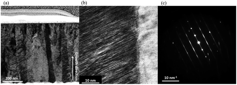
The as-deposited Ni–Mo–W (Fig. 3(a)) thin films consist of densely packed columnar grains with an average width of 55 nm (Fig. 3(b)). The vast majority of the grains comprise finely spaced stripes, most of which are slanted with respect to the loading axis; the average deviation from the loading axis is 23.5°, ranging from 2° to 54° (Fig. 3(c)). The cross-sectional micrograph of the annealed sample (Fig. 3(d)) shows columnar grain width and planar fault orientation that are very close to those of the as-deposited sample (Fig. 3(e) and (f)). A closer look at the stripes reveals that the planar fault spacing in both as-deposited and annealed Ni–Mo–W thin films are mostly in angstrom scale (Fig. 4(a) and (c)). The selected area electron diffraction (SAED) patterns of both as-deposited and annealed Ni–Mo–W thin films show intense streaks penetrating the spots, alluding to the high density of SFs (Fig. 4(b) and (d)).Tensile and fatigue behavior of Ni–Mo–W thin films
The representative stress–strain curves of as-deposited and annealed Ni–Mo–W thin films are plotted in Fig. 5(a). As-deposited Ni–Mo–W thin films show an average tensile strength of 2.83 GPa and an elastic modulus (the initial loading slope) of 170 GPa. The annealed batch has a comparable average tensile strength (2.81 GPa), but the average modulus is slightly higher than that of the as-deposited film (192 GPa). Fig. 5(b) portrays separate sets of tensile test data, in which the films were loaded up to 2600 MPa, fully unloaded, then reloaded until fracture. As-deposited Ni–Mo–W thin film's reloading segment falls between the initial loading and unloading segments, demonstrating an anelastic behavior typically observed in dual-phase steels51–53 and ufg/nc fcc metal thin films.42,54–56 Meanwhile, the annealed specimen shows a different mechanical response, where the unloading segment returns to the origin, and the reloading segment coincides with the initial loading path. The implications of anelasticity in the films are elaborated in the Discussion. | ||
| Fig. 5 (a) Representative stress–strain curves of Ni–Mo–W thin films without unloading. (b) Stress–strain curves of Ni–Mo–W thin films that were fully unloaded and reloaded until failure. | ||
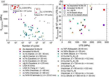
Stress-life (S–N) curves of as-deposited and annealed Ni–Mo–W thin films were obtained from tension–tension fatigue tests (Fig. 6(a)). The S–N curves of nc and ufg Ni,57–60 nc Ni-based alloys,61–64 and nt Cu-based metallic thin films65,66 are also plotted together. For clarity, a subset of representative curves has been chosen; S–N curves of all referenced materials can be found in the ESI.† Since the materials of interest were tested using different load ratios, the stress amplitudes have been adjusted with the modified Goodman relation:
Fig. 6(b) is presented to evaluate whether the superior fatigue behavior of Ni–Mo–W thin films solely originates from their high tensile strengths. The y-axis is the Goodman stress amplitude of the films at 105 cycles normalized by their UTS. The tensile and fatigue strengths of all materials in the legend are juxtaposed. As-deposited Ni–Mo–W thin film's normalized fatigue strength is 77%, well above those of nc Ni and nt Cu-based metals spanning from 17 to 54%. Only nc NiFe alloys have similar normalized strengths. Heat treatment has deteriorated the normalized fatigue strength from 77% to 38%, yet the performance is comparable to that of most other metals plotted together. Both Ni–Mo–W thin films possess an excellent combination of tensile and fatigue strengths, and their fatigue strength cannot be simply ascribed to the high tensile strength.
Microstructural analysis of as-deposited Ni–Mo–W thin film after cyclic loading
A portion of as-deposited Ni–Mo–W subjected to 7.8 × 10⁵ cycles, cyclically loaded between 236.5 and 2365 MPa, was lifted-out near the fracture plane for cross-sectional TEM imaging. The columnar grain morphology and its average width were retained (54 ± 13 nm; Fig. 7(a)) after deformation. The same holds for HRTEM images and SAED patterns: planar fault spacing, its misalignment with respect to the loading direction (23.2 ± 13.4°), and the streaks at diffraction spots were unchanged. Similarly, the specimen that fractured after 1001 cycles, cyclically loaded between 260 and 2600 MPa, showed virtually identical characteristics; the columnar grain width was 58 ± 12 nm, and planar fault orientation was 23.2 ± 10.4°.Discussion
General fatigue behavior of Ni–Mo–W thin films
As-deposited Ni–Mo–W thin film exhibits an abrupt transition from high cycle fatigue (HCF; 105 cycles or above) to low cycle fatigue (LCF; 104 cycles or below) at stress amplitudes of 2240 MPa or above (Fig. 6(a)). Considering that LCF is generally governed by early plastic deformation, the data indicates that significant plastic strain accumulation comes into play at and above that stress level. Annealed Ni–Mo–W on the other hand exhibits a transition from LCF to HCF with a steep gradient. There were no changes in the slope of cyclic load-displacement curves of as-deposited and annealed Ni–Mo–W thin films even when the fracture was imminent (see ESI†), implying that the material did not undergo cyclic hardening or softening. This behavior is in contrast to cyclic hardening and/or softening observed in nc Ni,67,68 driven by dislocation activities or time-dependent deformation.Post-mortem characterization of LCF and HCF fractured thin films did not reveal noticeable microstructural changes (Fig. 7). The absence of dislocation activity and other unusual characteristics near the fracture plane indicates that fatigue failure has occurred in a severely localized manner, such that the responsible deformation mechanism could not come into effect uniformly throughout the microstructure. Based on the aforementioned observations, we can deduce that Ni–Mo–W thin films have excellent crack initiation life at the expense of poor crack propagation life. That is, cracks would propagate instantaneously upon nucleation at a specific “weakest link of the chain” due to localized plastic strain accumulation.
Disparity in the fatigue strengths of as-deposited and annealed specimens
As shown in Fig. 3, there were no conspicuous changes in the texture and microstructure of Ni–Mo–W thin films before and after annealing. A more detailed analysis of the in-plane microstructures of Ni–Mo–W thin films was conducted to examine the presence of nano-scale precipitates. This characterization was deemed necessary as precipitates could exacerbate stress concentration during cyclic loading without adversely affecting the tensile strength.69 However, no signs of precipitates could be identified in the annealed thin films, and there are no macroscopic changes in the in-plane TEM images and SAED patterns after heat treatment (Fig. 8). The SAED ring patterns only include the indices of fcc Ni, and barely any spots deviate from the rings. Overall, all textural and microstructural characterizations conducted in this work preclude the emergence of precipitates in annealed Ni–Mo–W. | ||
| Fig. 8 (a) & (b) In-plane TEM image of as-deposited Ni–Mo–W and the polycrystal's SAED pattern. (c) & (d) Equivalent images of annealed Ni–Mo–W. | ||
Surface asperities are another commonly reported cause of diminished fatigue strength of metals.35 If the surface roughness of annealed Ni–Mo–W thin films is higher than that of its as-deposited counterpart, stress concentration would be a logical description of the disparity in the fatigue strength. However, the average roughness (Ra, in nm) values of as-deposited and annealed Ni–Mo–W thin films barely differ (Fig. 9; 4.17 nm for the as-deposited specimen, and 4.16 nm for the annealed specimen). This outcome rules out exacerbated stress concentration in annealed Ni–Mo–W thin films. In view of the above insights, it is more likely that the fatigue strength difference underlies in subtle changes in the microstructure and the resultant deformation mechanisms.
Possible fatigue failure mechanisms of Ni–Mo–W thin films
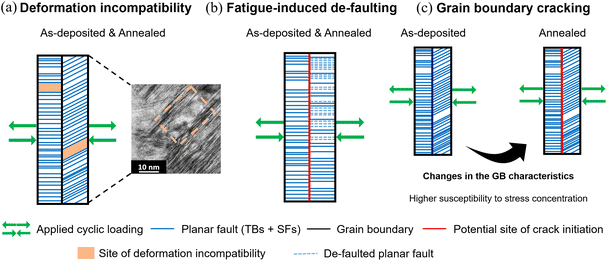
Nano-scale columnar grains and angstrom-scale planar faults would serve as powerful obstacles to dislocation movement in Ni–Mo–W thin films. In particular, both TBs and SFs offer pronounced strengthening as the spacing reduces down to a few nanometers.15,72,73 Impeding dislocation movement effectively delays fatigue crack initiation, and this suitably explains the superb crack initiation life of Ni–Mo–W thin films over the materials juxtaposed in Fig. 6. The poor crack propagation life can be conveniently rationalized by the macroscopically brittle behavior, but the detailed microscopic picture is unknown at this stage. Though the lack of microstructural changes after deformation obscures an accurate analysis, this section conjectures various deformation mechanisms in cyclically loaded Ni–Mo–W thin films based on analogous studies.On the other hand, the GB plasticity postulate can partially explain the gap. The mechanical response of Ni–Mo–W after annealing (Fig. 5(b)) implies that there were changes in the GB characteristics. One notable feature could be the presence of band-like or elliptical/granular nano-scale amorphous phases in the as-deposited films (Fig. S4†). The amorphous regions were identified along or near the GBs of as-deposited thin films, as illustrated in the ESI.† It is widely accepted that amorphous GB phases release shear stress accumulation by promoting GB sliding,77–79 and they are also known to promote steady fatigue crack growth and even plasticity distribution by diffusing the strain concentration at the GBs.80,81 Conversely, no evidence of similar nanostructures could be found in the micrographs of annealed Ni–Mo–W thin films. The lack of strain-accommodating regions could be a source of expedited stress concentration at the GBs of annealed thin films, but the scarcity of clear amorphous phases in the as-deposited films blurs a definitive analysis. Nevertheless, there could be room for annealing-induced changes in the GB characteristics in Ni–Mo–W thin films. Studies on the thermal stability of Ni–Mo–W systems characterized the structural evolution of GBs during heat-treatment at intermediate temperatures (400 °C to 800 °C).33,82 These studies revealed GB migration in Ni85Mo13W2 thin film annealed at 400 °C,33 and GB relaxation in Ni79.4Mo17W3.6 annealed at 525 °C and above.82 Considering these insights, we surmise that GBs in annealed Ni–Mo–W thin films can undergo structural evolution at a microscopic level. An in-depth characterization of the GBs in as-deposited and annealed Ni–Mo–W thin films will be conducted to shed light on the discrepancy, and how the discrepancy affects Ni–Mo–W thin film's susceptibility to stress concentration driven by cyclic loading.
Conclusion
This study investigated the fatigue behavior of freestanding sputter-deposited Ni–Mo–W thin films by conducting pseudo-load-controlled tension–tension fatigue tests and analyzing the microstructure. As-deposited films showed an extraordinary fatigue strength, lasting over 106 cycles at a Goodman stress amplitude of 2190 MPa. The microstructure of Ni–Mo–W remained robust even after cyclic deformation and annealing at 500 °C for 24 hours. The fatigue life of Ni–Mo–W has deteriorated after annealing, but it still exceeds 2 × 10⁵ cycles at a Goodman stress amplitude of 1050 MPa. Plausible origins of Ni–Mo–W's superb fatigue behavior were delineated in terms of planar fault–dislocation interactions and GB plasticity. Both hypotheses are justifiable accounts of fatigue failure in Ni–Mo–W, but the GB plasticity postulate better explains the fatigue strength disparity between the as-deposited and annealed specimens. The GB plasticity postulate links the disparity with structural evolution of GBs upon annealing, rendering the GBs in the annealed thin films more susceptible to stress concentration. A detailed TEM analysis is to be undertaken to substantiate this hypothesis. Ni–Mo–W thin films were already recognized for their high unidirectional strengths as well as thermal and mechanical stability, but this study consolidated their potential as nano-scale structural components by evaluating the reliability under fatigue. Further investigation could reveal whether Ni–Mo–W retains a similar level of fatigue performance under cyclic loading at elevated temperatures, and whether the responsible mechanism changes.Author contributions
JungHun Park: conceptualization, investigation, validation, writing – original draft, writing – review & editing. Yuhyun Park: investigation, methodology, resources, writing – review & editing. Sunkun Choi: methodology, software, writing – review & editing. Zhuo Feng Lee: formal analysis, methodology, writing – review & editing. Gi-Dong Sim: conceptualization, funding acquisition, supervision, writing – review & editing.Conflicts of interest
There are no conflicts to declare.Acknowledgements
This research has been supported by the National Research Foundation of Korea (NRF; NRF-2019M3D1A107922922, NRF-2023M2D2A1A0107814911), and the KAIST UP Program.References
- K. Lu, L. Lu and S. Suresh, Science, 2004, 324, 349–352 CrossRef PubMed.
- L. Lu, X. Chen, X. Huang and K. Lu, Science, 2009, 323, 607–610 CrossRef CAS PubMed.
- L. Lu, Y. Shen, X. Chen, L. Qian and K. Lu, Science, 2004, 304, 422–426 CrossRef CAS PubMed.
- R. T. Ott, J. Geng, M. F. Besser, M. J. Kramer, Y. M. Wang, E. S. Park, R. Lesar and A. H. King, Acta Mater., 2015, 96, 378–389 CrossRef CAS.
- C. Deng and F. Sansoz, Acta Mater., 2009, 57, 6090–6101 CrossRef CAS.
- X. Ke, J. Ye, Z. Pan, J. Geng, M. F. Besser, D. Qu, A. Caro, J. Marian, R. T. Ott, Y. M. Wang and F. Sansoz, Nat. Mater., 2019, 18, 1207–1214 CrossRef CAS PubMed.
- O. Anderoglu, A. Misra, H. Wang and X. Zhang, J. Appl. Phys., 2008, 103(9) DOI:10.1063/1.2913322.
- Y. Wang, E. Ma, R. Z. Valiev and Y. Zhu, Adv. Mater., 2004, 16, 328–331 CrossRef CAS.
- Y. Zhao, T. A. Furnish, M. E. Kassner and A. M. Hodge, J. Mater. Res., 2012, 27, 3049–3057 CrossRef CAS.
- D. Jang, C. Cai and J. R. Greer, Nano Lett., 2011, 11, 1743–1746 CrossRef CAS PubMed.
- A. M. Hodge, T. A. Furnish, C. J. Shute, Y. Liao, X. Huang, C. S. Hong, Y. T. Zhu, T. W. Barbee and J. R. Weertman, Scr. Mater., 2012, 66, 872–877 CrossRef CAS.
- K. Lu, Nat. Rev. Mater., 2016, 1, 16019 CrossRef CAS.
- Z. S. You, L. Lu and K. Lu, Acta Mater., 2011, 59, 6927–6937 CrossRef CAS.
- J. Wang, N. Li, O. Anderoglu, X. Zhang, A. Misra, J. Y. Huang and J. P. Hirth, Acta Mater., 2010, 58, 2262–2270 CrossRef CAS.
- Z. You, X. Li, L. Gui, Q. Lu, T. Zhu, H. Gao and L. Lu, Acta Mater., 2013, 61, 217–227 CrossRef CAS.
- X. Li, Y. Wei, L. Lu, K. Lu and H. Gao, Nature, 2010, 464, 877–880 CrossRef CAS PubMed.
- T. Zhu and H. Gao, Scr. Mater., 2012, 66, 843–848 CrossRef CAS.
- D. Jang, X. Li, H. Gao and J. R. Greer, Nat. Nanotechnol., 2012, 7, 594–601 CrossRef CAS PubMed.
- R. Su, D. Neffati, Y. Zhang, J. Cho, J. Li, H. Wang, Y. Kulkarni and X. Zhang, Mater. Sci. Eng., A, 2021, 803, 140696 CrossRef CAS.
- X. Feng, J. Zhang, K. Wu, X. Liang, G. Liu and J. Sun, Nanoscale, 2018, 10, 13329–13334 RSC.
- R. Su, D. Neffati, S. Xue, Q. Li, Z. Fan, Y. Liu, H. Wang, Y. Kulkarni and X. Zhang, Mater. Sci. Eng., A, 2018, 736, 12–21 CrossRef CAS.
- J. Li, J. Cho, J. Ding, H. Charalambous, S. Xue, H. Wang, X. Li Phuah, J. Jian, X. Wang, C. Ophus, T. Tsakalakos, R. Edwin García, A. K. Mukherjee, N. Bernstein, C. Stephen Hellberg, H. Wang and X. Zhang, Sci. Adv., 2019, 5, eaaw5519 CrossRef CAS PubMed.
- D. Zhang, J. Zhang, T. Xu, Y. Zhang, C. Che, D. Zhang and J. Meng, Mater. Sci. Eng., A, 2022, 845, 143238 CrossRef CAS.
- W. W. Jian, G. M. Cheng, W. Z. Xu, H. Yuan, M. H. Tsai, Q. D. Wang, C. C. Koch, Y. T. Zhu and S. N. Mathaudhu, Mater. Res. Lett., 2013, 1, 61–66 CrossRef CAS.
- C. A. C. Sequeira, D. S. P. Cardoso, L. Amaral, B. Šljukić and D. M. F. Santos, Corros. Rev., 2016, 34, 187–200 CrossRef CAS.
- M. Pröbstle, S. Neumeier, J. Hopfenmüller, L. P. Freund, T. Niendorf, D. Schwarze and M. Göken, Mater. Sci. Eng., A, 2016, 674, 299–307 CrossRef.
- G.-D. Sim, J. A. Krogstad, K. M. Reddy, K. Y. Xie, G. M. Valentino, T. P. Weihs and K. J. Hemker, Sci. Adv., 2017, 3, e1700685 CrossRef PubMed.
- G.-D. Sim, J. A. Krogstad, K. Y. Xie, S. Dasgupta, G. M. Valentino, T. P. Weihs and K. J. Hemker, Acta Mater., 2018, 144, 216–225 CrossRef CAS.
- Y. Park, S. Choi, K.H. Ryou, JH. Park, W. S. Choi, W.-S. Ko, P.-P. Choi and G.-D. Sim, manuscript in preparation.
- G. M. Valentino, P. P. Shetty, A. Chauhan, J. A. Krogstad, T. P. Weihs and K. J. Hemker, Scr. Mater., 2020, 186, 247–252 CrossRef CAS.
- G. M. Valentino, J. A. Krogstad, T. P. Weihs and K. J. Hemker, J. Alloys Compd., 2020, 833, 155024 CrossRef CAS.
- K. Kim, S. Park, T. Kim, Y. Park, G.-D. Sim and D. Lee, J. Alloys Compd., 2022, 919, 165808 CrossRef CAS.
- M. R. He, R. Zhang, R. Dhall, A. M. Minor and K. J. Hemker, Mater. Res. Lett., 2023, 11, 879–887 CrossRef CAS.
- G. M. Valentino, P. P. Shetty, J. A. Krogstad and K. J. Hemker, J. Microelectromech. Syst., 2020, 29, 329–337 CAS.
- R. I. Stephens and H. O. Fuchs, Metal fatigue in engineering, Wiley, 2001 Search PubMed.
- M. M. Saleem and H. Nawaz, Micro Nanosyst., 2019, 11, 11–33 CrossRef CAS.
- R. Schwaiger and O. Kraft, Acta Mater., 2003, 51, 195–206 CrossRef CAS.
- B. Merle and M. Göken, J. Mater. Res., 2014, 29, 267–276 CrossRef CAS.
- T. Kondo, X. C. Bi, H. Hirakata and K. Minoshima, Int. J. Fatigue, 2016, 82, 12–28 CrossRef CAS.
- A. Barrios, C. Kunka, J. Nogan, K. Hattar and B. L. Boyce, Small Methods, 2023, 7(7), 2201591 CrossRef CAS PubMed.
- H. D. Espinosa, B. C. Prorok and M. Fischer, A methodology for determining mechanical properties of freestanding thin ÿlms and MEMS materials, 2003, vol. 51 Search PubMed.
- H. Kim, J.-H. Choi, Y. Park, S. Choi and G.-D. Sim, J. Mech. Phys. Solids, 2023, 173, 105209 CrossRef CAS.
- Z. F. Lee, H. Ryu, J.-Y. Kim, H. Kim, J.-H. Choi, I. Oh and G.-D. Sim, Mater. Sci. Eng., A, 2024, 892, 146028, DOI:10.1016/j.msea.2023.146028.
- G.-D. Sim, J. H. Park, M. D. Uchic, P. A. Shade, S. B. Lee and J. J. Vlassak, Acta Mater., 2013, 61, 7500–7510 CrossRef CAS.
- C. Eberl, R. Thompson, D. Gianola and S. Bundschuh, Digital Image Correlation and Tracking with Matlab, 2012 Search PubMed.
- S. Choi, J. H. Park, Y. Park, H. Ryu, Z. F. Lee and G.-D. Sim, manuscript in preparation.
- G. A. Webster and A. N. Ezeilo, Int. J. Fatigue, 2001, 23, 375–383 CrossRef.
- B. Geetha Priyadarshini, S. Aich and M. Chakraborty, Bull. Mater. Sci., 2014, 37, 1265–1273 CrossRef CAS.
- C. Xiao, R. A. Mirshams, S. H. Whang and W. M. Yin, Mater. Sci. Eng., A, 2001, 301, 35–43 CrossRef.
- W. Nix and W. Cai, Imperfections in Crystalline Solids, Cambridge University Press, 1st edn, 2016 Search PubMed.
- D. Li and R. H. Wagoner, Acta Mater., 2021, 206, 116625 CrossRef CAS.
- H. Kim, C. Kim, F. Barlat, E. Pavlina and M. G. Lee, Mater. Sci. Eng., A, 2013, 562, 161–171 CrossRef CAS.
- E. J. Pavlina, M. G. Lee and F. Barlat, Metall. Mater. Trans. A, 2015, 46, 18–22 CrossRef CAS.
- R. P. Vinci, G. Cornella and J. C. Bravman, AIP, 2011, pp. 240–248 Search PubMed.
- G.-D. Sim and J. J. Vlassak, Scr. Mater., 2014, 75, 34–37 CrossRef CAS.
- I. Oh, H. Kim, H. Son, S. Nam, H. Choi and G.-D. Sim, Int. J. Plast., 2023, 161, DOI:10.1016/j.ijplas.2023.103515.
- T. Hanlon, Y. N. Kwon and S. Suresh, Scr. Mater., 2003, 49, 675–680 CrossRef CAS.
- P. Cavaliere, Int. J. Fatigue, 2009, 31, 1476–1489 CrossRef CAS.
- H. S. Cho, K. J. Hemker, K. Lian and J. Goettert, Technical Digest, in MEMS 2002 IEEE International Conference.
- Y. Yang, B. I. Imasogie, S. M. Allameh, B. Boyce, K. Lian, J. Lou and W. O. Soboyejo, Mater. Sci. Eng., A, 2007, 444, 39–50 CrossRef.
- N. M. Heckman, H. A. Padilla, J. R. Michael, C. M. Barr, B. G. Clark, K. Hattar and B. L. Boyce, Int. J. Fatigue, 2020, 134, 105472 CrossRef CAS.
- M. Y. Li, Z. X. Wang, B. Zhang, F. Liang, X. M. Luo and G. P. Zhang, Scr. Mater., 2023, 222, 114995 CrossRef CAS.
- S. Kobayashi, A. Kamata and T. Watanabe, in Journal of Physics: Conference Series, Institute of Physics Publishing, 2010, vol. 240 Search PubMed.
- B. L. Boyce and H. A. Padilla, Metall. Mater. Trans. A, 2011, 42, 1793–1804 CrossRef CAS.
- N. M. Heckman, M. F. Berwind, C. Eberl and A. M. Hodge, Acta Mater., 2018, 144, 138–144 CrossRef CAS.
- C. J. Shute, B. D. Myers, S. Xie, S. Y. Li, T. W. Barbee, A. M. Hodge and J. R. Weertman, Acta Mater., 2011, 59, 4569–4577 CrossRef CAS.
- B. Moser, T. Hanlon, K. S. Kumar and S. Suresh, Scr. Mater., 2006, 54, 1151–1155 CrossRef CAS.
- S. Cheng, J. Xie, A. D. Stoica, X. L. Wang, J. A. Horton, D. W. Brown, H. Choo and P. K. Liaw, Acta Mater., 2009, 57, 1272–1280 CrossRef CAS.
- K. Hockauf, M. F. X. Wagner, T. Halle, T. Niendorf, M. Hockauf and T. Lampke, Acta Mater., 2014, 80, 250–263 CrossRef CAS.
- Q. S. Pan, Q. H. Lu and L. Lu, Acta Mater., 2013, 61, 1383–1393 CrossRef CAS.
- Q. S. Pan, H. Zhou, Q. Lu, H. Gao and L. Lu, Nature, 2017, 551, 214–217 CrossRef CAS PubMed.
- H. Zhou, X. Li, S. Qu, W. Yang and H. Gao, Nano Lett., 2014, 14, 5075–5080 CrossRef CAS PubMed.
- W. W. Jian, G. M. Cheng, W. Z. Xu, C. C. Koch, Q. D. Wang, Y. T. Zhu and S. N. Mathaudhu, Appl. Phys. Lett., 2013, 103, 133108, DOI:10.1063/1.4822323.
- Q. Fang and F. Sansoz, Acta Mater., 2021, 212, 116925 CrossRef CAS.
- H. M. Ledbetter and R. P. Reed, J. Phys. Chem. Ref. Data, 1973, 2, 531–618 CrossRef CAS.
- A. J. Kalkman, A. H. Verbruggen and G. C. A. M. Janssen, Appl. Phys. Lett., 2001, 78, 2673–2675 CrossRef CAS.
- S. Guo and H. Sun, Acta Mater., 2021, 218, 117212, DOI:10.1016/j.actamat.2021.117212.
- K. Madhav Reddy, J. J. Guo, Y. Shinoda, T. Fujita, A. Hirata, J. P. Singh, J. W. McCauley and M. W. Chen, Nat. Commun., 2012, 3, 1052, DOI:10.1038/ncomms2047.
- I. Szlufarska, A. Nakano and P. Vashishta, Science, 2005, 309, 911–913 CrossRef CAS PubMed.
- A. Khalajhedayati, Z. Pan and T. J. Rupert, Nat. Commun., 2016, 7, 10802, DOI:10.1038/ncomms10802.
- J. D. Schuler, C. M. Barr, N. M. Heckman, G. Copeland, B. L. Boyce, K. Hattar and T. J. Rupert, JOM, 2019, 71, 1221–1232 CrossRef CAS.
- D. Zeng, J. Li, Y. Shi, X. Li and K. Lu, Nano Res., 2023, 16, 12800–12808 CrossRef CAS.
Footnote |
| † Electronic supplementary information (ESI) available. See DOI: https://doi.org/10.1039/d4nr01033g |
| This journal is © The Royal Society of Chemistry 2024 |


