DOI:
10.1039/D2DT03402F
(Paper)
Dalton Trans., 2023,
52, 1543-1550
Aerosol-assisted synthesis of titania-based spherical and fibrous materials with a rational design of mesopores using PS-b-PEO†
Received
20th October 2022
, Accepted 2nd December 2022
First published on 3rd December 2022
Abstract
Surfactant-assisted synthesis is a promising technique for the tailor-made design of highly porous metal oxide based nanomaterials. There has been a demand for the comprehensive design of their morphology, porous structure and crystallinity to extend potential applications using metal oxide based materials such as titania (TiO2). However, the porous structure is often deformed and/or destroyed during the process of crystallizing metal oxide frameworks. Herein, the aerosol-assisted synthesis of mesoporous TiO2 powders was conducted in the presence of high-molecular-weight poly(styrene)-block-poly(ethylene oxide) (PS-b-PEO), which improved the stability of the derivative mesoporous structure with an increase in the thickness of the TiO2 frameworks. To propose a rational synthetic route for stable and porous metal oxides, the resultant mesoporous structure and the textural morphology of the mesoporous TiO2 powders were surveyed using PS-b-PEO with different lengths of PS and PEO chains. By a judicious choice of the molecular structure of PS-b-PEO, the morphological design of the fully crystallized anatase phase of TiO2 from spherical to fibrous ones was achieved with control over the mesopore diameter.
Introduction
A wide variety of titania (TiO2) based materials with different structural features, such as crystalline phase, oxidative state, porosity, and morphology, have been investigated to explore unique functions as metal supported catalysts, photocatalysts, gas sensors, photovoltaic cells, rechargeable batteries, and so on.1–5 For this purpose, highly porous structures are interesting because more active sites are exposed to improve their properties.6–8 Surfactant-assisted mesoporous TiO2 is a potential candidate with the precise design of a wide range of structures, such as texture, morphology, mesostructure, and crystal structure.9–12 Such mesoporous structures are also helpful for enhancing the adsorption capacity of reactants and opening channels for mass transportation.13–15 However, as in the case of other metal oxides such as alumina,16,17 the synthesis of TiO2 based materials showing high ordering of surfactant-assisted mesopores, especially of powders, is quite difficult because starting chemicals such as the corresponding alkoxide and chloride are very reactive in the presence of water.7,18,19
By solving this fundamental issue so that such sol–gel reactions have to slow down, the evaporation-induced self-assembly (EISA) approach has been applied as one of the most powerful strategies for the rapid solidification of soluble metal oxide species with the self-assembly of amphiphilic organic molecules.7 According to the EISA approach, metal oxide powders can be recovered with the formation of uniform mesopores through a process of drying precursor solutions, and corresponding mesoporous films are fabricated by dip- and spin-coating techniques.6,20–24 In the case of mesoporous TiO2 films, sol–gel reactions proceed even after such solidification by spin-coating and then the resultant film is often cooled immediately in a freezer.24–26 In addition, utilization of an unreactive phosphate spacer has been proposed to reduce the opportunity for titanium oxo species to react after spin-coating.27 Application of the drying process seems to depend on experience because of the difficulty in controlling the evaporation rate of solvents (e.g., EtOH and H2O). To provide a general and reproducible method to synthesize highly porous TiO2, we have focused on a spray-drying process suitable for rapid solvent evaporation to prepare mesoporous metal oxide powders.18,28–30
A pioneering paper showed the usefulness of the spray-drying process to obtain spherical particles of highly porous metal oxides using poly(oxyethylene)-block-poly(oxypropylene)-block-poly(oxyethylene) (EOnPOmEOn) type triblock copolymers.18 Although a number of papers have been found for the synthesis of ordered mesoporous titania using EOnPOmEOn type triblock copolymers,18,31–34 the crystallinity of titania based frameworks has hardly been improved because the thickness of TiO2 frameworks is not sufficient around EOnPOmEOn templated mesopores of several nanometers in diameter. Thus, the thickness of TiO2 frameworks should be increased with a drastic enlargement in surfactant-assisted mesopores, as has already been confirmed in several cases of unique TiO2 films.35–39
In this study, we demonstrate the aerosol-assisted synthesis of a series of TiO2 powder samples using PS-b-PEO type diblock copolymers, investigate the crystallinity of TiO2 frameworks, explain the possibility of controlling pore size with a rational understanding of the basis of the molecular structure of PS-b-PEO, and provide an additional advance in the spray-drying process to obtain fiber-like mesoporous TiO2 by tuning the viscosity of the precursor solutions. The fibrous type with higher specific surface area and a larger pore volume will have potential as porous media for catalytic applications by using many active site and open channels.40–43
Experimental
Chemicals
Titanium tetraisopropoxide (TTIP), concentrated hydrochloric acid (conc. HCl, 35–37%), and tetrahydrofuran (THF, dehydrated) were purchased from FUJIFILM Wako Pure Chemical Co. A series of PS-b-PEO type diblock copolymers, e.g. PS40000-b-PEO53000 (Mw/Mn; 1.08) and PS58600-b-PEO71000 (Mw/Mn; 1.03), were purchased from Polymer Source Inc.
Aerosol-assisted synthesis of mesoporous TiO2 powders
Referring to the literature,44 a series of clear precursor solutions was prepared by using PS-b-PEO with different molecular structures. As a typical procedure, PS-b-PEO (0.08 mmol) was completely dissolved at 40 °C in THF (6.0 mL) for several minutes and then stirred for another 60 min. TTIP (0.6 mL) was added dropwise to the PS-b-PEO solution and combined with another THF (1.84 mL) solution containing conc. HCl (0.16 mL). The resultant solution was stirred for 60 min, spray-dried at an inlet temperature of 140 °C to recover a powder sample (Spray Dryer ADL311S-A, Yamato Scientific Co., Ltd) and calcined at 400 °C and higher for 3 h in a dry air flow at a heating rate of 2 °C min−1.
Characterization
To visualize the mesoporous structure and morphology, all the resultant mesoporous TiO2 powder samples were observed using field emission scanning electron microscopy (FE-SEM, HITACHI SU9000) and transmission electron microscopy (TEM, JEOL JEM-2010, operated at 200 kV). To evaluate porosity, N2 adsorption–desorption isotherms were recorded at 77 K using a volumetric gas sorption analyzer (Anton Paar – Quantachrome Instruments Autosorb-iQ). All the samples were heated at 110 °C for 6 h under a vacuum prior to the measurements. BET surface area and total pore volume were calculated using the adsorption data from P/P0 = 0.05 to 0.30 and the adsorption volume of N2 at P/P0 = 0.99, respectively. The pore size distribution curve was drawn from the desorption branch using the Barrett–Joyner–Halenda (BJH) method. To evaluate the crystallinity of TiO2 frameworks after calcination at 400 °C and higher in air, X-ray diffraction (XRD) patterns of the samples were recorded using a Rigaku RINT 2100 with monochromated Cu Kα radiation. To discuss the variation in resultant morphology, the viscosity of the precursor solutions was measured by using a Tuning-fork Vibro viscometer (A&D Company, Ltd, SV-1A).
Results and discussion
A variety of mesoporous TiO2 powder samples were synthesized using PS-b-PEO type asymmetric diblock copolymers through the spray-drying process of the corresponding precursor solutions. By changing the molecular structure of PS-b-PEO, the porosity of the resultant TiO2 powder samples was altered with the molecular weights of PS and PEO chains. Besides, the morphology was varied by considering the viscosity of the precursor solutions with an outstanding increase in the specific surface area (see Table 1). In this study, we demonstrated such significant insights by categorizing the synthesis of spherical particles and the control of pore size by considering the molecular structure of PS-b-PEO as well as the synthesis of fibrous particles by understanding the viscosity of the precursor solutions.
Table 1 Porosity and morphology of samples synthesized using a variety of PS-b-PEO type diblock copolymers
| PS-b-PEO |
S
BET (m2 g−1) |
V
total (cm3 g−1) |
D
BJH (nm) |
Morphology |
40![[thin space (1/6-em)]](https://www.rsc.org/images/entities/char_2009.gif) 000-b-53000 000-b-53000 |
71 |
0.34 |
19 |
Spherical |
| 18000-b-7500 |
103 |
0.26 |
8 |
Spherical, recessed |
| 35000-b-17000 |
88 |
0.40 |
18 |
Spherical |
| 51000-b-28000 |
63 |
0.36 |
25 |
Spherical |
| 18000-b-39000 |
55 |
0.18 |
4 |
Spherical |
| 58600-b-71000 |
189 |
0.65 |
16 |
Fibrous |
| 65000-b-97000 |
148 |
0.76 |
19 |
Fibrous |
Synthesis of spherical mesoporous TiO2 powders and its pore size variation
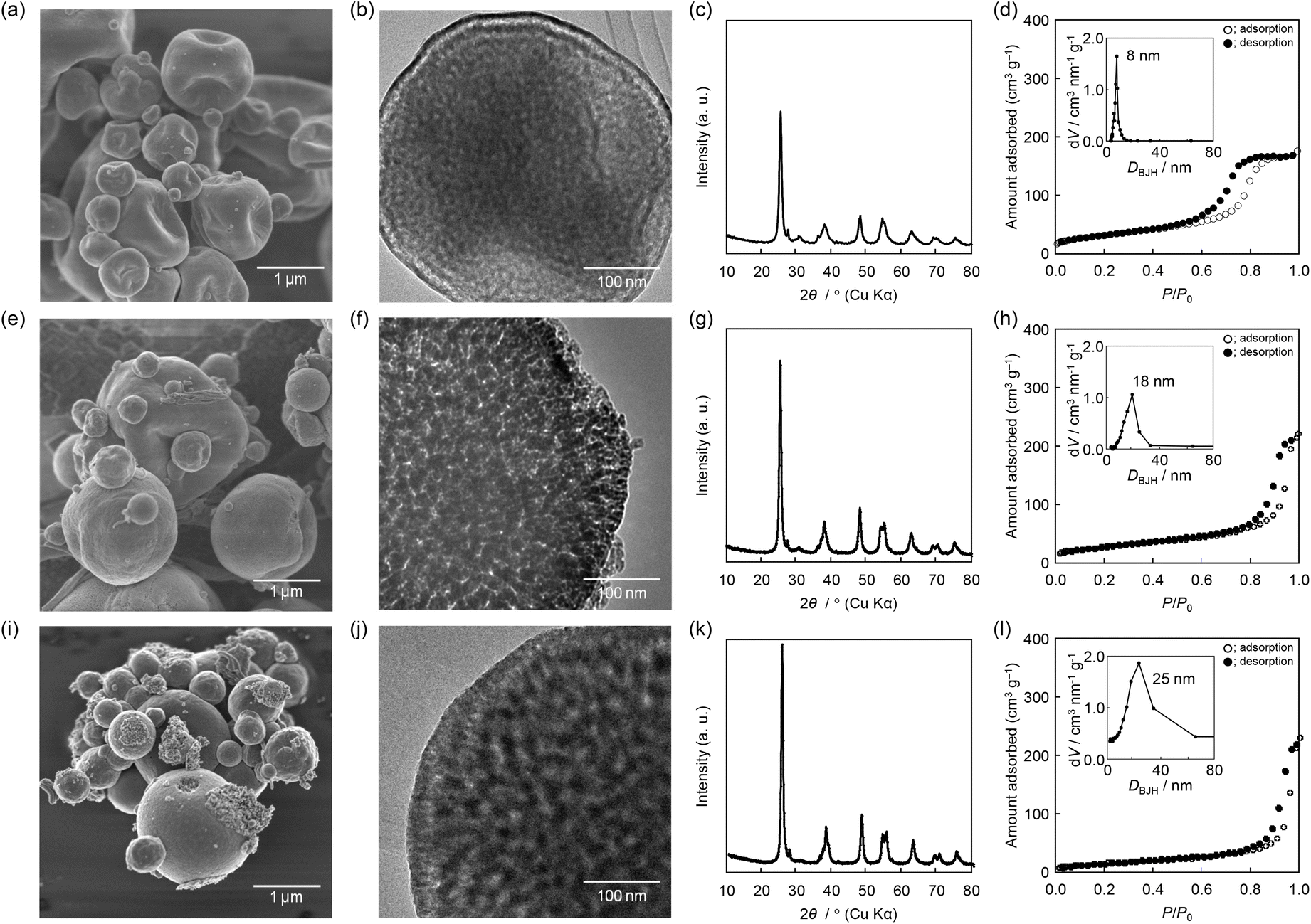
A mesoporous TiO2 powder sample was synthesized using PS40000-b-PEO53000 through the spray-drying process. Spherical particles, being typically recovered through the aerosol-assisted synthesis of mesoporous metal oxides,30,45 were observed in the low-magnification SEM image (Fig. 1a). The high-magnification SEM (Fig. 1b) and TEM images (Fig. 1c) showed the presence of uniform mesopores throughout the whole particle. The mesoporous structure was also characterized by using the data from the N2 adsorption–desorption isotherm (Fig. 1d) showing a steep increase in the adsorption at P/P0 > 0.8 with a hysteresis loop. The pore size distribution curve was centered at around 19 nm (Fig. 1d, inset). The specific surface area (71 m2 g−1) and the total pore volume (0.34 cm3 g−1) were higher than those observed for bulk TiO2.46,47 In addition, the XRD pattern showed that the TiO2 framework was sufficiently crystallized after calcination at 400 °C for 3 h in air (Fig. 1e). All the strong XRD peaks at 2θ = 25°, 38°, 48°, and 63° can be assigned to (101), (004), (200), and (204) reflections of the anatase phase of TiO2,48 which was related to the presence of the corresponding lattice fringes such as the (101) reflection by high-magnification TEM observation (Fig. 1f). Considering the presence of XRD peaks further assignable to the (105), (211), (116), (220) and (215) planes, the crystallinity is much higher than that of other mesoporous TiO2 based materials.18,31
 |
| | Fig. 1 (a) SEM, (b) high-magnification SEM images, (c) TEM image, (d) N2 adsorption–desorption isotherm with corresponding pore size distribution curve, (e) XRD pattern and (f) high-magnification TEM image of a sample prepared using PS40000-b-PEO53000. | |
A high molecular weight of PS-b-PEO generally induces the formation of larger mesopores.49,50 Considering the self-assembly of PS-b-PEO molecules with soluble metal species, PS moieties are aggregated as cores of the micelles and PEO chains interact with metal species and/or solvent surrounding the PS cores, suggesting that the molecular weight of the PS chain is related mainly to the resultant porosity, such as the size and volume of pores, and the PEO chain plays an important role in the control of textural morphology. Our previous study on the aerosol-assisted synthesis of mesoporous alumina (Al2O3) powders using PS-b-PEO revealed the importance of the PS/PEO ratio in changing the pore size.26,51 In this context, several PS-b-PEO type asymmetric diblock copolymers with similar molecular structure (PS > PEO, with the PS/PEO value ranging from 1.8 to 2.4) were utilized to control the mesopore size. All the other samples were fully crystallized as the anatase phase of titania (see Fig. 2c, g and k).
 |
| | Fig. 2 (a) SEM and (b) TEM images of a sample prepared using PS18000-b-PEO7500 and (c) its XRD pattern and (d) N2 adsorption–desorption isotherm with corresponding pore size distribution curve, as well as those of samples prepared using (e–h) PS35000-b-PEO17000 and (i–l) PS51000-b-PEO28000. | |
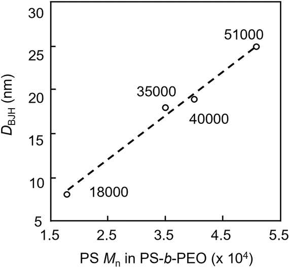
The SEM image of a mesoporous TiO2 powder sample prepared using PS18000-b-PEO7500 showed a recessed spherical morphology (Fig. 2a), though uniform mesopores were distributed throughout the whole particle (Fig. 2b). The morphology gradually changed into spherical and nicely shaped ones by utilizing longer PEO chains (Fig. 2e and i) as the pore size increased with the increase in the molecular weight of PS-b-PEO (Fig. 2f and j). As summarized in Table 1, the average pore size was controlled from ∼8 nm to ∼25 nm with the increase in the molecular weight of PS-b-PEO, mainly the PS chain (Fig. 3).50,51 Considering such an expansion of surfactant-assisted mesopores, a tendency to decrease the specific surface area (from 103 m2 g−1 to 63 m2 g−1) but with an increase in total pore volume (from 0.26 cm3 g−1 to 0.40 cm3 g−1) is quite reasonable.52,53 A mesoporous TiO2 powder sample was additionally prepared using a different kind of PS-b-PEO (PS < PEO, PS/PEO = 0.46) like PS18000-b-PEO39000. As expected, the morphology was a well-shaped one using PS-b-PEO with a longer PEO chain, though the mesoporous structure was altered to that showing a H2 hysteresis loop.54
 |
| | Fig. 3 Relationship between PS Mn in PS-b-PEO and DBJH. | |
Synthesis of fibrous mesoporous TiO2 powders and its understanding
Even in the synthesis with the spray-drying process, we observed the formation of a much bulkier powder sample showing a fibrous morphology in the presence of PS58600-b-PEO71000. The sample was characterized by SEM, TEM, XRD, etc., indicating the successful recovery of the fibrous sample with the formation of unique mesopores (Fig. 4a and b). The fibrous sample fully contained spherically shaped mesopores (Fig. 4c); the specific surface area was 189 m2 g−1 and the larger pore volume was 0.65 cm3 g−1 (Fig. 4d), higher than those observed for the spherical ones. In this case, an enhancement in the porosity would be achieved with an increase in the pore connectivity.26 The unique fibrous morphology should arise from an extension of the PEO chain, but using PS-b-PEO gave a small molecular structure (PS < PEO, with the PS/PEO value less than 1.0). A fibrous mesoporous TiO2 powder sample was also recovered by using PS65000-b-PEO97000. The specific surface area (148 m2 g−1) and total pore volume (0.76 cm3 g−1) were very high. All the XRD patterns showed complete crystallization of TiO2 frameworks to its anatase phase after calcination at 400 °C for 3 h in air (Fig. 4e). The average pore sizes were ∼16 nm (PS58600-b-PEO71000) and ∼19 nm (PS65000-b-PEO97000), somewhat smaller than expected. This tendency would come from the preferential formation of ellipsoidal mesopores deviating from soft-templated spherical pores by restriction during the formation of a fibrous morphology (Fig. 4f).26,55 The SEM image showed that the length of the fiber ranged from 5 μm to 10 μm with a diameter of 0.2–1.0 μm. In addition, spherical (PS40000-b-PEO53000) and fibrous (PS58600-b-PEO71000) samples were typically calcined at higher temperatures like 700 °C. Both samples were transformed to nonporous spherical and fibrous morphologies with the grain growth of the rutile phase (Fig. S1†). It was much easier to transform the crystal phase from anatase to rutile of the fibrous sample than the spherical sample.
 |
| | Fig. 4 (a) SEM, (b) high-magnification SEM images, (c) TEM image, (d) N2 adsorption–desorption isotherm with corresponding pore size distribution curve, (e) XRD pattern and (f) high-magnification TEM image of a sample prepared using PS58600-b-PEO71000. | |
In a spray-drying process, the atomizer generates droplets from a precursor solution to minimize the surface energy and accelerate solvent evaporation from the droplets.30,45 The morphology of the resultant powder sample is strongly related to the initial diameter of the droplets. The droplet diameter (Dd) can be described as follows:56 Among these three factors, the viscosity fluctuates with increasing concentration of the PEO chain,49,57,58 which may be vitally attributed to the morphological change from spherical to fibrous.
Kf,
Q,
n – equipment constants (pressure, carrier gas velocity,
etc.),
ρ,
a – density and its power constant,
σ,
b – surface tension and its power constant,
μ,
c – viscosity and its power constant.
The viscosity of a precursor solution can be decreased very easily by diluting with the same solvent as the original precursor solution. To investigate an effect of viscosity, the precursor solution containing PS65000-b-PEO97000 was diluted with different volumes of THF (originally 8 mL, diluted by addition to total 16 mL, 32 mL and 64 mL) followed by spray drying. By increasing the amount of THF, the viscosity was dramatically decreased from 19.8 mPa s to 1.75 mPa s (Fig. 5). The morphology was then changed from fibrous to fibrous/spherical particles with dimples and aggregated particles (Fig. 6a, d, g and j). Droplets of a high-viscosity precursor solution were changed into ellipsoidal ones through evaporation-induced solidification during the formation of such a fibrous morphology. In contrast, small droplets are produced from a low-viscosity precursor solution, which may provoke a geometrical crashing in the self-assembling process, resulting in the formation of an aggregated foam-like morphology. The N2 adsorption–desorption isotherms are displayed with specific surface areas and total pore volumes (Fig. 6c, f, i and l). Related to the lower porosity of particles with dimples and aggregated particles, the presence of PS-b-PEO templated mesopores was not adequately confirmed, though cavitation was observed in the desorption branches. As in the case of mesoporous TiO2 powders showing spherical and fibrous morphologies, all the XRD patterns revealed full crystallization to the anatase phase of titania by calcination under similar conditions (Fig. 6b, e, h and k).
 |
| | Fig. 5 Viscosity of precursor solution and morphological structure of product with various surfactant concentrations. | |
 |
| | Fig. 6 (a) SEM image, (b) XRD pattern and (c) N2 adsorption–desorption isotherm with corresponding pore distribution curve of a sample synthesized using PS65000-b-PEO97000 dissolved in 8 mL of THF, and those totalling (d–f) 16 mL; (g–i) 32 mL and (j–l) 64 mL. | |
Conclusions
A diverse series of mesoporous TiO2 particles was prepared in the presence of PS-b-PEO type amphiphilic organic molecules, being advantageous for designing large-sized mesopores, through the spray-drying process, with control over pore size and variation between fiber-like and almost spherical morphologies. With sufficient thickness of TiO2 frameworks, the frameworks can be fully crystallized through calcination even at 400 °C in air. In particular, fibrous mesoporous TiO2 powders showed higher specific surface area and larger pore volume. As illustrated in Scheme 1 as the significant finding in this study, the pore size mainly varies with the PS chain length of PS-b-PEO. A unique hierarchical mesoporous fibrous sample is successfully obtained by extending the PEO chain related to the increase in viscosity of a precursor solution, expecting the usage of fully crystalized and highly porous metal oxide media for emerging applications.
 |
| | Scheme 1 Schematic illustration for the control of pore size and morphology of mesoporous TiO2 powders using PS-b-PEO. | |
Conflicts of interest
There are no conflicts to declare.
Acknowledgements
This work was supported by the project (JPNP18016) commissioned by the New Energy and Industrial Technology Development Organization (NEDO).
References
- W. Li, Z. Wu, J. Wang, A. A. Elzatahry and D. Zhao, Chem. Mater., 2013, 26, 287–298 CrossRef.
- X. Chen and S. S. Mao, Chem. Rev., 2007, 107, 2891–2959 CrossRef CAS PubMed.
- A. Fujishima, T. N. Rao and D. A. Tryk, J. Photochem. Photobiol., C, 2000, 1, 1–21 CrossRef CAS.
- A. L. Linsebigler, G. Lu and J. T. Yates Jr., Chem. Rev., 1995, 95, 735–758 CrossRef CAS.
- Y. Kuwahara and H. Yamashita, J. Mater. Chem., 2011, 21, 2407–2416 RSC.
- S. Agarwala, M. Kevin, A. S. Wong, C. K. Peh, V. Thavasi and G. W. Ho, ACS Appl. Mater. Interfaces, 2010, 2, 1844–1850 CrossRef CAS PubMed.
- R. Zhang, A. A. Elzatahry, S. S. Al-Deyab and D. Zhao, Nano Today, 2012, 7, 344–366 CrossRef CAS.
- J. Tang, Y. Wu, E. W. McFarland and G. D. Stucky, Chem. Commun., 2004, 14, 1670–1671 RSC.
- K. Wang, M. Janczarek, Z. Wei, T. Raja-Mogan, M. Endo-Kimura, T. M. Khedr, B. Ohtani and E. Kowalska, Catalysts, 2019, 9, 1054 CrossRef CAS.
- M. T. Noman, M. A. Ashraf and A. Ali, Environ. Sci. Pollut. Res. Int., 2019, 26, 3262–3291 CrossRef CAS PubMed.
- K. Nakata and A. Fujishima, J. Photochem. Photobiol., C, 2012, 13, 169–189 CrossRef CAS.
- C. Yu, J. Fan, B. Tian and D. Zhao, Chem. Mater., 2004, 16, 889–898 CrossRef CAS.
- A. Taguchi and F. Schüth, Microporous Mesoporous Mater., 2005, 77, 1–45 CrossRef CAS.
- A. A. Ismail, D. W. Bahnemann, L. Robben, V. Yarovyi and M. Wark, Chem. Mater., 2009, 22, 108–116 CrossRef.
- H. Shibata, T. Ogura, T. Mukai, T. Ohkubo, H. Sakai and M. Abe, J. Am. Chem. Soc., 2005, 127, 16396–16397 CrossRef CAS PubMed.
- H. Maruoka, A. Tomita, Z. Liu and T. Kimura, Langmuir, 2018, 34, 13781–13787 CrossRef CAS PubMed.
- H. Maruoka and T. Kimura, New J. Chem., 2019, 43, 7269–7274 RSC.
- C. K. Tsung, J. Fan, N. Zheng, Q. Shi, A. J. Forman, J. Wang and G. D. Stucky, Angew. Chem., Int. Ed., 2008, 47, 8682–8686 CrossRef CAS PubMed.
- D. M. Antonelli and J. Y. Ying, Angew. Chem., Int. Ed. Engl., 1995, 34, 2014–2017 CrossRef CAS.
- V. Malgras, Y. Shirai, T. Takei and Y. Yamauchi, J. Am. Chem. Soc., 2020, 142, 15815–15822 CrossRef CAS PubMed.
- Q. Lian, M. Z. Mokhtar, D. Lu, M. Zhu, J. Jacobs, A. B. Foster, A. G. Thomas, B. F. Spencer, S. Wu, C. Liu, N. W. Hodson, B. Smith, A. Alkaltham, O. M. Alkhudhari, T. Watson and B. R. Saunders, ACS Appl. Mater. Interfaces, 2020, 12, 18578–18589 CrossRef CAS PubMed.
- J. H. Pan, X. S. Zhao and W. I. Lee, Chem. Eng. J., 2011, 170, 363–380 CrossRef CAS.
- P. Yang, D. Zhao, D. I. Margolese, B. F. Chmelka and G. D. Stucky, Chem. Mater., 1999, 11, 2813–2826 CrossRef CAS.
- C.-W. Wu, T. Ohsuna, M. Kuwabara and K. Kuroda, J. Am. Chem. Soc., 2006, 128, 4544–4545 CrossRef CAS PubMed.
- T. Kimura, Y. Yamauchi and N. Miyamoto, Chem. – Eur. J., 2010, 16, 12069–12073 CrossRef CAS PubMed.
- T. Kimura, Chem. – Asian J., 2011, 6, 3236–3242 CrossRef CAS PubMed.
- T. Kimura, Y. Yamauchi and N. Miyamoto, Chem. – Eur. J., 2011, 17, 4005–4011 CrossRef CAS PubMed.
- A. B. D. Nandiyanto, T. Ogi, W.-N. Wang, L. Gradon and K. Okuyama, Adv. Powder Technol., 2019, 30, 2908–2924 CrossRef.
- J. Choi, K. S. Yoo and J. Kim, Korean J. Chem. Eng., 2018, 35, 2480–2486 CrossRef CAS.
- R. Vehring, Pharm. Res., 2008, 25, 999–1022 CrossRef CAS PubMed.
- M. Pal, L. Wan, Y. Zhu, Y. Liu, Y. Liu, W. Gao, Y. Li, G. Zheng, A. A. Elzatahry, A. Alghamdi, Y. Deng and D. Zhao, J. Colloid Interface Sci., 2016, 479, 150–159 CrossRef CAS PubMed.
- M. A. Abdolahi Sadatlu and N. Mozaffari, Sol. Energy, 2016, 133, 24–34 CrossRef CAS.
- S. K. Das, M. K. Bhunia and A. Bhaumik, Dalton Trans., 2010, 39, 4382–4390 RSC.
- C. Yu, J. C. Yu and M. Chan, J. Solid State Chem., 2009, 182, 1061–1069 CrossRef CAS.
- D. Chandra, M. Bekki, M. Nakamura, S. Sonezaki, T. Ohji, K. Kato and T. Kimura, J. Mater. Chem., 2011, 21, 5738–5744 RSC.
- D. Chandra, T. Ohji, K. Kato and T. Kimura, Phys. Chem. Chem. Phys., 2011, 13, 12529–12535 RSC.
- T. Kimura, Macromol. Rapid Commun., 2013, 34, 423–430 CrossRef CAS PubMed.
- T. Kimura, J. Mater. Chem. A, 2014, 2, 10688–10696 RSC.
- T. Kimura, M. Shintate and N. Miyamoto, Chem. Commun., 2015, 51, 1230–1233 RSC.
- A. I. Hochbaum, R. Chen, R. D. Delgado, W. Liang, E. C. Garnett, M. Najarian, A. Majumdar and P. Yang, Nature, 2008, 451, 163–167 CrossRef CAS PubMed.
- K. Lim, Y.-M. Jo, J.-W. Yoon and J.-H. Lee, J. Mater. Chem. A, 2019, 7, 24919–24928 RSC.
- Y. Zhu, J. Li, M. Wan and L. Jiang, Macromol. Rapid Commun., 2008, 29, 239–243 CrossRef CAS.
- Z. Yu, H. Liu, M. Zhu, Y. Li and W. Li, Small, 2021, 17, e1903378 CrossRef PubMed.
- S. Yin, L. Song, S. Xia, Y. Cheng, N. Hohn, W. Chen, K. Wang, W. Cao, S. Hou and P. Müller-Buschbaum, Small Methods, 2020, 4, 1900689 CrossRef CAS.
- A. B. D. Nandiyanto and K. Okuyama, Adv. Powder Technol., 2011, 22, 1–19 CrossRef CAS.
- S. Wu, X. Li, Y. Tian, Y. Lin and Y. H. Hu, Chem. Eng. J., 2021, 406, 126747 CrossRef CAS.
- S. K. Parayil, H. S. Kibombo, L. Mahoney, C.-M. Wu, M. Yoon and R. T. Koodali, Mater. Lett., 2013, 95, 175–177 CrossRef CAS.
- X. Zhang, W. Liao, W. Mu, D. Zheng, Y. Zhou, B. Xue, W. Liu, Z. Lin and Y. Deng, J. Mater. Chem. A, 2014, 2, 11035–11039 RSC.
- B. Q. Kim, Y. Jung, M. Seo and S. Q. Choi, Langmuir, 2018, 34, 10293–10301 CrossRef CAS PubMed.
- R. B. Cheyne and M. G. Moffitt, Langmuir, 2006, 22, 8387–8396 CrossRef CAS PubMed.
- M. S. Islam, R. Wakabayashi and T. Kimura, Dalton Trans., 2021, 50, 7191–7197 RSC.
- H. Maruoka and T. Kimura, Bull. Chem. Soc. Jpn., 2019, 92, 1859–1866 CrossRef CAS.
- T. Kimura and H. Maruoka, Chem. Commun., 2019, 55, 10003–10006 RSC.
- M. Thommes, Chem. Ing. Tech., 2010, 82, 1059–1073 CrossRef CAS.
- T. Kimura and Y. Yamauchi, Langmuir, 2012, 28, 12901–12908 CrossRef CAS PubMed.
- W.-N. Wang, A. Purwanto, I. W. Lenggoro, K. Okuyama, H. Chang and H. D. Jang, Ind. Eng. Chem. Res., 2008, 47, 1650–1659 CrossRef CAS.
- J. Buitenhuis and S. Förster, J. Chem. Phys., 1997, 107, 262–272 CrossRef CAS.
- M. Karunakaran, S. P. Nunes, X. Qiu, H. Yu and K.-V. Peinemann, J. Membr. Sci., 2014, 453, 471–477 CrossRef CAS.
Footnote |
| † Electronic supplementary information (ESI) available: XRD patterns and N2 adsorption–desorption isotherms of spherical/fibrous samples after calcination at 700 °C, with their SEM and TEM images. See DOI: https://doi.org/10.1039/d2dt03402f |
|
| This journal is © The Royal Society of Chemistry 2023 |
Click here to see how this site uses Cookies. View our privacy policy here. 
 ,
Ryutaro
Wakabayashi
,
Ryutaro
Wakabayashi