
 Open Access Article
 This Open Access Article is licensed under a Creative Commons Attribution-Non Commercial 3.0 Unported Licence
A double-step emulsification device for direct generation of double emulsions†
Yu-Kai
Lai
 ab,
Adam S.
Opalski
a,
Piotr
Garstecki
a,
Ladislav
Derzsi
*a and
Jan
Guzowski
*a
ab,
Adam S.
Opalski
a,
Piotr
Garstecki
a,
Ladislav
Derzsi
*a and
Jan
Guzowski
*a
aInstitute of Physical Chemistry, Polish Academy of Sciences, Warsaw, Poland. E-mail: jguzowski@ichf.edu.pl
bLaboratory for MEMS Applications, IMTEK – Department of Microsystems Engineering, University of Freiburg, Georges-Koehler-Allee 103, 79110 Freiburg, Germany
First published on 8th June 2022
Abstract
In microfluidic step emulsification, the size of droplets generated in the dripping regime is predominantly determined by the nozzle's height and only weakly depends on the applied flow rates or liquid properties. While the generation of monodisperse emulsions at high throughput using step emulsifiers has been well established, the generation of double emulsions, i.e., liquid core–shell structures, is still challenging. Here, we demonstrate a novel double-step emulsification method for the direct generation of multi-core double-emulsions and provide a predictive model for the number of cores. While the mechanism of the formation of the core droplets or empty shell droplets follows the well-established scenario of simple step emulsification, the formation of double-emulsion droplets is strongly affected by the presence of the cores. Passing of the cores through the narrowing neck of the shell postpones shell pinch-off. In particular, we demonstrate that our system can be used for the generation of arbitrary large, tightly packed droplet clusters consisting of a controllable number of droplets. Finally, we discuss the options of upscaling the system for high-throughput generation of tailored double emulsions.
1 Introduction
Double-emulsions consist of droplets encapsulating smaller immiscible droplets. In particular, water-in-oil-in-water (W/O/W) double emulsions find a wide range of applications in multiple fields of industry, including food,1 cosmetic, and pharmaceutical products.2 Controlling the structure of double-emulsion droplets is crucial for their use; many applications require a single core with a thin shell for encapsulation and culturing of cells in core droplets which then provides optimal nutrient and waste exchange with the external environment,3,4 while droplets with multiple cores attract interest as vehicles for material engineering5 and chemical communication,6 or as smart capsules.7 By polymerizing the core droplet in single-core structures, monodisperse solid beads with tunable properties can be rapidly prepared.8Traditionally double emulsions are generated by bulk methods such as high-shear mixing or sonication.9 The process has to be optimized to avoid unwanted single emulsions, but even then it yields polydisperse double emulsion droplets with little control over the internal structure.10 Droplet microfluidics provides an attractive alternative as it offers a precise control over the size and number of core and shell droplets.11 There are shear-driven microfluidic emulsification methods that produce double emulsions with the size of the core and the shell as well as the number of the cores being adjustable. Various geometries have been demonstrated for this purpose, such as double flow-focusing channels,12–16 serial T-junctions17,18 and nested circular capillaries.19–25 All these methods offer good control over the size and structure of the double droplets, but the droplet generators are difficult to efficiently parallelize as the size of the droplets generated in these geometries is sensitive to even small changes in the applied flow rates.26,27
A promising approach to overcome this limitation is the so-called step emulsification which is characterized by a much weaker dependence of the size of the droplets on the applied flow rates.28,29 Step emulsification takes advantage of capillary pressure imbalance as a liquid finger confined in a shallow channel enters a deep reservoir. The sudden release of confinement leads to a rapid expansion of the liquid fingertip. The decreasing Laplace pressure in a forming droplet pulls more liquid from the confined area resulting in the formation of a neck and ultimately the droplet pinch-off.30 At sufficiently low capillary numbers the process depends only on the geometry of the nozzle and not, in particular, on the applied flow rates or viscosities or the surface tension.31–35 The critical capillary number below which such a dripping mode is stable36 depends on the applied width-to-height ratio of the microchannel.37,38 Since the robust emulsification mechanism is hard-wired into the geometry of the device, step emulsification is considered a suitable method for up-scaling of droplet generation by parallelizing the droplet generating units.31–33,39–42 It is worth mentioning that in a majority of applications the continuous phase does not require precise flow control and in many cases it is even at rest. In high-throughput applications the generated droplets need to be carried away from the step in order to avoid droplet crowding.31–33,39,40 This can be achieved by exploiting the flow of the continuous phase,32,39,40 separation based on density difference42,43 or their combination.44
Generation of double-emulsions using step emulsification is possible but much more challenging. To date the only approach to produce double emulsion droplets in microfluidic step-height geometries relies on the use of so-called tandem systems, composed of two chips connected via tubing,45,46 where a monodisperse emulsion generated in the first chip is re-injected into the second chip for re-emulsification. Tandem emulsification has also been demonstrated in a PDMS device by bonding two step emulsifiers on top of each other.47 However, the transfer of droplets between the independent emulsifiers increases the chance of core droplet coalescence due to their collisions48 and, in general, de-synchronizes core- and shell-droplet generators which may impact core-number distribution in the final double-emulsion drops.
Here, for the first time, we demonstrate a double-step emulsification method which allows for the direct (non-tandem) generation of double emulsion droplets with a tunable internal structure. We propose a geometry consisting of two consecutive steps, where the core droplets formed in the first step are directly encapsulated by the shell phase in the consecutive step (see Fig. 1). The process results in the generation of double-emulsion drops with multiple cores in a single shell. We demonstrate that tuning the flow rates of the two dispersed phases allows for control over the number and size of the liquid cores in double-emulsion droplets. In particular, we demonstrate the generation of drops with a large number of cores, N ∼ 20, and rationalize the dependence of N on the core volume fraction ϕ upon ϕ approaching the close-packing limit, a regime not investigated in previous studies. Our results may help in engineering high-throughput systems for passive generation of monodisperse double emulsions with well controlled structures.
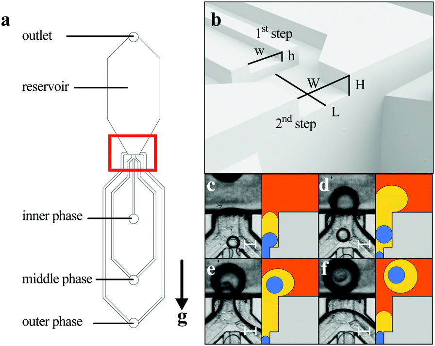 | ||
| Fig. 1 (a) General layout of the microfluidic network employed in the experiments. The red box indicates the double-step geometry; the g direction indicates gravity. (b) 3D rendering of the geometry. (c)–(e) Experimental images and cross-sectional sketches illustrating the formation of double emulsion droplets. Scale bar: 100 μm. | ||
2 Materials and methods
2.1 Liquids
We used three immiscible liquid phases to generate W/O/O double emulsions. As the outer phase we used FC-40 (3 M,USA) with 1 wt% of PFPE-PEG-PFPE surfactant (Chemipan, Poland), as the middle phase we used n-hexadecane with 2 wt% Span 80 (Sigma-Aldrich, Germany) or its mixture with silicone oil, AP 100 (Sigma-Aldrich, Germany) (0–70% w/w) (see Table 1), and as the core phase we used distilled water with dye (Congo red). The role of a surfactant (SPAN 80) in the silicone oil/hexadecane mixture was to stabilize the aqueous droplets against coalescence. To overcome the problem of wetting the channel walls by the dispersed middle- and core phases we rendered the channel walls fluorophilic by applying the commercial fluorinated coating Novec 1720 (3 M, USA).| Phase | Liquids | Case | ρ [g cm3] | η [mPa s] | γ water [mN m−1] | γ FC-40 [mN m−1] |
|---|---|---|---|---|---|---|
| Inner phase | Distilled water with congo red | — | 0.997 | 0.829 ± 0.023 | — | — |
| Hexadecane 2% w/w Span 80 | A | 0.770 | 2.999 ± 0.009 | 4.56 ± 0.10 | 5.80 ± 0.14 | |
| Middle phase | Hexadecane/AP 100/Span 80 (48![[thin space (1/6-em)]](https://www.rsc.org/images/entities/char_2009.gif) :50:2 w/w) :50:2 w/w) |
B | 0.908 | 8.653 ± 0.043 | 3.85 ± 0.02 | 5.88 ± 0.03 |
| Hexadecane/AP 100/Span 80 (28:70:2 w/w) |
C | 0.963 | 14.381 ± 0.001 | 2.60 ± 0.04 | 5.77 ± 0.09 | |
| Outer phase | FC-40 with 1% PFPE-PEG-PFPE | — | 1.855 | 3.935 ± 0.013 | — | — |
2.2 Microfluidic device and experiment
Experiments were carried out in devices made of either polycarbonate (PC) or poly(dimethylsiloxane) (PDMS). A network of microfluidic channels was milled in a polycarbonate plate using a CNC milling machine (Ergwind, Poland) and either bonded to another PC plate (to form a PC device) or used as a template for the fabrication of a PDMS master which allowed the fabrication of multiple replicas of the device. Channel dimensions were measured using an optical profilometer (CountourGT-K, Bruker, USA). To fabricate the master, PDMS (10:1 w/w elastomer and cross-linker) was poured onto a PC plate with milled channels, baked for an hour at 75 °C and peeled off. The mold was silanized for 3 hours at 20 mbar with tridecafluoro(-1,1,2,2,-tetrahydrooctyl)-1-trichlorosilane (Alfa Aesar, Germany). Such a mold was then filled with PDMS and baked for an hour at 75 °C, ad then the cured PDMS was peeled off. After punching the holes with a needle the PDMS cast was bound to glass using oxygen plasma treatment. Surface modification was achieved by filling the channel with Novec 1720 (3 M, USA) and baking the device at 130 °C for 30 minutes. We note that, after micromilling, the walls of the channels remained rough, with roughness of the order of 5 μm (see Fig. S2, ESI†). Such surface roughness is actually known to improve the effectiveness of the fluorophilic coating of polycarbonate with Novec 1720.28 In order to avoid even partial wetting of channel walls by the dispersed phases, first we filled our devices with the outer phase and only then introduced the to-be dispersed phases.
In all microfluidic experiments neMESYS syringe pumps (Cetoni, Germany) were used to inject liquids at flow rates ranging from 0.1 μl min−1 to 15 μl min−1. Droplet formation on-chip was recorded using a monochrome fast camera (1024PCI, K100, Photron, Japan). The droplet diameters were measured either manually using ImageJ or automatically with dedicated software.49 The droplet volume and diameter in the confined geometry were calculated assuming a filled-torus morphology, which has been shown to accurately describe the shape of a droplet flattened by the walls of a microchannel (see Fig. S1, ESI†).
3 Results
3.1 Microfluidic devices
The geometry of the device we used for emulsification is presented in Fig. 1a and b. The key features are the heights/depths of the inlet channel and the plateau (h = 42 μm, H = 98 μm, respectively), their widths (w = 162 μm and W = 345 μm, respectively), and the length of the plateau (L = 280 μm). According to previous research,37,41 the diameter of the generated droplets at either step was expected to range from 3 to 4 times the height of the preceding channel, i.e., 3h–4h for core droplets and 3H–4H for shell droplets, and we considered this fact while designing our device. Since in most of the applications double-emulsion drops with thin shells are preferred, the ratio H/h should be kept possibly small. However, when H/h < 2, the size of the droplets generated at the first step depends not only on h but also on H.30 To ensure that both steps feature simple step emulsification, we keep H/h ≥ 2. Here, we used the device with H/h ratio equal 2.3. Note that the dimensions of the outer channels supplying the external phase do not affect droplet formation (Fig. 1a). These channels serve only to deliver the outer phase to the reservoir prior to the experiment but do not operate during the experiment.3.2 Dimensionless parameters
In order to study the dynamics of droplet formation, we characterized our system by several variables such as the flow rates of the inner and middle phases, Qi and Qm, respectively. We denote the viscosities of the inner, middle, and outer liquid phases as ηi, ηm, and ηo, the densities as ρi, ρm, and ρo, respectively, and the relevant interfacial tensions as γi,m, and γm,o. We also introduce the core droplet volume fraction defined as ϕ = Qi/(Qi + Qm).We further introduce the capillary numbers Cai = ηiUi/γi,m and Cam = ηmUm/γm,o, of the inner and middle phases, respectively, with the linear velocities Ui = Qi/(wh) and Um = Qm/(WH) and the Bond numbers, Boi = (ρi − ρm)gDi2/γi,m and Bom = (ρo − ρm)gDm2/γm,o where Di and Dm are the diameters of the droplets of the inner phase (in the middle phase) and middle phase (in the outer phase; here, without cores in the middle phase). The diameters were measured by image analysis (see Fig. S2, ESI†). We found that Boi was of the order 10−3 meaning that the effects of gravity were negligible in the generation of core droplets. In contrast, Bom was in the range 10−1–100 depending on the actual size of the droplets. In any case, we expect that, except for very large double-emulsion drops, gravity also had a minor impact on drop formation (we discuss the validity of the model in the Discussion section).
3.3 Decoupling shell and core droplet formation
To compare the operation of our double-step droplet generator with the operation of standard single-step generators described previously in the literature, we first studied the formation of single droplets at the first or the second step with only two liquid phases.First, we removed the outer liquid (FC-40) from the system and blocked the inlet port of the outer phase. Before injecting the inner (core) phase, we waited until the shell phase completely filled the reservoir, and then, keeping the shell phase flowing, we turned on the flow of the inner phase. We recorded the droplet generation and measured the diameter of core droplets Di formed in the first step as a function of the flow rates of the inner and middle phases (see Fig. 2). We observed a stable generation of monodisperse inner phase droplets up to Qi = 5 μl min−1 (corresponding to  ), above which transition to the jetting regime (also known as the balloon regime35) took place. The measured diameter of the inner phase droplets, Di increased by approximately 12% in this regime which is typical for step emulsifiers.30,32,37,41 We note that, due to the density of the droplet phase being higher than that of the external phase, during these experiments the microfluidic device had to be flipped, with the nozzle facing upwards, so we could take advantage of buoyancy to remove the generated droplets from the nozzle. However, this proved not to be always sufficient, particularly in cases B and C (see Table 1), where the density difference between the oil and aqueous phases was small. Therefore, in this series of experiments we let the middle phase liquid flow at Qm, which we changed between 1 and 10 μm min−1. We plotted the diameter of the generated core droplets as the function of Qm and Qi, and found that the flow of the middle phase did not affect the formation of core droplets. For a fixed Qi we observed only a minor non-systematic variation of the droplet size Di which did not exceed 7% (Fig. 2b).
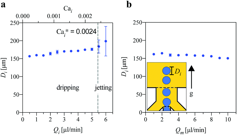 | ||
| Fig. 2 The diameter of the core droplets (Di) generated in the first step inside the middle phase without the supply of the outer phase as a function of the flow rate of (a) the inner phase Qi (at Qm = 5 μl min−1) and (b) the middle phase Qm with Qi held constant. (Qi = 1 μl min−1). Sub-panel: a schematic representation of the experimental setup. | ||
Next, we measured the diameter of empty shell droplets Dm, i.e. droplets of the middle phase formed in the second step in the absence of the inner phase. Droplets broke off into the reservoir filled with the outer phase liquid (FC-40), which was at rest and the formed droplets were carried away from the step by buoyancy. For all viscosities ηm we observed a slight increase of the droplet diameter Dm with the applied flow rate Qm until the transition to the jetting regime at around  (Fig. 3b).
 | ||
| Fig. 3 (a) Diameter of the shell droplet (Dm) generated in the second step as a function of the flow rate Qm of the middle phase for various types of the middle phase with the inner phase turned off, Qi = 0. (b and c) The same data plotted as a function of (b) the capillary number Cam and (c) the modified capillary number Cam,mod. Sub-panel: a schematic representation of the experimental setup. | ||
As we expected, in the case of the core droplets and the empty shell droplets, the mechanism of droplet formation and the dripping to jetting transition follow the classic scenario of the simple step emulsification well-described in the literature. By the “classic scenario” we mean the situation in which the dispersed phase pushed through a shallow and wide rectangular microchannel enters a deep reservoir, and the sudden release of confinement compels the tip of the liquid finger to rapidly round off in order to minimize the area of the liquid–liquid interface. The resulting pressure drop in the microchannel leads to the formation of a neck which eventually breaks. Such interfacial-tension driven mechanism of droplet breakup results in the size of droplets being predominantly set by the geometry of the device, in particular the height of the shallow inlet channel. Empirical studies37,41 showed that in order to precisely predict the size of the generated droplets (Fig. 3b) the viscosity of the continuous phase also needs to be taken into account. Accordingly, we also investigated the dependence of the size of the droplets Dm formed at the second step on the modified capillary number defined as Cam,mod := (ηo+α'ηm)Um/γm,o with a numerical constant α' depending on the geometry of the device. We found that, within the dripping regime, and with the constant adjusted to α' = 0.5, Dm was determined solely by Cam,mod (Fig. 3c), a consideration that remains in line with the previous studies.37 In the next section, we present and discuss new results considering the generation of double-emulsion droplets at the double-step geometry.
3.4 Formation of double emulsion droplets
We now consider the case in which the flows of both the inner and middle phases are turned on, and the reservoir is filled with the third, outer phase liquid (Fig. 4). We observe that the presence of the third liquid phase in the outlet reservoir has little effect on the formation of the core droplets in the first step. We also observe only a minor variation of the core diameter Di with Qi (Fig. 4a) and Qm or upon changing the type of the middle phase. | ||
| Fig. 4 (a) The diameter Di of the core droplets as a function of Qi for various Qm and various types of the middle phase. (b) The corresponding diameter DDE of the double-emulsion drops as a function of the net flow rate Qi + Qm. Subpanel: a schematic representation of the experimental setup. (c) DDE as a function of the effective capillary number Caeff. The solid line is the theoretical prediction (eqn (15) with β = 380, D0 = 305 μm). Inset: Caeff ∈ [0, 0.0052]. Symbols in the blue/red/green color correspond to middle phase fluids A, B, and C. | ||
The most significant change can be observed in the formation of the middle phase droplets in the second step as the growing droplet is filled not only by the middle phase but also by the core droplets. We observed a significant increase in the diameter DDE of the double emulsion (DE) droplets as compared to the diameter of the empty shell droplets observed earlier. The increase in the diameter can be associated with postponed pinch-off which results from the core droplets continuously passing through the plateau between the two steps towards the forming shell droplet, recurringly expanding the neck. Previously, it was reported37,38 that the increasing flow rate of the disperse phase postpones droplet break-up at the step as the dynamic pressure within the neck increases, thus stabilizing the neck. Here, the situation is similar with the difference that the dynamic pressure in the thinning neck is increased periodically every time a core droplet passes through it. The periodic oscillation of the width of the neck is shown in Fig. 5b. Building on previous works describing the dynamics of simple step emulsification and combining it with our experimental observations, in the following paragraph we provide a quantitative model of the above described ‘postponed dripping’. In particular, we predict the diameter of the DE droplets and the number of the encapsulated cores as a function of the applied flow rates in the whole regime of available volume fractions.
 | ||
| Fig. 5 (a) A scheme showing the widening of the neck upon core passing through it; black dashed line indicates the steps. The outer phase is not colored for visibility. (b) Dashed line: time evolution of the neck width wneck(t) of the shell phase at Qi = 1 μl min−1 and Qm = 2.5 μl min−1. Solid line: time evolution of the time-averaged neck width wav(t) (see eqn (11)) obtained as a running average of wneck(t) with the averaging window T = 0.3 s (roughly 3 x the period of oscillations). Time t = 0 marks the moment when the tip of the dispersed phase reaches the edge of the step (ts). (c) The running average wav(t) for various flow rates of the inner phase Qi at Qm = 2.5 μl min−1. (d) Data from panels (c) plotted versus the rescaled time t* = t (1 − ϕ/ϕ0), according to eqn (13) with ϕ0 = 0.6. | ||
3.5 Theoretical model of postponed dripping
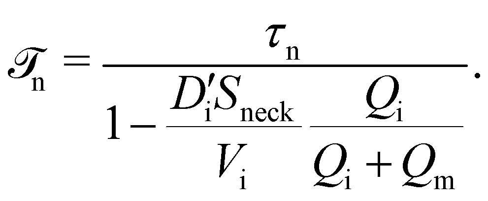
![[scr T, script letter T]](https://www.rsc.org/images/entities/b_char_e533.gif) n.
First, we consider the generation of the drops of the middle phase in the case without cores. We denote by t* the ‘stage of thinning’ of the neck of the middle phase, where t* = 0 corresponds to the formation of the neck and t* = τn to neck breakup. More precisely, after Crestel et al.,41 we define τn = tb − ts, where tb is the time of neck breakup and ts is the time of neck formation, i.e., the moment in which the meniscus passes the edge of the step.
n.
First, we consider the generation of the drops of the middle phase in the case without cores. We denote by t* the ‘stage of thinning’ of the neck of the middle phase, where t* = 0 corresponds to the formation of the neck and t* = τn to neck breakup. More precisely, after Crestel et al.,41 we define τn = tb − ts, where tb is the time of neck breakup and ts is the time of neck formation, i.e., the moment in which the meniscus passes the edge of the step.
Now, in the case with the cores, our experimental observations (Fig. 5b) suggest that the neck temporarily widens each time a core passes through it. We assume that such widening proportionally postpones neck breakup by a time interval Δti corresponding to the transit of the core through the thinnest part of the neck. Accordingly, the actual time ![[scr T, script letter T]](https://www.rsc.org/images/entities/char_e533.gif) n of thinning of the neck in the case with the cores can be calculated as
n of thinning of the neck in the case with the cores can be calculated as
 | (1) |
| Δt* = Ti − Δti. | (2) |
From eqn (1) and (2) we obtain
 | (3) |
The transit time Δti can be approximated as
 | (4) |
 is the apparent diameter of the core droplets flattened by the upper and lower wall at the plateau between the steps. We will use the approximation Uneck = (Qi + Qm)/Sneck where Sneck is the average area of the neck cross-section over the time at which the cores transit the neck, that is
| Sneck = Hw0, | (5) |
| w0 = 〈wneck(t)〉 [t1,tN]. | (6) |
Since Ti = Vi/Qi, where Vi is the volume of a single core and  , we then substitute Δti and Ti in eqn (3) and obtain
 | (7) |
 | (8) |
 | (9) |
 | (10) |
 , and Sneck are nearly independent of the applied flow rates, ϕ0 can be treated as a constant depending only on the geometry of the junction. Actually, ϕ0 can be acquired directly from the experiment via measuring the average values Vi,
  , Sneck.
Next, we turn to the evolution of the neck width wneck(t) from the moment of neck formation at t = 0 to its breakup at t = n. In the experiment we observe that w(t) consists of fluctuations around a slowly varying envelope wav(t), where
| wav(t) = 〈wneck(t′)〉[t,t+T], | (11) |
| wav(t) = w*(t*), | (12) |
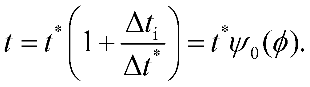 | (13) |
We can verify the scaling formula in eqn (12) using the actual experimental data wneck(t) (Fig. 5). To this end, we first determine ϕ0 using eqn (10) in which we estimate Sneck (eqn (5) and (6)) by taking t1 as the first local minimum and tN as the last local maximum of wneck(t). Via averaging the data in the cases with various Qi, at Qm = 2.5 μl min−1 and Qm = 4 μl min−1 (the data for Qm = 2.5 μl min−1 are shown in Fig. 5c), we obtain w0 ≃ 0.17 mm which leads to Sneck ≃ 0.17 mm2. By using image analysis we also find  ± 9 μm (the average apparent core diameter in the cases with Qi ∈ {0.25, 0.5, 1.0} μl min−1 and Qm ∈ {2.5, 4.0} μl min−1) from which (via applying the nodoid model, see the ESI†) we find Vi ≃ 1.68 ± 0.09 nl. Those values eventually lead to the estimation of ϕ0 ≃ 0.60 ± 0.03 which allows testing the scaling proposed in eqn (13). In fact, we find that the experimental data for wneck plotted for various Qi and Qm values against the rescaled time t*/ψ0(ϕ) (eqn (13)) with ϕ0 = 0.6 tend to collapse onto a single master envelope (Fig. 5d).
n = τnψ0(ϕ). This assumption leads to| VDE = V0 + (Qi + Qm)τnψ0(ϕ). | (14) |
Next, drawing from previous works considering step emulsification,37,41 we assume 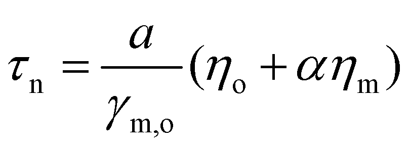 where a and α are model constants (a has the dimension of length and α is non-dimensional). We also introduce the net velocity of the dispersed phases at the second step, UDE = (Qi + Qm)/(HW) which, considering that VDE = πDDE3/6, allows us to rewrite eqn (14) in the form
| DDE = D0 (1 + βCaeff)1/3, | (15) |
| Caeff = ψ0(ϕ)(ηo + αηm)UDE/γm,o | (16) |
In agreement with those predictions, the experimentally measured diameters of double-emulsion drops DDE for various Qi and Qm values plotted as a function of Caeff upon adjusting α = 0.5 and β = 380 collapse onto a master curve (Fig. 4c). Note that we find α = α′ (the latter appears in the definition of Cam,mod in Section 3.3.) which confirms the consistency of our model. Further, we find that the scaling proposed in eqn (15) holds for the effective capillary numbers Caeff below approximately 5 × 10⁻³. In this regime fitting of eqn (15) to the collapsed data leads to D0 = 305 μm from which we also find a = 167.5 mm. For higher Caeff we observe that DDE (Caeff) grows faster than expected, resembling the transition to jetting (see Fig. 2 and 3). Remarkably, the data for various viscosities of the middle phase ηm collapse even in the 'jetting' regime, unlike in the case of simple middle-phase dripping (i.e., in the case without the cores, see Fig. 3c). However, the data collapse seems to fail at Caeff > 0.012 (which typically corresponds to the cases with ϕ ≥ 0.5). The outliers in this regime have DDE values smaller than those expected from the master curve which might be attributed to the faster pinch-off of very large drops, e.g., due to their buoyancy (in fact, we checked that in these cases the Bond number approaches unity) or as a result of the irregularities in the feeding of the core droplets into the shell at long times.
 | (17) |
 | (18) |
To calculate the map N(Qi, Qm) we take the value V0 = πD03/6 = 14.9 nl (where D0 is taken from the fit to the data DDE(Caeff) based on eqn (15) as discussed above) and estimate τn by extrapolating the experimentally measured wneck(t) towards wneck = 0 in the cases with the inner phase switched off, Qi = 0 μl min−1, Qm = {2.5; 4} μl min−1 (we take the average of the two measurements); we obtain τn ≃ 0.22 s. With these values we find good agreement between eqn (17) (Fig. 6b) and the experimentally measured N (Fig. 6a) without any additional fitting parameters. We note, in particular, that the theoretical formula (eqn (17)) predicts N = ∞ for ϕ ≥ ϕ0 (black region in Fig. 6b). In the experiment we indeed observe no double-emulsion drop pinch-off in this regime (Fig. 6a) but rather the formation of an indefinitely growing double-emulsion ‘tongue’.
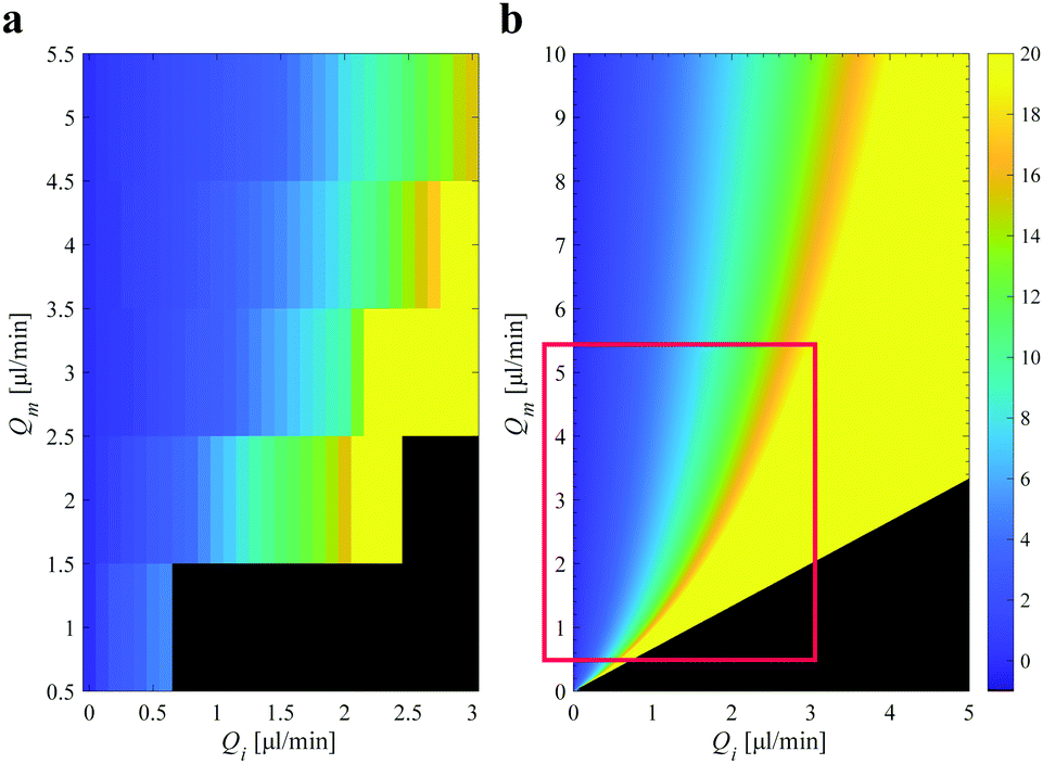 | ||
| Fig. 6 (a) Experimental measurements: the heat map representing the number N of core droplets in the double-emulsion drops generated for various Qi and Qm using the middle fluid Case A. The colors indicate interpolated experimental data. The actual data points N were collected at Qi ∈ [0, 3] and Qm ∈ [0.5, 5.5] μl min−1, at intervals 0.5 and 1.0 μl min−1, respectively, in each case as an average of 11 generated double-emulsion drops. The black region corresponds to N > 20. (b) Theoretical prediction of the proposed model (eqn (17) with V0 = 14.9 nl, Vi = 1.68 nl, Q0= 4.1 μl min−1, reflecting typical experimental conditions). The black region corresponds to a diverging N (‘N = ∞’). The red box indicates the experimental regime considered in panel (a). | ||
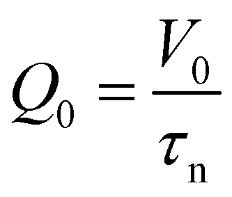
Furthermore, we note that for large volume fractions ϕ, i.e., for ϕ approaching the threshold ϕ0, the scaling function ψ(ϕ) diverges, whereas Qi/Q0 remains around or less than unity (Qi ∈ [1, 3] μl min−1 and Q0 ≃ 4.1 μl min−1), which means that the first term in eqn (17) can be neglected. In such a case we obtain:
 | (19) |
 | (20) |
Dividing eqn (19) by Qm and multiplying by  we obtain
 | (21) |
In particular, eqn (21) can be used to collapse the experimental data N(Qi) obtained for various Qm values (Fig. 7a) onto a single master curve (Fig. 7b). In principle, one should expect the scaling to be valid only for sufficiently large volume fractions. Despite that, we observe reasonable collapse over nearly the entire range of applied volume fractions. We take  (eqn (20)), calculated based on the values of Vi and τn estimated from the experiments described above.
 | ||
Fig. 7 (a) The number N of cores in the double-emulsion drops as a function of Qi for various Qm. (b) N rescaled with  as a function of the volume fraction ϕ = Qm/(Qi + Qm). The solid line is the theoretical prediction (eqn (21) with as a function of the volume fraction ϕ = Qm/(Qi + Qm). The solid line is the theoretical prediction (eqn (21) with  μl min−1 reflecting the typical experimental conditions). (c) Images of the DE droplets with various numbers of core droplets. Scale bar: 100 μm. μl min−1 reflecting the typical experimental conditions). (c) Images of the DE droplets with various numbers of core droplets. Scale bar: 100 μm. | ||
4 Discussion and conclusion
In this work we study the formation of double emulsion (DE) drops with one or multiple cores using a double-step emulsifier. We find the the size of the cores Di is roughly independent of the applied flow rates Qi and Qm (of the core and shell phases, respectively), whereas the size of the double-emulsiondrops DDE (Fig. 4) and the number of the encapsulated cores N (Fig. 6 and 7) depend on both Qi and Qm. In particular, we find that N diverges upon the volume fraction of the cores ϕ = Qi/(Qi + Qm) approaching the threshold ϕ0 (for the studied geometry we find ϕ0 = 0.60), see Fig. 6a. We refer to such behavior as ‘postponed dripping’: in this regime close-packing of the cores postpones double-emulsion drop pinch-off. In particular, we find periodic widening of the shell neck upon cores transiting the plateau between the two steps. Based on this observation we establish a scaling function ψ0 (ϕ), depending parametrically on ϕ0, which describes the effective delay in the necking timen (eqn (8)) and allows us to define the effective capillary number Caeff (eqn (16)). We further derive formulas for DDE(Caeff) (eqn (15)) and N(Qi, Qm) which remain in good agreement with the experimental data. In particular, we also find that (at moderate ϕ) N/Qm is a function only of ϕ (eqn (21)).
We note that our model is based on the ‘proportionality hypothesis’ which we introduce as an empirical assumption, following the observation of similar shapes of the envelopes wav(t) describing the running average of the neck width versus time for various volume fractions (Fig. 5c and d). One possible way of justifying such an assumption is that, in step emulsification, the dynamics of neck thinning is driven predominantly by the surface tension and, in particular, that the necking time is nearly independent of the applied flow rates, a fact known from previous studies.37,41 Accordingly, in the case without the cores, it is the shape of the neck at a given moment in time that determines the dynamics of further neck thinning. In the case with the cores, when a core enters the neck, the thinning stops, and then continues after the core leaves the neck.
While the model works remarkably well in a wide range of volume fractions ϕ, it neglects the impact of buoyancy of the DE drop on its pinch-off. We expect buoyancy to actually accelerate pinch-off which then should lead to the generation of DE drops smaller than those predicted using eqn (15) (and the number of cores N smaller than those predicted using eqn (17)). The impact of buoyancy could be expected to be particularly strong in the case of larger drops N ∼ 20 (which are expected to occur at ϕ close to ϕ0) for which the Bond number could approach or even exceed unity.
Furthermore, in the actual experiments the cores are not always perfectly packed, even at high ϕ, which leads to local fluctuations in the packing fraction. Since the neck thins very quickly in the time between the transit of the subsequent cores (see Fig. 5), any irregularities in the rate of feeding of the cores to the neck may result in DE drop pinch-off. Accordingly, the local fluctuations may lead to the generation of finite DE drops even for the average ϕ being at or above the threshold ϕ0. As a consequence the generated DE drops are smaller than those predicted by the theory and slightly polydisperse. Indeed, we observe larger error bars of the measured DDE for ϕ close to ϕ0, see Fig. 7b. Here, we do not investigate the distribution of the DE drop sizes in detail. However, we have recently provided a detailed analysis of such distributions for a related system generating DE droplets via flow-focusing.50
We note that the previous studies on tandem step emulsification predominantly considered either low-ϕ regimes45 or different step geometries which did not support the stabilization of the neck by the cores.46 The main novelty of our work is providing the rationale behind the generation of DE drops in the high-ϕ regime51 allowing for the controlled generation of structures with large numbers of close-packed cores (N > 10), i.e., for the formation of soft-granular aggregates. The demonstrated microfluidic formulation of such structures is an important step towards the fabrication of mesoscale functional materials such as multi-compartment capsules or mesoporous microbeads with exciting new applications, e.g., in tissue engineering.5
We suppose that changing the geometry by varying W or w as long as w/h > 2.6 and W/H > 2.6 would have little effect on the operation of the device since in this regime the sizes of the droplets are set predominantly by the heights of the channels (h and H).37,38,41,52 Another parameter of interest is the ratio of the interfacial tensions of the shell and of the cores, γm,o/γi,m, which in our experiments was in the range [1,2]. However, if this ratio was much larger than unity, γm,o/γi,m ≫ 1, one could expect the inner droplets to comply with the shape of the thinning neck rather than to expand the neck. In such a case, one would expect the effect of postponed dripping to be less pronounced. Accordingly, one could speculate that the critical volume fraction ϕ0 should increase with an increase in γm,o/γi,m.
Considering the range of available droplet sizes, we find that, in agreement with the previous literature,37,38,41 the core size Di is set predominantly by the height of the supply channel h, with Di ∼ 4h (see Fig. 2). It has been shown previously that step emulsification enables generation of droplets with diameters ranging from submicrons to 1000 μm.34,42 In order to achieve DE droplets with micron-sized cores one would need to fabricate ultra-shallow channels with h in the submicron regime. Such small dimensions are outside the reach of micromilling, but could be achieved using, e.g., photolithography followed by etching.34 Considering the upper limit of the size of the clusters, we find that, as already noted, the number N of encapsulated cores tends to grow indefinitely upon ϕ approaching the threshold ϕ0 (Fig. 7b), yet with increasing fluctuations.
Next, considering the throughput of the device, from our experimental data (Fig. 4b) it follows that the maximal available flow rates Qi + Qm are typically of the order of several μl min−1 and therefore are not excessive. However, we believe that the generation of double emulsions could be upscaled via parallel integration of multiple double-step nozzles. The advantage of the proposed geometry would be the relatively weak dependence of the size of the generated DE drops on the applied flow rates or their fluctuations, at least for ϕ by a safe margin below the threshold ϕ0 (here, for ϕ < 0.5, see Fig. 7b).
In this work, as a proof of concept, we have studied the generation of water-in-oil-in-oil (W/O/O) droplets whereas many applications such as, e.g., cell encapsulation, require W/O/W droplets. In order to generate W/O/W double emulsions one would need to apply selective hydrophilic/hydrophobic surface modification of the double-step geometry. Such modification would require very precise hydrophilicity patterning. In particular, the water inlet channel and the vertical wall behind the first step would need to be modified to be hydrophobic, while the rest of the device, hydrophilic (Fig. S3, ESI†). Such selective modification is challenging (we have not attempted it in this work) which may be a limitation of our device. Anyway, we expect our quantitative considerations considering the postponed dripping regime (Section 3.5) to remain valid independent of the type of emulsion (W/O/O or W/O/W), as long as the droplets do not wet the walls of the channels.
Last but not least, the more complex geometries similar to the double-step geometry presented here, but with multiple steps, could also facilitate direct generation of multiple emulsions consisting of three or more immiscible phases, in a manner similar to the methods exploiting multiple co-axial capillaries or multiple serial flow-focusing junctions.20,53
Conflicts of interest
There are no conflicts to declare.Acknowledgements
The authors thank Airit Agasty for viscosity measurements, Agnieszka Koszewska for providing samples, and Patryk Adamczuk and Karol Patyrak for the microfabrication of the devices. This publication is part of a project that received funding from the European Union's Horizon 2020 research and innovation programme under the Marie Skłodowska-Curie grant agreement No. 711859 and scientific work funded by the financial resources for science in the years 2017–2021 awarded by the Polish Ministry of Science and Higher Education for the implementation of an international co-financed project. L. D. was supported by the Foundation for Polish Science within the project TEAM NET (POIR.04.04.00-00-16ED/18). A. S. O. and P. G. were supported by the Foundation for Polish Science within the project TEAM TECH (POIR.04.04.00-00-2159/16 00). A. S. O. acknowledges funding from the National Science Centre, Poland, within the project 2016/23/N/ST4/01020. J. G. was supported by the Foundation for Polish Science within the project First TEAM (POIR.04.04.00-00-26C7/16-00).References
- M. G. Santos, F. T. Bozza, M. Thomazini and C. S. Favaro-Trindade, Food Chem., 2015, 171, 32–39 CrossRef CAS PubMed.
- J. J. Liau, S. Hook, C. A. Prestidge and T. J. Barnes, Eur. J. Pharm. Biopharm., 2015, 97, 15–21 CrossRef CAS.
- C.-H. Choi, H. Wang, H. Lee, J. H. Kim, L. Zhang, A. Mao, D. J. Mooney and D. A. Weitz, Lab Chip, 2016, 16, 1549–1555 RSC.
- H. F. Chan, Y. Zhang and K. W. Leong, Small, 2016, 12, 2720–2730 CrossRef CAS.
- M. Costantini, J. Guzowski, P. J. Żuk, P. Mozetic, S. De Panfilis, J. Jaroszewicz, M. Heljak, M. Massimi, M. Pierron, M. Trombetta, M. Dentini, W. Święszkowski, A. Rainer, P. Garstecki and A. Barbetta, Adv. Funct. Mater., 2018, 28, 1870133 CrossRef.
- J. Guzowski, K. Gizynski, J. Gorecki and P. Garstecki, Lab Chip, 2016, 16, 764–772 RSC.
- G. Villar, A. D. Graham and H. Bayley, Science, 2013, 340, 48–52 CrossRef CAS PubMed.
- E. Y. Liu, S. Jung, D. A. Weitz, H. Yi and C.-H. Choi, Lab Chip, 2018, 18, 323–334 RSC.
- B. Tal-Figiel, Chem. Eng. Res. Des., 2007, 85, 730–734 CrossRef CAS.
- G. Muschiolik and E. Dickinson, Compr. Rev. Food Sci. Food Saf., 2017, 16, 532–555 CrossRef CAS.
- J. Guzowski, S. Jakiela, P. M. Korczyk and P. Garstecki, Lab Chip, 2013, 13, 4308 RSC.
- F.-C. Chang and Y.-C. Su, J. Micromech. Microeng., 2008, 18, 065018 CrossRef.
- M. Seo, C. Paquet, Z. Nie, S. Xu and E. Kumacheva, Soft Matter, 2007, 3, 986 RSC.
- A. R. Abate, A. T. Krummel, D. Lee, M. Marquez, C. Holtze and D. A. Weitz, Lab Chip, 2008, 8, 2157 RSC.
- J. Yan, W.-A. Bauer, M. Fischlechner, F. Hollfelder, C. Kaminski and W. Huck, Micromachines, 2013, 4, 402–413 CrossRef.
- A. Vian, B. Reuse and E. Amstad, Lab Chip, 2018, 18, 1936–1942 RSC.
- S. Okushima, T. Nisisako, T. Torii and T. Higuchi, Langmuir, 2004, 20, 9905–9908 CrossRef CAS.
- T. Nisisako, S. Okushima and T. Torii, Soft Matter, 2005, 1, 23 RSC.
- A. S. Utada, E. Lorenceau, D. R. Link, P. D. Kaplan, H. A. Stone and D. A. Weitz, Science, 2005, 308, 537–541 CrossRef CAS.
- L.-Y. Chu, A. Utada, R. Shah, J.-W. Kim and D. Weitz, Angew. Chem., 2007, 119, 9128–9132 CrossRef.
- H. C. Shum, D. Lee, I. Yoon, T. Kodger and D. A. Weitz, Langmuir, 2008, 24, 7651–7653 CrossRef CAS.
- D. Lee and D. A. Weitz, Small, 2009, 5, 1932–1935 CrossRef CAS PubMed.
- S.-H. Kim, J. W. Kim, J.-C. Cho and D. A. Weitz, Lab Chip, 2011, 11, 3162–3166 RSC.
- H. C. Shum, Y.-j Zhao, S.-H. Kim and D. A. Weitz, Angew. Chem., Int. Ed., 2011, 50, 1648–1651 CrossRef CAS PubMed.
- B. Herranz-Blanco, E. Ginestar, H. Zhang, J. Hirvonen and H. A. Santos, Int. J. Pharm., 2017, 516, 100–105 CrossRef CAS PubMed.
- T. Nisisako, T. Ando and T. Hatsuzawa, Lab Chip, 2012, 12, 3426 RSC.
- M. B. Romanowsky, A. R. Abate, A. Rotem, C. Holtze and D. A. Weitz, Lab Chip, 2012, 12, 802 RSC.
- A. S. Opalski, K. Makuch, Y.-K. Lai, L. Derzsi and P. Garstecki, Lab Chip, 2019, 19, 1183–1192 RSC.
- F. Dutka, A. S. Opalski and P. Garstecki, Lab Chip, 2016, 16, 2044–2049 RSC.
- R. Dangla, E. Fradet, Y. Lopez and C. N. Baroud, J. Phys. D: Appl. Phys., 2013, 46, 114003 CrossRef.
- S. Sugiura, M. Nakajima, S. Iwamoto and M. Seki, Langmuir, 2001, 17, 5562–5566 CrossRef CAS.
- S. Sugiura, M. Nakajima and M. Seki, Langmuir, 2002, 18, 5708–5712 CrossRef CAS.
- S. Sugiura, M. Nakajima, N. Kumazawa, S. Iwamoto and M. Seki, J. Phys. Chem. B, 2002, 106, 9405–9409 CrossRef CAS.
- L. Shui, A. van den Berg and J. C. T. Eijkel, Microfluid. Nanofluid., 2011, 11, 87–92 CrossRef.
- Z. Li, A. M. Leshansky, L. M. Pismen and P. Tabeling, Lab Chip, 2015, 15, 1023–1031 RSC.
- M. Hein, J.-B. Fleury and R. Seemann, Soft Matter, 2015, 11, 5246–5252 RSC.
- N. Mittal, C. Cohen, J. Bibette and N. Bremond, Phys. Fluids, 2014, 26, 082109 CrossRef.
- A. Montessori, M. Lauricella, E. Stolovicki, D. A. Weitz and S. Succi, Phys. Fluids, 2019, 31, 021703 CrossRef.
- A. Ofner, D. G. Moore, P. A. Rühs, P. Schwendimann, M. Eggersdorfer, E. Amstad, D. A. Weitz and A. R. Studart, Macromol. Chem. Phys., 2017, 218, 1600472 CrossRef.
- S. Sahin and K. Schroën, Lab Chip, 2015, 15, 2486–2495 RSC.
- E. Crestel, L. Derzsi, H. Bartolomei, J. Bibette and N. Bremond, Phys. Rev. Fluids, 2019, 4, 073602 CrossRef.
- E. Stolovicki, R. Ziblat and D. A. Weitz, Lab Chip, 2018, 18, 132–138 RSC.
- Y.-T. Kao, T. S. Kaminski, W. Postek, J. Guzowski, K. Makuch, A. Ruszczak, F. von Stetten, R. Zengerle and P. Garstecki, Lab Chip, 2020, 20, 54–63 RSC.
- M. Schulz, S. Probst, S. Calabrese, A. R. Homann, N. Borst, M. Weiss, F. von Stetten, R. Zengerle and N. Paust, Molecules, 2020, 25, 1914 CrossRef CAS PubMed.
- M. L. Eggersdorfer, W. Zheng, S. Nawar, C. Mercandetti, A. Ofner, I. Leibacher, S. Koehler and D. A. Weitz, Lab Chip, 2017, 17, 936–942 RSC.
- A. Ofner, I. Mattich, M. Hagander, A. Dutto, H. Seybold, P. A. Rühs and A. R. Studart, Adv. Funct. Mater., 2019, 29, 1806821 CrossRef.
- S. Nawar, J. K. Stolaroff, C. Ye, H. Wu, D. T. Nguyen, F. Xin and D. A. Weitz, Lab Chip, 2020, 20, 147–154 RSC.
- N. Bremond, A. R. Thiam and J. Bibette, Phys. Rev. Lett., 2008, 100, 024501 CrossRef PubMed.
- Z. Z. Chong, S. B. Tor, A. M. Gañán-Calvo, Z. J. Chong, N. H. Loh, N.-T. Nguyen and S. H. Tan, Microfluid. Nanofluid., 2016, 20, 66 CrossRef.
- M. Bogdan, A. Montessori, A. Tiribocchi, F. Bonaccorso, M. Lauricella, L. Jurkiewicz, S. Succi and J. Guzowski, Phys. Rev. Lett., 2022, 128, 128001 CrossRef CAS PubMed.
- J. Guzowski and P. Garstecki, Phys. Rev. Lett., 2015, 114, 188302 CrossRef PubMed.
- A. Montessori, M. Lauricella, S. Succi, E. Stolovicki and D. Weitz, Phys. Rev. Fluids, 2018, 3, 072202 CrossRef.
- A. R. Abate and D. A. Weitz, Small, 2009, 5, 2030–2032 CrossRef CAS.
Footnote |
| † Electronic supplementary information (ESI) available. See DOI: https://doi.org/10.1039/d2sm00327a |
| This journal is © The Royal Society of Chemistry 2022 |