DOI:
10.1039/D2RA04705E
(Paper)
RSC Adv., 2022,
12, 24752-24759
Up-conversion luminescence properties with temperature change of strontium tungstate phosphors
Received
28th July 2022
, Accepted 26th August 2022
First published on 31st August 2022
Abstract
Thermally stable SrWO4:[Er3+]/[Yb3+] upconversion phosphors were synthesized. X-ray diffraction analysis indicated a crystalline inorganic phosphor material with a tetragonal structure having a clear peak in the (112) phase, which is the main peak. The upconversion phosphor was synthesized using a precursor prepared by co-precipitation and sintered at 800 °C. When the phosphor was excited by a 980 nm laser with a pumping power of 200 mW, a strong green light was emitted. As the concentration of Er3+ ions increased, it was observed that the emission intensity decreased due to concentration quenching. The changes in the intensity of luminescence according to the pumping power are due to a two-photon process. As the temperature increased, the green emission intensity of the up-conversion phosphor increased. This was thought to be a phenomenon caused by efficient energy transfer between Yb3+ and Er3+ ions by the SrWO4 host with negative thermal expansion. A composite was prepared by mixing phosphor powder and PDMS, that could be used for temperature sensing.
1 Introduction
Up-conversion phosphors are materials that emit light by fusing low-energy photons and converting them into high-energy photons, and these phosphors are attracting attention in various fields such as sunlight, bio-emerging, and phosphor applications.1,2 Existing down-conversion phosphors are mainly based on Stokes emission, which generates low energy long wavelength visible light from high energy short wavelengths such as the ultraviolet region.3,4 In contrast to this Stokes emission, upconversion phosphors have an anti-Stokes-emission mechanism in which long-wavelength photons fuse to produce short-wavelength photons.5,6 By using an up-conversion phosphor, light in the infrared region can be converted into visible light, or light in the visible region can be converted into ultraviolet light.7,8 This can be considered a phenomenon that violates the laws of thermodynamics, but the up-conversion phenomenon is possible because it is an emission phenomenon that occurs by the fusion of energy of two or more photons, not by a single photon.9,10 Research on up-conversion phosphors has been actively conducted using inorganic materials doped with rare earth ions. Up-conversion is caused by a luminescence phenomenon, due to the double excitation of photons in inorganic materials with a long lifetime transition energy region.11,12 By absorbing the primary photon, the excited electron with the transition energy band absorbs an additional photon before falling to the ground state and is excited to a higher energy level.13,14 Pavani et al. synthesized SrLaAl3O7 co-doped with Er3+ and Yb3+ by the facile Pechini method. The phosphor excited at 980 nm showed strong green emission by the 4I13/2 → 4I15/2 transition. In addition, the visible spectrum of 528, 548, and 660 nm was shown, and the up-conversion mechanism was reported.15 Lim et al. synthesized a yellow-emitting phosphor when excited at 980 nm by co-doping Yb3+ and Ho3+ with LiNaCaLa(MoO4)3 quadruple molybdate as a host, by the microwave sol–gel method.16 Deng et al. doped the GaAlO3:Er3+, Yb3+ phosphor with Lu3+ and Ga3+ ions by co-precipitation. Ga3+ is Al3+ and Lu3+ is Gd3+ substituted, and a change in the intensity of the up-conversion green emission according to the structural change in the host was reported.17 Barrera et al. synthesized an up-conversion phosphor by doping Er3+ and Yb3+ ions into a KY(WO4)2 host using the Pechini method. The phosphor excited at 980 nm showed emission spectra of 530, 550, and 660 nm. A change in emission intensity according to the change in the concentration of doped rare earth ions, and a change in the intensity of the excitation light, were observed.18 Pandey et al. prepared Y2O3 up-conversion phosphors co-doped with Ho3+ and Yb3+ ions by the combustion route. When excited with a 980 nm diode laser, the color changed according to the doping concentration of Ho3+ ions, as shown in the CIE color coordinates, and a visible light spectrum of strong green and relatively weak red and blue was observed.19 Various synthesis methods and doping with rare earth ions have been performed to synthesize upconverted phosphors and to investigate their luminescence properties. In this study, phosphors were synthesized by co-doping with Er3+ and Yb3+ rare earth (RE) ions using strontium tungstate (SrWO4), which has chemical and thermal stability, as a host. The precursor was prepared by co-precipitation and heat-treated at 800 °C. Changes in the crystal structure and luminescence characteristics of the synthesized phosphor after doping with rare earth ions were investigated. In addition, a change in luminescence characteristics according to the power and temperature of the excitation light was observed, and it was suggested that the composite could be used for temperature sensing.
2 Experimental
2.1. Synthesis of SrWO4:[Er3+]/[Yb3+] by co-precipitation and sintering
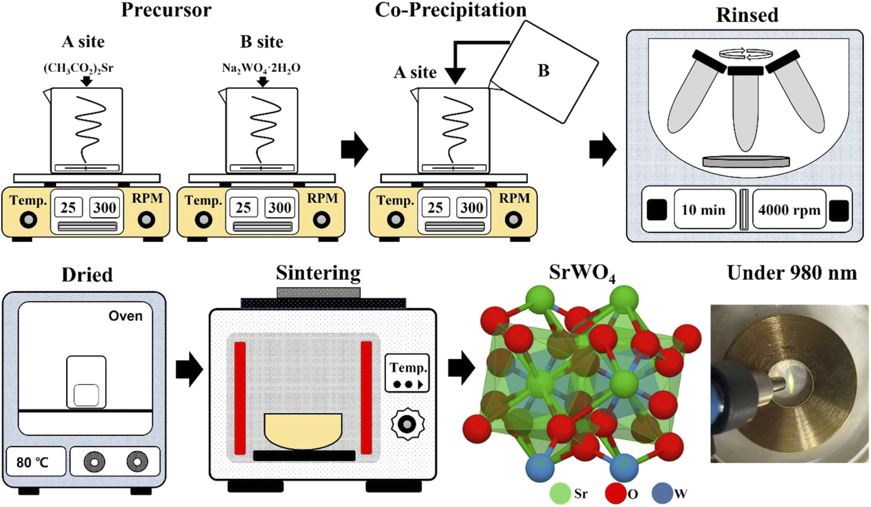
Starting materials. Barium acetate ((CH3CO2)2Sr, Sigma-Aldrich), sodium tungstate (Na2WO4·2H2O, Sigma-Aldrich), ytterbium(III) nitrate pentahydrate (Yb(NO3)3·5H2O, Yb3+), and erbium(III) nitrate pentahydrate (Er(NO3)3·5H2O, Er3+). 10 mmol of (CH3CO2)2Sr was dissolved in beaker “A” containing 100 ml of distilled water. In beaker “B”, 10 mmol of Na2WO4·2H2O was dissolved in 100 ml distilled water (Fig. 1). The solution that was completely dissolved in beaker “B” was slowly poured into a stirring beaker “A” and remained there for about 20 minutes. After that, powder was recovered using a centrifuge (4000 rpm, 5 min), and the powder was prepared by rinsing with distilled water 3 times to remove any remaining sodium. The powder was dried in an oven at 80 °C for 16 hours. The up-conversion phosphor was synthesized with SrWO4 as a host, the precursor made by simultaneously adding Yb(NO3)3·5H2O and Er(NO3)3·5H2O to beaker “A”. The prepared precursor was sintered at 800 °C until the SrWO4:[Er3+]/[ Yb3+] phosphor was synthesized. Rare earth [RE] ions of Yb3+ were fixed at 0.5 mmol, and the amount of Er3+ (0.05, 0.1, 0.15, 0.2, 0.25, 0.3, 0.4, 0.5 mmol) added changed ([Er3+]/[Yb3+] ∼ 0.1, 0.2, 0.3, 0.4, 0.6, 0.8, 1).20,21
 |
| | Fig. 1 Experimental procedure for preparing the SrWO4:[Er3+]/[Yb3+] up-conversion phosphors. | |
2.2. Characterization
The crystal structure of the synthesized phosphor powder was measured using an X-ray diffraction apparatus (XRD, X'Pert PRO MPD, 40 kV, 30 mA) having Cu-Kα radiation (wavelength: 1.5406 Å) at a scan rate of 4° min−1 at a diffraction angle of 10° to 70°. The size and microscopic surface shape of the crystal grains were photographed with a field emission scanning electron microscope (FE-SEM, CZ, MIRA I LMH, TESCAN). To measure the fluorescence spectrum by up-conversion, a semiconductor pulse laser (TCLDM9, Thorlabs) that emitted an output of 200 mW at a wavelength of 980 nm as excitation, and a spectrometer (HR4000, Ocean Optics) with a photomultiplier connected were used to measure the emission spectrum. To analyze the fluorescence mechanism by up-conversion, the energy absorption and energy transfer processes in the excited state were analyzed by changing the intensity of the pulsed laser and measuring the changes in the intensity of the fluorescence.
2.3. Fabrication of a temperature sensing composite
The synthesized SrWO4:[Er3+]/[Yb3+] up-conversion phosphors were mixed with 0.1 g each and 1 g of polydimethylsiloxane (PDMS) and cast in a square mold. After curing in an oven at 80 °C for 2 hours, the prepared composite was placed on a hot plate, excited at 980 nm, and the change in luminescence intensity according to temperature change was photographed.
3 Results & discussion
3.1. Structure and morphology of SrWO4:[Er3+]/[Yb3+] up-conversion phosphors
Fig. 2(a) shows the XRD patterns of the samples prepared by co-precipitation at room temperature and calcined at 800 °C. The synthesized SrWO4 was consistent with the ICDD card no. 01-089-2568, and the tetragonal structure (a = 5.40 Å, b = 5.40 Å, c = 11.91 Å, space group I41/a) could be confirmed. A strong peak at 27.65° was detected, which was the (112) phase, the main peak of SrWO4. In addition, the phases (101), (004), (200), (211), (204), (220), (116), (312), (224), (008), and (440) were also observed. In the SrWO4:[Er3+]/[Yb3+] samples doped with rare earth ions for synthesis as up-conversion phosphors, the main peak (112) phase was also clearly observed. As the amount of rare earth ions increased (Er3+), the detection position of the main peak (112) phase shifted, but no secondary phase was found. Using the change in the detection position of the main peak-in (112), the change in the lattice constant was calculated by substituting it into the Bragg's equation (2d![[thin space (1/6-em)]](https://www.rsc.org/images/entities/char_2009.gif) sinθ = nλ)22 and is shown in Fig. 2(b). The lattice constant of pure SrWO4 was calculated to be 0.2901 nm, and the lattice constant decreased as the added amount of Er3+ ions increased (SrWO4: 0.2901 nm, SrWO4:[Er3+]/[Yb3+] ∼ 0.1:0.2898 nm SrWO4:[Er3+]/[Yb3+] ∼ 0.4:0.2896 nm). Although this is a small change, due to the change in the lattice constant it means that rare earth ions were doped in the lattice. Ortega et al. observed a shift of 2 theta angles on (111), the main peak of CeO2. It was reported that this was due to doping with rare earth ions having a large ionic radius (Ce4+ = 0.97 Å, Eu3+ = 1.066 Å, La3+ = 1.16 Å).23 Ortega et al. observed a shift of 2 theta angles on (111), the main peak of CeO2. It was reported that this was due to doping of rare earth ions with a large ionic radius. In this study, 2 theta angles on the main peak (112) were shifted by doping rare earth ions with relatively small ionic radii, and the lattice constant was changed, due to co-doping (Sr2+ = 1.18 Å, Yb3+ = 0.868 Å, Er3+ = 0.89 Å).24,25 Fig. 3 shows the FE-SEM image and energy dispersive X-ray spectroscopy (EDS) component analysis of the synthesized SrWO4:[Er3+][Yb3+] sample. The sample had a shape like a dumbbell as it was spread out widely with round ends in the form of a long cylinder. It had a size of about 4.08 μm in the longitudinal direction and about 1.38 μm in the transverse direction. Ryu et al. observed the change in the surface shape of the particles according to the molar ratio of Sr2+ and WO42−- when synthesizing crystalline SrWO4 by the co-precipitation method. As the value of [WO42−]/[Sr2+] increased, the rod-like shape changed to dumbbell and spherical shapes. This phenomenon was explained as a process in which the aggregation and growth directions of particles, etc., started from both ends of the particles and eventually changed to a spherical shape due to the self-assembly reaction.26
sinθ = nλ)22 and is shown in Fig. 2(b). The lattice constant of pure SrWO4 was calculated to be 0.2901 nm, and the lattice constant decreased as the added amount of Er3+ ions increased (SrWO4: 0.2901 nm, SrWO4:[Er3+]/[Yb3+] ∼ 0.1:0.2898 nm SrWO4:[Er3+]/[Yb3+] ∼ 0.4:0.2896 nm). Although this is a small change, due to the change in the lattice constant it means that rare earth ions were doped in the lattice. Ortega et al. observed a shift of 2 theta angles on (111), the main peak of CeO2. It was reported that this was due to doping with rare earth ions having a large ionic radius (Ce4+ = 0.97 Å, Eu3+ = 1.066 Å, La3+ = 1.16 Å).23 Ortega et al. observed a shift of 2 theta angles on (111), the main peak of CeO2. It was reported that this was due to doping of rare earth ions with a large ionic radius. In this study, 2 theta angles on the main peak (112) were shifted by doping rare earth ions with relatively small ionic radii, and the lattice constant was changed, due to co-doping (Sr2+ = 1.18 Å, Yb3+ = 0.868 Å, Er3+ = 0.89 Å).24,25 Fig. 3 shows the FE-SEM image and energy dispersive X-ray spectroscopy (EDS) component analysis of the synthesized SrWO4:[Er3+][Yb3+] sample. The sample had a shape like a dumbbell as it was spread out widely with round ends in the form of a long cylinder. It had a size of about 4.08 μm in the longitudinal direction and about 1.38 μm in the transverse direction. Ryu et al. observed the change in the surface shape of the particles according to the molar ratio of Sr2+ and WO42−- when synthesizing crystalline SrWO4 by the co-precipitation method. As the value of [WO42−]/[Sr2+] increased, the rod-like shape changed to dumbbell and spherical shapes. This phenomenon was explained as a process in which the aggregation and growth directions of particles, etc., started from both ends of the particles and eventually changed to a spherical shape due to the self-assembly reaction.26
 |
| | Fig. 2 (a) XRD patterns of synthesized SrWO4 and SrWO4:[Er3+]/[Yb3+] powders and (b) change in d(112) spacing. | |
 |
| | Fig. 3 FE-SEM image and EDS analysis of the SrWO4:[Er3+]/[Yb3+] powder. | |
3.2. Luminescence characteristics of SrWO4:[Er3+]/[Yb3+] up-conversion phosphors
Fig. 4(a) shows the photoluminescence (PL) spectrum of SrWO4:[Er3+]/[Yb3+] phosphors prepared by co-precipitation at 800 °C. When the sample was excited with a 980 nm semiconductor laser, the pump power was fixed at 200 mW. The synthesized up-conversion phosphor showed a strong green light emission signal at 520–560 nm and a relatively weak red signal spectrum at 640–680 nm. The green emission bands were centered at 529 and 543, corresponding to the (2H11/2, 4S3/2) → 4I15/2 transitions, and the red emission band was centered at 657 nm, corresponding to the 4F9/2 → 4I15/2 transitions of Er3+ ions.27,28 It is well known that Yb3+ and Er3+ rare earth ions co-doped into a SrWO4 host are absorbed by the Yb3+ ions of 980 nm from the outside, and then energy is transferred to the Er3+ ions, with emission peaks in the green and red spectrum, respectively, from the Er3+ ions.29 Yb3+ ions are suitable co-activators because of their large absorption cross-sectional area and the high concentration at which quenching occurs, and most of the infrared energy is absorbed by Yb3+ ions. In Fig. 4(b), when [Er3+]/[Yb3+] ∼ 0.1, the integrated area of the PL spectrum was the highest, and thus strong emissions were shown. As the concentration of Er3+ ions increased, PL intensity decreased, and it is considered that the energy absorbed due to the concentration quenching caused by excessive rare earth doping is caused by inefficient transfer. The [Er3+]/[Yb3+] ∼ 0.1 sample, which showed the strongest up-conversion luminescence at a laser power of 980 nm at 200 mW, exhibited a change in up-conversion luminescence characteristics according to the change in laser power, and is shown in Fig. 5(a). The laser power was varied from 75 to 280 mW. A band of strong green emission was still observed, and the intensity of the emission spectrum also increased as the laser power increased (Fig. 5(b)). The up-conversion process induces an energy transfer (ET) process, in which energy absorbed by Yb3+ ions is transferred to Er3+ ions, and an excited state absorption (ESA) process by the transfer of additional energy to the excited Er3+ ions. The ESA process occurs in a single ion, while the ET process occurs when two rare earth ions are involved. The SrWO4[Er3+]/[Yb3+] ∼ 0.1 phosphor to which Er3+ and Yb3+ ions are added absorbs photons from a 980 nm laser, and Yb3+ ions at the 2F7/2 level are excited to the 2F5/2 level. The excited Yb3+ ions are adjacent to Er3+ ions, which excite the Er3+ ions to the 4I11/2 level through ET (2F5/2 (Yb3+) + 4I15/2 (Er3+) → 2F7/2 (Yb3+) + 4I11/2 (Er3+)) and return to the ground state.30–32 When an up-conversion phosphor with a wavelength of 980 nm is pumped by an excitation source, the Er3+ ions are excited to the 4I11/2 level through the ET1 process, and the GSA process, as shown in Fig. 5(c). The lifetime of the 4I11/2 level is long enough that electrons are occupied at the 4F7/2 level of the Er3+ ion by the ET3 (2F5/2 (Yb3+) + 4I11/2 (Er3+) → 2F7/2 (Yb3+) + 4F7/2 (Er3+)) process and the ESA1 process by the Yb3+ ion excited by the absorption of another photon.33 Another way electrons can be occupied at the 4F7/2 level is the cross-relaxation process that occurs between adjacent Er3+ ions. One of the two Er3+ ions in the 4I11/2 level interacts with an adjacent ion to gain energy and move to 4F7/2, and the other ion loses energy and transitions to the ground state 4I15/2 level.34 By this process, two green lights are emitted at a wavelength of 529 nm by the 2H11/2 → 4I15/2 transition and 543 nm by the 4S3/2 → 4I15/2 transition. In the observed red spectrum, electrons are occupied at the 4I13/2 level by non-radiative transition from the 4I11/2 level, and the electrons are occupied at the 4F9/2 level through the ET2 and ESA2 processes. Then, red light of 657 nm is emitted by 4F9/2 → 4I15/2 transition.35 The intensity of fluorescence by up-conversion is related to the fluorescence intensity of pump excitation light by the following equation:36where, I is the up-conversion emission intensity, P is the laser power intensity, and n is the number of pumping photons required to excite the upper emitting state. The n is the number of excitation photons involved in the upward conversion process for fluorescence emission, and it can be obtained from the slope of the graph by the logarithm of the pumping light intensity and the logarithmic value of the fluorescence intensity. As shown in Fig. 5(d), the slope values of 529 nm (1.95) and 543 nm (1.74) of green emission were greater than 1. This is because the emission of green fluorescence is due to a two-photon process involving two excitation photons. The slope of 657 nm of red fluorescence is 1.34. After the above-mentioned ground state electron absorbs the first photon, it is excited to the 4I11/2 level, and then occupies the 4I13/2 level by non-radiative transition, and through the ET2 process. This is a two-photon process that emits red light with a transition of 4F9/2 → 4I15/2 by an ESA2 process that absorbs a second photon.37,38
 |
| | Fig. 4 (a) PL spectra under 980 nm and (b) change in PL intensity according to Er3+ concentration at 200 mW in the SrWO4:[Er3+]/[Yb3+] up-conversion phosphors. | |
 |
| | Fig. 5 (a) PL spectra, (b) change in PL intensity, (c) schematic energy transfer process, (d) linear fitting 529 nm, 543 nm, and 657 nm intensity according to pump power under 980 nm of SrWO4:[Er3+]/[Yb3+] ∼ 0.1 up-conversion phosphors. | |
3.3. SrWO4:[Er3+]/[Yb3+] up-conversion phosphors applied for temperature sensing
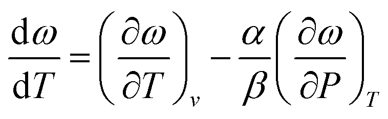
0.1 g of the synthesized up-conversion SrWO4:[Er3+]/[Yb3+] ∼ 0.1 phosphor powder was placed in a copper holder and immersed in a silicone oil bath. The silicone oil bath was placed on a hot plate and the temperature was raised (25–250 °C). After checking the temperature of the powder with an infrared thermometer, a change in luminescence intensity according to temperature change was observed. The spectrum and integrated up-conversion photoluminescence intensity are shown in Fig. 6(a) and (b). In general, it has been reported that the luminescent properties of up-conversion and down-conversion phosphors are reduced due to thermal quenching by external temperature. Ju et al. synthesized a down-converted phosphor doped with Sm3+ rare earth ions using SrWO4 as a host, and observed a change in luminescence properties by heating it from room temperature to 200 °C. As the temperature increased, the thermal quenching phenomenon was observed very weakly. It was observed that the luminescence intensity rather increased at 190 °C, and it was reported that SrWO4 is an excellent material as a thermally stable host.39 Liao et al. synthesized the Sc2(MoO4)3:Yb/Er up-conversion phosphor, and reported the change in luminescence characteristics according to temperature increase, to be negative thermal expansion of the host.40 This phenomenon is caused by the radiative trapping of Yb3+. This is because lattice shrinkage shortens the distance of Yb3+/Er3+ at high temperatures and promotes the radiative trapping of Yb3+. In phosphorescent materials co-doped with Yb3+/Er3+, Yb3+ acts not only as a radiation trap to store energy, but also as a sensitizer to transfer energy to Er3+. These radiation traps may promote the release of Er3+. Also, the distance of Yb3+/Er3+ becomes shorter as the temperature rises. The ET process between the sensitizer (Yb3+) and the activator (Er3+) is usually thought to be caused by dipole interactions. Its ET efficiency is proportional to r−6 (where r is the donor–acceptor distance). Therefore, ET efficiency can be significantly improved at high temperatures.40 Desgreniers et al. observed changes in Raman signals by giving CaWO4, SrWO4, and BaWO4 changes in temperature (10–300 K) and pressure (910 kbar). As the temperature and pressure increased, no significant change was observed in the high frequency region. However, in the WO4(Z)–WO4(Z) stretching region, CaWO4 shifted in phonon energy and frequency due to external environmental factors. However, SrWO4 did not react sensitively, and the frequency shift hardly shifted by about 1–2 cm−1. Using this change, the thermal expansion of the crystal was calculated using the equation below,41| |
 | (2) |
where α and β stand for the thermal expansion and isothermal compressibility of the solid. For crystals of SrWO4, the thermal expansion was calculated to be negative. Because the ionic bond between the covalent bonds within the tetrahedron. Furthermore, anomalous temperature behavior for the least energetic stretching vibrations is revealed through negative values and cannot be explained by the hierarchy of bonds theory.41
 |
| | Fig. 6 (a) PL spectra of changing temperature and (b) integrated PL intensity under 980 nm at 200 mW of SrWO4:[Er3+]/[Yb3+] ∼ 0.1 up-conversion phosphors. | |
The SrWO4:[Er3+]/[Yb3+] up-conversion phosphor synthesized in this work is also considered to have a stronger luminescence intensity as the temperature increases due to efficient energy transfer between rare earth ions by negative thermal expansion to a thermally stable SrWO4 host. To check whether the synthesized up-conversion phosphor could be used as a temperature sensor, a composite was prepared by mixing PDMS and the phosphor powder. The prepared composite was placed on a hot plate, the temperature was raised, and a 980 nm laser was fixed at 200 mW and irradiated. The temperature was checked with an infrared thermometer on the composite. The produced composite was easily bent by fingers, and it was confirmed that the size of the laser circle reflected on the surface became clearer as the temperature increased, and its size gradually increased. Not only in the powder state, but also in the composite made by mixing with the polymer, the luminescence intensity increased with the rise in temperature. These characteristics suggest the potential for a device that can directly emit a visual danger signal with an increase in temperature (Fig. 7).
 |
| | Fig. 7 Photograph of flexible composite under 980 nm at 100 mW, according to the temperature. | |
4 Conclusion
The precursor was prepared at room temperature by co-precipitation, and crystalline SrWO4 calcined at 800 °C was synthesized. In order to synthesize an up-conversion phosphor material, SrWO4:[Er3+]/[Yb3+] were synthesized using the same experimental procedure, by co-doping with rare earth ions Yb3+ and Er3+ using SrWO4 as a host. The synthesized powder exhibited a main peak (112) phase in the X-ray diffraction analysis and had a tetragonal structure. Doping with rare earth ions shifted the 2 theta position of the main peak and decreased the lattice constant. With these changes, it was possible to confirm the change in the crystal structure due to the addition of rare earth ions. The surface of the long cylindrical shaped synthesized powder sample was observed by FE-SEM in the longitudinal direction, and particles with a size of about 4 μm were confirmed. When the synthesized phosphor powder was excited with a 200 mW pump power of a 980 nm laser, a strong green spectrum (529 nm, 543 nm) was confirmed. A relatively weak red spectrum at 657 nm was also observed. As the concentration of Er3+ ions increased, the luminescence intensity decreased due to the concentration quenching phenomenon. When the intensity of the laser pumping energy was changed, a green fluorescence emission was observed, indicating a two-photon process through an excited state absorption process and an energy transfer process. In addition, the synthesized SrWO4:[Er3+]/[Yb3+] up-conversion phosphor showed an increase in luminescence intensity according to the increase in temperature. The host SrWO4 showed thermal stability due to negative thermal expansion, and energy transfer between Yb3+ and Er3+ was efficient. It is a phenomenon that has been formed. For application as a temperature sensor, it was confirmed that when a composite was produced by mixing with PDMS and then subjected to a thermal change, as with the powder, the luminescence intensity increased as the temperature increased, so that it could be used as a material for a temperature sensor that could be identified by the naked eye.
Data availability
The data presented in this study are available on request from the corresponding author.
Conflicts of interest
The authors declare no conflict of interest.
Acknowledgements
This research was supported by Basic Science Research Program through the National Research Foundation of Korea (NRF) funded by the Ministry of Education (NRF-2020R1F1A1072676).
References
- H. J. M. A. A. Zijlmans, J. Bonnet, J. Burton, K. Kardos, T. Vail, R. S. Niedbala and H. J. Tanke, Anal. Biochem., 1999, 267, 30–36, DOI:10.1006/abio.1998.2965.
- A. Rapaport, J. Milliez, M. Bass, A. Cassanho and H. Jenssen, J. Disp. Technol., 2006, 2, 68–78, DOI:10.1109/JDT.2005.863781.
- J. Kundu, Y. Ghosh, A. M. Dennis, H. Htoon and J. A. Hollingsworth, Nano Lett., 2012, 12, 3031–3037, DOI:10.1021/nl3008659.
- B. S. Richards, Sol. Energy Mater. Sol. Cells, 2006, 90, 1189–1207, DOI:10.1016/j.solmat.2005.07.001.
- G. Blasse and A. Bril, J. Chem. Phys., 1967, 47, 5139–5145, DOI:10.1063/1.1701771.
- S. Adachi, J. Lumin., 2018, 202, 263–281, DOI:10.1016/j.jlumin.2018.05.053.
- Y. Zhou, Y. Cheng, Q. Huang, J. Xu, H. Lin and Y. Wang, J. Mater. Chem. C, 2021, 9, 222–223, 10.1039/d0tc05759b.
- J. H. Zeng, T. Xie, Z. H. Li and Y. Li, Cryst. Growth Des., 2007, 7, 2774–2777, DOI:10.1021/cg070477n.
- V. Vaiano, O. Sacco, G. Iervolino, D. Sannino, P. Ciambelli, R. Liguori, E. Bezzeccheri and A. Rubino, Appl. Catal., B, 2015, 176–177, 594–600, DOI:10.1016/j.apcatb.2015.04.049.
- B. Qu, Y. Jiao, S. He, Y. Zhu, P. Liu, J. Sun, J. Lu and X. Zhang, J. Alloys Compd., 2016, 658, 848–853, DOI:10.1016/j.jallcom.2015.11.024.
- A. Shalav, B. S. Richards and M. A. Green, Sol. Energy Mater. Sol. Cells, 2007, 91, 829–842, DOI:10.1016/j.solmat.2007.02.007.
- A. M. Darwish, S. Moore, A. Mohammad, D. Alexander, T. Bastian, W. Dorlus, S. Sarkisov, D. Patel, P. Mele, B. Koplitz and D. Hui, Composites, Part B, 2017, 109, 82–90, DOI:10.1016/j.compositesb.2016.10.053.
- A. M. Darwish, S. Wilson, A. Balckwell, K. Taylor, S. S. Sarkisov, D. N. Patel and B. Koplitz, Ammonia Sensor Based on Polymer-Inorganic Nano-Composite Thin Film Upconversion Light Emitter Prepared by Double-Beam Pulsed Laser Deposition, 2015 Search PubMed.
- L. D. Carlos, R. A. S. Ferreia, V. Z. Bermudez, B. Julian-Lopez and P. Escribano, Chem. Soc. Rev., 2011, 40, 536–549, 10.1039/c0cs00069h.
- K. Pavani, J. Suresh Kumar, K. Srikanth, M. J. Soares, E. Pereira, A. J. Neves and M. P. F. Graça, Sci. Rep., 2017, 7, 1–15, DOI:10.1038/s41598-017-17725-z.
- C. S. Lim, Trans. Electr. Electron. Mater., 2018, 20, 60–66, DOI:10.1007/s42341-018-0083-z.
- T. Deng, S. Yan, X. Jiang and Q. Zhang, Int. J. Opt., 2019, 2019, 1–6, DOI:10.1155/2019/4814793.
- E. W. Barrera, Q. Madueño, F. J. Novegil, A. Speghini and M. Bettinelli, Opt. Mater., 2018, 84, 354–359, DOI:10.1016/j.optmat.2018.07.022.
- A. Pandey, V. K. Rai, R. Dey and K. Kumar, Mater. Chem. Phys., 2013, 139, 483–488, DOI:10.1016/j.matchemphys.2013.01.043.
- J. Jung, J. Kim, Y. Shim, D. Hwang and C. S. Son, Materials, 2020, 13, 4165, DOI:10.3390/ma13184165.
- J. Jung, S. Yi, D. Hwang and C. Son, Materials, 2021, 14, 3717, DOI:10.3390/ma14133717.
- J. Ruan, T. Yuan, Y. Pang, S. Luo, C. Peng, J. Yang and S. Zheng, Carbon, 2018, 126, 9–16, DOI:10.1016/j.carbon.2017.09.099.
- P. P. Ortega, B. Hangai, H. Moreno, L. S. R. Rocha, M. A. Ramírez, M. A. Ponce, E. Longo and A. Z. Simões, J. Alloys Compd., 2021, 888, 161517, DOI:10.1016/j.jallcom.2021.161517.
- X. Wang, Y. Wang, Y. Bu, X. Yan, J. Wang, P. Cai, T. Vu and H. J. Seo, Sci. Rep., 2017, 7, 43383, DOI:10.1038/srep43383.
- R. D. Shannon, Acta Crystallogr., Sect. A: Found. Adv., 1976, 32, 751–767, DOI:10.1107/S0567739476001551.
- E. Ryu and Y. Huh, Mater. Lett., 2008, 62, 3081–3083, DOI:10.1016/j.matlet.2008.01.108.
- Y. Hu, X. Liang, Y. Wang, E. Liu, X. Hu and J. Fan, Ceram. Int., 2015, 41, 14545–14553, DOI:10.1016/j.ceramint.2015.07.171.
- J. Zhao, Y. Sun, X. Kong, L. Tian, Y. Wang, L. Tu, J. Zhao and H. Zhang, J. Phys. Chem. B, 2008, 112, 15666–15672, DOI:10.1021/jp805567k.
- W. A. Pisarski, L. Grobelny, J. Pisarska, R. Lisiecki and W. Ryba-Romanowski, J. Alloys Compd., 2011, 509, 8088–8092, DOI:10.1016/j.jallcom.2011.05.056.
- B. Wei, L. Z. Zhang, Z. B. Lin and G. F. Wang, Mater. Res. Innovations, 2007, 11, 154–157, DOI:10.1179/143307507X225605.
- G. Xiang, X. Liu, W. Liu, B. Wang, Z. Liu, S. Jiang, X. Zhou, L. Li, Y. Jin and J. Zhang, J. Am. Ceram. Soc., 2020, 103, 2540–2547, DOI:10.1111/jace.16939.
- M. Erdem and B. Sitt, Opt. Mater., 2015, 46, 260–264, DOI:10.1016/j.optmat.2015.04.029.
- G. A. Fernández-Alcober and P. Shumyatsky, J. Algebra, 2018, 500, 19–29, DOI:10.1016/j.jalgebra.2016.07.041.
- G. Bilir, A. Kaya, H. Cinkaya and G. Eryürek, Spectrochim. Acta, Part A, 2016, 165, 183–190, DOI:10.1016/j.saa.2016.04.042.
- P. Woźny, M. Runowski and S. Lis, J. Lumin., 2019, 209, 321–327, DOI:10.1016/j.jlumin.2019.02.008.
- M. Guan, H. Zheng, L. Mei, M. S. Molokeev, J. Xie, T. Yang, X. Wu, S. Huang and Z. Huang, J. Am. Ceram. Soc., 2015, 98, 1182–1187, DOI:10.1111/jace.13415.
- M. A. Chamarro and R. Cases, Infrared to visible upconversion of Er3+ ions in Yb3+ doped fluorohafnate glasses, Elsevier BV, 1990 Search PubMed.
- X. Xia, A. Volpi, J. Y. D. Roh, M. C. De Siena, D. R. Gamelin, M. P. Hehlen and P. J. Pauzauskie, J. Lumin., 2021, 236, 118006, DOI:10.1016/j.jlumin.2021.118006.
- Z. JU, R. WEI, J. MA, C. PANG and W. LIU, J. Alloys Compd., 2010, 507, 133–136, DOI:10.1016/j.jallcom.2010.07.138.
- J. Liao, M. Wang, F. Lin, Z. Han, B. Fu, D. Tu, X. Chen, B. Qiu and H. Wen, Nat. Commun., 2022, 13, 2090, DOI:10.1038/s41467-022-29784-6.
- S. Desgreniers, S. Jandl and C. Carlone, J. Phys. Chem. Solids, 1984, 45, 1105–1109, DOI:10.1016/0022-3697(84)90004-0.
|
| This journal is © The Royal Society of Chemistry 2022 |
Click here to see how this site uses Cookies. View our privacy policy here. 
 Open Access Article
 This Open Access Article is licensed under a Creative Commons Attribution-Non Commercial 3.0 Unported Licence
 *b
*b