DOI:
10.1039/D2RA02224A
(Paper)
RSC Adv., 2022,
12, 18144-18153
Effect of tertiary amino groups in the hydrophobic segment of an amphiphilic block copolymer on zinc phthalocyanine encapsulation and photodynamic activity†
Received
6th April 2022
, Accepted 14th June 2022
First published on 21st June 2022
Abstract
Polymer micelles are promising nanocarriers for hydrophobic photosensitizers of photodynamic therapy (PDT). Poly(styrene-co-(2-(N,N-dimethylamino)ethyl acrylate))-block-poly(polyethylene glycol monomethyl ether acrylate) (P(St-co-DMAEA)-b-PPEGA; 1) was prepared via reversible addition and fragmentation chain transfer (RAFT) polymerization as a carrier for a zinc phthalocyanine (ZnPc) photosensitizer to be used in PDT. The DMAEA-unit composition in the P(St-co-DMAEA) segment was adjusted to 0.40 molar ratio, which caused a sharp increase in water-solubility when the pH decreased from 7.4 to 5.0. The polymer 1 micelle size distribution also shifted to lower when the pH decreased, whereas this change was not observed in PSt-co-PPEGA (2), which was previously reported. The UV-vis spectrum of the ZnPc-loaded micelles of polymer 1 exhibited relatively sharp Q bands, comparable to those measured in DMSO, indicating good compatibility of the condensed core with ZnPc. ZnPc-loaded micelles of polymer 1 exerted excellent photocytotoxicity in the MNNG-induced mutant of the rat murine RGM-1 gastric epithelial cell line (RGK-1). In contrast, the ZnPc-loaded micelles of polymer 2 were completely inactive under the same conditions. Fluorescence from the RGK-1 cells treated with ZnPc-loaded micelles of polymer 1 was observed after 4 h of co-incubation, while no fluorescence was observed in cells treated with ZnPc-loaded micelles of polymer 2. These results indicate that the pH-responsive nature and good compatibility with ZnPc exhibited by the polymer 1 micelles are essential characteristics of ZnPc carriers for efficient photodynamic therapy.
Introduction
Photodynamic therapy (PDT) is a minimally-invasive cancer therapy using photosensitizers and light that is currently applied to esophageal cancer and non-small cell lung cancer.1–8 After a photosensitizer is administered to a patient and accumulates at the tumor site, photoirradiation is used to generate a cytotoxic reactive oxygen species (ROS). Several mechanisms have been suggested to kill tumor cells, such as initiating the apoptosis cascade and vascular shutdown. In addition to being precisely controlled by photoirradiation, tumor-accumulating photosensitizers regulate legions to eliminate malignancies. Limited light availability in the deeper tissue is a major drawback in PDT treatment. Effective photosensitizers must absorb light in the near-infrared (NIR) region,9–11 due to the fact that longer wavelength increases tissue transparency.
Zinc phthalocyanine (ZnPc) derivatives have many advantages such as high absorption ability in the first near-infrared (NIR-I) window, good ROS-generation efficiency, and excellent photostability.12–14 However, ZnPc derivatives are highly hydrophobic and tend to aggregate which decreases their photosensitizing ability. Therefore, many attempts have been made to increase the water solubility of phthalocyanine derivatives. The most straightforward approach is the conjugation of hydrophilic elements such as sulfo,15–19 carboxyl,19–21 amino,22 ammonio,20,23–26 hydroxy,27 guanidyl,28,29 and morpholine groups,30 as well as polyethylene glycol moieties31–37 at peripheral and non-peripheral positions. This strategy also affords additional functionalities such as tumor-targeting ligands, including carbohydrates38–42 and peptides.43–48 However, this approach frequently requires laborious synthetic work, and the resulting photosensitizers are very costly.
Another promising approach is the use of drug delivery system (DDS) technology. This allows us to design the photosensitizer and carrier separately to meet photophysical and physiological requirements, respectively. Among the many types of carriers for PDT photosensitizers, polymer micelles are very promising nanomaterials because of their biocompatibility and synthetic availability. Several ZnPc-loaded polymer micelles have been prepared using amphiphilic block copolymers such as poly(ethylene glycol)-b-poly(L-lactide) (PEG-b-PLLA),49–51 poly(ethylene glycol)-b-poly(ε-caprolacton) (PEG-b-PCL),52 and more highly sophisticated polymers.53–55 ZnPc can be physically trapped in the hydrophobic core of the polymer micelle. However, most ZnPc-loaded polymer micelles show severely broadened UV-vis spectra, indicating the formation of ZnPc aggregates. Aggregation changes the photophysical properties of ZnPc molecules, and deactivates the photodynamic effect.56,57 Previously, we demonstrated the synthesis of polystyrene-b-poly(polyethylene glycol monomethyl ether acrylate) (PSt-b-PPEGA) and the preparation of ZnPc-loaded polymer micelles.58 The efficiency of encapsulating ZnPc depended on the molecular architecture of PSt-b-PPEGA. Disappointingly, the photocytotoxicity of the resulting ZnPc-loaded polymer micelles was very low in HeLa cells. Similar to their behavior in other polymer micelles, ZnPc molecules are strongly aggregated in the polymer micelles, as evidenced by the UV-vis spectra. In addition, the ZnPc-loaded polymer micelles were kinetically frozen and lacked the ability to release ZnPc. Hence, the ZnPc molecules trapped in the polymer micelles were photochemically inactivated. This might have caused the low photocytotoxicity of ZnPc-loaded polymer micelles. Therefore to achieve successful PDT treatment, it is crucial that carriers physically encapsulate ZnPc and prevent its aggregation.
Recently, Borovkov et al. reported that a mixture of a π-accepting solvent such as chlorobenzene and a nitrogenous base such as triethylamine dissolves ZnPc to make a clear solution with molecular dispersion.59 In addition, we demonstrated that the side group of the hydrophobic chain affects the aggregating behavior of ZnPc in the polymer micelles.60 These two findings inspired us to investigate the molecular dispersion of ZnPc in polymer micelles with the hydrophobic chains consisting of repeating units with aromatic rings and tertiary amines. In addition, tertiary amines, with pKa values approximately 6.5–7.0, can be used as a pH-sensitive element between the extracellular physiological environment (pH = 7.4) and acidic organelles (pH = 5.0) during cellular internalization. In this study, we synthesized a pH-responsive amphiphilic block copolymer using 2-(N,N-dimethylamino)ethyl acrylate (DMAEA) as a pH-sensitive element, and preparation of the ZnPc-loaded polymer micelles. The pH-responsive properties, aggregating behavior of ZnPc, and photocytotoxicity of the polymer micelles are discussed.
Results and discussion
Design of pH-responsive hydrophobic chain
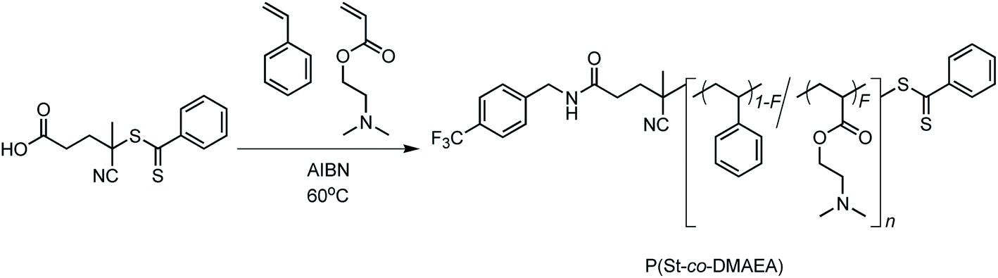
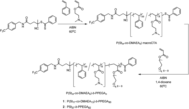
To provide a pH-response ability, we designed a copolymer of styrene and DMAEA as a hydrophobic segment. First, an appropriate molar fraction of DMAEA units in the copolymer, P(St-co-DMAEA), was investigated to exhibit a large solubility change during the pH shift from 7.4 to 5.0. Six copolymers were prepared by bulk RAFT polymerization using 4-cyano-4-(thiobenzoyl)thiopentanoic acid (CPADB) as a chain transfer agent, as shown in Scheme 1.
 |
| | Scheme 1 Synthesis of P(St-co-DMAEA) for solubility test via RAFT polymerization. | |
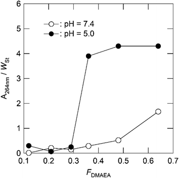
The mole fractions of the DMAEA units (FDMAEA), number average molecular mass (Mn), and dispersity (Mw/Mn) of the resulting copolymers are listed in Table 1. The FDMAEA values varied from 0.12 to 0.64, while the Mn values were relatively constant at approximately 104, except for the case of FDMAEA = 0.64. The relative water solubility was examined in phosphate-buffered saline (PBS), and PBS adjusted to pH 5.0, by adding HCl. The amount of the copolymer dissolved in the buffered solution was quantified by the absorbance at 264 nm and divided by the weight fraction of styrene units to determine the water solubility of the copolymer. Fig. 1 shows the plots of the relative water solubility as a function of the FDMAEA value of the copolymer. The relative water solubility gradually increased with an increase in the FDMAEA value at pH 7.4. In contrast, the relative water solubility suddenly increased at an FDMAEA of approximately 0.3 at pH 5.0. This suggests that the copolymer FDMAEA of approximately 0.3 exerts a large water-solubility change between the physiological environment (pH = 7.4) and acidic intracellular organelles such as endosomes and lysosomes (pH = 5.0). Therefore, we choose the FDMAEA value of 0.4 for the hydrophobic P(St-co-DMAEA) segment for the pH-responsive amphiphilic block copolymer.
Table 1 Synthesis of P(St-co-DMAEA) for solubility test via RAFT polymerizationa
| Run no. |
fDMAEAb |
Yieldc (%) |
FDMAEAd |
Mn (Mw/Mn)e |
| Bulk polymerization; [St + DMAEA]0/[CPADB]0/[AIBN]0 = 500/1/0.2; polym. temp., 60 °C; polym. time, 24 h. Mole fraction of DMAEA in the feed. Isolated yield. Mole fraction of DMAEA unit in the copolymer. Number average molecular mass and dispersity determined by SEC using polystyrene standards. |
| 1 |
0.10 |
15 |
0.12 |
10![[thin space (1/6-em)]](https://www.rsc.org/images/entities/char_2009.gif) 500 (1.07) 500 (1.07) |
| 2 |
0.20 |
14 |
0.21 |
9900 (1.08) |
| 3 |
0.30 |
20 |
0.29 |
13600 (1.09) |
| 4 |
0.40 |
8 |
0.36 |
10200 (1.14) |
| 5 |
0.60 |
21 |
0.48 |
8300 (1.40) |
| 6 |
0.80 |
22 |
0.64 |
6200 (1.55) |
 |
| | Fig. 1 Relative solubility of P(St-co-DMAEA) in PBS (pH = 7.4; open circle) and in PBS adjusted to a pH of 5.0 by adding HCl (filled circle) as a function of the FDMAEA value. | |
Synthesis of P(St-co-DMAEA)-b-PPEGA
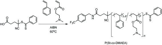
The amphiphilic block copolymer, P(St-co-DMAEA)-b-PPEGA, was synthesized by RAFT polymerization of styrene with DMAEA, followed by chain extension with PEGA, as shown in Scheme 2.
 |
| | Scheme 2 Synthesis of P(St17-co-DMAEA13)-b-PPEGA90 (1) and PSt37-b-PPEGA82 (2) via RAFT polymerization. | |
It should be noted that we used trifluoromethyl-labeled CPADB (CF3-CPADB) as a chain transfer agent to determine the degree of polymerization (DPn) of each repeating unit by the combination of 1H and 19F NMR spectroscopies. First, a statistical copolymer, P(St-co-DMAEA) macroCTA, was synthesized by bulk RAFT polymerization of styrene with DMAEA using CF3-CPADB and azobisisobutyronitrile (AIBN) as a chain transfer agent and initiator, respectively, at 60 °C for 24 h. The DPn values of styrene and DMAEA of the resulting polymer were 16 and 12, respectively, indicating the FDMAEA value was 0.43 (Table 2).
Table 2 Synthesis of P(Stm-co-DMAEAk) macroCTA via RAFT polymerizationa
| Run no. |
fDMAEAb |
xStc |
xDMAEAc |
Yieldd (%) |
ke |
me |
Mn,NMRf |
Mn (Mw/Mn)g |
| Bulk polymerization; [St + DMAEA]0/[CF3-CPADB]0/[AIBN]0 = 200/1/0.1; polym. temp., 60 °C; polym. time, 24 h. Mole fraction of DMAEA in the feed. Conversions of St (xSt) and DMAEA (xDMAEA) determined by 1H NMR spectroscopy. Isolated yield. Number average degree of polymerization of St (m) and DMAEA (k) determined by 1H and 19F NMR spectroscopies using cross reference. Number average molecular mass calculated using the k and m values considering the mass of CF3-CPADB. Number average molecular mass and dispersity determined by SEC using polystyrene standards. |
| 1 |
0.40 |
0.29 |
0.13 |
7 |
16 |
12 |
3400 |
2800 (1.10) |
| 2 |
0 |
0.27 |
— |
12 |
37 |
0 |
4300 |
3700 (1.08) |
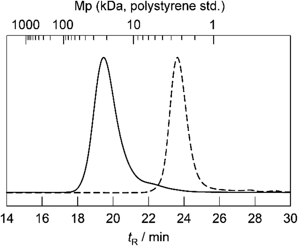
For comparison, a homopolymer, PSt macroCTA, was also synthesized by a similar procedure. The resulting macroCTA was extended with PEGA using AIBN as an initiator in 1,4-dioxane at 60 °C for 24 h to produce block copolymer P(St-co-DMAEA)-b-PPEGA and PSt-b-PPEGA. Fig. 2 shows the SEC traces of P(St-co-DMAEA) macroCTA and P(St-co-DMAEA)-b-PPEGA. The SEC trace of P(St-co-DMAEA)-b-PPEGA clearly shifted toward a higher molecular mass than that of P(St-co-DMAEA) macroCTA without significant broadening.
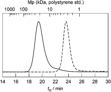 |
| | Fig. 2 SEC traces of P(St16-co-DMAEA12) macroCTA (broken line) and polymer 1 (solid line). | |
Fig. 3 shows the 1H DOSY spectrum of P(St-co-DMAEA)-b-PPEGA, in which the styrene units exhibit a diffusion coefficient identical to that of the PEGA units. These results indicate the formation of a block copolymer, P(St-co-DMAEA)-b-PPEGA.
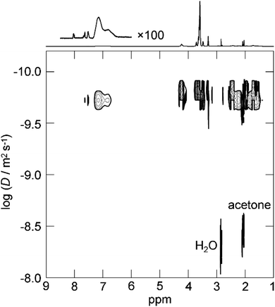 |
| | Fig. 3 1H DOSY spectrum of polymer 1 in acetone-d6 at 298 K. | |
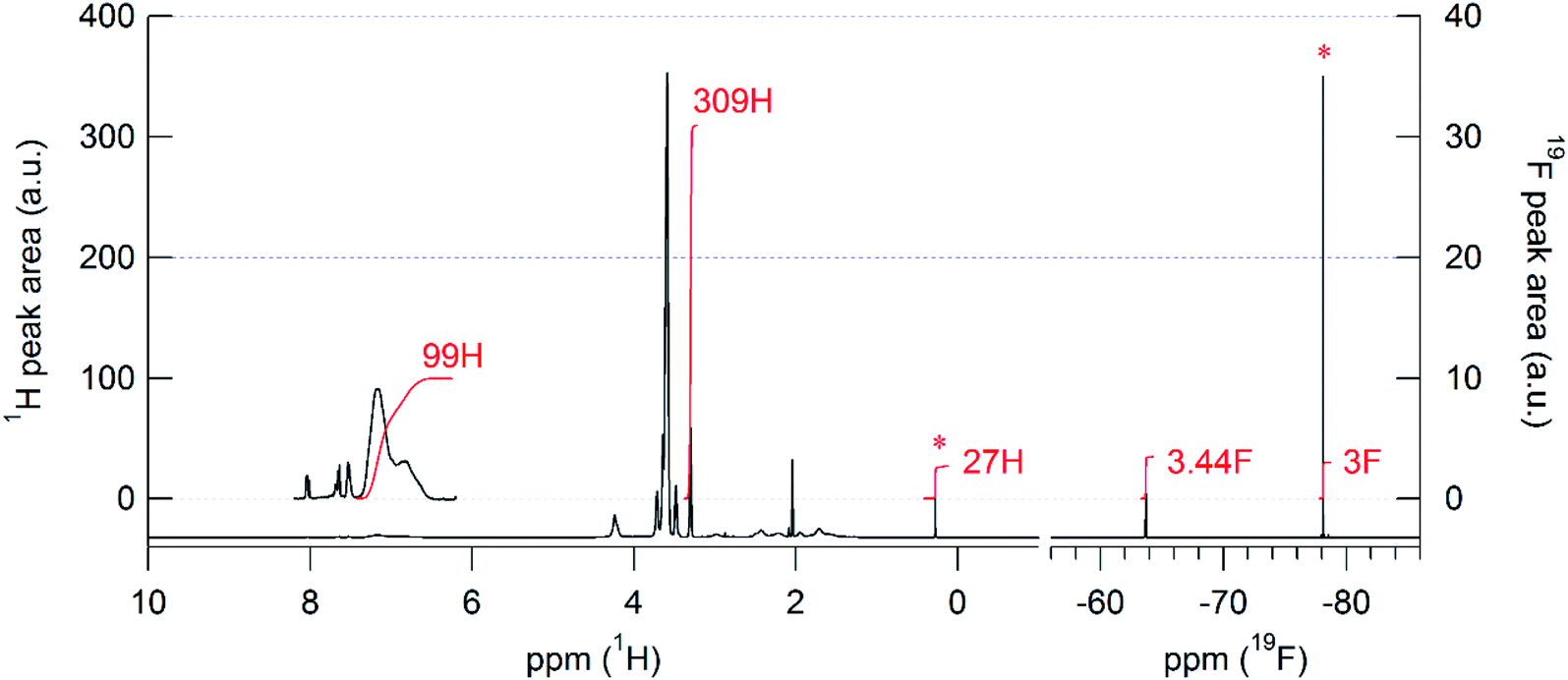
Fig. 4 shows the 1H and 19F NMR spectra of P(St-co-DMAEA)-b-PPEGA in acetone-d6 mixed with tris(trimethylsilyl)-2,2,2-trifluoroethoxysilane (CF3CH2OTTMS) as an internal cross-reference for peak area normalization between these two spectra. The trifluoromethyl group at the initiating end was unequivocally identified in the 19F NMR spectrum owing to the nuclear specificity of NMR spectroscopy. The 19F NMR spectrum affords the ratio of polymer to CF3CH2OTTMS, while the 1H NMR spectrum affords the ratio of each repeating unit to CF3CH2OTTMS. Therefore, the combination of these two spectra afforded DPn values for each repeating unit. This 1H–19F cross-reference technique is helpful for determining the DPn and Mn values by end group analysis, especially in the case that the polymer affords very intense peaks in the 1H NMR spectrum, such as a PPEGA segment.
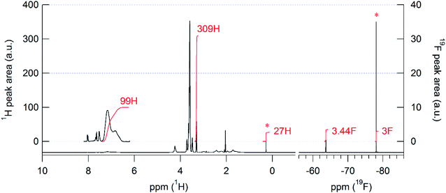 |
| | Fig. 4 1H (left panel) and 19F NMR spectra (right panel) of 1 in acetone-d6. The asterisks indicate the peaks from CF3CH2OTTMS as an internal cross reference for peak area normalization between the 1H and 19F NMR spectra. | |
Table 3 summarizes the DPn values for each repeating unit of the block copolymers. Finally, we obtained a pH-responsive block copolymer, P(St17-co-DMAEA13)-b-PPEGA90 (hereinafter denoted as 1), and the block copolymer, PSt37-b-PPEGA82 (hereinafter denoted as 2), as shown in Scheme 2. The pH-responsive nature of the resulting block copolymer was examined by acid–base titration. Fig. 5 shows the titration curves of polymers 1 and 2. The curve for polymer 1 had an inflection point at pH of approximate 7 due to the buffering capacity of DMAEA units, while polymer 2 showed no buffering nature.
Table 3 Synthesis of P(St17-co-DMAEA13)-b-PPEGA90 (1) and PSt37-b-PPEGA82 (2) via chain extension with PEGAa
| Symble |
xPEGAb |
Yieldc (%) |
md |
kd |
nd |
Mn,NMRe |
Mn (Mw/Mn)f |
| [PEGA]0/[P(Stk-co-DMAEAm) macroCTA]0/[AIBN]0 = 100/1/0.2; polym. solv., 1,4-dioxane; polym. temp., 60 °C; polym. time, 24 h. Conversion of PEGA (xPEGA) determined by 1H NMR spectroscopy. Isolated yield. Number average degree of polymerization of St (m), DMAEA (k) and PEGA (n) determined by 1H and 19F NMR spectroscopies using cross reference. Number average molecular mass calculated using the k, m and n values considering the mass of CF3-CPADB. Number average molecular mass and dispersity determined by SEC using polystyrene standards. |
| 1 |
0.49 |
43 |
17 |
13 |
90 |
47400 |
16000 (1.25) |
| 2 |
0.68 |
57 |
37 |
0 |
82 |
44000 |
15000 (1.21) |
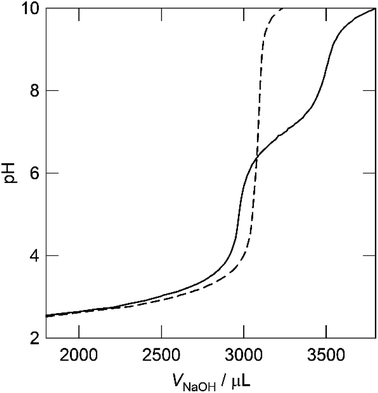 |
| | Fig. 5 pH titration curves of polymers 1 (solid line) and 2 (broken line). | |
Preparation and characterization of polymer micelles
Polymer micelles were prepared by the dialysis method (Chart 1).
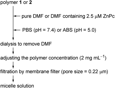 |
| | Chart 1 Flow chart for preparation of polymer micelle solution. | |
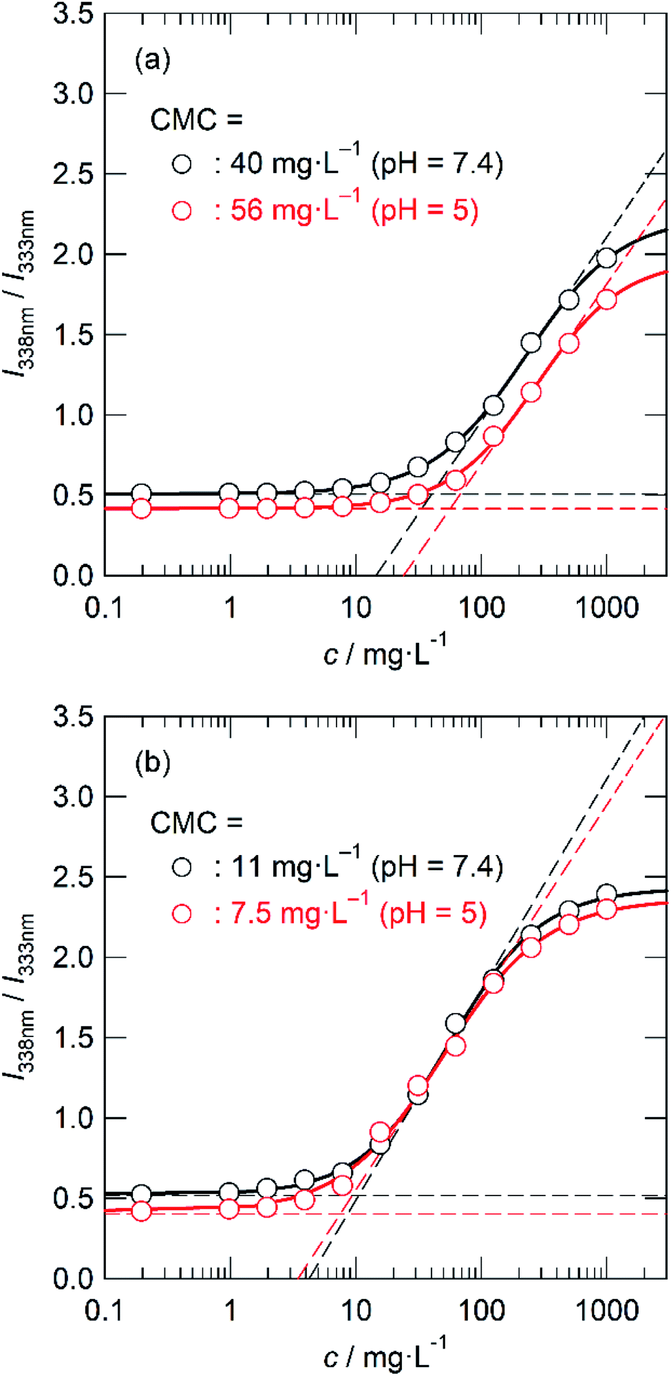
Briefly, polymer 1 or 2 was dissolved in DMF and slowly added to the same amount of PBS, then DMF was removed by dialysis to afford a clear solution without any precipitates. After adjusting the polymer concentration to 2 mg mL−1, the solution was filtered through a membrane filter (pore size of 0.22 μm). The critical micelle concentration (CMC) in PBS (pH = 7.4) and acetate buffered saline (ABS, pH = 5.0) was estimated by the fluorometric method using pyrene as a hydrophobic fluorescent probe. Fig. 6 shows plots of the fluorescence intensity ratio (I338/I333) as a function of the logarithm of the polymer concentration.
 |
| | Fig. 6 I338/I333 values as a function of the concentration of polymers 1 (a) and 2 (b) in PBS (black, pH = 7.4) and ABS (red, pH = 5). [Pyrene] = 1 mM. | |
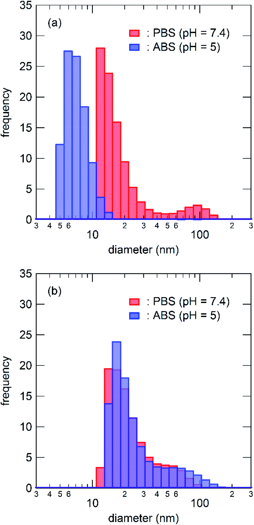
Interestingly, the plots of polymer 1 in ABS slightly shifted toward higher concentrations than those measured in PBS, while no such shift was observed for polymer 2. The CMC value was determined by the crossing point of the baseline and the tangent at the inflection point of the best-fitted sigmoidal curve. The CMC value of polymer 1 was determined to be 40 mg L−1 in PBS and changed to 56 mg L−1 in ABS. Significant changes due to the pH switch were also observed in dynamic light scattering (DLS) measurements. Fig. 7 shows the size distribution of the micelles of polymers 1 and 2 in PBS and ABS at a polymer concentration of 1 mg mL−1. The size distribution of the micelles of polymer 1 shifted to lower when the pH changed from 7.4 to 5.0. In contrast, no changes were observed for polymer 2. These results suggest that DMAEA units act as pH-responsive moieties to destabilize the polymeric micelles under acidic conditions.
 |
| | Fig. 7 Size distribution of polymer 1 micelle (a) and polymer 2 micelle (b) in PBS (red, pH = 7.4) and ABS (blue, pH = 5). The polymer concentration was 1000 mg L−1. | |
Zinc phthalocyanine (ZnPc), a highly hydrophobic photosensitizer, was used as the payload of these polymer micelles. ZnPc-loaded micelles were prepared by a procedure similar to that of free micelles except for the use of a DMF solution of ZnPc instead of pure DMF. After dialysis and adjustment of the polymer concentration, insoluble ZnPc precipitates were removed by filtration with a membrane filter (pore size 0.22 μm). The concentrations of ZnPc in the micelle solutions were determined spectrofluorometrically and are listed in Table 4.
Table 4 Preparation of the micelle solutions of polymers 1 and 2a
| Run no. |
In preparation |
Micelle solution |
| Polymerb |
[ZnPc]c (μM) |
Bufferd |
[ZnPc]e (μM) |
E.E.f (%) |
D50%g (nm) |
| [Polymer] = 2 mg mL−1. 1, P(St17-co-DMAEA13)-b-PPEGA90; 2, PSt37-b-PPEGA82. Concentration of ZnPc in DMF for dissolving the polymer. PBS, phosphate buffered saline (pH 7.4); ABS, acetate-buffered saline (pH 5.0). Concentration of ZnPc in the micelle solution determined by spectrofluorometry. Encapsulation efficiency defined as E.E. (%) = ([ZnPc]micelle × 5)/([ZnPc]DMF × 2) × 100. Median diameter, as determined by DLS. |
| 1 |
1 |
0 |
PBS |
0 |
— |
18 |
| 2 |
1 |
0 |
ABS |
0 |
— |
7 |
| 3 |
2 |
0 |
PBS |
0 |
— |
20 |
| 4 |
2 |
0 |
ABS |
0 |
— |
20 |
| 5 |
1 |
2.5 |
PBS |
0.73 |
73 |
79 |
| 6 |
1 |
2.5 |
PBS |
0.63 |
63 |
n.d. |
| 7 |
1 |
2.5 |
PBS |
0.52 |
52 |
n.d. |
| 8 |
2 |
2.5 |
PBS |
0.28 |
28 |
33 |
| 9 |
2 |
2.5 |
PBS |
0.58 |
58 |
n.d. |
| 10 |
2 |
2.5 |
PBS |
0.44 |
44 |
n.d. |
Triplicate experiments suggest that the encapsulation efficiency (E.E.) did not depend on the presence of DMAEA units, and ranged from 28% to 73%. Fig. 8 shows the UV-vis spectra of ZnPc-loaded polymer micelles in PBS. The ZnPc-loaded micelles of polymer 2 showed a broadened absorption band, indicating that ZnPc molecules were tightly aggregated in the hydrophobic core. In contrast, the ZnPc-loaded micelles of polymer 1 showed Q bands similar to those recorded in dimethyl sulfoxide (DMSO). Therefore, the micelles of polymer 1 afford the microenvironment to dissolve ZnPc in a monomeric form. The difference in the microenvironment of the polymer micelles crucially affects the photophysical and photodynamic effects of ZnPc.
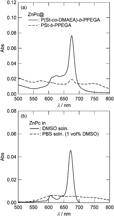 |
| | Fig. 8 UV-vis spectra of ZnPc-loaded micelles of polymers 1 ([ZnPc] = 0.52 μM; solid line) and 2 ([ZnPc] = 0.44 μM) in PBS (a) and ZnPc in DMSO and PBS containing 1% DMSO ([ZnPc] = 0.5 μM; b). | |
In vitro photocytotoxicity of ZnPc-loaded polymer micelles
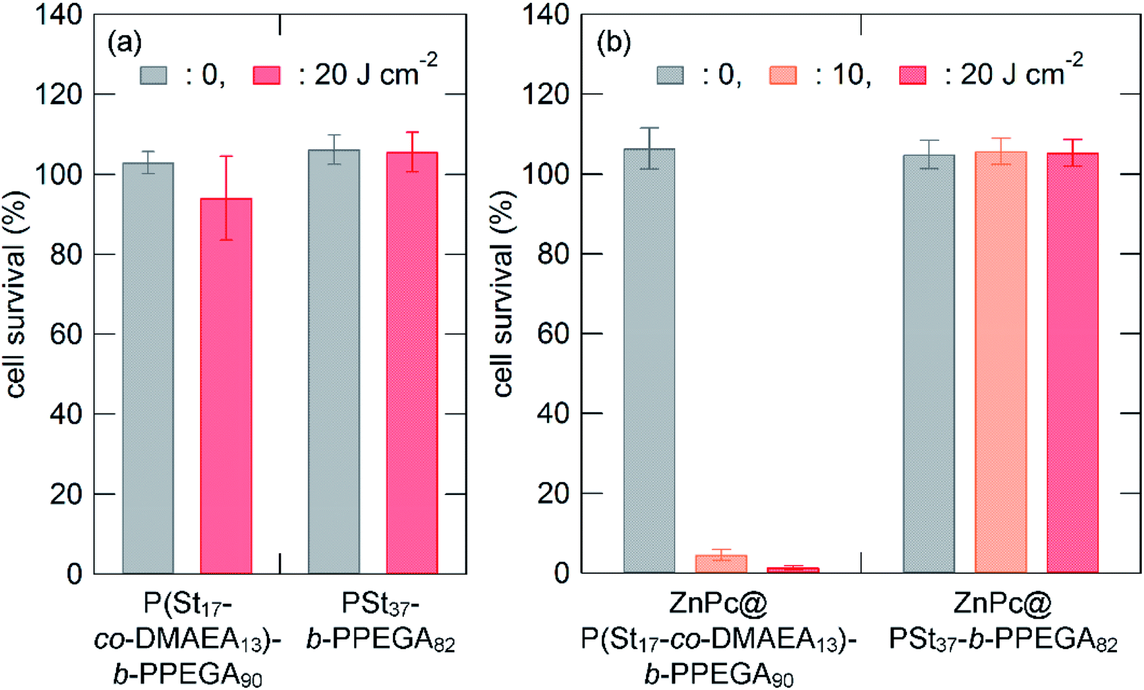
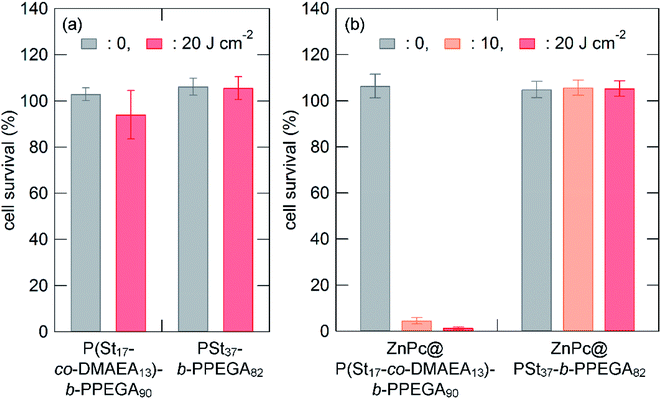
The in vitro photocytotoxicity of ZnPc-loaded micelles was examined in MNNG-induced mutant of the rat murine RGM-1 gastric epithelial cell line (RGK-1).61 RGK-1 cells were co-incubated with the micelles in a mixture of PBS and culture medium (1/1, v/v) for 24 h. After removing the excess amount of the drug, the cells were photoirradiated with a 100 W halogen lamp equipped with a Y-50 cut filter. The cell survival rate was determined by WST-8 assay and normalized by the value for no drug treatment with photoirradiation. Fig. 9a shows the cell survival rate after treatment with the payload-free micelles of polymers 1 and 2 at a concentration of 1 mg mL−1 as a function of the light dose. Fig. 9a indicates that the micelles of 1 and 2 essentially have no cytotoxicity regardless of photoirradiation. The cell survival rates after the ZnPc-loaded polymer micelle treatment are shown in Fig. 9b. ZnPc-loaded micelles of 1 and 2 did not show cytotoxicity without photoirradiation (light dose of 0 J cm−2). ZnPc-loaded micelles of 2 did not show cytotoxicity even upon photoirradiation at a light dose of 20 J cm−2. This can be expected because of the very low ZnPc concentration in comparison to that used in our previous study.58 In contrast, ZnPc-loaded micelles of 1 exerted excellent photocytotoxicity under the same condition; almost complete annihilation was achieved upon photoirradiation of 20 J cm−2 with a ZnPc dose of only 0.05 μM.
 |
| | Fig. 9 In vitro dark and photocytotoxicity of polymers 1 and 2 (a; [polymer] = 1000 mg L−1) and their ZnPc-loaded micelles of polymers 1 and 2 (b; [ZnPc] = 0.05 μM) in RGK-1 cells as a function of the light dose. The photoirradiation was provided by a 100-W halogen lamp equipped with a Y-50 cutoff filter (λ > 500 nm). The values are the mean ± standard deviation of six replicate experiments. | |
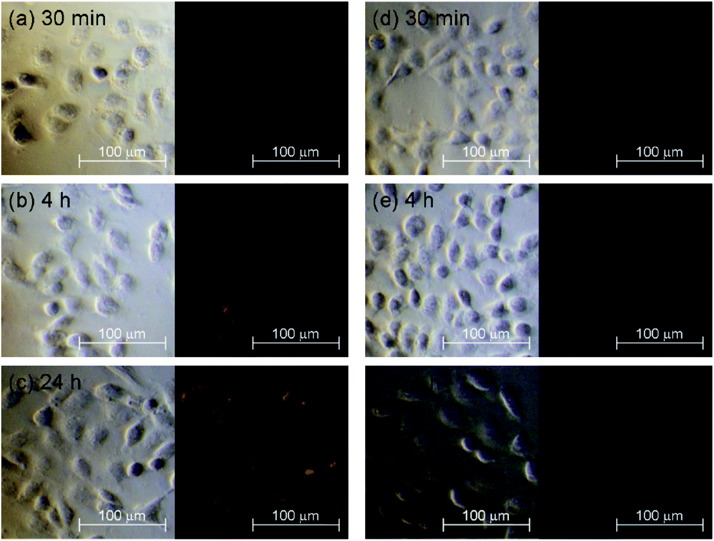
Fig. 10 shows the images of RGK-1 cells co-incubated with ZnPc-loaded polymer micelles captured by fluorescence microscopy. The red fluorescence from the cells treated with ZnPc-loaded micelles of 1 gradually increased with co-incubation time. In contrast, almost no fluorescence was observed in the cells treated with 2. This distinct difference in the photophysical behavior in the cellular microenvironment affected the photocytotoxicity of the ZnPc-loaded micelles.
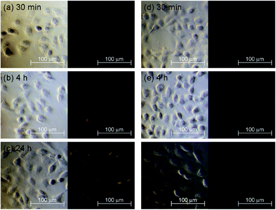 |
| | Fig. 10 Bright field (left panel) and fluorescence images (right panel) of RGK-1 cells incubated in the presence of ZnPc-loaded micelles of polymers 1 (a, 30 min; b, 4 h; c, 24 h) and 2 (d, 30 min; e, 4 h; f, 24 h). | |
The photodynamic behavior of ZnPc-loaded micelles of 1 and 2 should be related to the fate of polymer micelles co-incubated with cells, that is, cellular uptake via endocytosis, endosome escape, and ZnPc release or conservation in polymer micelles. The pH-responsive nature of 1 enhances endosome escape and facilitates ZnPc release. However, another mechanism is also possible. If the polymer micelle is stable enough in the cellular microenvironment, ZnPc encapsulated in the micelles of 1 must be photochemically active rather than that in 2. The first hypothesis is a line along with the usual drug delivery regime, in which polymer micelles act as simple carriers for photosensitizers. However, the latter hypothesis is beyond the simple drug delivery regime; polymer micelles provide a suitable microenvironment for photosensitizers. The key is the kinetic properties of polymer micelles, which are not yet fully understood, especially in very complicated microenvironments such as the cytosol. To address this issue, a study on the behavior of micelles of 1 is in progress in our laboratory using Förster resonance energy transfer (FRET) and aggregation-induced emission (AIE) dye labeling techniques.
Conclusions
Amphiphilic block copolymers, P(St-co-DMAEA)-b-PPEGA (1) and PSt-b-PPEGA (2), were successfully prepared by successive RAFT polymerizations. The mole fraction of styrene units in P(St-co-DMAEA) of 1 was 0.4, which caused the water solubility to increase when the pH decreased from 7.4 to 5.0. Polymers 1 and 2 both formed micelles in PBS. The unique nature of 1 was evidenced by DLS and the UV-vis measurements. DLS results revealed the pH sensitivity of the micelles of 1: the size distribution of the micelle of 1 dramatically shifted lower with a decrease in the pH, while that of 2 did not change. The UV-vis spectrum of ZnPc-loaded micelles of 1 indicated the excellent compatibility of the core components of the micelle with ZnPc: the Q bands of ZnPc-loaded micelle of 1 were very similar to those observed in DMF, while those of 2 were severely broadened. ZnPc-loaded micelle of 1 exerted excellent photocytotoxicity in RGK-1 cells and showed distinct photochemical behavior in the cellular microenvironment compared to 2. The enhanced PDT effect of polymer 1 micelles should be related to its pH-responsive nature and good compatibility with ZnPc.
Conflicts of interest
There are no conflicts to declare.
Acknowledgements
The authors wish to thank Prof. Hidenori Okuzaki, University of Yamanashi for support with the DLS measurements. This work was supported by JSPS KAKENHI (grant numbers 21550156, 25410126, and 17K05877).
Notes and references
- T. C. Pham, V. N. Nguyen, Y. Choi, S. Lee and J. Yoon, Chem. Rev., 2021, 121, 13454–13619 CrossRef CAS PubMed.
- A. Escudero, C. Carrillo-Carrión, M. C. Castillejos, E. Romero-Ben, C. Rosales-Barrios and N. Khiar, Mater. Chem. Front., 2021, 5, 3788–3812 RSC.
- S. Wang, X. Wang, L. Yu and M. Sun, Photodiagn. Photodyn. Ther., 2021, 34, 102254 CrossRef CAS PubMed.
- B. Mansoori, A. Mohammadi, M. A. Doustvandi, F. Mohammadnejad, F. Kamari, M. F. Gjerstorff, B. Baradaran and M. R. Hamblin, Photodiagn. Photodyn. Ther., 2019, 26, 395–404 CrossRef CAS PubMed.
- M. Lan, S. Zhao, W. Liu, C.-S. Lee, W. Zhang and P. Wang, Adv. Healthcare Mater., 2019, 8, 1900132 CrossRef PubMed.
- S. Kwiatkowski, B. Knap, D. Przystupski, J. Saczko, E. Kędzierska, K. Knap-Czop, J. Kotlińska, O. Michel, K. Kotowski and J. Kulbacka, Biomed. Pharmacother., 2018, 106, 1098–1107 CrossRef CAS PubMed.
- S. Yano, S. Hirohara, M. Obata, Y. Hagiya, S. Ogura, A. Ikeda, H. Kataoka, M. Tanaka and T. Joh, J. Photochem. Photobiol., C, 2011, 12, 46–67 CrossRef CAS.
- J. P. Celli, B. Q. Spring, I. Rizvi, C. L. Evans, K. S. Samkoe, S. Verma, B. W. Pogue and T. Hasan, Chem. Rev., 2010, 110, 2795–2838 CrossRef CAS PubMed.
- T. L. Rapp and C. A. DeForest, Adv. Drug Delivery Rev., 2021, 171, 94–107 CrossRef CAS PubMed.
- C. Ash, M. Dubec, K. Donne and T. Bashford, Laser Med. Sci., 2017, 32, 1909–1918 CrossRef PubMed.
- P. Avci, A. Gupta, M. Sadasivam, D. Vecchio, Z. Pam, N. Pam and M. R. Hamblin, Semin. Cutaneous Med. Surg., 2013, 32, 41–52 Search PubMed.
- P. C. Lo, M. S. Rodríguez-Morgade, R. K. Pandey, D. K. P. Ng, T. Torres and F. Dumoulin, Chem. Soc. Rev., 2020, 49, 1041–1056 RSC.
- L. P. Roguin, N. Chiarante, M. C. García Vior and J. Marino, Int. J. Biochem. Cell Biol., 2019, 114, 105575 CrossRef CAS PubMed.
- C. M. Allen, W. M. Sharman and J. E. Van Lier, J. Porphyrins Phthalocyanines, 2001, 5, 161–169 CrossRef CAS.
- T. Ikeuchi, J. Mack, T. Nyokong, N. Kobayashi and M. Kimura, Langmuir, 2016, 32, 11980–11985 CrossRef CAS PubMed.
- R. Liang, S. You, L. Ma, C. Li, R. Tian, M. Wei, D. Yan, M. Yin, W. Yang, D. G. Evans and X. Duan, Chem. Sci., 2015, 6, 5111–5518 Search PubMed.
- N. Cauchon, H. Ali, H. M. Hasséssian and J. E. van Lier, Photochem. Photobiol. Sci., 2010, 9, 331–341 CrossRef CAS PubMed.
- S. Kudrevich, N. Brasseur, C. La Madeleine, S. Gilbert and J. E. van Lier, J. Med. Chem., 1997, 40, 3897–3904 CrossRef CAS PubMed.
- J. Griffiths, J. Schofield, M. Wainwright and S. B. Brown, Dyes Pigm., 1997, 33, 65–78 CrossRef CAS.
- J. Kollar, M. Machacek, M. Halaskova, J. Lenco, R. Kucera, J. Demuth, M. Rohlickova, K. Hasonova, M. Miletin, V. Novakova and P. Zimcik, J. Med. Chem., 2020, 63, 7616–7632 CrossRef CAS PubMed.
- H. Yao, K. Xu, J. Zhou, L. Zhou and S. Wei, ACS Biomater. Sci. Eng., 2020, 6, 450–462 CrossRef CAS PubMed.
- X. Li, X. H. Peng, B. D. Zheng, J. Tang, Y. Zhao, B. Y. Zheng, M. R. Ke and J. D. Huang, Chem. Sci., 2018, 9, 2098–2104 RSC.
- S. Yan, Q. Huang, X. Song, Z. Chen, M. Huang and J. Zhang, RSC Adv., 2019, 9, 24560–24567 RSC.
- M. Li, B. Mai, A. Wang, Y. Gao, X. Wang, X. Liu, S. Song, Q. Liu, S. Wei and P. Wang, RSC Adv., 2017, 7, 40734–40744 RSC.
- M. Machacek, A. Cidlina, V. Novakova, J. Svec, E. Rudolf, M. Miletin, R. Kučera, T. Simunek and P. Zimcik, J. Med. Chem., 2015, 58, 1736–1749 CrossRef CAS PubMed.
- A. Wang, Y. Li, L. Zhou, L. Yuan, S. Lu, Y. Lin, J. Zhou and S. Wei, J. Photochem. Photobiol., B, 2014, 141, 10–19 CrossRef CAS PubMed.
- M. Hu, N. Brasseur, S. Z. Yildiz, J. E. van Lier and C. C. Leznoff, J. Med. Chem., 1998, 41, 1789–1802 CrossRef CAS PubMed.
- X. Shi, Q. Zhan, Y. Li, L. Zhou and S. Wei, Mol. Pharmaceutics, 2020, 17, 190–201 CrossRef CAS PubMed.
- B. R. Vummidi, F. Noreen, J. Alzeer, K. Moelling and N. W. Luedtke, ACS Chem. Biol., 2013, 8, 1737–1746 CrossRef CAS PubMed.
- M. Kucinska, P. Skupin-Mrugalska, W. Szczolko, L. Sobotta, M. Sciepura, E. Tykarska, M. Wierzchowski, A. Teubert, A. Fedoruk-Wyszomirska, E. Wyszko, M. Gdaniec, M. Kaczmarek, T. Goslinski, J. Mielcarek and M. Murias, J. Med. Chem., 2015, 58, 2240–2255 CrossRef CAS PubMed.
- W. Wang, J. Wang, G. Hong, L. Mao, N. Zhu and T. Liu, Biomacromolecules, 2021, 22, 4284–4294 CrossRef CAS PubMed.
- R. O. Ogbodu, B. Nitzsche, A. Ma, D. Atilla, A. G. Gürek and M. Höpfner, J. Photochem. Photobiol., B, 2020, 208, 111915 CrossRef CAS PubMed.
- W. Kuzyniak, E. A. Ermilov, D. Atilla, A. G. Gürek, B. Nitzsche, K. Derkow, B. Hoffmann, G. Steinemann, V. Ahsen and M. Höpfner, Photodiagn. Photodyn. Ther., 2016, 13, 148–157 CrossRef CAS PubMed.
- X. Jia, F. F. Yang, J. Li, J. Y. Liu and J. P. Xue, J. Med. Chem., 2013, 56, 5797–5805 CrossRef CAS PubMed.
- B. Zhao, W. Duan, P. C. Lo, L. Duan, C. Wu and D. K. P. Ng, Chem.–Asian J., 2013, 8, 55–59 CrossRef CAS PubMed.
- H. Li, F. R. Fronczek and M. G. H. Vicente, Tetrahedron Lett., 2011, 52, 6675–6678 CrossRef CAS PubMed.
- S. Tuncel, F. Dumoulin, J. Gailer, M. Sooriyaarachchi, D. Atilla, M. Durmuş, D. Bouchu, H. Savoie, R. W. Boyle and V. Ahsen, Dalton Trans., 2011, 40, 4067–4079 RSC.
- A. Galstyan, K. Riehemann, M. Schäfers and A. Faust, J. Mater. Chem. B, 2016, 4, 5683–5691 RSC.
- S. G. Kimani, T. A. Shmigol, S. Hammond, J. B. Phillips, J. I. Bruce, A. J. MacRobert, M. V. Malakhov and J. P. Golding, Photochem. Photobiol., 2013, 89, 139–149 CrossRef CAS PubMed.
- J. Y. Liu, P. C. Lo, W. P. Fong and D. K. P. Ng, Org. Biomol. Chem., 2009, 7, 1583–1591 RSC.
- C. F. Choi, J. D. Huang, P. C. Lo, W. P. Fong and D. K. P. Ng, Org. Biomol. Chem., 2008, 6, 2173–2181 RSC.
- A. O. Ribeiro, J. P. C. Tomé, M. G. P. M. S. Neves, A. C. Tomé, J. A. S. Cavaleiro, Y. Iamamoto and T. Torres, Tetrahedron Lett., 2006, 47, 9177–9180 CrossRef CAS.
- E. Y. Xue, R. C. H. Wong, C. T. T. Wong, W.-P. Fong and D. K. P. Ng, RSC Adv., 2019, 9, 20652–20662 RSC.
- L. Luan, W. Fang, W. Liu, M. Tian, Y. Ni, X. Chen and X. Yu, Org. Biomol. Chem., 2016, 14, 2985–2992 RSC.
- E. Ranyuk, N. Cauchon, K. Klarskov, B. Guérin and J. E. van Lier, J. Med. Chem., 2013, 56, 1520–1534 CrossRef CAS PubMed.
- L. Li, Z. Luo, Z. Chen, J. Chen, S. Zhou, P. Xu, P. Hu, J. Wang, N. Chen, J. Huang and M. Huang, Bioconjugate Chem., 2012, 23, 2168–2172 CrossRef CAS PubMed.
- M. R. Ke, S. L. Yeung, W. P. Fong, D. K. P. Ng and P. C. Lo, Chem.–Eur. J., 2012, 18, 4225–4233 CrossRef CAS PubMed.
- M. Sibrian-Vazquez, J. Ortiz, I. V. Nesterova, F. Fernández-Lázaro, A. Sastre-Santos, S. A. Soper and M. G. H. Vicente, Bioconjugate Chem., 2007, 18, 410–420 CrossRef CAS PubMed.
- Ł. Lamch, J. Kulbacka, M. Dubińska-Magiera, J. Saczko and K. A. Wilk, Photodiagn. Photodyn. Ther., 2019, 34, 480–491 CrossRef PubMed.
- Ł. Lamch, W. Tylus, M. Jewgiński, R. Latajka and K. A. Wilk, J. Phys. Chem. B, 2016, 120, 12768–12780 CrossRef PubMed.
- Ł. Lamch, J. Kulbacka, J. Pietkiewicz, J. Rossowska, M. Dubińska-Magiera, A. Choromańska and K. A. Wilk, J. Photochem. Photobiol., B, 2016, 160, 185–197 CrossRef PubMed.
- J. Cao, X. Gao, M. Cheng, X. Niu, X. Li, Y. Zhang, Y. Liu, W. Wang and Z. Yuan, Nano Lett., 2019, 19, 1665–1674 CrossRef CAS PubMed.
- W. Yu, M. Ye, J. Zhu, Y. Wang, C. Liang, J. Tang, H. Tao and Y. Shen, Nanomedicine, 2018, 14, 1099–1110 CrossRef CAS PubMed.
- S. Wang, F. Yuan, K. Chen, G. Chen, K. Tu, H. Wang and L. Q. Wang, Biomacromolecules, 2015, 16, 2693–2700 CrossRef CAS PubMed.
- M. N. Sibata, A. C. Tedesco and J. M. Marchetti, Eur. J. Pharm. Sci., 2004, 23, 131–138 CrossRef CAS PubMed.
- I. R. Calori, C. C. Jayme, L. T. Ueno, F. B. C. Machado and A. C. Tedesco, Spectrochim. Acta, Part A, 2019, 214, 513–521 CrossRef CAS PubMed.
- C. Felip-León, O. Martínez-Arroyo, S. Díaz-Oltra, J. F. Miravet, N. Apostolova and F. Galindo, Bioorg. Med. Chem. Lett., 2018, 28, 869–874 CrossRef PubMed.
- M. Obata, S. Tanaka, H. Mizukoshi, E. Ishihara, M. Takahashi and S. Hirohara, J. Polym. Sci., Part A: Polym. Chem., 2018, 56, 560–570 CrossRef CAS.
- N. Y. Borovkov, E. G. Odintsova, V. E. Petrenko and A. M. Kolker, RSC Adv., 2019, 9, 33969–33975 RSC.
- M. Obata, S. Masuda, M. Takahashi, K. Yazaki and S. Hirohara, Eur. Polym. J., 2021, 147, 110325 CrossRef CAS.
- O. Shimokawa, H. Matsui, Y. Nagano, T. Kaneko, T. Shibahara, A. Nakahara, I. Hyodo, A. Yanaka, H. J. Majima, Y. Nakamura and Y. Matsuzaki, In Vitro Cell. Dev. Biol.: Anim., 2008, 44, 26–30 CrossRef CAS PubMed.
|
| This journal is © The Royal Society of Chemistry 2022 |
Click here to see how this site uses Cookies. View our privacy policy here. 
 Open Access Article
 This Open Access Article is licensed under a Creative Commons Attribution-Non Commercial 3.0 Unported Licence
 *a,
Eika Ishiharaa and
Shiho Hiroharab
*a,
Eika Ishiharaa and
Shiho Hiroharab