DOI:
10.1039/D1BM01559A
(Paper)
Biomater. Sci., 2022,
10, 114-123
A targeted self-assembling photosensitizer nanofiber constructed by multicomponent coordination†
Received
6th October 2021
, Accepted 3rd November 2021
First published on 19th November 2021
Abstract
Employing a peptide-based supramolecular photosensitizer nanofiber that combines the flexibility of a self-assembling short peptide and high spatiotemporal precision is a promising approach in photodynamic therapy (PDT). Herein, we developed a versatile multicomponent and multifunctional coordination self-assembling photosensitizer nanofiber based on the combination of a diphenylalanine (FF) short peptide, cell penetrating peptide 44 (CPP44) and 5-(4-aminophenyl)-10,15,20-triphenyl porphine (TPP-NH2), resulting in CPP44-FF-TPP-NH2 nanofibers (CFTNFs). Transmission electron microscopy observations showed the filamentous morphology of CFTNFs. Compared with free TPP-NH2, CFTNFs exhibited a higher cell uptake ability in HepG2 cells and a better tumor targeting ability in in vivo experiments. Furthermore, CFTNFs induced apoptosis and necrosis of more HepG2 cells in vitro and showed higher tumor growth inhibitory activity in vivo. In summary, these results indicated that CFTNFs could lead to greatly enhanced photodynamic treatment efficacy. Moreover, our study provides new opportunities for the development of peptide-based multicomponent coordination self-assembling photosensitizer nanofibers to enhance tumor-specific delivery and the anticancer efficiency.
Introduction
Photodynamic therapy (PDT) is a clinical treatment modality wherein a photosensitizer (PS) is combined with oxygen and a laser of specific wavelength to exert cytotoxicity towards neoplastic diseases.1 There are three main mechanisms by which PDT mediates tumor destruction: direct tumour-cell killing, vascular damage and immune response.2 PDT has attracted broad attention due to its minimally invasive nature, high spatiotemporal precision and limited side effects.3 Porphyrin, a class of superior photosensitizers, possesses unique advantages including relatively high 1O2 quantum yield, a propensity to accumulate in malignant cells and minimally invasive photoactivation with light.4–6 However, the wider application of porphyrin in clinical treatment is limited by its aqueous insolubility,7 low cellular uptake, tumor off-target effect and skin phototoxicity.8,9 These limitations instigated the development of supermolecular nanocarrier delivery platforms10–14 that improved porphyrin's pharmacokinetic profiles,15 biomedically relevant photophysical properties and tumor-targeting ability.16
Cell-penetrating peptides (CPPs) are able to cross biological membranes without cytotoxicity,17 and they are natural and excellent carriers to transport cargos into cells.18 However, targeted delivery of cargos to specific sites by delivery systems is the most critical issue.19,20 Surprisingly, tumor-homing cell-penetrating peptides can be screened via phage display technology, which possesses the capability of both tumor-homing and internalization.21 Kondo and co-workers22 obtained several tumor-homing CPPs through functional screening, among which CPP44 could more easily penetrate HepG2 cells than other cell lines, ascribed to the high expression of M160.23 Compared to non-targeted CPPs, CPP44 could non-invasively target transport molecules to HepG2 cancer cells more efficiently without cytotoxicity. Aromatic short peptides exhibit outstanding advantages in self-assembly into supramolecular structures, such as facile structural programmability, inherent biocompatibility, easy preparation and versatile functionality.24–27 Yan et al.28 developed photothermal nanodots by self-assembling short peptide–porphyrin, which showed excellent inhibition of tumors in vivo. Bai et al.29 reported versatile photodynamic nanoparticles based on aromatic peptide-modulated self-assembly of porphyrin derivatives, and the nanoPSs have shown enhanced PDT efficacy leading to complete tumor eradication.
In light of this, we developed a versatile multicomponent and multifunctional coordination self-assembling photosensitizer nanofiber based on the combination of an FF aromatic short peptide, CPP44 and TPP-NH2, resulting in CPP44-FF-TPP-NH2 nanofibers (CFTNFs). The FF short peptide, as a building block,30,31 assists porphyrin to show flexible self-assembly capabilities, while CPP44 mainly serves as a targeting unit to improve the tissue targeting of the nanofibers. Intriguingly, the self-assembling CFTNFs not only showed excellent phototoxicity against HepG2 cells in vitro, but also showed optimized tumor targeting ability, improved tumor accumulation and enhanced tumor growth inhibition. Our study provides a promising strategy to develop peptide-based multicomponent coordination self-assembling photodynamic nanomaterials for precision therapy.
Results and discussion
Synthesis and characterization of CFTNFs
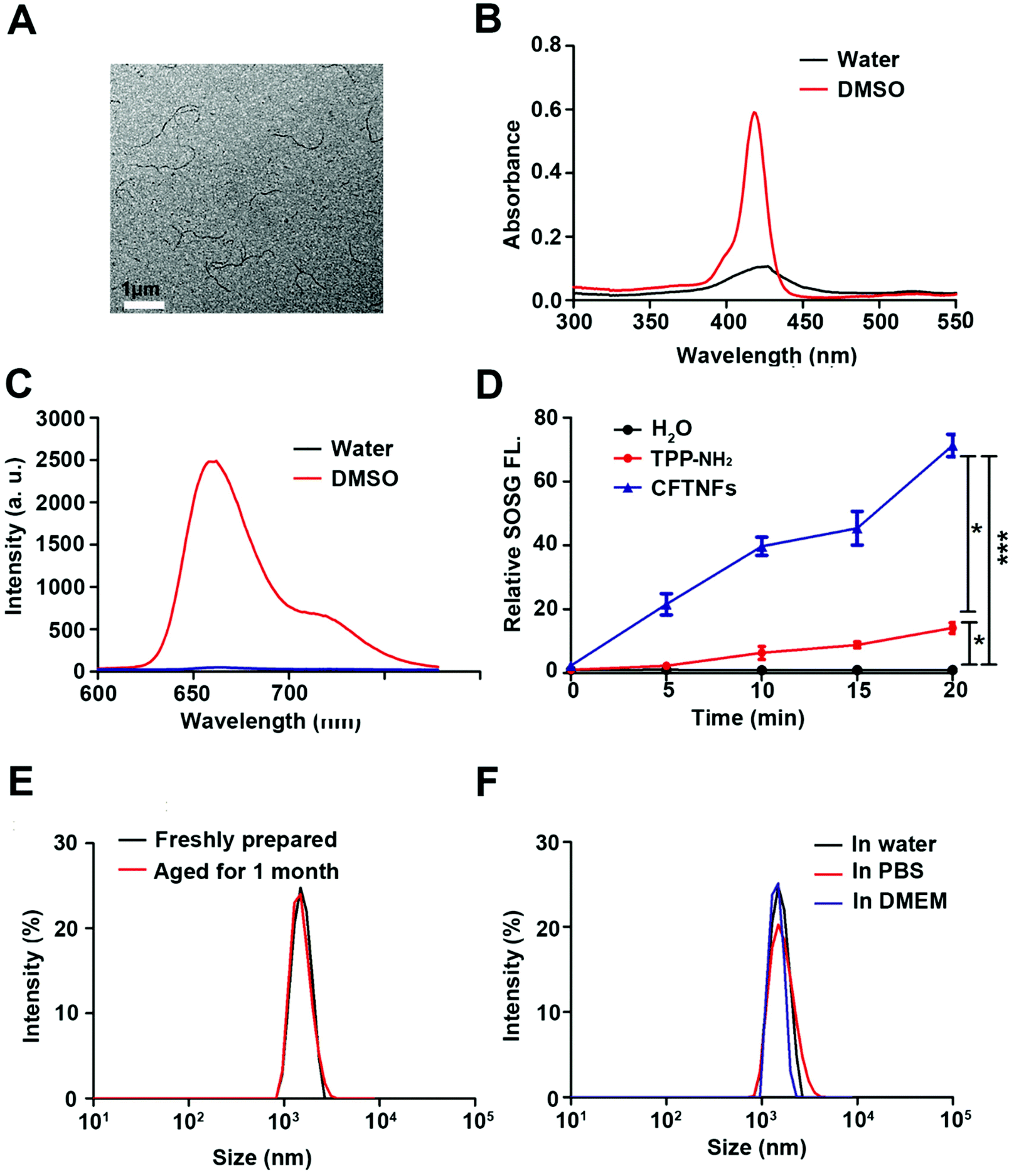
The diphenylalanine peptide is extracted from Alzheimer's β-amyloid polypeptide, which can often be designed into nanomaterials as the self-assembly motif.32 CPP44 is a tumour lineage-homing cell-penetrating peptide.22 Our group synthesized CPP44-FF-TPP-NH2 through the standard solid-phase peptide synthesis (Fig. S1†). In Fig S2 and S3,† the MALDI-TOF MS results indicated that highly purified conjugates were obtained after purification by RP-HPLC. Because of the amphiphilicity, CPP44-FF-TPP-NH2 could spontaneously assemble into nanostructured CFTNFs in DMSO–H2O (1![[thin space (1/6-em)]](https://www.rsc.org/images/entities/char_2009.gif) :9, v/v) solution. The morphology of CFTNFs was characterized by TEM at the concentration of 3 mg mL−1, and they showed a uniform nanofibrous structure with a width of 10–20 nm and the length in micrometers (Figs 1A and S4†). Besides, we also explored the morphology of CFTNFs at higher concentrations and found that they aggregated at a concentration of 30 mg mL−1 to form fibrous superstructures with a width of 60–70 nm fibers (Fig. S5†). In addition, dynamic light scattering (DLS) profiles revealed that the average size of CFTNFs prepared at the concentration of 3 mg mL−1 was 2458 nm (Fig. 1A). A bathochromic shift and a broadening were found for Soret bands in the absorption spectra of aggregate CFTNFs in water as compared with those of monomeric CPP44-FF-TPP-NH2 in DMSO (Fig. 1B). As shown in Fig. 1C, the fluorescence of CFTNFs in water was completely quenched, implying that the molecules underwent aggregation. These results all confirmed the successful construction of CFTNFs. We also tested the DLS size profiles of CFTNFs aged 1 month. Compared with fresh CFTNFs, the DLS profiles did not change significantly, indicating that CFTNFs were stable over time (Fig. 1E). Moreover, as shown in Fig. 1F, CFTNFs showed excellent colloidal stability in water, PBS and DMEM, indicating great potential for biomedical applications.
:9, v/v) solution. The morphology of CFTNFs was characterized by TEM at the concentration of 3 mg mL−1, and they showed a uniform nanofibrous structure with a width of 10–20 nm and the length in micrometers (Figs 1A and S4†). Besides, we also explored the morphology of CFTNFs at higher concentrations and found that they aggregated at a concentration of 30 mg mL−1 to form fibrous superstructures with a width of 60–70 nm fibers (Fig. S5†). In addition, dynamic light scattering (DLS) profiles revealed that the average size of CFTNFs prepared at the concentration of 3 mg mL−1 was 2458 nm (Fig. 1A). A bathochromic shift and a broadening were found for Soret bands in the absorption spectra of aggregate CFTNFs in water as compared with those of monomeric CPP44-FF-TPP-NH2 in DMSO (Fig. 1B). As shown in Fig. 1C, the fluorescence of CFTNFs in water was completely quenched, implying that the molecules underwent aggregation. These results all confirmed the successful construction of CFTNFs. We also tested the DLS size profiles of CFTNFs aged 1 month. Compared with fresh CFTNFs, the DLS profiles did not change significantly, indicating that CFTNFs were stable over time (Fig. 1E). Moreover, as shown in Fig. 1F, CFTNFs showed excellent colloidal stability in water, PBS and DMEM, indicating great potential for biomedical applications.
 |
| | Fig. 1 Characterization of CFTNFs. (A) TEM image of CFTNFs at a concentration of 3 mg mL−1. Scale bar: 1 μm. (B) UV–vis absorption spectra of CFTNFs in water and CPP44-FF-TPP-NH2 in DMSO. (C) Fluorescence emission spectra of CFTNFs in water and CPP44-FF-TPP-NH2 in DMSO. (D) Photodynamic effects of TPP-NH2 and CFTNFs evaluated by recording their SOSG fluorescence (FL) intensity upon exposure to a 630 nm laser (0.3 W cm−2). The error bars are based on triplicate measurements. The data are expressed as mean ± SD (n = 3). p-Values were calculated by Student's t-test: ***p < 0.001, **p < 0.01, or *p < 0.05 and ns (no significant difference). (E) DLS size profiles of freshly prepared and aged CFTNFs. (F) DLS size profiles of CFTNFs in water, PBS and DMEM. | |
ROS generation and observation
The singlet oxygen-generating capability of CFTNFs under light irradiation was evaluated using a commercial detection probe Singlet Oxygen Sensor Green (SOSG). Compared with free TPP-NH2, CFTNFs showed higher singlet oxygen production capacity when irradiated with a 630 nm laser at 0.3 W cm−2 for various time intervals (Fig. 1D). These results demonstrated that CFTNFs could promote the production of ROS and serve as efficient photodynamic nanofibers for PDT. To evaluate the intracellular ROS-generating capability of CFTNFs under laser irradiation was identified from 2′,7′-dichlorofluorescin diacetate (DCFH-DA). Compared with the PBS group, strong green fluorescence was observed in the CFTNF group under irradiation for 5 min (Fig. 2). However, there was negligible green fluorescence in CFTNFs without laser irradiation, which was consistent with the results in Fig. 1D. These results indicated that CFTNFs could generate extracellular and intracellular ROS during the laser irradiation.
 |
| | Fig. 2 CLSM observation of ROS generation of HepG2 cells incubated with CFTNFs with or without irradiation. Blue, DAPI; green, DCFH-DA. | |
Cellular uptake
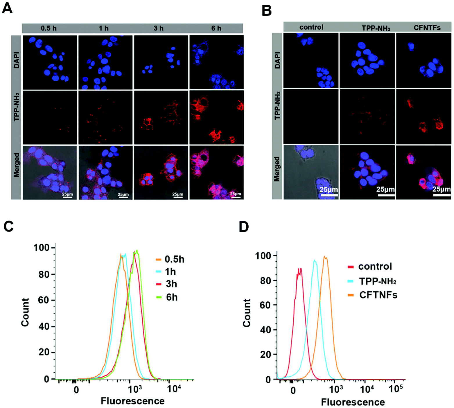
We explored the time-dependent cellular uptake behaviors of CFTNFs by confocal laser scanning microscopy (CLSM) and flow cytometry (FCM) towards HepG2 cells. The images show that the CFTNF fluorescence was almost entirely located in the cytoplasm and the fluorescence intensity gradually increased with time (Figs 3A and S6†). The flow cytometry analysis indicated that the CFTNF fluorescence signal increased with the incubation time (Fig. 3C). These results showed that CFTNFs could be efficiently engulfed in a time-dependent manner. Moreover, we also investigated the cellular uptake of free TPP-NH2 and CFTNFs by HepG2 cells. Compared with free TPP-NH2, CFTNFs exhibited a higher internalizing behavior, as shown in CLSM images (Fig. 3B). Flow cytometry analysis revealed that CFTNF treatment showed higher fluorescence intensity than free TPP-NH2 treatment (Fig. 3D), suggesting that the tumor-homing peptide-based nanofibers can efficiently enhance the cellular uptake behavior of cancerous cells.
 |
| | Fig. 3 Intracellular internalization profiles of TPP-NH2 and CFTNFs. The cell uptake study of HepG2 cells incubated with CFTNFs for 0.5, 1, 3 and 6 h by (A) CLSM observation and (C) flow cytometry. Cellular uptake behaviors of HepG2 cells after different treatments: control, TPP-NH2 and CFTNFs for 4 h by (B) CLSM observation and (D) flow cytometry. Blue, DAPI; red, CFTNFs. Scale bar, 25 μm. | |
Fluorescence cellular images were used to demonstrate the selectivity of CFTNFs. Briefly, 1 × 10⁻⁶M of CFTNFs were incubated with each cell line for 1 h, followed by washing out of extracellular CFTNFs, after which the cells were examined by CLSM. As shown in Fig. S7,† CFTNFs selectively penetrated all cancer cells, especially HepG2 cells, and weak fluorescence signals were observed in non-cancer cells. Following these results, we confirmed the selective uptake of CFTNFs in cancer cells and normal cells.
In vitro cellular toxicity
After confirming that CFTNFs can generate intracellular ROS, the cytotoxicities of CFTNFs and TPP-NH2 were evaluated via the CCK-8 assay. As shown in Fig. 4A and B, HepG2 cells and Hek293t cells were incubated with different concentrations of CFTNFs or TPP-NH2 for 30 min, respectively. Interestingly, the IC50 values of CFTNFs and TPP-NH2 were 0.398 μM and >30 μM for HepG2 cells with irradiation, respectively, while for Hek293t cells, the IC50 values of CFTNFs and TPP-NH2 were 0.997 μM and >30 μM with irradiation, respectively. There was a significant difference in IC50 between HepG2 cells and Hek293t cells. These results suggested that CFTNFs boosted the cancer cells’ selective uptake. CFTNFs also showed a better photodynamic effect than free TPP-NH2 under 630 nm laser irradiation, which was consistent with the results of ROS generation capacity analysis. In contrast, both CFTNFs and TPP-NH2 showed negligible change in cell viability without the 630 nm laser irradiation, indicating excellent biocompatibility. Taken together, these results indicated that CFTNFs possessed higher phototoxicity, lower dark cytotoxicity and better cancer cell selective uptake, favoring cancer PDT treatment. As shown in Fig. S8,† the cytotoxic activity of CFTNFs on HepG2 cells was time dependent. With CFTNF treatment for 0.5 h and 2 h, the IC50 values were 0.398 μM and 0.285 μM, respectively. However, when the incubation time was extended to 24 h, the IC50 value was greatly reduced to 0.109 μM. The results shown in Fig. 4C are consistent with the results in Fig. 4A, indicating that CFTNFs possess ability of selective uptake between cancer cells and non-cancer cells. Moreover, the Fluor 647-Annexin V/PI method was used to determine the apoptosis of HepG2 cells by flow cytometry. As illustrated in Fig. 4D, the control treatment showed negligible apoptotic cells with or without irradiation, verifying that the laser intensity was safe for cells. Then, without laser irradiation, both CFTNF and TPP-NH2 treatment also showed hardly any apoptotic cells. However, the flow cytometry analysis showed that the percentages of apoptotic cells induced by CFTNFs and TPP-NH2 were 58.5% and 17.22% with the 630 nm laser irradiation for 5 min, respectively. These results show that CFTNFs induced HepG2 cell apoptosis only under the condition of laser irradiation and CFTNFs showed a much higher apoptotic rate than TPP-NH2 with laser irradiation, in agreement with the cytotoxicity results. Furthermore, the HepG2 cells were also stained with Calcein-AM and PI to visually detect the PDT effect. The red fluorescence of PI and the green fluorescence of Calcein-AM represented dead and live cells, respectively. HepG2 cells were treated with 1 μM CFTNFs and TPP-NH2 for 4 h. As anticipated, the CFTNF group exhibited red stain after 5 min of irradiation, while the TPP-NH2 group remained green, as observed in the control group (Fig. 4E), suggesting that stronger photodamage occured in the CFTNF group. These results may be due to higher cellular uptake and stronger intracellular ROS production with CFTNFs. Taken together, the results of ROS production, CCK-8 assay, flow cytometry assay and Calcein-AM and PI staining assay were consistent, revealing that CFTNFs could serve as excellent photodynamic nanofibers for cancer treatment.
 |
| | Fig. 4
In vitro PDT evaluation of TPP-NH2 and CFTNFs against HepG2 cells and Hek293t cells. (A) Photodynamic and dark cytotoxicity of CFTNFs in HepG2 and Hek293t cells by the CCK-8 assay. (B) Photodynamic and dark cytotoxicity of TPP-NH2 in HepG2 cells and Hek293t cells by the CCK-8 assay. (C) Photodynamic and dark cell viability of various cancer cells and normal cells treated with 0.47 μM CFTNFs by the CCK-8 assay. (D) Cell apoptosis in HepG2 cells treated with TPP-NH2 or CFTNFs using flow cytometry. Untreated cells were used as controls. (E) Fluorescence microscopy images of HepG2 cells stained with Calcein-AM and PI upon different treatments. Green, Calcein AM, red: PI. | |
In vivo imaging and biodistribution of CFTNFs
The intrinsic red fluorescence of TPP-NH2 is useful for tumor imaging and evaluating the in vivo distribution of CFTNFs in HepG2 tumor-bearing nude mice using an in vivo imaging system. As shown in Fig. 5A, we observed strong fluorescent signals in the tumor site 1 h after intravenous injection of CFTNFs. Remarkably, the fluorescence signal in the tumor increased over a prolonged period of time, reaching a maximum at 10 h. The fluorescence intensity was maintained until 24 h, which demonstrated an excellent tumor retention time. However, free TPP-NH2 as a control did not show tumor targeting. After 10 h intravenous injection, the mice in the TPP-NH2-treatment group and CFTNF-treatment group were sacrificed and the main organs (heart, liver, spleen, lung and kidney) were excised to visually evaluate the tumor-targeting efficacy and tissue distribution. Compared to free TPP-NH2, CFTNFs showed stronger fluorescence intensity at the tumor site. Nevertheless, free TPP-NH2 mainly accumulated in the liver and kidney (Fig. 5B). The same as free TPP-NH2, CFTNFs were also captured and metabolized by the liver and kidney, resulting in strong fluorescence signals of CFTNFs in the liver and kidney.33
 |
| | Fig. 5
In vivo imaging and biodistribution of TPP-NH2 and CFTNFs. (A) In vivo fluorescence imaging of HepG2 tumor-bearing mice at varying time points after intravenous injection of TPP-NH2 or CFTNFs. (B) Ex vivo fluorescence images of the major organs and tumors of mice after intravenous injection of free TPP-NH2 or CFTNFs over a period of 10 h (n = 2). | |
Photodynamic therapeutic efficacy of CFTNFs in vivo
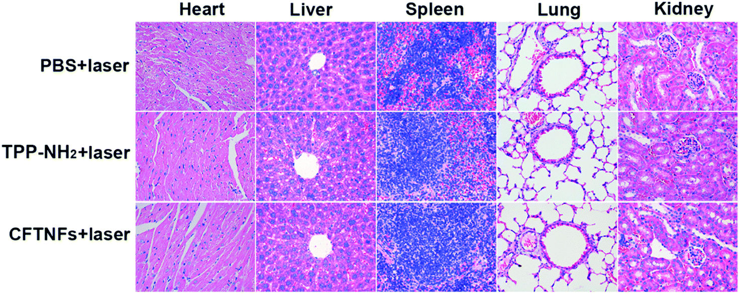
Based on the results of in vivo imaging, we confirmed that CFTNFs possessed the ability of accumulation and retention in tumor sites. Then, the photodynamic anticancer efficacy of CFTNFs was explored after intravenous injection. The experimental observation lasted for 10 days and the treatment was conducted at the 1st, 3rd and 5th day, whereafter CFTNF- and TPP-NH2-treated mice were irradiated with a 630 nm laser at 0.3 W cm−2 for 10 min, at 10 h after injection. We recorded the average tumor volumes and body weights every other day for 10 days. As shown in Fig. S9,† the tumors of the CFTNF + laser treated group were successfully ablated, leaving a dark burned scar at tumor sites, but the tumors of the other groups showed no significant change. Notably, CFTNFs + laser treated tumors were fully suppressed over the course of three treatments, while the injection of CFTNFs only showed negligible influence on the tumor growth (Fig. 6A). The TPP-NH2 + laser-treated tumors showed a moderate inhibition in growth, and also showed slow growth over the treatment period. Besides, on the last day of observation, the tumor weights (Fig. 6C) and images (Fig. 6D) were obtained after removing them from the mice. The CFTNF + laser-treated tumors were the lightest and smallest among all of the treatments. Moreover, there were no significant body weight losses (Fig. 6B), suggesting that the treatment did not induce systemic toxicity. To further evaluate the biocompatibility of CFTNFs, we harvested the main organs, including the heart, liver, spleen, lung and kidney, at the end of observation. Hematoxylin and eosin staining of organs was performed for histopathological examination. There were no apparent histopathological abnormalities in these tissues in any of the groups (Fig. 7), indicating that CFTNFs are safe for application in vivo. These results confirm that CFTNFs are efficient and safe photodynamic anticancer nanofibers.
 |
| | Fig. 6
In vivo therapeutic efficacy of PDT. Tumor volumes with different treatments are shown. Black arrows present the injection time points (A). Body weight changes of each group during treatments (B). Changes in the tumor weights following each treatment (C). Dissected tumors after the administration of different treatments (D). The data are expressed as means ± SD (n = 3); p-values were calculated by Student's t-test: ***p < 0.001, **p < 0.01, or *p < 0.05 and ns (no significant difference). | |
 |
| | Fig. 7 Histological section of heart, liver, spleen, lung and kidney tissues after treatment with PBS + laser, TPP-NH2 + laser and CFTNFs + laser by intravenous injection into HepG2 tumor-bearing mice. H&E staining: 400× magnification. | |
Conclusion
In summary, we have developed a targeted self-assembling photosensitizer nanofiber based on multicomponent and multifunctional peptide–motif coordination for PDT. Compared to free TPP-NH2, CFTNFs could generate more extracellular and intracellular singlet oxygen during the laser irradiation, possessed excellent phototoxicity against HepG2 cells in vitro and showed selective cell uptake. Extensive in vivo evaluations of CFTNFs showed remarkable tumor-targeting specificity and excellent inhibition of growth of the tumors. The strategy of combining a self-assembling short peptide, a cell-penetrating peptide and a small molecule photosensitizer to construct excellent photosensitive nanomaterials could be a powerful concept. Moreover, this may pave the way for designing efficient and intelligent photodynamic therapeutic agents and may eventually advance toward clinical treatment of a variety of cancers.
Materials and methods
Rink amide-AM resin and Fmoc-amino acids were obtained from GL Biochem (Shanghai, China). HOBt and HCTU were purchased from GL Biochem (Shanghai, China). DMSO was purchased from Sigma Aldrich Chemical Company. Carbon-coated copper grids were purchased from Zhongjingkeji Technology (Beijing, China). TPP-NH2 was obtained from J&K Scientific Ltd (analytical grade, only for research). DCFH-DA and a Cell Counting Kit-8 were purchased from Beyotime (Shanghai, China). A Calcein-AM/PI double-staining kit was obtained from Maokang Biotechnology Co. Ltd. Dulbecco's Modified Eagle's Medium (DMEM, containing 4.5 g L−1 glucose, 100 units per mL penicillin, 100 μg mL−1 streptomycin) and fetal bovine serum (FBS) were purchased from Gibco. All other reagents were of analytical grade.
Synthesis of CPP44 and CPP44-FF-TPP-NH2 conjugates
CPP44 (Ac-KRPTMRFRYTWNPMK-amide) and CPP44-FF-TPP-NH2 (TPP-GFFY-GPG-CPP44) were synthesized on Rink amide-AM resin using the Fmoc solid-phase peptide synthesis method on a 0.2 mmol scale. After the resins were dried under vacuum, the dried resins containing the protected amino acid sequences were used in the coupling reaction with TPP-NH2.
DA-TPP.
To a solution of TPP-NH2 (100 mg, 0.16 mmol) in DMF (1 mL) was added diglycolic anhydride (55 mg, 0.48 mmol). The solution was stirred at room temperature in the dark. After the mixture was stirred for 24 h, CHCI3 (10 mL) was added, and then 150 mL distilled water was added to the reaction mixture. The product was extracted using CHCI3 (10 mL × 3) through a separating funnel. The combined organic phase was washed with water (100 mL × 3) and evaporated, and finally dried under vacuum to yield 100 mg (84%) of DA-TPP.
CPP44-FF-TPP-NH2.
To a solution of DA-TPP (0.04 mmol) in DMF (3 mL) were added DIPEA (0.1 mmol), HOBt (0.1 mmol), and TBTU (0.1 mmol). After the mixture was reacted for 20 min, CPP44-FF peptidyl resin (0.2 mmol) was introduced into a plastic synthesizer. The reaction mixture was shaken for 24 h at room temperature and then filtered to yield a green resin. Peptides were cleaved using trifluoroacetic acid/thioanisole/anisole (95/3/2, v/v) and, after 150 min, precipitated in cold diethyl ether. The crude products were purified using RP-HPLC (C18, 4.6 × 250 mm) on the basis of a 0.1% TFA/acetonitrile gradient from 20% to 60% in 35 min. Then, the purified products were evaluated using MALDI-TOF MS (AB, SCIEX).
Preparation of CFTNF nanofibers
CFTNF nanofibers were prepared as follows: 400 μL of a DMSO solution of CPP44-FF-TPP-NH2 (30 mg mL−1) was mixed with 3600 μL of pure water, and the obtained dispersive CPP44-FF-TPP-NH2 was aged for 24 h and then dialyzed against water. For dialysis, 4 mL of CPP44-FF-TPP-NH2 solution was sealed in a dialysis bag (molecular weight cut-off: 5 kDa) and immersed in 2 L of pure water for 48 h. Initially, the water was replaced every 2 hours, and the water was replaced a total of 6 times. Finally, CPP44-FF-TPP-NH2 was transferred to ultra-centrifugal filters (molecular weight cut-off: 10 kDa) and centrifuged at 10000 rpm for 20 min. Then, CFTNFs retained in the upper tubes of the filters were collected and re-dispersed with other solvents.
Characterization of CFTNF nanofibers
The CFTNF (3 mg mL−1) morphology was observed by transmission electron microscopy (TEM; Tecnai G20 F20, America). The hydrodynamic diameters were measured in water solution (3 mg mL−1) by dynamic light scattering (DLS, Malvern, UK). Ultraviolet–visible spectroscopy measurements were recorded on a Jasco UV-2450 spectrophotometer. Fluorescence spectroscopy measurements were conducted on a Jasco F-7100 spectrofluorometer.
Cell culture
HepG2, MCF-7, Hek293t, LO2 and L929 cell lines were obtained from the American Type Culture Collection and cultured in a humidified atmosphere (5% CO2) at 37 °C. The medium included 10% FBS and 1% antibiotics (penicillin–streptomycin, 10000 U mL−1).
Detection of the singlet oxygen generation capacity of CFTNFs in HepG2 cells
HepG2 cells were incubated with CFTNFs (3 × 10⁻⁶M in terms of TPP-NH2) for 6 h. Then, the medium was removed and the cells were washed with PBS buffer; 50 × 10−6M DCFH-DA was added and incubated for 20 min. Then the cells were washed thrice with PBS buffer and irradiated with a 630 nm laser at 0.3 W cm−2 for 5 min. After irradiation, the cells were fixed with 4% paraformaldehyde solution and stained with 4′,6-diamidino-2-phenylindole (DAPI) before imaging with a CLSM.
Using the singlet oxygen sensor green (SOSG) to detect the generation of singlet oxygen by CFTNFs (3 × 10⁻⁶M TPP-NH2), which were mixed with the SOSG fluorescent probe (5 × 10⁻⁶M), and then irradiated with a 630 nm laser at 0.3 W cm−2 for various intervals, the SOSG fluorescence intensity of each sample was recorded with a microreader (Variskan, Thermofisher) with an excitation wavelength of 504 nm and an emission wavelength of 525 nm.
In vitro cytotoxicity test of CFTNFs
The cytotoxic effect of CFTNFs was evaluated via a commercial assay using the cell counting kit-8 (CCK-8). The cells were seeded into 96-well culture plates with a density of 1 × 10⁶ cells per well in DMEM medium for 24 h. The cells were treated with 300–7500 nM of TPP-NH2 or CFTNFs for 30 min. Then, the cells were irradiated or not irradiated with a 630 nm laser at 0.3 W cm−2 for 5 min and further incubated for 4 h in the dark. In order to evaluate the cell viability, the cells were incubated with the DMEM medium containing 10% CCK-8 solution for 0.5–4 h and the absorbance (450 nm) was measured with a microplate reader.
Flow cytometry assays
Flow cytometry was employed for TPP-NH2- and CFTNF-induced apoptosis analysis. Briefly, HepG2 cells were seeded (2 × 10⁵ cells per well) in 6-well plates and cultured at 37 °C and 5% CO2 for 24 h. Then, the cells were washed twice with PBS, trypsinized and resuspended in a medium containing 1 × 10⁻⁶M TPP-NH2 and CFTNFs and incubated for 4 h. HepG2 cells without treatment and only irradiated with a 630 nm laser were used as the control. Then, the cell pellet was lightly washed three times with cold PBS and stained with a Fluor 647-Annexin V/PI apoptotic detection kit according to the manufacturer's instructions. The fluorescence was measured using flow cytometry (BD, USA).
In vitro cellular uptake of CFTNFs
The cellular uptake of CFTNFs in HepG2 cells was investigated by CLSM and flow cytometry. The cells were seeded in a 6-well plate at a density of 2 × 10⁵ cells per well and incubated in DMEM medium for 24 h. Then, the culture medium was replaced with fresh medium containing 3 × 10⁻⁶M CFTNFs and incubated for 0.5 h, 1 h, 3 h, and 6 h in the dark, respectively. Thereafter, the cells were washed three times with PBS and fixed with 4% paraformaldehyde for 10 min at room temperature. DAPI was used to stain the cell nuclei (diluted 1:1000 in DMEM). Intracellular localization was observed using a laser scanning confocal microscope (LSM710, Carl Zeiss Jena, Germany).
The cellular uptake of the samples was quantified by flow cytometry. Briefly, the cells were seeded in a 6-well plate at a density of 2 × 10⁵ cells per well and incubated in DMEM medium for 24 h. Then, the cells were washed twice with PBS, trypsinized and resuspended in a medium containing 3 × 10⁻⁶M CFTNFs and incubated for 0.5 h, 1 h, 3 h, and 6 h in the dark, respectively. Thereafter, the cell pellet was lightly washed three times with cold PBS and re-suspended in 0.1 mL PBS.
Calcein-AM/PI double staining
HepG2 cells were seeded in a 12-well plate and incubated for 24 h. Cells were washed twice with PBS after treatment with 1 × 10⁻⁶M for 4 h and then 500 μL of a dye mixture containing 2 × 10⁻⁶M Calcein-AM and 4 × 10⁻⁶M PI in PBS was added to each well. Fluorescence was visualized immediately using fluorescence microscopy (Olympus FV1000).
In vivo imaging
HepG2 tumor-bearing nude mice were intravenously injected with 100 μL of TPP-NH2 or CFTNFs for in vivo imaging. After anesthetizing with 4% chloral hydrate, the real-time fluorescence images of TPP-NH2 in mice were observed using a Small-Animal Imaging System (Caliper-PE in vivo imaging system spectrum) at an excitation wavelength of 400 nm and an emission wavelength of 620 nm.
In vivo antitumor growth of CFTNFs
All animal studies were approved by the Institutional Animal Care and Use Committee of Hunan Normal University and the National Institutes of Health guidelines for animal experiments were followed. 5–6 weeks old Male BALB/c-nu mice were purchased from the Slac & Jingda Corporation of Laboratory Animals, Changsha, China. To build the subcutaneous HepG2 tumor model, mice were inoculated with 2 × 10⁶ HepG2 cells to establish xenograft tumors. The HepG2 tumor-bearing mice were used for further experiments when the tumor volume reached 150 mm3. 18 mice were randomized into 6 groups: (a) PBS, (b) PBS + laser, (c) TPP-NH2 (3.125 mg kg−1), (d) TPP-NH2 + laser (3.125 mg kg−1), (e) CFTNFs (17.2 mg kg−1 of TPP-NH2), and (f) CFTNFs + laser (17.2 mg kg−1 of TPP-NH2). The mice were intravenously injected with 100 μL of PBS, TPP-NH2 or CFTNFs. At 10 h after injection, the mice in groups (b), (d) and (f) were irradiated with a 630 nm laser (0.3 W) for 10 min. The mice were subjected to similar treatment on days 1, 3 and 5. Free TPP-NH2 was dissolved in a mixture consisting of DMSO, polyethylene glycol 400 and tween 80 (2.5:30:2.5, v/v) in water. All of the mouse weights and tumor volumes were recorded every 2 days until day 10 post-treatment. After 10 days, the mice were sacrificed and photographed, and the primary tumors and major organs were extracted and washed with cold PBS. The tumor volume was calculated using the following formula: tumor volume (mm3) = (length × width2)/2 (n = 3).
Abbreviations
| SPPS | Solid-phase peptide synthesis |
| DMF | dimethyl formamide |
| TPP-NH2 | 5-(4-aminophenyl)-10,15,20-triphenyl porphine |
| DCFH-DA | 2′,7′-dichlorofluorescein diacetate |
| SOSG | Singlet Oxygen Sensor Green |
| TFA | trifluoroacetic acid |
| RP-HPLC | reverse-phase high-pressure liquid chromatography |
| HCTU | 5-chloro-1-[bis(dimethylamino)methylene]-1H-benzotriazolium3-oxide hexafluorophosphate |
| HOBT | 1-hydroxybenzotriazole |
| PI | propidium iodide |
| CCK-8 | Cell Counting Kit-8 |
| DLS | dynamic light scattering |
| TEM | transmission electron microscopy |
| H&E | Hematoxylin and eosin |
Author contributions
Qianqian Zhang and Jiawei Xu contributed equally to this work. Most of the peptides were synthesized, purified, and characterized by Qianqian Zhang and Jiawei Xu. Qianqian Zhang organized and ran most of the bioactivity experiments. Jiawei Xu ran some of the bioactivity experiments. Jiayi Peng did some chemical synthesis work and bioactivity experiments. Qianqian Zhang and Zhonghua Liu wrote the manuscript. All authors reviewed the manuscript.
Conflicts of interest
There are no conflicts to declare.
Acknowledgements
This research was financially supported by the National Science Foundation of China (General Program: 32071262, 31770832, 31570782), the Science and Technology Innovation Program of Hunan Province (2020RC4023), and the National Science Foundation of China (31872718).
References
- D. Dolmans, D. Fukumura and R. K. Jain, Nat. Rev. Cancer, 2003, 3, 380–387 CrossRef CAS PubMed.
- A. C. Kübler, Photochem. Photobiol., 2005, 20, 37–45 Search PubMed.
- Z. Zhou, J. Song, L. Nie and X. Chen, Chem. Soc. Rev., 2016, 45, 6597–6626 RSC.
- D. Dolmans, F. Dai and R. K. Jain, Nat. Rev. Cancer, 2003, 3, 380–387 CrossRef CAS.
- M. Ethirajan, Y. Chen, P. Joshi and R. K. Pandey, Chem. Soc. Rev., 2011, 40, 340–362 RSC.
- M. A. Rajora, J. Lou and G. Zheng, Chem. Soc. Rev., 2017, 46, 6433–6469 RSC.
- X. Xue, A. Lindstrom and Y. Li, Bioconjugate Chem., 2019, 30, 1585–1603 CrossRef CAS PubMed.
- A. Maier, F. Tomaselli, V. Matzi, M. Woltsche, U. Anegg, B. Fell, P. Rehak, H. Pinter and F. Smolle-Jüttner, Lasers Surg. Med., 2010, 30, 12–17 CrossRef.
- V. A. Schwarz, S. D. Klein, R. Hornung, R. Knochenmuss and H. Walt, Lasers Surg. Med., 2001, 29, 252–259 CrossRef CAS.
- S. Li, Q. Zou, Y. Li, C. Yuan, R. Xing and X. Yan, J. Am. Chem. Soc., 2018, 140, 10794–10802 CrossRef CAS PubMed.
- H. Zhang, K. Liu, S. K. Li, X. Xin, S. l. Yuan and G. H. Ma, ACS Nano, 2018, 12, 8266–8276 CrossRef CAS PubMed.
- K. Liu, R. Xing, Q. Zou, G. Ma, H. Möhwald and X. H. Yan, Angew. Chem., Int. Ed., 2016, 55, 3036–3039 CrossRef CAS PubMed.
- E. Huynh, J. F. Lovell, B. L. Helfield, M. Jeon, C. Kim, D. E. Goertz, B. C. Wilson and Z. Gang, J. Am. Chem. Soc., 2012, 134, 16464–16467 CrossRef CAS PubMed.
- J. F. Lovell, C. S. Jin, E. Huynh, H. Jin, C. Kim, J. L. Rubinstein, W. Chan, W. Cao, L. V. Wang and Z. Gang, Nat. Mater., 2011, 10, 324–332 CrossRef CAS.
- Y. Li, T. Y. Lin, Y. Luo, Q. Liu, W. Xiao, W. Guo, D. Lac, H. Zhang, C. Feng and S. Wachsmann-Hogiu, Nat. Commun., 2014, 5, 4712 CrossRef CAS.
- J. Shi, T. Liu, J. Chen, G. David, J. David, B. Wilson, F. Wang and G. Zheng, Theranostics, 2011, 1, 363–370 CrossRef CAS PubMed.
- F. Wang, Y. Wang, X. Zhang, W. Zhang, S. Guo and F. Jin, J. Controlled Release, 2014, 174, 126–136 CrossRef CAS.
- E. Koren and V. P. Torchilin, Trends Mol. Med., 2012, 18, 385–393 CrossRef CAS.
- S. Lucky, K. Soo and Y. Zhang, Chem. Rev., 2015, 115, 1990–2042 CrossRef CAS PubMed.
- M. Abbas, Q. Zou, S. Li and X. Yan, Adv. Mater., 2017, 29, 1605021 CrossRef.
- J. Enbäck and P. Laakkonen, Biochem. Soc. Trans., 2007, 35, 780–783 CrossRef PubMed.
- E. Kondo, K. Saito, Y. Tashiro, K. Kamide, S. Uno, T. Furuya, M. Mashita, K. Nakajima, T. Tsumuraya and N. Kobayashi, Nat. Commun., 2012, 3, 951 CrossRef PubMed.
- J. Gronlund, L. Vitved, M. Lausen, K. Skjodt and U. Holmskov, J. Immunol., 2000, 165, 6406–6415 CrossRef CAS.
- J. Wang, K. Liu, R. Xing and X. Yan, Chem. Soc. Rev., 2016, 45, 5589–5604 RSC.
- L. Adler-Abramovich and E. Gazit, Chem. Soc. Rev., 2014, 43, 6881–6893 RSC.
- K. Ariga, Q. Ji, T. Mori, M. Naito, Y. Yamauchi and H. Abe, Chem. Soc. Rev., 2013, 42, 6322–6345 RSC.
- G. Qi, Y. Gao, L. Wang and H. Wang, Adv. Mater., 2018, 30, 1703444 CrossRef PubMed.
- Q. Zou, M. Abbas, L. Zhao, S. Li and X. Yan, J. Am. Chem. Soc., 2017, 139, 1921–1927 CrossRef CAS PubMed.
- J. Li, A. Wang, L. Zhao, Q. Dong, M. Wang, H. Xu, X. Yan and S. Bai, ACS Appl. Mater. Interfaces, 2018, 10, 28420–28427 CrossRef CAS PubMed.
- K. Tao, A. Levin, L. Adler-Abramovich and E. Gazit, Chem. Soc. Rev., 2016, 45, 3935–3953 RSC.
- Q. Zou, L. Zhang, X. Yan, A. Wang, G. Ma, J. Li, H. Möhwald and S. Mann, Angew. Chem., Int. Ed., 2014, 53, 2366–2370 CrossRef CAS.
- M. Reches and E. Gazit, Science, 2003, 300, 625–627 CrossRef CAS PubMed.
- D. Sun, J. Ding, C. Xiao, J. Chen, X. Zhuang and X. Chen, ACS Appl. Mater. Interfaces, 2014, 6, 21202–21214 CrossRef CAS PubMed.
Footnotes |
| † Electronic supplementary information (ESI) available: Figs S1–S7 (PDF). See DOI: 10.1039/d1bm01559a |
| ‡ These authors contributed equally to this paper. |
|
| This journal is © The Royal Society of Chemistry 2022 |
Click here to see how this site uses Cookies. View our privacy policy here. 
 *ab
*ab