 Open Access Article
Creative Commons Attribution 3.0 Unported Licence
Identification of β-strand mediated protein–protein interaction inhibitors using ligand-directed fragment ligation†
Zsófia
Hegedüs‡
 a,
Fruzsina
Hóbor
 bc,
Deborah K.
Shoemark
 e,
Sergio
Celis
 a,
Lu-Yun
Lian
 d,
Chi H.
Trinh
 bc,
Richard B.
Sessions
 e,
Thomas A.
Edwards
 bc and
Andrew J.
Wilson
 *ab
*ab
aSchool of Chemistry, University of Leeds, Woodhouse Lane, Leeds LS2 9JT, UK. E-mail: a.j.wilson@leeds.ac.uk
bAstbury Centre for Structural Molecular Biology, University of Leeds, Woodhouse Lane, Leeds LS2 9JT, UK
cSchool of Molecular and Cellular Biology, University of Leeds, Woodhouse Lane, Leeds LS2 9JT, UK
dInstitute of Systems, Molecular and Integrative Biology, University of Liverpool, Liverpool L69 3BX, UK
eSchool of Biochemistry, Biomedical Sciences Building, University of Bristol, Bristol, BS8 1TD, UK
First published on 6th January 2021
Abstract
β-Strand mediated protein–protein interactions (PPIs) represent underexploited targets for chemical probe development despite representing a significant proportion of known and therapeutically relevant PPI targets. β-Strand mimicry is challenging given that both amino acid side-chains and backbone hydrogen-bonds are typically required for molecular recognition, yet these are oriented along perpendicular vectors. This paper describes an alternative approach, using GKAP/SHANK1 PDZ as a model and dynamic ligation screening to identify small-molecule replacements for tranches of peptide sequence. A peptide truncation of GKAP functionalized at the N- and C-termini with acylhydrazone groups was used as an anchor. Reversible acylhydrazone bond exchange with a library of aldehyde fragments in the presence of the protein as template and in situ screening using a fluorescence anisotropy (FA) assay identified peptide hybrid hits with comparable affinity to the GKAP peptide binding sequence. Identified hits were validated using FA, ITC, NMR and X-ray crystallography to confirm selective inhibition of the target PDZ-mediated PPI and mode of binding. These analyses together with molecular dynamics simulations demonstrated the ligands make transient interactions with an unoccupied basic patch through electrostatic interactions, establishing proof-of-concept that this unbiased approach to ligand discovery represents a powerful addition to the armory of tools that can be used to identify PPI modulators.
Introduction
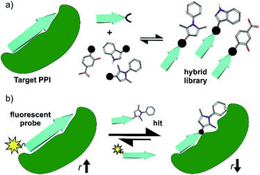
Protein–protein interactions (PPIs) play an essential role in the majority of biological processes and therefore represent arbiters of health and disease.1,2 Chemical probes for PPIs offer tremendous opportunities to understand PPIs and therefore biological mechanisms, whilst providing starting points for drug-discovery.3 Conventional ligand discovery methods (e.g. fragment screening and elaboration) have delivered PPI modulators for a number of targets4–6 leading to clinically approved drugs such as Venetoclax (ABT-199).7 However, despite the enormous opportunity offered by PPIs, methods to identify modulators of PPIs remain underdeveloped. Peptide interacting motifs represent promising templates for design given they are likely to mediate a significant proportion of PPIs;8 however, selecting the most appropriate scaffold to inhibit a specific PPI is highly dependent on the properties of the target interaction and varies in difficulty across PPI classes.9,10 There has been considerable success in developing generic approaches to mimic the α-helix and inhibit α-helix mediated PPIs (e.g. using peptides, constrained peptides and peptidomimetics), but less so for other PPI topographies.11,12 In particular, β-strand/β-sheet mediated PPIs exhibit a more complex topography and have proven more challenging; these interfaces are shallower and more elongated with backbone hydrogen bonding contributing significantly to the binding energy.13,14 Scaffolds that mimic β-strands/β-sheets15–20 have hitherto seen limited application21–23 in PPI inhibitor discovery.24–29In this work, we take a novel approach to β-strand mimicry whereby a truncated protein-interacting motif is combined with a small-molecule fragment (Fig. 1a), to generate a functional mimetic,30i.e. a mimic that reproduces binding, but may not necessarily mimic structure. As a screening approach, this offers the advantage of exploiting a peptide with intrinsic target affinity as an anchor to permit identification of weakly binding fragments. A related principle has been exploited in the REPLACE strategy whereby – aided by in silico methods – key residues of known peptide ligands were “mutated” for molecular fragments to identify inhibitors of conventional drug discovery targets e.g. kinases.31–35 Subsequently, extension of this concept to α-helix mediated PPIs has been achieved by screening peptide-small molecule hybrids in silico, then preparing promising candidates for experimental validation using click chemistry.36–38 In this work, we instead use reversible hydrazone formation39 between peptide hydrazones and an aldehyde library together with dynamic ligation screening40,41 to identify peptide-fragment hybrids. Such dynamic, template assisted methods shift thermodynamic equilibria in favour of the highest affinity ligands40–44 and can target unoccupied binding sites45 in an unbiased manner; they have proven successful for discovery of active site inhibitors,46–49 but not been applied to PPI inhibitor discovery.
As a model β-strand mediated PPI, we use the GKAP/SHANK-PDZ50 which, plays a role in the organization of synaptic protein complexes and has been linked to several neuronal disorders.51 There are >250 PDZ domains with several therapeutically relevant proteins known to exhibit their function through a PDZ mediated interaction.52–54 Ligand discovery for PDZ domains has proven challenging (e.g. fragment-based screening was unsuccessful for the PDZ domain of PSD-95![[thin space (1/6-em)]](https://www.rsc.org/images/entities/char_2009.gif) 55), with the majority of potent ligands based on protein,56,57 peptide58,59 or peptidomimetic scaffolds27,60–62 and limited examples of small-molecules.63–66 This rendered the GKAP/SHANK-PDZ interaction as a stringent test for our dynamic ligation screening approach. Using a three-residue sequence from the GKAP ligand, C- and N-terminal hydrazones were generated, then peptide-fragment hybrid assembly under neutral conditions performed in the presence of protein as a template and directly coupled to a fluorescence-based biophysical assay (Fig. 1b). Characterization of the most promising hits using ITC, NMR, X-ray crystallography and molecular dynamics simulations established the hits as selective SHANK1 PDZ ligands (in comparison to PSD-95) with comparable potency to the GKAP PDZ-binding motif (PBM) (Ac-EAQTRL-COOH) from which they were derived. Potency was achieved by binding to a cluster of basic residues proximal to the peptide binding site on the PDZ domain. These results thus establish dynamic ligation screening as a powerful tool to identify β-strand peptide-fragment mimetics as PPI inhibitors and broaden the scope of design strategies for PPI inhibitor discovery.
55), with the majority of potent ligands based on protein,56,57 peptide58,59 or peptidomimetic scaffolds27,60–62 and limited examples of small-molecules.63–66 This rendered the GKAP/SHANK-PDZ interaction as a stringent test for our dynamic ligation screening approach. Using a three-residue sequence from the GKAP ligand, C- and N-terminal hydrazones were generated, then peptide-fragment hybrid assembly under neutral conditions performed in the presence of protein as a template and directly coupled to a fluorescence-based biophysical assay (Fig. 1b). Characterization of the most promising hits using ITC, NMR, X-ray crystallography and molecular dynamics simulations established the hits as selective SHANK1 PDZ ligands (in comparison to PSD-95) with comparable potency to the GKAP PDZ-binding motif (PBM) (Ac-EAQTRL-COOH) from which they were derived. Potency was achieved by binding to a cluster of basic residues proximal to the peptide binding site on the PDZ domain. These results thus establish dynamic ligation screening as a powerful tool to identify β-strand peptide-fragment mimetics as PPI inhibitors and broaden the scope of design strategies for PPI inhibitor discovery.
Results and discussion
Hybrid library design
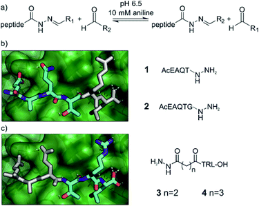
We chose acylhydrazone bond formation to covalently link fragments to peptide anchors (Fig. 2a). In the presence of anilines, the reaction can be performed close to neutral pH, allowing the screening to take place close to a physiologically relevant pH and temperature with the protein as a template for product formation.67,68 Moreover, at neutral pH acylhydrazones are stable, facilitating subsequent hit validation.69,70 Given the commercial availability of a large array of aldehydes, we thus designed the hybrids to be formed from peptide hydrazides.Peptide anchors were designed based on the key features of the interaction between the wild type GKAP sequence and SHANK1 PDZ.50 Class I PDZ domains bind to consensus sequences with a Thr/Ser in the -2 position from the C-terminus, with the C-terminus hydrophobic residue and free carboxylate being the main determinants of affinity.71,72 The plasticity of PDZ domains allows the accommodation of various hydrophobic side-chains at the C terminus of the peptide,73 which we hypothesized to be an ideal target site for hydrophobic fragments; for SHANK1, Leu dominates for C-terminal carboxylates and Phe for non-C-terminal sequences.74 This led to compounds 1 and 2 (Fig. 2b and Scheme S1†), directing the fragments toward the C-terminal hydrophobic pocket on SHANK1 PDZ. On the other hand, compounds 3 and 4 contained the TRL-COOH core sequence and the hydrazide functionality was attached to the N-terminus of the peptide through a 2 or 3 carbon atom linker (Fig. 2c and Scheme S2†), allowing exploration of the protein surface for secondary binding sites further away from the key residues. To ease purification, all peptide hydrazides were reacted with benzaldehyde, yielding the corresponding acylhydrazones (1-, 2-, 3-, 4-A001).
A diversity-based selection was performed on commercially available aldehyde fragments obeying guidelines for fragment-based drug design,75 resulting in a small library of 129 compounds (A001–A129, Table S1 and Fig. S1†). Acylhydrazone formation was tested in the presence of 10 mM aniline67 at pH 6.5 using a 5-fold excess of the competing aldehydes, which revealed fast and complete exchange of the benzaldehyde motif (Fig. S2†), reaching equilibrium within 24 hours. To allow us to perform the hybrid formation and screening in the presence of the target protein in a single step, it was necessary to first establish that SHANK1 PDZ was still able to bind to its natural ligand under the same conditions. For this, FITC labelled GKAP was used and the fluorescence anisotropy measurements gave an initial KD of ∼1 μM, which did not significantly change over the course of 24 hours (Fig. S3†).
Library screening
Prior to screening, the intrinsic inhibitory potency of the peptide hydrazones 1-, 2-, 3-, 4-A001 was determined (Fig. S4†); they were no more active than the parent peptides. Then, hybrid formation and screening were performed by mixing each of these compounds at 10 or 50 μM concentration with 5 equivalents of the aldehydes in a 384 well microtiter plate. After 24 hours of equilibration in the presence of SHANK1 PDZ protein, FITC-Ahx-TRL-OH was added as competitor. In all assays, the GKAP PBM sequence (Ac-EAQTRL-COOH), Ac-TRL-OH and a buffer control were used, which facilitated determination of threshold values for the hit compounds (Fig. 3a, S5 and S6†). Any hybrid that exceeded the activity of its parent compound was considered an improvement caused by the replacement of the fragment.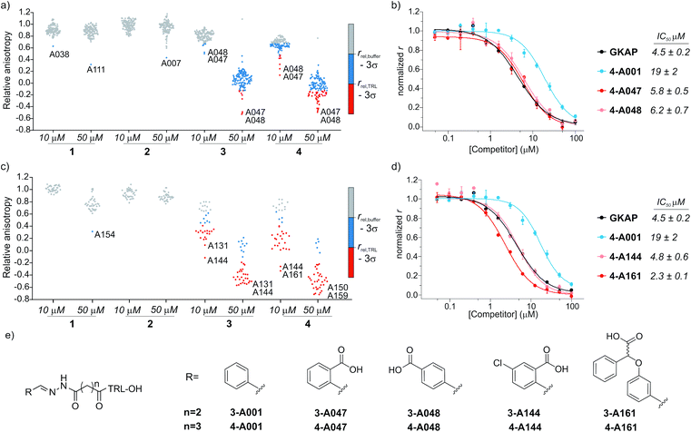 | ||
| Fig. 3 Results of the fluorescence anisotropy competition screening for compounds 1–4. (a) Screening results for hydrazones formed from 1–4 using the diverse screening library (A001-A129). (b) FA competition curves of the best hybrid hits. (c) Screening results using the extended library including near neighbors (A130-165) for the initial hits. (d) FA competition curves for hybrids from the extended screen. All screening and validation assays were performed in 50 mM NH4Ac buffer, pH 6.5, 10 mM aniline using 1 μM final protein concentration and 10 nM FITC-Ahx-TRL-COOH as tracer at room temperature. Compounds 1-, 2-, 3-, 4-A001 were incubated individually with 5 equivalents of fragments in the presence of SHANK1 PDZ for 24 hours. Anisotropy is expressed relative to the control experiment where no competitor is present. Hit thresholds were defined based on the relative anisotropy of the (i) buffer control (rbuffer − 3σ) and (ii) the TRL control (rTRL − 3σ). Compounds below these thresholds are coloured blue and red respectively. Labels indicate the two best aldehyde fragment hits from the individual screens. For individual values and FA validation for additional hits, see ESI Fig. S5–S12.† (e) Structures of the synthesized hit compounds. The absolute configuration and enantiomeric purity of fragment A161 is unknown. | ||
The two different truncation strategies had profoundly different hit rates, reflecting the affinity of the parent compounds. The removal of the key C terminal residue of GKAP did not result in detectable binding affinity for the parent compounds 1-A001 and 2-A001 (Fig. S4†); our hypothesis was that a hybrid with a fragment effectively mimicking the truncated sequence could result in detectable affinity. Our library did not contain such fragments; hit rates were low, perhaps reflecting the challenge in attaining the correct orientation and composition of functionality needed to faithfully mimic the key interactions afforded by the natural truncated sequence. The solitary confirmed hit (Fig. S7†) appeared to interfere with the assay and was not considered further.
Conversely, using compounds 3-A001 and 4-A001 with intrinsic potency (Fig. S4†) resulted in a higher hit rate (Fig. 3a, S5 and S6†), with 51 hybrids exceeding the activity of Ac-TRL-COOH, which was the core motif in both hybrid sequences. The most promising hits were re-tested in order to validate the screening assay (Fig. 3b, S8 and S9†). The IC50 values measured for the best hits were in the range of 5–6 μM, a 10-fold improvement compared to the parent Ac-TRL-COOH sequence, indicating that covalent attachment of the fragment contributes favourably to interaction with the protein. Interestingly, the most potent hybrids formed from 3 and 4 contained the same carboxylic acid functionalized aromatic aldehyde fragments (A047 and A048, Fig. 3b), indicating that these types of fragments might play an important role in binding.
Based on this observation, we extended the aldehyde library to near neighbours of these fragments and selected an additional 36 carboxylic acid functionalized aromatic aldehydes (A130-165, Fig. S1 and Table S1†) with which to carry out a further screen. In this extended screen, the hit rate increased significantly (Fig. 3c, d and S10–S12†), confirming that an acidic binding motif is generic for favourable binding and confirming they could not have been identified without conjugation to the anchor peptides.
In the subsequent validation, the IC50 values of the best hits (formed from compound 4 and A144 or A161) reached or slightly exceeded the binding affinity of full length GKAP peptide, indicating that the fragment can restore the affinity of the truncated sequence to that of the wild-type peptide. Significantly, all but one of the aldehyde fragments showed no inhibitory capability on their own in the assay (Fig. S13†).
Structure–activity relationships
To establish structure–activity relationships, the most active hybrids were synthesized individually (Fig. 3e and Scheme S2†) and subjected to further characterization by isothermal titration calorimetry (ITC) (see Fig. S14†). The truncation of the GKAP sequence (KD = 2 ± 0.3 μM) to its core TRL motif (KD = 22 ± 3 μM) resulted in a decrease in affinity (Fig. 4 and Table 1). This was accompanied by the loss of binding enthalpy but also resulted in a more favourable entropy of binding (Table 1), presumably due to the loss of rotatable bonds on amino acid removal. Hydrazones formed from benzaldehyde and this core motif (compounds 3-A001 and 4-A001) revealed slightly increased affinity compared to the TRL motif (Fig. 4 and Table 1), which might be the result of favourable interaction of the aromatic fragment or the hydrazone bond itself with the protein. Hybrid hits in which the fragments contained the additional carboxylic acid functional group resulted in increased binding affinity in every case (Fig. 4 and Table 1), which confirmed the importance of the acidic group and that these fragments actively contribute to SHANK1 binding. Interestingly, hybrids with the C3 linker (compound 4 hydrazones) showed more favourable enthalpic contributions and slightly higher affinity in comparison to compounds having the C2 linker (compound 3) when the same fragment was attached. This indicated that the more flexible linker allows for a better orientation of the fragment leading to a more favourable enthalpy of binding, but also increases the entropic cost of binding, leading only to a moderate difference in the overall binding energy.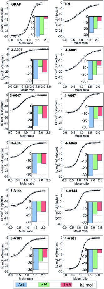 | ||
| Fig. 4 Hit validation by ITC. Fitted thermograms and thermodynamic signatures for the tested peptides and hydrazones. ITC data were acquired in 25 mM Tris, pH 7.5 buffer containing 150 mM NaCl at 25 °C by injecting 0.75–1 mM peptide or hydrazone solution into 75–150 μM protein in the cell. For individual raw ITC data and fitted thermograms see Fig. S14.† | ||
| n | K D (μM) | ΔH (kJ mol−1) | ΔS (J mol−1 K−1) | |
|---|---|---|---|---|
| GKAP | 1.07 ± 0.01 | 2.0 ± 0.3 | −29.6 ± 0.4 | 9.7 |
| TRL | 1.04 ± 0.02 | 23 ± 2 | −19.3 ± 0.5 | 24.1 |
| 3-A001 | 1.04 ± 0.04 | 16 ± 3 | −9.7 ± 0.6 | 59.0 |
| 4-A001 | 1.00 ± 0.03 | 14.8 ± 0.8 | −20.9 ± 0.2 | 22.2 |
| 3-A047 | 0.94 ± 0.01 | 8 ± 1 | −11.5 ± 0.4 | 59.1 |
| 4-A047 | 1.06 ± 0.01 | 4.6 ± 0.2 | −17.1 ± 0.1 | 44.8 |
| 3-A048 | 1.03 ± 0.01 | 10 ± 1 | −14.1 ± 0.3 | 48.4 |
| 4-A048 | 0.99 ± 0.01 | 3.1 ± 0.2 | −22.3 ± 0.2 | 30.7 |
| 3-A144 | 1.00 ± 0.02 | 7.1 ± 0.9 | −14.4 ± 0.2 | 50.1 |
| 4-A144 | 1.03 ± 0.01 | 3.1 ± 0.2 | −18.3 ± 0.1 | 44.1 |
| 3-A161 | 0.84 ± 0.01 | 4.8 ± 0.4 | −18.0 ± 0.3 | 41.3 |
| 4-A161 | 1.00 ± 0.00 | 1.54 ± 0.05 | −28.0 ± 0.1 | 17.3 |
Based on its affinity and the thermodynamic signature, compound 4-161 was identified as the best mimic of the wild-type sequence. It should be noted that the fragment in this hybrid has unknown enantiomeric purity, therefore the binding affinity might be the average of the two diastereoisomers. To compare the effect of fragments on binding energy we calculated group efficiency values (GE) (Tables S2–S4†). These data illustrate the potential of the dynamic ligation approach to rapidly explore structure affinity relationships to identify the efficient fragments. Moreover, the replacement of the N terminal EAQ amino acids of GKAP to completely non-peptidic motifs resulted in similar GE values indicating this strategy can also provide molecules that are less peptidic in nature, while keeping the original potency of the native ligand. It is worth noting that although compound 4-161 possessed the highest binding affinity, the aldehyde fragment in compounds 4-A048 or 4-A144 have slightly higher group efficiencies (Tables S2–S4†) and may therefore represent equally reasonable starting points for further optimization.
To obtain more insight on the molecular nature of the interaction between the peptide-fragment hybrids and SHANK1 PDZ, co-crystal structures were solved for compounds 3-A047, 4-A047, 3-A048, and 4-A048 with SHANK1 PDZ at resolutions of 1.5–2.2 Å (Table S5†). In all structures, hybrids bound to the anticipated binding site with a similar conformation to the wild type sequence within the core amino acid sequence (RMSD values between 0.18–0.32, Fig. S15–S19 and Table S6†), indicating that the peptide component of the hybrid indeed fulfils the role of an anchor directing fragments towards the protein surface. In all hybrids, the hydrazone bond existed in an E configuration with the aromatic ring of the fragment co-planar with the hydrazone. The N terminal glutamate of the wild-type GKAP interacts with R679, thus we anticipated that the carboxylate of the hybrids might reproduce this same interaction. However, the N terminal part of the hybrids showed different binding behaviour in comparison with the wild type sequence. In some cases, we observed multiple conformations of the same compound having different orientations of the fragments relative to the peptide component presumably allowed by the conformational flexibility of the linker region. The more favourable entropy of binding (Table 1) supports the hypothesis that linker flexibility leads to a “fuzzier” mode76 of interaction (see below) although the carboxylate exhibited a slight preference for interaction with R736 (Fig. S19†) which differs from the wild-type GKAP peptide (see above) and likely arises from a combination of hybrid flexibility and the propensity of R736 to engage in effective charge-reinforced hydrogen-bonding interactions.
It is noteworthy that the linker flexibility resulted in high B-factors with some missing density around the fragment in most of the structures. Although this leads to slight uncertainty about the exact location of the fragments, we hypothesize that the key carboxylic acid moiety moves dynamically across a positively charged patch formed by R679, R736 and R743 on the protein surface (Fig. 5), making transient charge-reinforced hydrogen-bonding and cation–π interactions which is also consistent with a more favourable entropy of interaction. Finally, in the crystal structures where strong electron density allowed determination of the location of the fragment, we observed that this motif interacted with a loop of a symmetry related protein in the crystals (Fig. S17 and S18†). To probe this behaviour further, we determined the rotational correlation time (τc) of the SHANK1 PDZ protein in the absence/presence of ligand using NMR T1 and T2 relaxation experiments (Fig. S20†). These analyses indicated similar τc values for apo-SHANK1 and SHANK1 in the presence of these compounds, meaning that the interaction in the X-ray structure most likely arises from a crystal packing specific interaction.
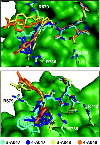 | ||
| Fig. 5 Superimposed crystal structure of 3-A047 (cyan, PDB: 6YWZ), 4-A047 (blue, PDB: 6YX1), 3-A048 (yellow, PDB: 6YX0) and 4-A048 (orange, PDB: 6YX2) in complex with SHANK1 PDZ (green) showing two different orientations of the complexes. The core TRL sequence binds in a similar manner to the native wild-type ligand in both crystallographic protomers whilst the orientation of the acylhydrazone fragment component varies for each ligand and appears to switch interactions dynamically with a series of basic residues (R679, R736, R743) at the periphery of the β-strand binding cleft on the PDZ domain. | ||
To provide support for the hypothesis that the hybrids target the cationic patch on the surface of SHANK1, we performed molecular dynamics simulations on the SHANK1-compound 3-A048 complex using the crystal structure as a starting point. Over the course of three repeats of 200 ns, the aromatic carboxylate of the fragment indeed moves across the surface of the SHANK1 protein, making transient contacts with R679, R736 and R743, as well as intramolecular interaction onto the arginine in the peptide ligand to a lesser extent (see: ESI, Movie S1†). The distance between the carboxylate and these residues plotted against time over the course of the simulations (Fig. S21†) further indicates that the aromatic carboxylate is able to make transient non-covalent contacts in turn with each of the positively charged side-chains of the three arginine residues with a preference for R736.
Since Class I PDZ domains bind to consensus sequences,71,72 we set out to further assess the selectivity of the peptide-fragment hybrids against another Class I PDZ domain. We compared the ability to bind to SHANK1 PDZ against the ability to bind to the PDZ domain of PSD-95, a therapeutically important target in neuronal diseases and cancer.58 We monitored the binding using chemical shift perturbations in the 1H–15N HSQC spectra of the PDZ domain of PSD-95 (Fig. S22, S23, Tables S7 and S8†). These analyses indicated weak binding with EC50 = 2.4 ± 0.3 mM for 4-A047 (Fig. S24†), which is comparable with the affinity of GKAP towards PSD-95 (Table S7†). The observed weak potency of GKAP and the peptide-acylhydrazone fragment hybrids towards PSD-95 likely derives from its preference for Val or Ile at the C-terminus of its ligands.77 However, PSD-95 also lacks the patch of cationic residues proximal to the C-terminal carboxylate binding site. Such a result provides confidence that the dynamic ligation screening approach used here can encode and retain selectivity.
Conclusions
The identification of inhibitors of β-sheet mediated PPI interfaces, which are generally shallow and elongated, is challenging. Here, we used dynamic ligation screening to identify peptide-fragment hybrids linked through an acylhydrazone bond, that are able to functionally mimic a β-strand and inhibit its PPI using GKAP/SHANK1 PDZ as a model interaction. The identified ligands were shown to bind with comparable potency to the GKAP PBM (Ac-EAQTRL-COOH) ligand from which they were derived and were selective when tested against an alternative PDZ domain. Crystallographic studies supported by molecular dynamics analyses indicated that the fragment portion of the hybrids was able to reinforce SHANK1 recognition by engaging in transient charge-reinforced and cation–π interactions on the protein surface. The observation of this distinct binding mode provides new insight on the molecular recognition of SHANK1 towards its peptide/protein ligands that can inform chemical probes development – a focus of our future studies.More generally, our approach is advantageous in that it allows: (i) use of commercially available aldehyde fragments to build a small, diverse library then extend it to near neighbors based on initial hits; (ii) ligation to be performed under conditions where the reaction is reversible, and is compatible with the screening assay; (iii) validate hits easily without the need for a further synthetic step, thus providing useful insights on structure–activity relationships; and (iv) explore unoccupied binding sites on a protein surface in an unbiased manner to generate ligands with target affinity and selectivity. Significantly, whilst conventional fragment based approaches and reversible protein-directed fragment discovery in the form of disulfide-tethering have been used to develop modulators of PPIs,78–80 the use of ligand-directed fragment discovery to develop modulators of PPIs has not been widely developed. Thus, these proof-of concept results exemplified here point to future application of dynamic-ligation screening to identify and optimize PPI modulators more broadly.
Author contributions
Z. H., T. A. E. and A. J. W. conceived and designed the research program, Z. H. designed studies and performed the research with support from F. H. and L.-Y. L. (NMR), S. C. (synthesis) and C. H. T. (crystallography). D. K. S. and R. B. S. performed molecular dynamics analyses. The manuscript was written by Z. H. and A. J. W. with contributions from all authors. Z. H. prepared all figures.Conflicts of interest
There are no conflicts to declare.Acknowledgements
This project has received funding from the European Union's Horizon 2020 research and innovation programme under the Marie Skłodowska-Curie grant agreement no. MSCA-IF-2016-749012. This work was supported by EPSRC (EP/N035267/1). AJW holds a Royal Society Leverhulme Trust Senior Fellowship (SRF/R1/191087). We acknowledge Diamond Light Source for time on Beamline I03, I04 and I24 under Proposal mx19248 and the authors would like to thank the staff of these beamlines for assistance with data collection. We thank the Advanced Computing Research Centre at Bristol for provision of High Performance Computing.Notes and references
- R. Nussinov and C. J. Tsai, Cell, 2013, 153, 293–305 CrossRef CAS.
- O. Keskin, A. Gursoy, B. Ma and R. Nussinov, Chem. Rev., 2008, 108, 1225–1244 CrossRef CAS.
- C. H. Arrowsmith, J. E. Audia, C. Austin, J. Baell, J. Bennett, J. Blagg, C. Bountra, P. E. Brennan, P. J. Brown, M. E. Bunnage, C. Buser-Doepner, R. M. Campbell, A. J. Carter, P. Cohen, R. A. Copeland, B. Cravatt, J. L. Dahlin, D. Dhanak, A. M. Edwards, S. V. Frye, N. Gray, C. E. Grimshaw, D. Hepworth, T. Howe, K. V. M. Huber, J. Jin, S. Knapp, J. D. Kotz, R. G. Kruger, D. Lowe, M. M. Mader, B. Marsden, A. Mueller-Fahrnow, S. Müller, R. C. O'Hagan, J. P. Overington, D. R. Owen, S. H. Rosenberg, B. Roth, R. Ross, M. Schapira, S. L. Schreiber, B. Shoichet, M. Sundström, G. Superti-Furga, J. Taunton, L. Toledo-Sherman, C. Walpole, M. A. Walters, T. M. Willson, P. Workman, R. N. Young and W. J. Zuercher, Nat. Chem. Biol., 2015, 11, 536–541 CrossRef CAS.
- S. A. Andrei, E. Sijbesma, M. Hann, J. Davis, G. O'Mahony, M. W. D. Perry, A. Karawajczyk, J. Eickhoff, L. Brunsveld, R. G. Doveston, L. G. Milroy and C. Ottmann, Expert Opin. Drug Discovery, 2017, 12, 925–940 CrossRef CAS.
- M. R. Arkin, Y. Tang and J. A. Wells, Chem. Biol., 2014, 21, 1102–1114 CrossRef CAS.
- D. E. Scott, A. R. Bayly, C. Abell and J. Skidmore, Nat. Rev. Drug Discovery, 2016, 15, 533–550 CrossRef CAS.
- A. W. Roberts, M. S. Davids, J. M. Pagel, B. S. Kahl, S. D. Puvvada, J. F. Gerecitano, T. J. Kipps, M. A. Anderson, J. R. Brown, L. Gressick, S. Wong, M. Dunbar, M. Zhu, M. B. Desai, E. Cerri, S. H. Enschede, R. A. Humerickhouse, W. G. Wierda and J. F. Seymour, N. Engl. J. Med., 2016, 374, 311–322 CrossRef CAS.
- P. Tompa, N. E. Davey, T. J. Gibson and M. M. Babu, Mol. Cell, 2014, 55, 161–169 CrossRef CAS.
- Y. Ivarsson and P. Jemth, Curr. Opin. Struct. Biol., 2019, 54, 26–33 CrossRef CAS.
- X. Ran and J. E. Gestwicki, Curr. Opin. Chem. Biol., 2018, 44, 75–86 CrossRef CAS.
- H. I. Merritt, N. Sawyer and P. S. Arora, Pept. Sci., 2020, 112, e24145 CAS.
- V. K. Outlaw, J. T. Lemke, Y. Zhu, S. H. Gellman, M. Porotto and A. Moscona, J. Am. Chem. Soc., 2020, 142, 2140–2144 CrossRef CAS.
- A. M. Watkins and P. S. Arora, ACS Chem. Biol., 2014, 9, 1747–1754 CrossRef CAS.
- P.-N. Cheng, J. D. Pham and J. S. Nowick, J. Am. Chem. Soc., 2013, 135, 5477–5492 CrossRef CAS.
- E. A. German, J. E. Ross, P. C. Knipe, M. F. Don, S. Thompson and A. D. Hamilton, Angew. Chem., Int. Ed., 2015, 54, 2649–2652 CrossRef CAS.
- H. Lingard, J. T. Han, A. L. Thompson, I. K. H. Leung, R. T. W. Scott, S. Thompson and A. D. Hamilton, Angew. Chem., Int. Ed., 2014, 53, 3650–3653 CrossRef CAS.
- M. Matsumoto, S. J. Lee, M. L. Waters and M. R. Gagné, J. Am. Chem. Soc., 2014, 136, 15817–15820 CrossRef CAS.
- L. Fülöp, I. M. Mándity, G. Juhász, V. Szegedi, A. Hetényi, E. Wéber, Z. Bozsó, D. Simon, M. Benko, Z. Király and T. A. Martinek, PLoS One, 2012, 7, e39485 CrossRef.
- N. C. Thomas, G. J. Bartlett, D. N. Woolfson and S. H. Gellman, J. Am. Chem. Soc., 2017, 139, 16434–16437 CrossRef CAS.
- A. G. Kreutzer and J. S. Nowick, Acc. Chem. Res., 2018, 51, 706–718 CrossRef CAS.
- C. W. Kang, M. P. Sarnowski, S. Ranatunga, L. Wojtas, R. S. Metcalf, W. C. Guida and J. R. Del Valle, Chem. Commun., 2015, 51, 16259–16262 RSC.
- M. P. Sarnowski, C. W. Kang, Y. M. Elbatrawi, L. Wojtas and J. R. Del Valle, Angew. Chem., Int. Ed., 2017, 56, 2083–2086 CrossRef CAS.
- J. Zheng, A. M. Baghkhanian and J. S. Nowick, J. Am. Chem. Soc., 2013, 135, 6846–6852 CrossRef CAS.
- N. G. Angelo and P. S. Arora, J. Am. Chem. Soc., 2005, 127, 17134–17135 CrossRef CAS.
- P. Rzepecki, H. Gallmeier, N. Geib, K. Cernovska, B. König and T. Schrader, J. Org. Chem., 2004, 69, 5168–5178 CrossRef CAS.
- A. G. Jamieson, D. Russell and A. D. Hamilton, Chem. Commun., 2012, 48, 3709–3711 RSC.
- M. C. Hammond, B. Z. Harris, W. A. Lim and P. A. Bartlett, Chem. Biol., 2006, 13, 1247–1251 CrossRef CAS.
- P. N. Cheng, C. Liu, M. Zhao, D. Eisenberg and J. S. Nowick, Nat. Chem., 2012, 4, 927–933 CrossRef CAS.
- R. Zutshi, J. Franciskovich, M. Shultz, B. Schweitzer, P. Bishop, M. Wilson and J. Chmielewski, J. Am. Chem. Soc., 1997, 119, 4841–4845 CrossRef CAS.
- M. Pelay-Gimeno, A. Glas, O. Koch and T. N. Grossmann, Angew. Chem., Int. Ed., 2015, 54, 8896–8927 CrossRef CAS.
- G. Kontopidis, M. J. Andrews, C. McInnes, A. Plater, L. Innes, S. Renachowski, A. Cowan and P. M. Fischer, ChemMedChem, 2009, 4, 1120–1128 CrossRef CAS.
- S. Liu, P. N. Premnath, J. K. Bolger, T. L. Perkins, L. O. Kirkland, G. Kontopidis and C. McInnes, J. Med. Chem., 2013, 56, 1573–1582 CrossRef CAS.
- P. N. Premnath, S. N. Craig, S. Liu and C. McInnes, Bioorg. Med. Chem. Lett., 2016, 26, 3754–3760 CrossRef CAS.
- M. J. I. Andrews, G. Kontopidis, C. McInnes, A. Plater, L. Innes, A. Cowan, P. Jewsbury and P. M. Fischer, ChemBioChem, 2006, 7, 1909–1915 CrossRef CAS.
- P. N. Premnath, S. N. Craig, S. Liu, E. L. Anderson, A. I. Grigoroudis, G. Kontopidis, T. L. Perkins, M. D. Wyatt, D. L. Pittman and C. McInnes, J. Med. Chem., 2015, 58, 433–442 CrossRef CAS.
- A. M. Beekman, M. A. O'Connell and L. A. Howell, Angew. Chem., Int. Ed., 2017, 56, 10446–10450 CrossRef CAS.
- A. M. Beekman, M. M. D. Cominetti, S. J. Walpole, S. Prabhu, M. A. O'Connell, J. Angulo and M. Searcey, Chem. Sci., 2019, 10, 4502–4508 RSC.
- L. Howell and A. Beekman, RSC Chem. Biol., 2021 10.1039/D0CB00148A.
- D. K. Kölmel and E. T. Kool, Chem. Rev., 2017, 117, 10358–10376 CrossRef.
- M. Y. Unver, R. M. Gierse, H. Ritchie and A. K. H. Hirsch, J. Med. Chem., 2018, 61, 9395–9409 CrossRef CAS.
- M. Jaegle, E. L. Wong, C. Tauber, E. Nawrotzky, C. Arkona and J. Rademann, Angew. Chem., Int. Ed., 2017, 56, 7358–7378 CrossRef CAS.
- M. K. Chung, P. S. White, S. J. Lee, M. L. Waters and M. R. Gagné, J. Am. Chem. Soc., 2012, 134, 11415–11429 CrossRef CAS.
- M. K. Chung, P. S. White, S. J. Lee, M. R. Gagné and M. L. Waters, J. Am. Chem. Soc., 2016, 138, 13344–13352 CrossRef CAS.
- M. K. Chung, S. J. Lee, M. L. Waters and M. R. Gagné, J. Am. Chem. Soc., 2012, 134, 11430–11443 CrossRef.
- D. Rooklin, A. E. Modell, H. Li, V. Berdan, P. S. Arora and Y. Zhang, J. Am. Chem. Soc., 2017, 139, 15560–15563 CrossRef CAS.
- M. F. Schmidt, A. El-Dahshan, S. Keller and J. Rademann, Angew. Chem., Int. Ed., 2009, 48, 6346–6349 CrossRef CAS.
- M. F. Schmidt, A. Isidro-Llobet, M. Lisurek, A. El-Dahshan, J. Tan, R. Hilgenfeld and J. Rademann, Angew. Chem., Int. Ed., 2008, 47, 3275–3278 CrossRef CAS.
- M. Mondal, N. Radeva, H. Fanlo-Virgós, S. Otto, G. Klebe and A. K. H. Hirsch, Angew. Chem., Int. Ed., 2016, 55, 9422–9426 CrossRef CAS.
- S. Ng, E. Lin, P. I. Kitov, K. F. Tjhung, O. O. Gerlits, L. Deng, B. Kasper, A. Sood, B. M. Paschal and P. Zhang, J. Am. Chem. Soc., 2015, 137, 5248–5251 CrossRef CAS.
- Y. J. Im, J. H. Lee, S. H. Park, S. J. Park, S. H. Rho, G. B. Kang, E. Kim and S. H. Eom, J. Biol. Chem., 2003, 278, 48099–48104 CrossRef CAS.
- P. Monteiro and G. Feng, Nat. Rev. Neurosci., 2017, 18, 147–157 CrossRef CAS.
- A. Ernst, B. A. Appleton, Y. Ivarsson, Y. Zhang, D. Gfeller, C. Wiesmann and S. S. Sidhu, J. Mol. Biol., 2014, 426, 3509–3519 CrossRef CAS.
- F. Ye and M. Zhang, Biochem. J., 2013, 455, 1–14 CrossRef CAS.
- M. Zhang and W. Wang, Acc. Chem. Res., 2003, 36, 530–538 CrossRef CAS.
- P. J. Hajduk, J. R. Huth and S. W. Fesik, J. Med. Chem., 2005, 48, 2518–2525 CrossRef CAS.
- Y. Ivarsson, R. Arnold, M. McLaughlin, S. Nim, R. Joshi, D. Ray, B. Liu, J. Teyra, T. Pawson, J. Moffat, S. S. C. Li, S. S. Sidhu and P. M. Kim, Proc. Natl. Acad. Sci. U. S. A., 2014, 111, 2542–2547 CrossRef CAS.
- C. Rimbault, K. Maruthi, C. Breillat, C. Genuer, S. Crespillo, V. Puente-Muñoz, I. Chamma, I. Gauthereau, S. Antoine, C. Thibaut, F. W. J. Tai, B. Dartigues, D. Grillo-Bosch, S. Claverol, C. Poujol, D. Choquet, C. D. Mackereth and M. Sainlos, Nat. Commun., 2019, 10, 1–20 CrossRef CAS.
- A. Bach, B. H. Clausen, M. Møller, B. Vestergaard, C. N. Chi, A. Round, P. L. Sørensen, K. B. Nissen, J. S. Kastrup, M. Gajhede, P. Jemth, A. S. Kristensen, P. Lundström, K. L. Lambertsen and K. Strømgaard, Proc. Natl. Acad. Sci. U. S. A., 2012, 109, 3317–3322 CrossRef CAS.
- M. Sainlos, C. Tigaret, C. Poujol, N. B. Olivier, L. Bard, C. Breillat, K. Thiolon, D. Choquet and B. Imperiali, Nat. Chem. Biol., 2011, 7, 81–91 CrossRef CAS.
- D. G. Udugamasooriya, S. C. Sharma and M. R. Spaller, ChemBioChem, 2008, 9, 1587–1589 CrossRef CAS.
- A. Piserchio, G. D. Salinas, T. Li, J. Marshall, M. R. Spaller and D. F. Mierke, Chem. Biol., 2004, 11, 469–473 CrossRef CAS.
- A. Bach, J. N. N. Eildal, N. Stuhr-Hansen, R. Deeskamp, M. Gottschalk, S. W. Pedersen, A. S. Kristensen and K. Strømgaard, J. Med. Chem., 2011, 54, 1333–1346 CrossRef CAS.
- C. N. Chi, A. Bach, K. Strømgaard, S. Gianni and P. Jemth, BioFactors, 2012, 38, 338–348 CrossRef CAS.
- T. S. Thorsen, K. L. Madsen, N. Rebola, M. Rathje, V. Anggono, A. Bach, I. S. Moreira, N. Stuhr-Hansen, T. Dyhring, D. Peters, T. Beuming, R. Huganir, H. Weinstein, C. Mulle, K. Strømgaard, L. C. B. Rønn and U. Gether, Proc. Natl. Acad. Sci. U. S. A., 2010, 107, 413–418 CrossRef CAS.
- J. Saupe, Y. Roske, C. Schillinger, N. Kamdem, S. Radetzki, A. Diehl, H. Oschkinat, G. Krause, U. Heinemann and J. Rademann, ChemMedChem, 2011, 6, 1411–1422 CrossRef CAS.
- J. Shan, X. Zhang, J. Bao, R. Cassell and J. J. Zheng, Chem. Biol. Drug Des., 2012, 79, 376–383 CrossRef CAS.
- V. T. Bhat, A. M. Caniard, T. Luksch, R. Brenk, D. J. Campopiano and M. F. Greaney, Nat. Chem., 2010, 2, 490–497 CrossRef CAS.
- D. Larsen, A. M. Kietrys, S. A. Clark, H. S. Park, A. Ekebergh and E. T. Kool, Chem. Sci., 2018, 9, 5252–5259 RSC.
- J. Kalia and R. T. Raines, Angew. Chem., Int. Ed., 2008, 47, 7523–7526 CrossRef CAS.
- T. Bunyapaiboonsri, O. Ramström, S. Lohmann, J. M. Lehn, L. Peng and M. Goeldner, ChemBioChem, 2001, 2, 438–444 CrossRef CAS.
- F. Ye and M. Zhang, Biochem. J., 2013, 455, 1–14 CrossRef CAS.
- A. A. Ibarra, G. J. Bartlett, Z. Hegedüs, S. Dutt, F. Hobor, K. A. Horner, K. Hetherington, K. Spence, A. Nelson, T. A. Edwards, D. N. Woolfson, R. B. Sessions and A. J. Wilson, ACS Chem. Biol., 2019, 14, 2252–2263 CAS.
- J. H. Lee, H. J. Park, S. J. Park, H. J. Kim and S. H. Eom, Biochem. Biophys. Res. Commun., 2011, 407, 207–212 CrossRef CAS.
- N. E. Davey, M. H. Seo, V. K. Yadav, J. Jeon, S. Nim, I. Krystkowiak, C. Blikstad, D. Dong, N. Markova, P. M. Kim and Y. Ivarsson, FEBS J., 2017, 284, 485–498 CrossRef CAS.
- H. Jhoti, G. Williams, D. C. Rees and C. W. Murray, Nat. Rev. Drug Discovery, 2013, 12, 644 CrossRef CAS.
- R. Pricer, J. E. Gestwicki and A. K. Mapp, Acc. Chem. Res., 2017, 50, 584–589 CrossRef CAS.
- N. A. Cohen, J. E. Brenman, S. H. Snyder and D. S. Bredt, Neuron, 1996, 17, 759–767 CrossRef CAS.
- J. M. Ostrem, U. Peters, M. L. Sos, J. A. Wells and K. M. Shokat, Nature, 2013, 503, 548–551 CrossRef CAS.
- N. Wang, C. Y. Majmudar, W. C. Pomerantz, J. K. Gagnon, J. D. Sadowsky, J. L. Meagher, T. K. Johnson, J. A. Stuckey, C. L. Brooks, J. A. Wells and A. K. Mapp, J. Am. Chem. Soc., 2013, 135, 3363–3366 CrossRef CAS.
- E. Sijbesma, K. K. Hallenbeck, S. Leysen, P. J. De Vink, L. Skóra, W. Jahnke, L. Brunsveld, M. R. Arkin and C. Ottmann, J. Am. Chem. Soc., 2019, 141, 3524–3531 CrossRef CAS.
Footnotes |
| † Electronic supplementary information (ESI) available. Protein-ligand co-crystal structure data is available via the protein data bank under accession numbers: 6YWZ, 6YX1, 6YX0 and 6YX2. For ESI and crystallographic data in CIF or other electronic format see DOI: 10.1039/d0sc05694d |
| ‡ Present address: Department of Medical Chemistry, Faculty of Medicine, University of Szeged, 8. Dóm tér, Szeged, 6720, Hungary. |
| This journal is © The Royal Society of Chemistry 2021 |

