
 Open Access Article
 This Open Access Article is licensed under a
 Creative Commons Attribution 3.0 Unported Licence
Recent advances in plasmonic nanocavities for single-molecule spectroscopy
Nicolò
Maccaferri
 a,
Grégory
Barbillon
b,
Alemayehu Nana
Koya
c,
Guowei
Lu
d,
Guillermo P.
Acuna
*e and
Denis
Garoli
*cf
a,
Grégory
Barbillon
b,
Alemayehu Nana
Koya
c,
Guowei
Lu
d,
Guillermo P.
Acuna
*e and
Denis
Garoli
*cf
aDepartment of Physics and Materials Science, University of Luxembourg, 162a avenue de la Faïencerie, L-1511 Luxembourg, Luxembourg
bEPF-Ecole d'Ingénieurs, 3 bis rue Lakanal, 92330 Sceaux, France
cIstituto Italiano di Tecnologia, Via Morego 30, 16163 Genova, Italy
dState Key Laboratory for Mesoscopic Physics, Collaborative Innovation Center of Quantum Matter, Peking University, Beijing 100871, China. E-mail: guillermo.acuna@unifr.ch
eDépartement de Physique - Photonic Nanosystems, Université de Fribourg, CH-1700 Fribourg, Switzerland
fFaculty of Science and Technology, Free University of Bozen-Bolzano, Piazza università 1, 39100 Bolzano, Italy. E-mail: denis.garoli@unibz.it
First published on 5th November 2020
Abstract
Plasmonic nanocavities are able to engineer and confine electromagnetic fields to subwavelength volumes. In the past decade, they have enabled a large set of applications, in particular for sensing, optical trapping, and the investigation of physical and chemical phenomena at a few or single-molecule levels. This extreme sensitivity is possible thanks to the highly confined local field intensity enhancement, which depends on the geometry of plasmonic nanocavities. Indeed, suitably designed structures providing engineered local optical fields lead to enhanced optical sensing based on different phenomena such as surface enhanced Raman scattering, fluorescence, and Förster resonance energy transfer. In this mini-review, we illustrate the most recent results on plasmonic nanocavities, with specific emphasis on the detection of single molecules.
 Nicolò Maccaferri | Nicolò Maccaferri is sub-group leader of the Ultrafast Condensed Matter Physics Group at the University of Luxembourg. He received his PhD in Physics in 2016 from the University of the Basque Country (Spain). Before joining the University of Luxembourg, he worked as a researcher in Spain, Sweden and Italy. The main objective of his research is to develop novel concepts in materials science by investigating the physical properties of multi-functional metamaterials of high technological interest, in particular for information processing, opto-electronics and biomedical applications, using optical frequency- and time-resolved spectroscopy techniques, multi-physics numerical methods and bottom-up/top-down fabrication techniques. |
 Grégory Barbillon | Grégory Barbillon completed his PhD in Physics (2007) with the greatest distinction at the University of Technology of Troyes (France). Then, he obtained his Habilitation (HDR) in Physics (2013) from the University of Paris Sud (Orsay, France). He has been Professor of Physics at the Faculty of Engineering “EPF-Ecole d'Ingénieurs” (Sceaux, France) since his appointment in September 2017. His research interests are focused on plasmonics, nano-optics, non-linear optics, biosensing, optical sensing, condensed matter physics, nanophotonics, nanotechnology, surface enhanced spectroscopies, sum frequency generation spectroscopy, materials chemistry, physical chemistry, and fluorescence. |
 Alemayehu Nana Koya | Alemayehu Nana Koya is a postdoctoral researcher at the Italian Institute of Technology (IIT), Genova, Italy. He received his PhD in Optics from Changchun University of Science and Technology (China) in 2017. Prior to joining IIT, he had been working for the Chinese Academy of Sciences as a postdoctoral researcher and visiting scientist. Alemayehu's research focuses on exploring the fundamentals of photonic and plasmonic nanomaterials for enhancing optical trapping, biosensing, and nanolasing applications. |
 Guowei Lu | Guowei LU completed his PhD in Optics (2006) at the Institute of Physics, Chinese Academy of Sciences (China). He has been Associate Professor at the School of Physics, Peking University (Beijing, China) since his appointment in January 2009. His research interests are focused on nanophotonics and onics such as surface/tip enhanced spectroscopy, time-resolved photoemission electron microscopy, light emission of metallic nanostructures, single molecule spectroscopy, and nano-sensors. |
 Guillermo P. Acuna | Since 2018 Guillermo Pedro Acuna has been a Full Professor at the Physics department of the University of Fribourg where he leads the Photonic Nanosystems group. He has pioneered the use of the DNA origami technique for nanophotonics focusing on the fabrication of optical antennas for enhanced spectroscopies. Prof. Acuna obtained his physics diploma from the Universidad de Buenos Aires (2005) and his PhD degree from the LMU München (2010). In 2018, Prof. Acuna obtained a Full Professor position at the University of Rostock. His main interests are nanophotonics, plasmonics, DNA nanotechnology, nanoscopy, single molecule techniques and sensing. |
 Denis Garoli | Since 2020 Denis Garoli has been an Assistant Professor at the applied science department of the University of Bozen where he works in the Environmental Sensing and Flexible Electronics group. He is also an affiliated researcher at Italian Institute of Technology where he works on the fabrication of plasmonic nanopores for enhanced spectroscopies. Prof. Garoli obtained his PhD degree from the University of Padova (2008). His main interests are nanophotonics, plasmonics, DNA nanotechnology, nanoscopy, single molecule techniques and sensing. He is now the coordinator of H2020-FET Open DNA-Fairylight project. |
Introduction
Single-molecule spectroscopy is a central topic in nanoscience, and tremendous applications have been developed so far, from sequencing and trapping1 to sub-nm control of quantum effects.2–5 In parallel, during the last decade, metallic plasmonic nanocavities were extensively investigated as transducers for enhanced sensing,6,7 optical trapping,8 single-molecule imaging9 and extreme nanophotonics.10 Plasmonic nanocavities enable the confinement of visible and near-infrared light to subwavelength volumes (typically a few tens of nm3) simultaneously, and the amplification of optical field intensity by several orders of magnitude.3,11 This local field intensity enhancement is possible thanks to the resonant excitation of surface plasmon polaritons generated from the coupling between the external electromagnetic (EM) radiation and the conduction electrons inside the metallic material. Thus, plasmonic nanocavities provide a powerful solution for reducing effective mode volumes and achieve, at the sub-nanometer scale, spatial control of the coupling with a single molecule in close proximity.12,13 Confining light to a cavity is then used to enhance the interaction between the optical field and low dimensional materials, including small molecules,14 2D materials,15 quantum dots,16 nanoparticles17,18 or quantum emitters5 passing through or diffusing within the cavity.A typical plasmonic nanocavity can be realized by coupling two nanostructures in a dimer-like fashion with a nanometer (or even sub-nanometer) gap. Alternatively, a dimer-like nanocavity can be achieved by placing a nanostructure above a metallic layer and separating the two building blocks by using a very thin (few nm) dielectric spacer. In this arrangement, also known as a nanoparticle-on-mirror (NPoM)10,19 cavity, the metallic nanostructure interacts with its image induced on the other side of the metallic layer. This image-charge configuration generates a plasmonic hotspot centered between the nanoparticle and the metallic substrate. NPoM nanocavities, which can support multiple resonances, exhibit deep sub-diffraction mode volumes below 10−7 (λ/n)3 (where λ is the incident radiation wavelength and n is the refractive index of the cavity).20,21 Finally, nanocavities can also be fabricated by engraving nanoholes of different shapes such as bow-tie, rectangular or circular, in thin metallic films.22 These geometries have been proved to be powerful platforms for many applications such as trapping and manipulation of nano-objects,8,23 bio-sensing,24–26 enhancement of the Raman signal of small molecules,27 and the realization of strong light–matter interactions.2,28 The present mini-review aims to collect the most recent results on various plasmonic nanocavity architectures, such as apertures in metallic films and zero-mode waveguides, pico-cavities with an atomic resolution, and nanocavities realized by using DNA-based nanofabrication techniques. In particular, we focus our attention on these architectures' extreme sensitivity capabilities to achieve single-molecule resolution.
Apertures in metallic films: from zero-mode waveguides to plasmonic nanopores
In this section, we target a particular type of plasmonic nanocavity, which is often used to perform single-molecule detection by means of fluorescence. This architecture, a dubbed zero-mode waveguide (ZMW) since it operates at wavelengths longer than its cut-off wavelength, is realized by engraving an aperture, usually a square- or circular-like nanohole (typically with a lateral size of 50–100 nm), in a thin metallic film. This configuration allows guiding visible EM radiation into a volume smaller than its wavelength and confining it at the bottom of the aperture (Fig. 1). This extreme EM field confinement can reduce the effective detection volume down to 10−21 liter, allowing parallel and rapid sensing of molecules at concentrations in the micromolar range,22,25,29–31 since the excitation of molecules outside this detection volume is screened by the metallic film. Furthermore, besides affecting the excitation rate of the molecules inside the zeptolitre detection volume, ZMWs can also modify the fluorescence photokinetics decay rates,32,33 improving the net detected photon count rate per molecule.22,34 Although the most explored ZMW geometry is a circular hole prepared on a metallic film,35 recently several groups investigated alternative ZMW designs. In particular, rectangular ZMWs realized either on Al or Au–Si bilayers (Fig. 1(a) and (b), respectively), have been proved to yield significant enhancement both in terms of fluorescence signals and volume reduction.22,29,30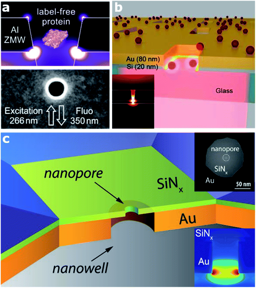 | ||
| Fig. 1 Various architectures of zero mode waveguide (ZMW) plasmonic nanoapertures. (a) Deep ultraviolet plasmonic enhancement of single protein autofluorescence in an Al ZMW. This figure has been reproduced from ref. 31 with permission from ACS Publications, copyright 2019. (b) A hybrid Au–Si zero mode waveguide for enhanced single molecule detection. This figure has been reproduced from ref. 30 with permission from the Royal Society of Chemistry, copyright 2019. (c) Enhanced single molecule fluorescence detection with a plasmonic nanowell–nanopore device architecture made of a nanowell fabricated in a gold film (orange) with a nanopore drilled in a freestanding Si3N4 membrane (light green). This figure has been reproduced from ref. 40 with permission from Wiley, copyright 2017. | ||
Alternatively, the use of apertures in metallic films and whose depth is partially engraved in a transparent substrate have been proved as a potential approach to increase the fluorescence of molecules.34 Current efforts in ZMW optimization are also devoted towards extending their working spectral range down to the UV, where several bio-molecules have intrinsic fluorescence, thus enabling label-free detection.26,31 Thanks to ZMW technology, a wide range of applications have been enabled, from DNA sequencing36 to enzymatic reactions.25 Moreover, the development of sensing architectures based on the ZMW concept paved the way for the realization of a new class of sensing platforms, so called solid-state nanopores,37 and consequently the subclass of plasmonic nanopores.1 Nanopore technology recently got massive attention, in particular for single molecule detection and sequencing.38–40 In the last few years, several groups investigated different configurations of plasmonic nanopores. A very smart as well as simple geometry has been proposed by Meller and co-workers, who realized a ZMW on a transparent thin Si3N4 membrane in a flow-through configuration by drilling a sub-10 nm hole in the membrane using a high-resolution transmission electron microscope40 (Fig. 1(c)). This platform enables enhanced single-molecule fluorescence detection and can be integrated with an electrical read-out of DNA translocation through the nanopore. Several optimizations or variations of this design have been reported. In terms of both local field confinement and enhancement, an outstanding example is represented by a bowtie antenna (dimer made of Au triangles) fabricated in close proximity to a solid-state nanopore.38
Spectroscopic techniques used in plasmonic-based single-molecule sensing
Although fluorescence is the most used spectroscopic method in ZMW and nanopore-based single-molecule experiments, other phenomena have been investigated with very interesting outcomes. For instance, Förster resonance energy transfer (FRET) enhancement has been demonstrated in a ZMW.25,26,41 The exploitation of the FRET mechanism in plasmonic nanopores has been recently proposed as an efficient approach to multiplex maximum fluorescence wavelength channels by means of life-time/intensity multiplexing.42,43 The latter can find application in nanopore protein sequencing, where the high number of distinct amino acids to be discriminated (20) makes the fluorescence-based sequencing far more challenging. Moreover, plasmonic nanocavities have been demonstrated to enable forbidden dipole–dipole FRET exchanges.24,44,45In the case of non-fluorescent molecules, surface-enhanced Raman scattering (SERS) spectroscopy performed on plasmonic nanocavities has been demonstrated to enable single-molecule sensitivity.46–51 The ability of SERS to detect these fingerprints with single-molecule sensitivity can be of paramount importance in plasmonic-based sequencing applications. In fact, by probing the SERS signals of four nucleotides and DNA oligonucleotides, the vibrational modes of single molecules very close (∼1 nm) to a metallic nanopore can be detected, owing to the enhanced electromagnetic fields.52 It is well-known that every molecule has its own Raman fingerprint, which is related to the building blocks of bio-molecules such as DNA, RNA, and proteins. In attempts to reach high and reproducible SERS signals, several types of plasmonic pores have been tested in recent years showing that the approach is potentially a winning strategy. As a significant example, recently van Dorpe and colleagues were able to detect DNA adsorbed inside a plasmonic nanoslit, reporting a spectroscopic library of nucleotides identified with single-molecule sensitivity.52
From nano to picocavities for high-resolution single-molecule imaging and spectroscopy
Visualizing single molecules with chemical recognition represents a fundamental target in nano-biotechnology. Vibrational spectroscopy based on tip-enhanced Raman scattering (TERS) allows accessing the spectral signals of molecular species very efficiently via the strong localized plasmonic fields produced at the tip apex.53 In this context, recently Jaculbia and co-workers reported nanocavity-based TERS as a versatile tool for single molecule chemical analysis at the nanoscale.47 Similarly, nanocavities can be exploited to reconstruct single molecules' spatial locations within a plasmonic hotspot with an accuracy of 1 nm, thus enabling nanoscopy of their vibrational signatures.54 Recently, Hou and colleagues were able to image individual vibrational modes at the Ångström level for a single Mg-porphine molecule, revealing distinct characteristics of the vibrational modes in real space (Fig. 2).55 The same group also demonstrated spatially and spectrally resolved photoluminescence imaging of a single phthalocyanine molecule, as well as the local mapping of the molecular exciton energy and linewidth, coupled to nanocavity plasmons in a tunnelling junction with a spatial resolution down to ∼8 Å.56 Similarly, Lee et al. reported similar results using TERS at the precisely controllable junction of a cryogenic ultrahigh-vacuum scanning tunneling microscope, showing that Ångström-scale resolution is attained at subatomic separation between the tip atom and a molecule in the quantum tunneling regime of plasmons.57 They were able to record the vibrational spectra of a single molecule, obtain images of normal modes and analyse at the atomistic level the intramolecular charges and currents driven by vibrations.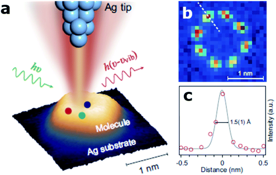 | ||
| Fig. 2 Ångström-resolved Raman images of vibrational modes for a single molecule by scanning Raman picoscopy (SRP). (a) The nanocavity defined by the silver tip and substrate generates a strong and highly confined plasmonic field, which is used for the excitation and emission enhancement of the Raman signals from a single molecule. (b) SRP image at 3072 cm−1 used for the estimation of spatial resolution. (c) Line profile of Raman signal intensities corresponding to the dashed line in (b), exhibiting a lateral spatial resolution down to 1.5(1) Å. This figure has been reproduced from ref. 55 with permission from Oxford Academic, copyright 2019. | ||
In this context, in pioneering work Baumberg and colleagues were able to place a self-assembled monolayer of biphenyl-4-thiol molecules sandwiched in a picocavity made of a gold nanoparticle on top of a gold film and able to localize light to volumes well below 1 nm3. This architecture was then used to experimentally record time-dependent Raman spectra from individual molecules at cryogenic temperature.58 They reported extreme optical confinement yielding a 100-fold enhancement, thus enabling optomechanical coupling between the cavity field and the vibrations of individual molecular bonds. In the same year they also showed that by scaling the cavity volume to less than 40 nm3, they could achieve room temperature strong coupling at the single-molecule level.59 Recently, the same group reported large-scale room-temperature single-molecule detection by using nanocavities to retrieve either the enhanced Raman scattering of the molecules (Fig. 3)60 or their fluorescence emission.4
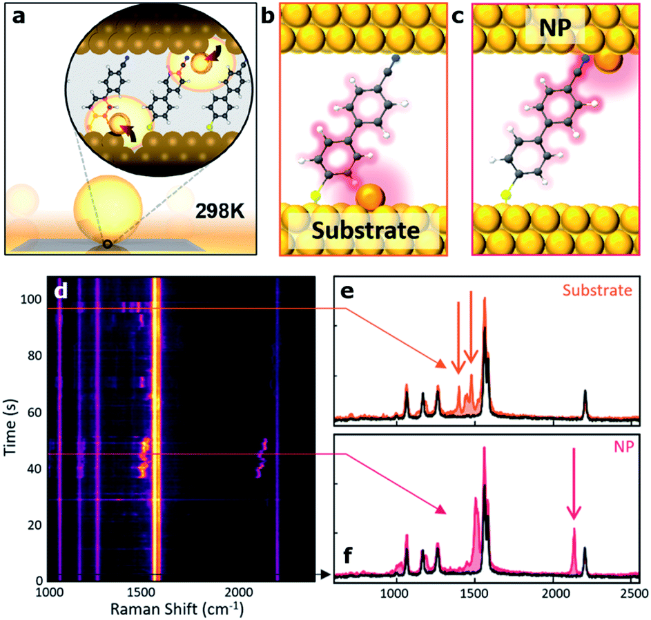 | ||
| Fig. 3 Room-temperature optical picocavities below 1 nm3 for accessing single-atom geometries. (a) Schematic of a nanoparticle-on-mirror (NPoM) geometry. The inset shows the formation of a picocavity by the movement of the surface atom to the adatom. (b, c) Schematic of picocavity adatoms on the Au substrate and nanoparticle facet, giving different interactions with 4-cyanobiphenyl-4-thiol (NC-BPT). (d) Consecutive SERS spectra showing transient peaks resulting from both forms of the picocavity. (e) SERS spectra for the picocavity adatom on the Au substrate and (f) picocavity adatom on the nanoparticle facet. This figure has been reproduced from ref. 60 with permission from ACS Publications, copyright 2018. | ||
Finally, it is worth mentioning here that narrow fingerprint Raman peaks are promising for biomedical analysis. Raman sensing based on a flow-through scheme is desirable for many practical applications, including lab-on-chip diagnostics. Recently, Huang et al. introduced a new scheme to achieve on demand control and delivery of single plasmonic nanoparticles, which can be functionalized with Raman tags. Using an analogous strategy and exploiting the enhanced optical field in a picocavity formed by translocating a gold nanoparticle coupled to a plasmonic nanohole, they were able to discriminate the SERS signal of single DNA bases in single oligonucleotides by an electro-plasmonic trapping mechanism.27 They also used this approach to detect single amino acid residues in polypeptides.61
Nanocavities realized by using DNA-based nanotechnology
Over the last decade, the DNA origami technique has been consolidated into the state-of-the-art approach for the self-assembly of nanophotonic structures.62,63 DNA origami is fabricated in a bottom-up manner by folding a “long” single-stranded DNA (ss-DNA) sequence (termed “scaffold”, ∼8000 bases long) into a predesigned shape with the help of approximately 200 “short” ss-DNA sequences (termed “staples”, ∼40 bases long and complementary to the scaffold sequence – see Fig. 4(a)). These structures can be used as breadboards where different species, including single-photon emitters such as organic fluorophore molecules and quantum dots, together with colloidal metallic nanoparticles (MNPs) of different shapes, materials and sizes, can be incorporated with nanometric accuracy and stoichiometric control64 to form nanocavities.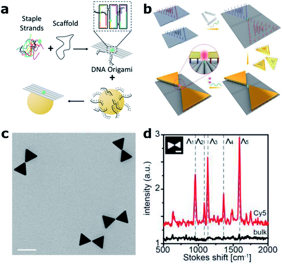 | ||
| Fig. 4 DNA origami based nanocavities. (a) Basic principle: a “long” ss-DNA sequence (Scaffold) is folded with the help of hundreds of “short” ss-DNA sequences into the predesigned shape. MNPs, previously functionalized with DNA, can be incorporated through DNA hybridization. (b) Bowtie antennas self-assembled onto DNA origami structures fabricated based on two rectangular DNA origami structures. A single Cy5 Raman active molecule is incorporated at the bowtie hotspot. (c) Corresponding TEM image (scale bar 50 nm) and (d) Raman spectra of an individual bowtie with a single Cy5 (red) and a bulk solution of Cy5 (black). (b)–(d) have been reproduced from ref. 76 with permission from Wiley, copyright 2018. | ||
Compared to conventional “top-down” nanofabrication techniques, i.e., electron or ion beam lithography, the DNA origami technique has three main advantages. First, it is a bottom-up self-assembly process in which billions of structures can be fabricated in a parallel fashion without the need of costly equipment. Second, it employs colloidal MNPs, which are less prone to surface defects and can be fabricated with higher uniformity than evaporated metallic structures leading to improved reproducibility and performance.65 Finally, with this technique, a single-photon emitter can be routinely placed in the hotspots of MNPs with nanometer precision, a key factor in controlling the coupling between single molecules and nanocavities.66,67 These advantages were initially exploited to revisit experiments on fluorescence-enhanced spectroscopy68,69 and SERS70–72 using dimer nanocavities made of Au or Ag, achieving enhancement values outperforming in some cases those obtained by using nanocavities fabricated with more complex top-down lithographic techniques.73 While most DNA origami based dimer nanocavities are based on spherical MNPs, anisotropic geometries such as gold nanorods were also demonstrated.74,75 One step further was recently taken by Ding's group by positioning and orienting triangular gold nanoplates onto two rectangular DNA origami structures in order to fabricate bowtie antennas76 (see Fig. 4(b) and (c)). The advancement introduced by the DNA origami fabrication technique is reflected by smaller and more homogenous gaps between the triangular plates reaching 5 ± 1 nm, which represents an extremely challenging gap to fabricate with lithographic techniques. Moreover, smaller gaps translate into a 200-fold higher electric field enhancement. However, the main advantage of this approach is that single Cy3 and Cy5 Raman active molecules could be placed at the hotspot of the bowtie antenna in order to demonstrate single-molecule SERS (Fig. 4(d)).
Another improvement recently enabled by the DNA origami technique is the possibility to tune the gap of dimer nanocavities. The groups led by Liedl and Lohmüller showed that the distance between two Au 40 nm MNPs self-assembled onto a DNA origami structure can be adjusted by increasing the cavity temperature, see Fig. 5(a). An increase of approximately 200 °C leads to a shrinking of the DNA molecule and thus to a reduction of the gap size from 2.5 to 1.4 nm.49 The gap thermal shrinking was monitored by studying the redshift in the scattering cross-section of the dimer nanocavity (Fig. 5(b)) and by SERS measurements of a single Cy3.5 molecule placed at the hotspot.
 | ||
| Fig. 5 Gap control and NPoM nanocavities. (a) Sketch of the Au MNP dimer bound to a rectangular DNA origami structure for thermal control of the nanocavity gap. (b) Scattering cross-section as the dimer gap is reduced by increasing the temperature. (c) Scattering spectra of an individual dimer structure modified with a single Cy3.5 molecule placed at the hot spot before (black) and after a first (green) and a second (red) round of a 10 s laser excitation (612 nm, ∼60 kW cm−2). This figure has been reproduced from ref. 49 with permission from ACS Publications, copyright 2016. (d) Fluorescence measurements on different samples in which the single molecule was laterally displaced. Values were normalized to the emission on bare glass. Inset: sketch of the fabrication approach of the NPoM using a DNA origami as a spacer. A single molecule can be placed at the NPoM hotspot with nanometer precision. This figure has been reproduced from ref. 77 with permission from ACS Publications, copyright 2018. | ||
An alternative approach for the fabrication of nanocavities, using the NPoM geometry, was proposed by Chikkaraddy and co-workers.77 A rectangular DNA origami structure was used as a spacer in order to self-assemble a single spherical Au MNP onto an Au layer forming an NPoM nanocavity (Fig. 5(c)). This approach enabled the subsequent incorporation of single molecules at a well-defined position within the NPoM cavity gap in contrast to initial studies where neither the position nor the stoichiometry of fluorescent molecules could be controlled.78 This level of position control was exploited to map the hotspot of the NPoM cavity by placing single Cy5 molecules at different positions within the 5 nm gap between an 80 nm gold MNP and a thick gold layer using a rectangular DNA origami (Fig. 5(d)). By performing fluorescence measurements at each position, the spatial profile of the local density of optical states was estimated with a resolution of approximately 2 nm.77
Conclusions
In summary, since their development, plasmonic nanocavities have gained growing interest due to their unique capabilities to confine and concentrate the EM field in the nanometer and sub-nanometer range. This effect has been exploited to apply spectroscopic techniques (e.g., SERS and fluorescence) to detect single molecules with a strong impact on biomedical applications such as real-time DNA sequencing. ZMWs and plasmonic nanopores still represent the most valuable platforms for single diffusing/translocating molecule detection at high concentrations in the micromolar range. They can be used not only in single-molecule spectroscopy, but also integrated with additional functionalities such as optical trapping and thermo-electro-phoretic effects. Moreover, advanced nanofabrication approaches enable the preparation of hybrid devices that integrate both solid-state and biological elements (such as organic coatings or functional proteins).79,80 Future development in nanocavities engineering might include the exploration of high-index dielectric materials for the fabrication of dimers,81,82 and their integration with MNPs.83 Other directions might include the combination of dimers and NPoM structures, so called dimers-on-film, in order to obtain narrow resonances through the superposition of bright and dark modes.84,85 DNA nanotechnology might also play a fundamental role in optimizing nanocavity hotspots. This paves the way for the combination of nanocavities with, for example, bio-assays for DNA sensing and diagnostics.86 Finally, we also envision a synergistic combination of the DNA origami technique with nanoapertures87,88 in order to control and address their occupation.Conflicts of interest
There are no conflicts to declare.Acknowledgements
N. M. acknowledges support from the Luxembourg National Research Fund (CORE Grant no. C19/MS/13624497 ‘ULTRON’) and from the FEDER Program (Grant no. 2017-03-022-19 ‘Lux-Ultra-Fast’). G. P. A. acknowledges support from the Swiss National Science Foundation (200021_184687) and through the National Center of Competence in Research Bio-Inspired Materials (NCCR, 51NF40_182881). The authors thank Morgane Loretan for helping to sketch the DNA origami-based nanocavity basic concept (Fig. 4(a)).Notes and references
- D. Garoli, H. Yamazaki, N. Maccaferri and M. Wanunu, Plasmonic Nanopores for Single-Molecule Detection and Manipulation: Toward Sequencing Applications, Nano Lett., 2019, 19(11), 7553–7562 CrossRef CAS.
- K. Santhosh, O. Bitton, L. Chuntonov and G. Haran, Vacuum Rabi Splitting in a Plasmonic Cavity at the Single Quantum Emitter Limit, Nat. Commun., 2016, 7, 11823 CrossRef.
- M. Urbieta, M. Barbry, Y. Zhang, P. Koval, D. Sánchez-Portal, N. Zabala and J. Aizpurua, Atomic-Scale Lightning Rod Effect in Plasmonic Picocavities: A Classical View to a Quantum Effect, ACS Nano, 2018, 12(1), 585–595 CrossRef CAS.
- O. S. Ojambati, R. Chikkaraddy, W. D. Deacon, M. Horton, D. Kos, V. A. Turek, U. F. Keyser and J. J. Baumberg, Quantum Electrodynamics at Room Temperature Coupling a Single Vibrating Molecule with a Plasmonic Nanocavity, Nat. Commun., 2019, 10(1), 1049 CrossRef.
- C. Ciracì, R. Jurga, M. Khalid and F. Della Sala, Plasmonic Quantum Effects on Single-Emitter Strong Coupling, Nanophotonics, 2019, 8(10), 1821–1833 Search PubMed.
- J. Liu, M. Jalali, S. Mahshid and S. Wachsmann-Hogiu, Are Plasmonic Optical Biosensors Ready for Use in Point-of-Need Applications?, Analyst, 2020, 145(2), 364–384 RSC.
- R. Verre, N. Maccaferri, K. Fleischer, M. Svedendahl, N. Odebo Länk, A. Dmitriev, P. Vavassori, I. V. Shvets and M. Käll, Polarization conversion-based molecular sensing using anisotropic plasmonic metasurfaces, Nanoscale, 2016, 8(20), 10576–10581 RSC.
- C. Zhan, G. Wang, J. Yi, J.-Y. Wei, Z.-H. Li, Z.-B. Chen, J. Shi, Y. Yang, W. Hong and Z.-Q. Tian, Single-Molecule Plasmonic Optical Trapping, Matter, 2020, 3(4), 1350–1360 CrossRef.
- Y. Zhang, Y. Luo, Y. Zhang, Y.-J. Yu, Y.-M. Kuang, L. Zhang, Q.-S. Meng, Y. Luo, J.-L. Yang, Z.-C. Dong and J. G. Hou, Visualizing coherent intermolecular dipole–dipole coupling in real space, Nature, 2016, 531, 623–627 CrossRef CAS.
- J. J. Baumberg, J. Aizpurua, M. H. Mikkelsen and D. R. Smith, Extreme Nanophotonics from Ultrathin Metallic Gaps, Nat. Mater., 2019, 18(7), 668–678 CrossRef CAS.
- A. Martín-Jiménez, A. I. Fernández-Domínguez, K. Lauwaet, D. Granados, R. Miranda, F. J. García-Vidal and R. Otero, Unveiling the Radiative Local Density of Optical States of a Plasmonic Nanocavity by STM, Nat. Commun., 2020, 11(1), 1021 CrossRef.
- Y. Zhang, Q.-S. Meng, L. Zhang, Y. Luo, Y.-J. Yu, B. Yang, Y. Zhang, R. Esteban, J. Aizpurua and Y. Luo, et al., Sub-Nanometre Control of the Coherent Interaction between a Single Molecule and a Plasmonic Nanocavity, Nat. Commun., 2017, 8(1), 15225 CrossRef CAS.
- Y. Zou, G. Song, R. Jiao, G. Duan and L. Yu, Strong Coupling between a Quasi-Single Molecule and a Plasmonic Cavity in the Trapping System, Nanoscale Res. Lett., 2019, 14, 74 CrossRef.
- A. B. Taylor and P. Zijlstra, Single-Molecule Plasmon Sensing: Current Status and Future Prospects, ACS Sens., 2017, 2(8), 1103–1122 CrossRef CAS.
- T. Wen, W. Zhang, S. Liu, A. Hu, J. Zhao, Y. Ye, Y. Chen, C. W. Qiu, Q. Gong and G. Lu, Steering Valley-Polarized Emission of Monolayer MoS2 Sandwiched in Plasmonic Antennas, Sci. Adv., 2020, 6(21), eaao0019 CrossRef CAS.
- J. M. Katzen, C. Tserkezis, Q. Cai, L. H. Li, J. M. Kim, G. Lee, G. R. Yi, W. R. Hendren, E. J. G. Santos and R. M. Bowman, et al., Strong Coupling of Carbon Quantum Dots in Plasmonic Nanocavities, ACS Appl. Mater. Interfaces, 2020, 12(17), 19866–19873 CrossRef CAS.
- P. Mestres, J. Berthelot, S. S. Aćimović and R. Quidant, Unraveling the Optomechanical Nature of Plasmonic Trapping, Light: Sci. Appl., 2016, 5(7), e16092 CrossRef CAS.
- J. T. Hugall, A. Singh and N. F. Van Hulst, Plasmonic Cavity Coupling, ACS Photonics, 2018, 5(1), 43–53 CrossRef CAS.
- G. C. Li, Q. Zhang, S. A. Maier and D. Lei, Plasmonic Particle-on-Film Nanocavities: A Versatile Platform for Plasmon-Enhanced Spectroscopy and Photochemistry, Nanophotonics, 2018, 7(12), 1865–1889 CAS.
- V. Devaraj, J.-M. Lee and J.-W. Oh, Distinguishable Plasmonic Nanoparticle and Gap Mode Properties in a Silver Nanoparticle on a Gold Film System Using Three-Dimensional FDTD Simulations, Nanomaterials, 2018, 8(8), 582 CrossRef.
- R. Chikkaraddy, X. Zheng, F. Benz, L. J. Brooks, B. de Nijs, C. Carnegie, M.-E. Kleemann, J. Mertens, R. W. Bowman, G. A. E. Vandenbosch, V. V. Moshchalkov and J. J. Baumberg, How Ultranarrow Gap Symmetries Control Plasmonic Nanocavity Modes: From Cubes to Spheres in the Nanoparticle-on-Mirror, ACS Photonics, 2017, 4(3), 469–475 CrossRef CAS.
- M. Baibakov, A. Barulin, P. Roy, J.-B. Claude, S. Patra and J. Wenger, Zero-Mode Waveguides Can Be Made Better: Fluorescence Enhancement with Rectangular Aluminum Nanoapertures from the Visible to the Deep Ultraviolet, Nanoscale Adv., 2020, 2, 4153–4160 RSC.
- A. N. Koya, J. Cunha, T. L. Guo, A. Toma, D. Garoli, T. Wang, S. Juodkazis, D. Cojoc and R. Proietti Zaccaria, Novel Plasmonic Nanocavities for Optical Trapping-Assisted Biosensing Applications, Adv. Opt. Mater., 2020, 8(7), 901481 CrossRef.
- M. Sanz-Paz, J. Wenger, N. F. van Hulst, M. Mivelle and M. F. Garcia-Parajo, Nanoscale Control of Single Molecule Förster Resonance Energy Transfer by a Scanning Photonic Nanoantenna, Nanophotonics, 2020, 9(12), 4021–4031 CAS.
- M. P. Goldschen-Ohm, D. S. White, V. A. Klenchin, B. Chanda and R. H. Goldsmith, Observing Single-Molecule Dynamics at Millimolar Concentrations, Angew. Chem., 2017, 129(9), 2439–2442 CrossRef.
- M. Baibakov, S. Patra, J. B. Claude, A. Moreau, J. Lumeau and J. Wenger, Extending Single-Molecule Förster Resonance Energy Transfer (FRET) Range beyond 10 Nanometers in Zero-Mode Waveguides, ACS Nano, 2019, 13(7), 8469–8480 CrossRef CAS.
- J.-A. Huang, M. Z. Mousavi, Y. Zhao, A. Hubarevich, F. Omeis, G. Giovannini, M. Schütte, D. Garoli and F. De Angelis, SERS Discrimination of Single DNA Bases in Single Oligonucleotides by Electro-Plasmonic Trapping, Nat. Commun., 2019, 10, 5321 CrossRef.
- P. Peng, Y. C. Liu, D. Xu, Q. T. Cao, G. Lu, Q. Gong and Y. F. Xiao, Enhancing Coherent Light-Matter Interactions through Microcavity-Engineered Plasmonic Resonances, Phys. Rev. Lett., 2017, 119, 233901 CrossRef.
- P. Ponzellini, X. Zambrana-Puyalto, N. Maccaferri, L. Lanzanò, F. De Angelis and D. Garoli, Plasmonic Zero Mode Waveguide for Highly Confined and Enhanced Fluorescence Emission, Nanoscale, 2018, 10(36), 17362–17369 RSC.
- X. Zambrana-Puyalto, P. Ponzellini, N. Maccaferri, E. Tessarolo, M. G. Pelizzo, W. Zhang, G. Barbillon, G. Lu and D. Garoli, A Hybrid Metal-Dielectric Zero Mode Waveguide for Enhanced Single Molecule Detection, Chem. Commun., 2019, 55(65), 9725–9728 RSC.
- A. Barulin, J. B. Claude, S. Patra, N. Bonod and J. Wenger, Deep Ultraviolet Plasmonic Enhancement of Single Protein Autofluorescence in Zero-Mode Waveguides, Nano Lett., 2019, 19(10), 7434–7442 CrossRef CAS.
- V. V. Klimov, Smart Design of Zero-Mode Waveguide Nanodevices for Effective Detection of Single-Molecule Fluorescence, Phys. Rev. Appl., 2019, 12, 014049 CrossRef CAS.
- P. Holzmeister, E. Pibiri, J. J. Schmied, T. Sen, G. P. Acuna and P. Tinnefeld, Quantum Yield and Excitation Rate of Single Molecules Close to Metallic Nanostructures, Nat. Commun., 2014, 5, 5356 CrossRef CAS.
- M. Wu, W. Liu, J. Hu, Z. Zhong, T. Rujiralai, L. Zhou, X. Cai and J. Ma, Fluorescence Enhancement in an Over-Etched Gold Zero-Mode Waveguide, Opt. Express, 2019, 27(13), 19002–19018 CrossRef CAS.
- R. M. Jamiolkowski, K. Y. Chen, S. A. Fiorenza, A. M. Tate, S. H. Pfeil and Y. E. Goldman, Nanoaperture Fabrication via Colloidal Lithography for Single Molecule Fluorescence Analysis, PLoS One, 2019, 14(10), 1–18 CrossRef.
- J. Larkin, R. Y. Henley, V. Jadhav, J. Korlach and M. Wanunu, Length-Independent DNA Packing into Nanopore Zero-Mode Waveguides for Low-Input DNA Sequencing, Nat. Nanotechnol., 2017, 12(12), 1169–1175 CrossRef CAS.
- L. Xue, H. Yamazaki, R. Ren, M. Wanunu, A. P. Ivanov and J. B. Edel, Solid-state nanopore sensors, Nat. Rev. Mater., 2020 DOI:10.1038/s41578-020-0229-6.
- D. V. Verschueren, S. Pud, X. Shi, L. De Angelis, L. Kuipers and C. Dekker, Label-Free Optical Detection of DNA Translocations through Plasmonic Nanopores, ACS Nano, 2019, 13(1), 61–70 CrossRef CAS.
- V. Jadhav, D. P. Hoogerheide, J. Korlach and M. Wanunu, Porous Zero-Mode Waveguides for Picogram-Level DNA Capture, Nano Lett., 2019, 19(2), 921–929 CrossRef.
- O. N. Assad, T. Gilboa, J. Spitzberg, M. Juhasz, E. Weinhold and A. Meller, Light-Enhancing Plasmonic-Nanopore Biosensor for Superior Single-Molecule Detection, Adv. Mater., 2017, 29(9), 1–9 CrossRef.
- J. De Torres, P. Ghenuche, S. B. Moparthi, V. Grigoriev and J. Wenger, FRET Enhancement in Aluminum Zero-Mode Waveguides, ChemPhysChem, 2015, 16(4), 782–788 CrossRef CAS.
- X. Zambrana-Puyalto, N. Maccaferri, P. Ponzellini, G. Giovannini, F. De Angelis and D. Garoli, Site-Selective Functionalization of Plasmonic Nanopores for Enhanced Fluorescence Emission Rate and Förster Resonance Energy Transfer, Nanoscale Adv., 2019, 1(6), 2454–2461 RSC.
- A. Spitaleri, D. Garoli, M. Schütte, H. Lehrach, W. Rocchia and F. De Angelis, Adaptive nanopores: a bioinspired label-free approach for protein sequencing and identification, 2020, DOI:10.1007/s12274-020-3095-z.
- J. De Torres, M. Mivelle, S. B. Moparthi, H. Rigneault, N. F. Van Hulst, M. F. García-Parajó, E. Margeat and J. Wenger, Plasmonic Nanoantennas Enable Forbidden Förster Dipole-Dipole Energy Transfer and Enhance the FRET Efficiency, Nano Lett., 2016, 16(10), 6222–6230 CrossRef CAS.
- J. Bohlen, Á. Cuartero-González, E. Pibiri, D. Ruhlandt, A. I. Fernández-Domínguez, P. Tinnefeld and G. P. Acuna, Plasmon-Assisted Förster Resonance Energy Transfer at the Single-Molecule Level in the Moderate Quenching Regime, Nanoscale, 2019, 11(16), 7674–7681 RSC.
- C.-Y. Li, S. Duan, J. Yi, C. Wang, P. M. Radjenovic, Z. Tian and J. Li, Real-Time Detection of Single-Molecule Reaction by Plasmon-Enhanced Spectroscopy, Sci. Adv., 2020, 6(24), eaba6012 CrossRef CAS.
- R. B. Jaculbia, H. Imada, K. Miwa, T. Iwasa, M. Takenaka, B. Yang, E. Kazuma, N. Hayazawa, T. Taketsugu and Y. Kim, Single-Molecule Resonance Raman Effect in a Plasmonic Nanocavity, Nat. Nanotechnol., 2020, 15(2), 105–110 CrossRef CAS.
- J. Langer, D. J. de Aberasturi, J. Aizpurua, R. A. Alvarez-Puebla, B. Auguié, J. J. Baumberg, G. C. Bazan, S. E. J. Bell, A. Boisen and A. G. Brolo, et al., Present and Future of Surface-Enhanced Raman Scattering, ACS Nano, 2020, 14(1), 28–117 CrossRef CAS.
- S. Simoncelli, E. M. Roller, P. Urban, R. Schreiber, A. J. Turberfield, T. Liedl and T. Lohmüller, Quantitative Single-Molecule Surface-Enhanced Raman Scattering by Optothermal Tuning of DNA Origami-Assembled Plasmonic Nanoantennas, ACS Nano, 2016, 10(11), 9809–9815 CrossRef CAS.
- C. Heck, Y. Kanehira, J. Kneipp and I. Bald, Placement of Single Proteins within the SERS Hot Spots of Self-Assembled Silver Nanolenses, Angew. Chem., Int. Ed., 2018, 57(25), 7444–7447 CrossRef CAS.
- A. Hubarevich, J.-A. Huang, G. Giovannini, A. Schirato, Y. Zhao, N. Maccaferri, F. De Angelis, A. Alabastri and D. Garoli, λ-DNA Through porous materials – Surface Enhanced Raman Scattering in a simple plasmonic nanopore, J. Phys. Chem. C, 2020, 124(41), 22663–22670 CrossRef CAS.
- C. Chen, Y. Li, S. Kerman, P. Neutens, K. Willems, S. Cornelissen, L. Lagae, T. Stakenborg and P. Van Dorpe, High Spatial Resolution Nanoslit SERS for Single-Molecule Nucleobase Sensing, Nat. Commun., 2018, 9(1), 1733 CrossRef.
- R. Zhang, Y. Zhang, Z. C. Dong, S. Jiang, C. Zhang, L. G. Chen, L. Zhang, Y. Liao, J. Aizpurua, Y. Luo, J. L. Yang and J. G. Hou, Chemical mapping of a single molecule by plasmon-enhanced Raman scattering, Nature, 2013, 498, 82–86 CrossRef CAS.
- M. J. Horton, O. S. Ojambati, R. Chikkaraddy, W. M. Deacon, N. Kongsuwan, A. Demetriadou, O. Hess and J. J. Baumberg, Nanoscopy through a Plasmonic Nanolens, Proc. Natl. Acad. Sci. U. S. A., 2020, 117(5), 2275–2281 CrossRef CAS.
- Y. Zhang, B. Yang, A. Ghafoor, Y. Zhang, Y.-F. Zhang, R.-P. Wang, J.-L. Yang, Y. Luo, Z.-C. Dong and J. G. Hou, Visually constructing the chemical structure of a single molecule by scanning Raman picoscopy, Natl. Sci. Rev., 2019, 6(6), 1169–1175 CrossRef CAS.
- B. Yang, G. Chen, A. Ghafoor, Y. Zhang, Y. Zhang, Y. Zhang, Y. Luo, J. Yang, V. Sandoghdar, J. Aizpurua, Z. Dong and J. G. Hou, Sub-nanometre resolution in single-molecule photoluminescence imaging, Nat. Photonics, 2020, 14, 693–699 CrossRef CAS.
- J. Lee, K. T. Crampton, N. Tallarida and V. A. Apkarian, Visualizing vibrational normal modes of a single molecule with atomically confined light, Nature, 2019, 568, 78–82 CrossRef CAS.
- F. Benz, M. K. Schmidt, A. Dreismann, R. Chikkaraddy, Y. Zhang, A. Demetriadou, C. Carnegie, H. Ohadi, B. de Nijs, R. Esteban, J. Aizpurua and J. J. Baumberg, Single-molecule optomechanics in “picocavities”, Science, 2016, 354, 726–729 CrossRef CAS.
- R. Chikkaraddy, B. de Nijs, F. Benz, S. J. Barrow, O. A. Scherman, E. Rosta, A. Demetriadou, P. Fox, O. Hess and J. J. Baumberg, Single-Molecule Strong Coupling at Room Temperature in Plasmonic Nanocavities, Nature, 2016, 535, 127–130 CrossRef CAS.
- C. Carnegie, J. Griffiths, B. de Nijs, C. Readman, R. Chikkaraddy, W. M. Deacon, Y. Zhang, I. Szabó, E. Rosta, J. Aizpurua and J. J. Baumberg, Room-Temperature Optical Picocavities below 1 Nm 3 Accessing Single-Atom Geometries, J. Phys. Chem. Lett., 2018, 9(24), 7146–7151 CrossRef CAS.
- J. Huang, M. Z. Mousavi, G. Giovannini, Y. Zhao, A. Hubarevich, M. A. Soler, W. Rocchia, D. Garoli and F. De Angelis, Multiplexed Discrimination of Single Amino Acid Residues in Polypeptides in a Single SERS Hot Spot, Angew. Chem., Int. Ed., 2020, 59(28), 11423–11431 CrossRef CAS.
- M. Pilo-Pais, G. P. Acuna, P. Tinnefeld and T. Liedl, Sculpting Light by Arranging Optical Components with DNA Nanostructures, MRS Bull., 2017, 42(12), 936–942 CrossRef CAS.
- B. Hampel, B. Liu, F. Nording, J. Ostermann, P. Struszewski, J. Langfahl-Klabes, M. Bieler, H. Bosse, B. Güttler and P. Lemmens, et al., Approach to Determine Measurement Uncertainty in Complex Nanosystems with Multiparametric Dependencies and Multivariate Output Quantities, Meas. Sci. Technol., 2018, 29(3), 35003 CrossRef.
- A. Kuzyk, R. Jungmann, G. P. Acuna and N. Liu, DNA Origami Route for Nanophotonics, ACS Photonics, 2018, 5(4), 1151–1163 CrossRef CAS.
- J. Lee, J. H. Huh, K. Kim and S. Lee, DNA Origami-Guided Assembly of the Roundest 60–100 Nm Gold Nanospheres into Plasmonic Metamolecules, Adv. Funct. Mater., 2018, 28(15), 1707309 CrossRef.
- K. Hübner, M. Pilo-Pais, F. Selbach, T. Liedl, P. Tinnefeld, F. D. Stefani and G. P. Acuna, Directing Single-Molecule Emission with DNA Origami-Assembled Optical Antennas, Nano Lett., 2019, 19(9), 6629–6634 CrossRef.
- A. F. Koenderink, Single-Photon Nanoantennas, ACS Photonics, 2017, 4(4), 710–722 CrossRef CAS.
- C. Vietz, I. Kaminska, M. Sanz Paz, P. Tinnefeld and G. P. Acuna, Broadband Fluorescence Enhancement with Self-Assembled Silver Nanoparticle Optical Antennas, ACS Nano, 2017, 11(5), 4969–4975 CrossRef CAS.
- I. Kaminska, J. Bohlen, S. Mackowski, P. Tinnefeld and G. P. Acuna, Strong Plasmonic Enhancement of a Single Peridinin–Chlorophyll a –Protein Complex on DNA Origami-Based Optical Antennas, ACS Nano, 2018, 12(2), 1650–1655 CrossRef CAS.
- J. Prinz, B. Schreiber, L. Olejko, J. Oertel, J. Rackwitz, A. Keller and I. Bald, DNA Origami Substrates for Highly Sensitive Surface-Enhanced Raman Scattering, J. Phys. Chem. Lett., 2013, 4(23), 4140–4145 CrossRef CAS.
- J. Prinz, C. Heck, L. Ellerik, V. Merk and I. Bald, DNA Origami Based Au–Ag-Core–Shell Nanoparticle Dimers with Single-Molecule SERS Sensitivity, Nanoscale, 2016, 8(10), 5612–5620 RSC.
- S. Tanwar, K. K. Haldar and T. Sen, DNA Origami Directed Au Nanostar Dimers for Single-Molecule Surface-Enhanced Raman Scattering, J. Am. Chem. Soc., 2017, 139(48), 17639–17648 CrossRef CAS.
- A. Puchkova, C. Vietz, E. Pibiri, B. Wünsch, M. Sanz Paz, G. P. Acuna and P. Tinnefeld, DNA Origami Nanoantennas with over 5000-Fold Fluorescence Enhancement and Single-Molecule Detection at 25 Μm, Nano Lett., 2015, 15(12), 8354–8359 CrossRef CAS.
- S. Pal, Z. Deng, H. Wang, S. Zou, Y. Liu and H. Yan, DNA Directed Self-Assembly of Anisotropic Plasmonic Nanostructures, J. Am. Chem. Soc., 2011, 133(44), 17606–17609 CrossRef CAS.
- T. Zhang, N. Gao, S. Li, M. J. Lang and Q. H. Xu, Single-Particle Spectroscopic Study on Fluorescence Enhancement by Plasmon Coupled Gold Nanorod Dimers Assembled on DNA Origami, J. Phys. Chem. Lett., 2015, 6(11), 2043–2049 CrossRef CAS.
- P. Zhan, T. Wen, Z. Wang, Y. He, J. Shi, T. Wang, X. Liu, G. Lu and B. Ding, DNA Origami Directed Assembly of Gold Bowtie Nanoantennas for Single-Molecule Surface-Enhanced Raman Scattering, Angew. Chem., Int. Ed., 2018, 57(11), 2846–2850 CrossRef CAS.
- R. Chikkaraddy, V. A. Turek, N. Kongsuwan, F. Benz, C. Carnegie, T. van de Goor, B. de Nijs, A. Demetriadou, O. Hess and U. F. Keyser, et al., Mapping Nanoscale Hotspots with Single-Molecule Emitters Assembled into Plasmonic Nanocavities Using DNA Origami, Nano Lett., 2018, 18(1), 405–411 CrossRef CAS.
- T. B. Hoang, G. M. Akselrod, C. Argyropoulos, J. Huang, D. R. Smith and M. H. Mikkelsen, Ultrafast Spontaneous Emission Source Using Plasmonic Nanoantennas, Nat. Commun., 2015, 6, 7788 CrossRef CAS.
- S. Patra, M. Baibakov, J. B. Claude and J. Wenger, Surface Passivation of Zero-Mode Waveguide Nanostructures: Benchmarking Protocols and Fluorescent Labels, Sci. Rep., 2020, 10, 5235 CrossRef CAS.
- G. Giovannini, M. Ardini, N. Maccaferri, X. Zambrana-Puyalto, G. Panella, F. Angelucci, R. Ippoliti, D. Garoli and F. De Angelis, Bio-Assisted Tailored Synthesis of Plasmonic Silver Nanorings and Site-Selective Deposition on Graphene Arrays, Adv. Opt. Mater., 2020, 8(4), 1901583 CrossRef CAS.
- M. Caldarola, P. Albella, E. Cortés, M. Rahmani, T. Roschuk, G. Grinblat, R. F. Oulton, A. V. Bragas and S. A. Maier, Non-Plasmonic Nanoantennas for Surface Enhanced Spectroscopies with Ultra-Low Heat Conversion, Nat. Commun., 2015, 6, 7915 CrossRef CAS.
- J. Cambiasso, G. Grinblat, Y. Li, A. Rakovich, E. Cortés and S. A. Maier, Bridging the Gap between Dielectric Nanophotonics and the Visible Regime with Effectively Lossless Gallium Phosphide Antennas, Nano Lett., 2017, 17(2), 1219–1225 CrossRef CAS.
- S. Sun, M. Li, Q. Du, C. E. Png and P. Bai, Metal-Dielectric Hybrid Dimer Nanoantenna: Coupling between Surface Plasmons and Dielectric Resonances for Fluorescence Enhancement, J. Phys. Chem. C, 2017, 121(23), 12871–12884 CrossRef CAS.
- G. C. Li, Y. L. Zhang, J. Jiang, Y. Luo and D. Y. Lei, Metal-Substrate-Mediated Plasmon Hybridization in a Nanoparticle Dimer for Photoluminescence Line-Width Shrinking and Intensity Enhancement, ACS Nano, 2017, 11(3), 3067–3080 CrossRef CAS.
- Y. Meng, Q. Zhang, D. Lei, Y. Li, S. Li, Z. Liu, W. Xie and C. W. Leung, Plasmon-Induced Optical Magnetism in an Ultrathin Metal Nanosphere-Based Dimer-on-Film Nanocavity, Laser Photonics Rev., 2020, 14(9), 2000068 CrossRef CAS.
- S. E. Ochmann, C. Vietz, K. Trofymchuk, G. P. Acuna, B. Lalkens and P. Tinnefeld, Optical Nanoantenna for Single Molecule-Based Detection of Zika Virus Nucleic Acids without Molecular Multiplication, Anal. Chem., 2017, 89(23), 13000–13007 CrossRef CAS.
- E. Pibiri, P. Holzmeister, B. Lalkens, G. P. Acuna and P. Tinnefeld, Single-Molecule Positioning in Zeromode Waveguides by DNA Origami Nanoadapters, Nano Lett., 2014, 14(6), 3499–3503 CrossRef CAS.
- A. Gopinath, E. Miyazono, A. Faraon and P. W. K. Rothemund, Engineering and Mapping Nanocavity Emission via Precision Placement of DNA Origami, Nature, 2016, 535, 401–405 CrossRef CAS.
| This journal is © The Royal Society of Chemistry 2021 |