
 Open Access Article
 This Open Access Article is licensed under a
 Creative Commons Attribution 3.0 Unported Licence
A facile approach to prepare silica hybrid, spin-crossover water-soluble nanoparticles as potential candidates for thermally responsive MRI agents†
Patroula
Gkolfi
a,
Dimitra
Tsivaka
b,
Ioannis
Tsougos
*b,
Katerina
Vassiou
c,
Ondřej
Malina
 *d,
Michaela
Polaskova
de,
Christina D.
Polyzou
*a,
Christos T.
Chasapis
f and
Vassilis
Tangoulis
*a
*d,
Michaela
Polaskova
de,
Christina D.
Polyzou
*a,
Christos T.
Chasapis
f and
Vassilis
Tangoulis
*a
aDepartment of Chemistry, Laboratory of Inorganic Chemistry, University of Patras, 26504, Patras, Greece. E-mail: chpolyzou@upatras.gr; vtango@upatras.gr
bDepartment of Medical Physics, University Hospital of Larissa, University of Thessaly, Biopolis, GR-41110 Larissa, Greece. E-mail: tsougos@med.uth.gr
cDepartment of Radiology, University Hospital of Larissa, University of Thessaly, Biopolis, GR-41110 Larissa, Greece
dRegional Centre of Advanced Technologies and Materials, Czech Advanced Technology and Research Institute (CATRIN), Palacký University Olomouc, Czech Republic. E-mail: ondrej.malina@upol.cz
eDepartment of Experimental Physics, Faculty of Science, Palacký University Olomouc, 17. Listopadu 1192/12, 771 46 Olomouc, Czech Republic
fNMR Facility, Instrumental Analysis Laboratory, School of Natural Sciences, University of Patras, 26504, Patras, Greece
First published on 2nd September 2021
Abstract
A reverse micelle method was used for the synthesis of water-soluble silica hybrid, spin-crossover (SCO) nanoparticles (NPs). MRI experiments provided temperature dependent T2 values, indicating their potential use as smart MRI agents, while lyophilization of NP dispersions in water yielded powders with a preserved but modified thermal hysteretic magnetic profile.
Spin-crossover (SCO) materials have been demonstrated as “newcomers” in the field of MRI as they tend to replace the classic Gadolinium-Based Contrast Agents (GBCAs) due to their association with nephrogenic systemic fibrosis (NSF).1–4 Super-Paramagnetic Iron Oxide Nanoparticles (SPION)5–9 and Paramagnetic CEST (ParaCEST) agents10–13 are also classified as effective MRI contrast agents14,15 displaying superior magnetic properties and biocompatibility.
SCO coordination polymers have been recently in the foreground since their magnetic properties in the solid state are attractive for their application as drug nanocarriers,16–20 in cancer treatment21,22 and as MRI contrast agents.23,24 SCO iron(II) compounds undergo a spin state change between LS (S = 0) and HS (S = 2) that is temperature dependent. These properties might be exploited in MRI for the detection of tumor cells in cancer as their usual temperature is higher than that of normal cells. However, when it comes to biological applications, these materials face strong limitations emerging mainly from their low solubility in biological media.25 Tsukiashi et al.26,27 reported for the first time water soluble spin-crossover (SCO) iron(II) nanoparticles (NPs) of the one-dimensional (1-D) coordination polymer [Fe(Htrz)3−3X(NH2trz)3X](BF4)2 (Htrz = 1,2,4-1H-triazole, NH2trz = 4-amino-1,2,4-triazole, and X = 0.1) coated with polyethylene glycol (PEG) for use as thermally-responsive MRI contrast agents. A very recent report by Cordani et al.28 describes how the obstacle of low solubility in water is surpassed when the polymeric chain is shortened, ending up in trinuclear oligomers. The synthesized [Fe3(R-trz)6]X6 trimers (R-trz = substituted triazole; X = counter anion) were used as synergistic adjuvants for pancreatic cancer.
In this context, we present the synthesis and spectroscopic characterization of silicon-modified doped NPs based on the already known 1-D SCO coordination polymer [FeII(Htrz)2(trz)](BF4). In the literature, there have been reports29–37 describing the magnetic properties of doped or/and silicon modified [FeII(Htrz)2(trz)](BF4) NPs but without any success in the preservation of their stability in aqueous media. To the best of our knowledge, this is the very first example of doped aminated silica hybrid SCO CPs at the nanoscale in the literature displaying stable aqueous colloidal dispersions. The effective modification of their surfaces with SiO2 is considered of great importance as the NPs create stable colloidal dispersions in water and thus are good candidates for potential application as MRI-contrast agents. The general formula of the synthesized NPs is [Fe(Htrz)2(trz)](BF4)@SiO2 (1); for the ligand substituted NPs: [Fe(Htrz)2.1(trz)0.8(NH2trz)0.1](BF4)1.2@SiO2 (2); for the ligand and metal substituted NPs : [Fe0.9Zn0.1(Htrz)2.1(trz)0.8(NH2trz)0.1] (BF4)1.2@SiO2 (3); for the ligand substituted NPs: [Fe(Htrz)2(trz)0.7 (NH2trz)0.3](BF4)1.3@SiO2 (4). All the NPs were synthesized at room temperature, using the reverse micelle technique (Fig. 1). To effectively shift the thermal hysteretic phenomenon, two different experimental procedures were followed: (a) the ligand substituted addition of the bulkier dopant NH2trz ligand leading to a strong destabilization of the crystal lattice and (b) the metal and ligand co-substituted experimental protocol using as dopants both the ZnII metal ion and the NH2trz ligand, which is presented for the first time. The nature of the surfactant medium and the ratio of concentrations [Fe]/[HTrz], [Fe/Zn]/[HTrz/NH2Trz] and [Fe]/[HTrz/NH2Trz] were chosen carefully to accommodate two important prerequisites: (i) sphere-like monodisperse nanoparticles with sizes smaller than 40 nm and (ii) gradual SCO magnetic behavior in the temperature range of 280–320 K. Two water-in-oil (w/o) microemulsions were mixed with the organic phase consisting of certain amounts of surfactant (Triton X-100) and co-surfactants (n-hexanol and cyclohexane). The latter reagents also play the role of organic solvents. The first step of hydrolysis/condensation of TEOS (tetraethyl orthosilicate) is initiated with the addition of 100 μL of TEOS in the aqueous phase consisting of the respective metal salts and ligands each dissolved in 0.5 mL of deionized H2O (Fig. 1). The second step of hydrolysis/condensation of APTES (3-aminopropyltriethoxy silane) or co-condensation of APTES/TEOS took place after mixing the two water-in-oil (w/o) microemulsions and leaving them under magnetic stirring for 24 hours (Fig. 1). In this step, either 100 μL of APTES were added to the final microemulsion to initiate a new condensation procedure (NPs 4) or 100 μL of APTES/100 μL of TEOS were used for the co-condensation procedure (NPs 1, 2, and 3). NPs 1 were prepared using the Fe(BF4)2·6H2O and HTrz reagents in a 1![[thin space (1/6-em)]](https://www.rsc.org/images/entities/char_2009.gif) :3 molar ratio in the aqueous phase. The polymer consists of two neutral Htrz ligands and one deprotonated trz ligand bridging the FeII metal ions and BF4− ions, compensating its total charge. Partial replacement—in a gradually increasing percentage—of Fe(BF4)2·6H2O and the deprotonated trz ligand by Zn(BF4)2·6H2O and the NH2trz ligand, respectively, was performed. More explicitly, partial replacement of 10% of the deprotonated trz ligand by the NH2trz derivative resulted in the formation of NPs 2. The doped polymer was synthesized using the Fe(BF4)2·6H2O, HTrz and NH2trz reagents in a 1:2.9:0.1 molar ratio. The doping reactions were continued in the next step by further replacement of 10% Fe(BF4)2·6H2O by the ZnII analogue. The molar ratio used for the preparation of NP3, after the addition of the Fe(BF4)2·6H2O, Zn(BF4)2·6H2O, HTrz and NH2trz reagents was 0.9:0.1:2.9:0.1 (the Fe/Zn ratio is confirmed by EDS analysis; Fig. S1†). NPs 4 are considered as the most doped ones since the replacement of trz by NH2trz reached the percentage of 30%. Fe(BF4)2·6H2O, HTrz and NH2trz are the reagents used in a 1:2.7:0.3 molar ratio. The proposed compositions for the above-mentioned NPs, based on elemental analysis, took into consideration the mechanism presented by Piedrahita-Bello et al.36 according to which the dopant NH2trz ligand selectively replaces the deprotonated (trz) ligand instead of the neutral (HTrz) ligand, increasing the portion of BF4− accordingly.
:3 molar ratio in the aqueous phase. The polymer consists of two neutral Htrz ligands and one deprotonated trz ligand bridging the FeII metal ions and BF4− ions, compensating its total charge. Partial replacement—in a gradually increasing percentage—of Fe(BF4)2·6H2O and the deprotonated trz ligand by Zn(BF4)2·6H2O and the NH2trz ligand, respectively, was performed. More explicitly, partial replacement of 10% of the deprotonated trz ligand by the NH2trz derivative resulted in the formation of NPs 2. The doped polymer was synthesized using the Fe(BF4)2·6H2O, HTrz and NH2trz reagents in a 1:2.9:0.1 molar ratio. The doping reactions were continued in the next step by further replacement of 10% Fe(BF4)2·6H2O by the ZnII analogue. The molar ratio used for the preparation of NP3, after the addition of the Fe(BF4)2·6H2O, Zn(BF4)2·6H2O, HTrz and NH2trz reagents was 0.9:0.1:2.9:0.1 (the Fe/Zn ratio is confirmed by EDS analysis; Fig. S1†). NPs 4 are considered as the most doped ones since the replacement of trz by NH2trz reached the percentage of 30%. Fe(BF4)2·6H2O, HTrz and NH2trz are the reagents used in a 1:2.7:0.3 molar ratio. The proposed compositions for the above-mentioned NPs, based on elemental analysis, took into consideration the mechanism presented by Piedrahita-Bello et al.36 according to which the dopant NH2trz ligand selectively replaces the deprotonated (trz) ligand instead of the neutral (HTrz) ligand, increasing the portion of BF4− accordingly.
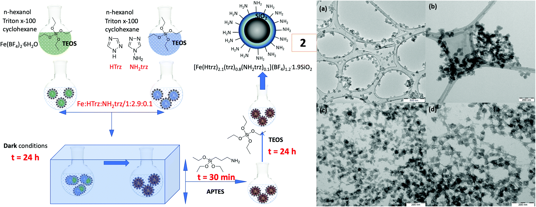 | ||
| Fig. 1 (left) Synthetic strategy for the preparation of NPs 2. (right) TEM images of NPs 1 (a), 2 (b), 3 (c) and 4 (d). | ||
The structure and detailed composition of the doped silicon-modified SCO NPs were determined by IR spectroscopy (Fig. S2†), X-ray powder diffraction (Fig. S3†) and elemental analysis (Table S1†). Particle dimensions and morphology were determined by transmission electron microscopy. Fig. S4† shows the size distribution for all the silicon-modified SCO NPs calculated based on the TEM images depicted in Fig. 2 and S5.† All the NPs are well-shaped adopting a sphere-like morphology accompanied by a very thin layer of SiO2 of a few nanometers. According to the Gaussian distributions (Fig. S4†) and TEM images (Fig. 2 and S5†) as well, the NPs 1 present a larger mean diameter (∼45 nm) than the rest of the NPs. Another interesting observation is that NPs 2 show an ultra-small diameter (∼10 nm) compared to NPs 3 and 4 with sizes close to 25 nm. Indeed, all the NPs present excellent homogeneity with few regions of minor aggregation due to the synthetic method chosen for their preparation.
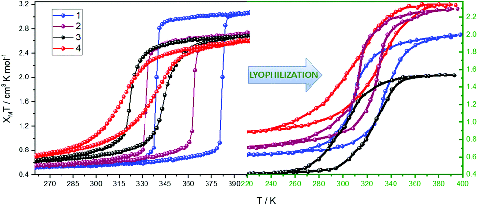 | ||
| Fig. 2 Thermal dependence of the χMT product for NPs 1–4 with a rate of 1 K min−1 along with the lyophilized analogues. | ||
It is remarkable to note that stable water colloidal dispersions of all NPs were prepared using ultrasonication (2 mg of NPs in 1 mL of H2O). The color of all the dispersions changed from purple to colorless over a period of several minutes, revealing the evolution of the SCO phenomenon in water from the LS state to a fraction of the HS state of the FeII ion (a yellow color is expected for a fully recovered HS state of the FeII ion). Interestingly, all dispersions obtained their initial purple color when stored in a refrigerator (−20 °C), presenting excellent reversibility. From all our synthetic efforts, it is important to underline the synthetic conditions under which stable aqueous dispersions are obtained. It was observed that in the “non-doping” reaction (NPs 1), where ascorbic acid was absent, the addition of a total of 300 μL of TEOS and 100 μL of APTES in a two-step hydrolysis/condensation procedure was necessary for the stabilization of the water dispersion. In the “doping” reactions (NPs 2, 3, and 4), where ascorbic acid is included, the two-step hydrolysis/condensation procedure can be performed with lower volumes: 200 μL of TEOS and 100 μL of APTES (NPs 4). When the above-mentioned volumes are further reduced, the behavior of the NPs in water changed dramatically. More explicitly, the color turned out to be yellowish without any possibility of the occurrence of the reversible process. In order to verify the reversibility of the SCO phenomenon in water, UV-vis measurements were carried out. Fig. S6† shows time-dependent UV-vis absorption spectra for NPs 1–4: (a) when the samples are dispersed for the first time in H2O and (b) during the thawing procedure (−20 °C – RT). In both cases, the absorption band around 280 nm—corresponding to an MLCT transition—showed a drastic reduction of its intensity as the samples switched from the LS to HS state of FeII accompanied by a color change in the dispersion from purple to colorless (Fig. S7 and S8†). The time duration of the SCO phenomenon calculated from the UV-vis experiments is presented in Table S3.†
DLS measurements of the aqueous dispersion of NPs (Fig. S9†) revealed a certain tendency to aggregate and their size increases to values close to 100 nm for NPs 1 and 50 nm for NPs 2. In the case of NPs 3 and 4, two distinctive size distributions are visible close to 20–30 nm and 200–300 nm. The estimated mean ζ-potential for all NPs has positive values greater than 30.0 mV (Fig. S10 and Table S4†), indicating the effective functionalization of their surface with amine groups, as well as the excellent quality of the colloid in water. Lyophilization of the latter aqueous dispersions resulted in powders with similar structural characteristics determined by IR spectroscopy (Fig. S2†) and X-Ray powder diffraction (Fig. S3a†). This observation may lead to the proposal that the aminated silica shell provides an effective shield to the NP core and furthermore stabilizes aqueous colloidal dispersions. A Pawley refinement was considered for all the NPs and their lyophilized analogues, and it was found that the powder patterns were consistent with the same unit cell and space group (Pnma) proposed for the [FeII(Htrz)2(trz)](BF4) system with small calculated differences (Fig. S3b, Table S2†).38,39
Temperature-dependent magnetic measurements were carried out on samples 1–4 as well as on their lyophilized analogues and are shown in Fig. 2. In each case, the second thermal cycle was taken into consideration as the first one is usually related to the dehydration of the sample. Based on the magnetic study, we can amplify the strong evidence—which is also supported by the literature29–37—that metal and ligand substitution (here referred to as “doping”) shifts the hysteresis clearly toward room temperature. The higher doping percentage is clearly the reason for the considerable shift of the hysteresis towards lower temperatures (NPs 3 and 4). We cannot also overlook the loss of hysteresis abruptness and narrowing of its width as the doping percentage is increased. Quite surprisingly, the lyophilized materials preserve the SCO hysteretic character, which is observed, to our knowledge, for the first time. A shift of the hysteresis and a further narrowing of its width have been observed only in the cases of lyophilized NPs 1 and 2, while for all the cases there is a suppression in the saturated χMT values (Table S5†) possibly due to the size effects and/or the hydrated form of the freeze-drying lyophilized samples. The SCO behavior exhibited by all the NPs was also confirmed through DSC measurements (Fig. S11†) as the critical temperatures coincide with those extracted from the magnetic study.
MRI T2 relaxometry studies are performed for the aqueous dispersions of all the NPs (Fig. S12–S18†). According to the thermal dependence of r2 relaxivity (Fig. 3), NPs 3 and 4 are promising candidates for potential use as thermally responsive T2 MRI contrast agents with r2 values in the range of 2.5–3.0 s−1 mM−1 which is consistent with the values for commercially available MRI contrast agents38 measured at 3 T in water. On the other hand, T1 relaxometry measurements (Fig. S19†) of NPs 3 and 4 also revealed temperature dependent T1 values that are significantly lower compared to those of GBCAs currently used for clinical diagnosis,40 in accordance with the recent studies of PEG-modified SCO NPs.27 The T1 and T2-w images of the phantom for NPs 3 and 4 at 20 °C and 50 °C at various inversion (TI) and echo (TE) times are shown in Fig. S18 and S19†, revealing their thermally responsive T2 character.
 | ||
| Fig. 3 Thermal dependence of the r2 relaxivity of NPs 1–4. The concentration used for the relaxometry studies was 3 mM. | ||
In conclusion, a two-step approach of the hydrolysis/condensation of TEOS/APTES is presented as an efficient synthetic path for the preparation of functional aminated silica hybrid SCO NPs of the general formula [Fe1−ZZnZ(Htrz)1+Y−X(trz)2−Y(NH2trz)X](BF4)Y@SiO2. Stable aqueous colloidal dispersions were prepared with a clear reversibility of the SCO phenomenon accompanied by a color change from colorless (HS state) to purple (LS state), while positive mean ζ-potential values greater than 30 mV were obtained for the SCO NPs, confirming an effective functionalization of their surface with amine groups as well as the excellent quality of the colloid in water. Lyophilization of the NP aqueous dispersions resulted in powders with similar structural characteristics, preserving the SCO hysteretic character. According to the MRI T2 relaxometry studies, the experimental doping protocols of (i) ligand substituted addition of the bulkier dopant NH2trz ligand (NPs 4) and (ii) metal and ligand co-substituted addition of ZnII/NH2trz (NPs 3) provide thermally responsive T2 MRI contrast agents. Overall, our present communication describes an effective experimental tool for the preparation of water-soluble SCO NPs and we are confident that this information will encourage the scientific community working on the biological applications of these materials.
Conflicts of interest
There are no conflicts of interest.Acknowledgements
This research was supported by a grant (80623) from the Research Committee of the University of Patras via the “C. CARATHEODORI” program. C. D. P. acknowledges the State Scholarships Foundation (IKY). This research is co-financed by Greece and the European Union (European Social Fund [ESF]) through the Operational Programme “Human Resources Development, Education and Lifelong Learning” in the context of the project “Reinforcement of Postdoctoral Researchers-2nd Cycle” (MIS-5033021), implemented by the State Scholarships Foundation (IKY). O.M. and M.P. would like to thank the following projects: the Research Infrastructure NanoEnviCz, supported by the Ministry of Education, Youth and Sports of the Czech Republic under Project No. LM2018124 and the ERDF/ESF project “Nano4Future” Development of Pre-applied Research in Nanotechnology and Biotechnology (no. CZ.02.1.01/0.0/0.0/17_048/0007323).Notes and references
- M. Edward, J. A. Quinn, S. Mukherjee, M. B. V. Jensen, A. G. Jardine, P. B. Mark and A. D. Burden, J. Pathol., 2008, 5, 584 CrossRef PubMed.
- H. H. Abujudeh, Radiology, 2009, 1, 81 CrossRef PubMed.
- R. N. Hellman, Semin. Nephrol., 2011, 3, 310 CrossRef PubMed.
- A. K. Abu-Alfa, Adv. Chronic Kidney Dis., 2011, 3, 188 CrossRef PubMed.
- C. Tassa and S. Y. Shaw, Acc. Chem. Res., 2011, 44, 842 CrossRef CAS PubMed.
- J. Gao, H. Gu and B. Xu, Acc. Chem. Res., 2009, 42, 1097 CrossRef CAS PubMed.
- Y. X. J. Wang, S. M. Hussain and G. P. Krestin, Eur. Radiol., 2001, 11, 2319 CrossRef CAS PubMed.
- C. Corot, P. Robert, J. M. Idee and M. Port, Adv. Drug Delivery Rev., 2006, 58, 1471 CrossRef CAS PubMed.
- U. I. Tromsdorf, O. T. Bruns, S. C. Salmen, U. Beisiegel and H. Weller, Nano Lett., 2009, 9, 4434 CrossRef CAS PubMed.
- I. R. Jeon, J. G. Park, C. R. Haney and T. D. Harris, Chem. Sci., 2014, 5, 2461 RSC.
- S. J. Dorazio, P. B. Tsitovich, K. E. Silters, J. A. Spernyak and J. R. Morrow, J. Am. Chem. Soc., 2011, 133, 14154 CrossRef CAS PubMed.
- S. J. Dorazio and J. R. Morrow, Inorg. Chem., 2012, 51, 7448 CrossRef CAS PubMed.
- S. J. Dorazio and J. R. Morrow, Eur. J. Chem., 2012, 2006 CAS.
- Z. Cai, C. Wu, L. Yang, D. Wang and H. Ai, ACS Biomater. Sci. Eng., 2020, 6, 2533 CrossRef CAS PubMed.
- J. Wahsner, E. M. Gale, A. Rodríguez-Rodríguez and P. Caravan, Chem. Rev., 2019, 119, 957 CrossRef CAS PubMed.
- Y. Hu, T. Lv, Y. Ma, J. Xu, Y. Zhang, Y. Hou, Z. Huang and Y. Ding, Nano Lett., 2019, 19, 2731 CrossRef CAS PubMed.
- M. Rezaei, A. Abbasi, R. Dinarvand, M. Jeddi-Tehrani and J. Janczak, ACS Appl. Mater. Interfaces, 2018, 10, 17594 CrossRef CAS PubMed.
- C. He, C. Poon, C. Chan, S. D. Yamada and W. Lin, J. Am. Chem. Soc., 2016, 138, 6010 CrossRef CAS PubMed.
- I. Imaz, M. Rubio-Martínez, L. García-Fernández, F. García, D. Ruiz-Molina, J. Hernando, V. Puntes and D. Maspoch, Chem. Commun., 2010, 46, 4737 RSC.
- X. Duan, C. Chan, W. Han, N. Guo, R. R. Weichselbaum and W. Lin, Nat. Commun., 2019, 10, 1899 CrossRef PubMed.
- J. Liu, H. Wang, X. Yi, Y. Chao, Y. Geng, L. Xu, K. Yang and Z. Liu, Adv. Funct. Mater., 2017, 27, 1703832 CrossRef.
- J. Liu, L. Tian, R. Zhang, Z. Dong, H. Wang and Z. Liu, ACS Appl. Mater. Interfaces, 2018, 10, 43493 CrossRef CAS PubMed.
- S. Suárez-García, N. Arias-Ramos, C. Frias, A. P. Candiota, C. Arús, J. Lorenzo, D. Ruiz-Molina and F. Novio, ACS Appl. Mater. Interfaces, 2018, 10, 38819 CrossRef PubMed.
- Y. Chen, K. Ai, J. Liu, X. Ren, C. Jiang and L. Lu, Biomaterials, 2016, 77, 198 CrossRef CAS PubMed.
- I. Bräunlich, A. Sánchez-Ferrer, M. Bauer, R. Schepper, P. Knüsel, J. Dshemuchadse, R. Mezzenga and W. Caseri, Inorg. Chem., 2013, 53, 3546 CrossRef PubMed.
- A. Tsukiashi, K. S. Min, H. Terasawa, S. Yoshinaga, M. Takeda, R. Ohtani, M. Nakamura, L. F. Lndoy and S. Hayami, Chem. Lett., 2018, 47, 598 CrossRef CAS.
- A. Tsukiashi, K. S. Min, H. Kitayama, H. Terasawa, S. Yoshinaga, M. Takeda, L. F. Lindoy and S. Hayami, Sci. Rep., 2018, 8, 14911 CrossRef PubMed.
- M. Cordani, E. Resines-Urien, A. Gamonal, P. Milán-Rois, L. Salmon, A. Bousseksou, J. S. Costa and A. Somoza, Antioxidants, 2021, 10, 66 CrossRef CAS PubMed.
- E. Coronado, J. R. Galán-Mascarós, M. Monrabal-Capilla, J. García-Martínez and P. Pardo-Ibáñez, Adv. Mater., 2007, 19, 1359 CrossRef CAS.
- J. R. Galán-Mascarós, E. Coronado, A. Forment-Aliaga, M. Monrabal-Capilla, E. Pinilla-Cienfuegos and M. Ceolin, Inorg. Chem., 2010, 49, 5706 CrossRef PubMed.
- S. Titos-Padilla, J. M. Herrera, X.-W. Chen, J. J. Delgado and E. Colacio, Angew. Chem., 2011, 123, 3348 CrossRef.
- M. Giménez-Marqués, M. L. García-Sanz de Larrea and E. Coronado, J. Mater. Chem. C, 2015, 3, 7946 RSC.
- J. M. Herrera, S. Titos-Padilla, S. J. a. Pope, I. Berlanga, F. Zamora, J. J. Delgado, K. V. Kamenev, X. Wang, A. Prescimone, E. K. Brechin and E. Colacio, J. Mater. Chem. C, 2015, 3, 7819 RSC.
- I. Suleimanov, J. S. Costa, G. Molnár, L. Salmon and A. Bousseksou, Chem. Commun., 2014, 50, 13015 RSC.
- S. Rat, M. Piedrahita-Bello, L. Salmon, G. Molnár, P. Demont and A. Bousseksou, Adv. Mater., 2018, 8, 1705275 CrossRef PubMed.
- M. Piedrahita-Bello, K. Ridier, M. Mikolasek, G. Molnár, W. Nicolazzi, L. Salmon and A. Bousseksou, Chem. Commun., 2019, 55, 4769 RSC.
- R. Torres-Cavanillas, L. Lima-Moya, F. D. Tichelaar, H. W. Zandbergen, M. Giménez-Marqués and E. Coronado, Dalton Trans., 2019, 48, 15465 RSC.
- A. Grosjean, P. Négrier, P. Bordet, C. Etrillard, D. Mondieig, S. Pechev, E. Ledraud, J.-F. Létard and P. Guionneau, Eur. J. Inorg. Chem., 2013, 5, 796 CrossRef.
- M. Palluel, L. E. Khoury, N. Daro, S. Buffière, M. Josse, M. Marchivie and G. Chastanet, Inorg. Chem. Front., 2021, 8, 3697 RSC.
- M. Rohrer, H. Bauer, J. Mintorovitch, M. Requardt and H. J. Weinmann, Invest. Radiol., 2005, 40, 715 CrossRef PubMed.
Footnote |
| † Electronic supplementary information (ESI) available: Synthetic details, UV/vis spectroscopy, P-XRD patterns, Differential Scanning Calorimetry (DSC), SEM/TEM microscopy, and additional figures. and tables. See DOI: 10.1039/d1dt02479e |
| This journal is © The Royal Society of Chemistry 2021 |