 Open Access Article
Creative Commons Attribution 3.0 Unported Licence
Tetra-arylborate lipophilic anions as targeting groups†
Kishore K.
Gaddale Devanna
a,
Justyna M.
Gawel
a,
Tracy A.
Prime
b,
Filip
Cvetko
b,
Cristiane
Benincá
b,
Stuart T.
Caldwell
 a,
Alexander
Negoda
c,
Andrew
Harrison
 b,
Andrew M.
James
b,
Evgeny V.
Pavlov
d,
Michael P.
Murphy
*be and
Richard C.
Hartley
b,
Andrew M.
James
b,
Evgeny V.
Pavlov
d,
Michael P.
Murphy
*be and
Richard C.
Hartley
 *a
*a
aSchool of Chemistry, University of Glasgow, Glasgow, G12 8QQ, UK. E-mail: Richard.Hartley@glasgow.ac.uk
bMRC Mitochondrial Biology Unit, Hills Road, University of Cambridge, CB2 0XY, UK. E-mail: mpm@mrc-mbu.cam.ac.uk
cDepartment of Physiology and Biophysics, Dalhousie University, Halifax, Nova Scotia, Canada
dNew York University, College of Dentistry, Department of Molecular Pathobiology, 345 East 24th Street, New York, NY 10010, USA
eDepartment of Medicine, University of Cambridge, Cambridge, UK
First published on 18th February 2021
Abstract
Tetraphenylborate (TPB) anions traverse membranes but are excluded from mitochondria by the membrane potential (Δψ). TPB-conjugates also distributed across membranes in response to Δψ, but surprisingly, they rapidly entered cells. They accumulated within lysosomes following endocystosis. This pH-independent targeting of lysosomes makes possible new classes of probe and bioactive molecules.
The ability to direct molecules to an appropriate location within the cell facilitates development of bioactive and probe compounds. For example, molecules are targeted to the mitochondrial matrix by conjugation to the lipophilic triphenylphosphonium cation.1–4 The large hydrophobic surface area of the triphenylphosphonium group enables rapid crossing of biological membranes by lowering the activation energy for movement through the membrane core.4,5 Furthermore, the positive charge drives extensive accumulation within the mitochondrial matrix due to the large membrane potential (Δψ negative inside).2,4,5
To deliver molecules to other cell compartments in a similar way, we explored conjugation to lipophilic anions. Just as the archetypal lipophilic cation is tetraphenylphosphonium (TPP) the corresponding lipophilic anion is tetraphenylborate (TPB). These molecules have identical radii (4.2 Å) and, apart from charge, are similar.6,7 Both are water soluble, facilitating biological uses, although TPB has a more negative free energy of hydration.8 Lipophilic ions bind to a potential energy well on the membrane surface, before flipping, to the corresponding potential energy well on the other side of the membrane, traversing the activation energy barrier of the membrane core (Fig. 1).6,7,9 Hence, TPB rapidly permeates phospholipid bilayers6,7,10,11 and its distribution is determined by the Δψ,12 resulting in exclusion from the mitochondrial matrix.
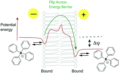 | ||
| Fig. 1 Membrane permeation by TPB lipophilic anions (adapted from ref. 9). | ||
TPB anions have been widely used in analytical chemistry,15 catalysis16 and solid electrolytes,17 and as substrates for oxidative18,19 and Suzuki-type cross-coupling.20,21 Despite their similar structure to TPP, and their complementary response to Δψ, the use of TPB anions to deliver compounds to different sub-cellular compartments has not been explored. This may be because the Δψ across the plasma membrane, which is negative inside, would be expected to disfavour intracellular delivery. There has been only one exploration of lipophilic anion distribution within cells, by conjugation of the monocarborane (1-carba-closo-dodecaborate) lipophilic anion to a porphyrin.13,14 Surprisingly, the monocarborane conjugate was taken up by cells,14 although the mechanism was not investigated. Thus, we investigated whether TPB anions could be used to deliver compounds, despite their negative charge, within the cell.
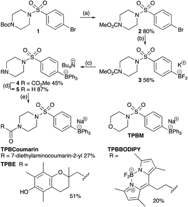
We synthesized a universal TPB-conjugate precursor 5 that could be derivatized through amide coupling to carboxylic acids. The key synthetic step was the addition of three identical phenyl substituents to an aryltrifluoroborate (Scheme 1).22 As tetraarylborates with electron-rich substituents are prone to oxidation and protodeborylation,23 we incorporated an electron-withdrawing sulfonamide. To assess intracellular distribution and functional properties we made a minimal TPB derivative with the sulfonamide piperazine replaced with a morpholine (TPBM), to avoid N-protonation targeting acidic compartments;24,25 a TPB conjugated to the chromanol group of α-tocopherol as a generic cargo (TPBE); and two fluorescent probes, TPBCoumarin and TPBBODIPY, to allow imaging.
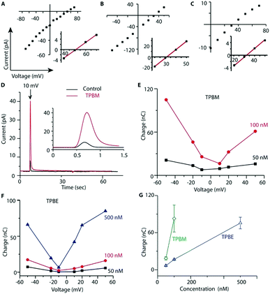
To see if TPB-conjugates could rapidly distribute across membranes in response to voltage we used a phospholipid black lipid membrane (BLM) system (Fig. 2). In agreement with previous observations,26 we detected increasing currents due to TPB crossing a BLM as a function of voltage (Fig. 2a). In the presence of a TPB gradient (1 μM![[thin space (1/6-em)]](https://www.rsc.org/images/entities/char_2009.gif) :
:![[thin space (1/6-em)]](https://www.rsc.org/images/entities/char_2009.gif) 10 μM) across a BLM the reversal potential was close to the theoretical Nernst equilibrium potential of 60 mV (Fig. 2a), demonstrating that the observed current is caused by TPB transfer across the bilayer. At higher TPB concentrations, but the same gradient, the current increased and the reversal potential shifted towards 0 mV, due to non-specific conductance caused by membrane disruption (Fig. 2b). TPBM disrupted the BLM at lower concentrations than TPB, leading to a larger non-specific conductance, hence the reversal potential for TPBM was considerably lower than 60 mV (Fig. 2c). The greater membrane disruption by TPBM compared to TPB is caused by its enhanced lipophilicity. Therefore, only low concentrations can be used and it was not possible to assess the membrane permeation of TPBM and TPBE directly. Instead, to estimate the amount of compound crossing the bilayer in response to a voltage we measured transient ionic currents induced by steps from 0 mV to defined voltages before/after addition of the compounds (Fig. 2d).
10 μM) across a BLM the reversal potential was close to the theoretical Nernst equilibrium potential of 60 mV (Fig. 2a), demonstrating that the observed current is caused by TPB transfer across the bilayer. At higher TPB concentrations, but the same gradient, the current increased and the reversal potential shifted towards 0 mV, due to non-specific conductance caused by membrane disruption (Fig. 2b). TPBM disrupted the BLM at lower concentrations than TPB, leading to a larger non-specific conductance, hence the reversal potential for TPBM was considerably lower than 60 mV (Fig. 2c). The greater membrane disruption by TPBM compared to TPB is caused by its enhanced lipophilicity. Therefore, only low concentrations can be used and it was not possible to assess the membrane permeation of TPBM and TPBE directly. Instead, to estimate the amount of compound crossing the bilayer in response to a voltage we measured transient ionic currents induced by steps from 0 mV to defined voltages before/after addition of the compounds (Fig. 2d).
In the absence of compounds the transient (capacitive) currents are determined by the membrane's dielectric properties. In the presence of a TPB-conjugates the transient currents are the sum of the capacitive current and that caused by anion redistribution.27 Charge transfer by the lipophilic anions crossing the bilayer, derived by subtracting the current in the absence of compound, depends on the compound concentration and the applied voltage (Fig. 2e and f). Combined data for a +50 mV voltage jump are shown in Fig. 2g. TPBM at 100 nM induced the largest charge flux, while higher concentrations caused non-specific conductance. The TPBE conductance was less (Fig. 2e and f). These results indicate that TPB-conjugates cross a BLM in a voltage-dependent manner.
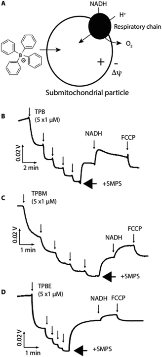
We next assessed whether TPB-conjugates crossed biological membranes in response to a Δψ using sub-mitochondrial particles (SMPs), which are inverted mitochondrial inner membrane vesicles (Fig. 3a). Proton pumping by the respiratory chain generates a positive-inside Δψ, which should drive uptake of lipophilic anions12 (Fig. 3a).
Using an ion-selective electrode (ISE) to measure TPB concentration28 showed its rapid uptake into SMPs upon induction of a Δψ by NADH, which was reversed by dissipating the Δψ with FCCP (Fig. 3b). Similarly, TPBM (Fig. 3c) and TPBE (Fig. 3d) also showed uptake into SMPs in response to Δψ. The greater hydrophobicity of TPBM and TPBE enhanced their membrane adsorption compared to TPB. Therefore, TPB-conjugates rapidly cross biological membranes in response to a Δψ and adsorb strongly to membranes.
Importantly, these attributes of TPB-conjugates are greatly enhanced over lipophilic cations due to the different interaction of lipophilic anions with biological membranes. This is primarily because of the large dipole potential from positive within the phospholipid bilayer core to negative at the surface6,7,9,29 which lowers the activation energy barrier for lipophilic anion transport, relative to cations, hence the membrane conductivity of TPB is ∼105-fold greater than that for TPP.6,7,9–11 Similarly, the binding constant for TPB to the potential energy well near the membrane surface is ∼103–104 fold greater than for TPP.6,7,9,29 Addition of a hydrophobic linker/cargo increases this membrane adsorption further still.
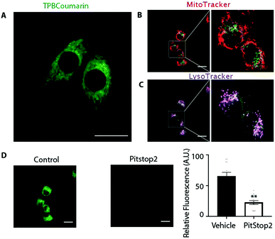
We next assessed the uptake and distribution of TPB-conjugates using a fluorescent probe, TPBCoumarin, with C2C12 cells by confocal laser scanning microscopy (Fig. 4a and Movie 1, ESI†). Despite its negative charge, TPBCoumarin was rapidly taken up by cells showing punctate staining, as was TPBBODIPY (Fig. S1 and Movie 2, ESI†). Parallel staining with MitoTracker (Fig. 4b) showed TPBCoumarin was not colocalizing with mitochondria while LysoTracker (Fig. 4c) showed partial colocalization with lysosomes. This suggests the punctate staining upon uptake of TPBCoumarin is due to uptake by endocytosis with initial localization within endosomes which then fuse with lysosomes.30 Supporting this, uptake was greatly decreased by Pitstop 2 (Fig. 4d), which inhibits clathrin-mediated endocytosis.31,32 This shows there is rapid initial uptake by endocytosis followed by redistribution to lysosomes. Critically, there was no co-localization with mitochondria, consistent with their exclusion from the negatively-charged matrix. Cell toxicity of TPBE was also negligible below 10 μM (Fig. S2, ESI†).
Despite their negative charge, and the negative-inside Δψ across the plasma membrane, TPB display a novel intracellular distribution. Most lysosome targeting is achieved through ion-trapping by protonation of weak base in the acidic lysosomes.24,25 The TPBBODIPY conjugates accumulate in the same way as TPBCoumarin (Fig. 4 and Fig. S1, Movies 1, 2, ESI†), showing that TPB-conjugates do not require such as site. Both TPBCoumarin and TPBBODIPY showed similar distribution in both HeLa and Cos7 cells showing the unique distribution was not dependent on the cell type (Fig. S3, ESI†). Their rapid endocytic uptake is likely due to the strong binding of the TPB moiety to the potential energy well on the membrane surface. Since TPB-conjugates could rapidly permeate the phospholipid bilayers of SMPs, there is also likely to be some uptake into cells directly through the plasma membrane. However, this is disfavoured by the plasma membrane potential (30–60 mV, negative inside). Any TPB-conjugates that do enter may be directed to the lysosomes in response to the negative-inside potential across the lysosomal membrane.33 The lack of co-localization with mitochondria is consistent with the expected thousand-fold exclusion due to the large Δψ (150–180 mV, negative inside) across the mitochondrial inner membrane.
In summary, we have generated a new targeting group that directs small molecules to the endosomal and lysosomal compartments within the cell in a way that complements current targeting methods that employ ion-trapping of weak bases. By manipulating hydrophobicity and incorporating cleavable linkers and membrane impermeant moieties it will be possible to fine tune the location and kinetics of cell distribution of bioactive molecules. This approach provides new opportunities to selectively manipulate and report on cell processes and to give a better understanding of the role of the lysosome in autophagy34,35 and in diseases such as cancer.36,37
This work was supported by the Medical Research Council UK (MC_U105663142), a Wellcome Trust Investigator award (110159/A/15/Z) to MPM, by BBSRC (BB/I012826/1) and Wellcome Trust Investigator (110158/Z/15/Z) awards to RCH, a University of Glasgow funded studentship to J. G. and from the Wellcome Trust PhD programme in metabolic and cardiovascular diseases (RG88195) for FC.
Conflicts of interest
There are no conflicts to declare.Notes and references
- M. P. Murphy and R. C. Hartley, Nat. Rev. Drug Discovery, 2018, 17, 865–886 CrossRef CAS.
- R. A. J. Smith, R. C. Hartley, H. M. Cocheme and M. P. Murphy, Trends Pharmacol. Sci., 2012, 33, 341–352 CrossRef CAS.
- L. F. Yousif, K. M. Stewart and S. O. Kelley, ChemBioChem, 2009, 10, 1939–1950 CrossRef CAS.
- J. Zielonka, J. Joseph, A. Sikora, M. Hardy, O. Ouari, J. Vasquez-Vivar, G. Cheng, M. Lopez and B. Kalyanaraman, Chem. Rev., 2017, 117, 10043–10120 CrossRef CAS.
- M. F. Ross, G. F. Kelso, F. H. Blaikie, A. M. James, A. Cocheme, T. Filipovska, T. R. Da Ros, R. A. Hurd, H. M. Smith and M. P. Murphy, Biochemistry (Moscow), 2005, 70, 222–230 CrossRef CAS.
- R. F. Flewelling and W. L. Hubbell, Biophys. J., 1986, 49, 531–540 CrossRef CAS.
- R. F. Flewelling and W. L. Hubbell, Biophys. J., 1986, 49, 541–552 CrossRef CAS.
- R. Scheu, B. M. Rankin, Y. Chen, K. C. Jena, D. Ben-Amotz and S. Roke, Angew. Chem., Int. Ed., 2014, 53, 9560–9563 CrossRef CAS.
- L. Wang, Annu. Rev. Biochem., 2012, 81, 615–635 CrossRef CAS.
- R. Benz, Biophys. J., 1988, 54, 25–33 CrossRef CAS.
- T. I. Rokitskaya, V. B. Luzhkov, G. A. Korshunova, V. N. Tashlitsky and Y. N. Antonenko, Phys. Chem. Chem. Phys., 2019, 21, 23355–23363 RSC.
- L. L. Grinius, A. A. Jasaitis, Y. P. Kadziauskas, E. A. Liberman, V. P. Skulachev, V. P. Topali, L. M. Tsofina and M. A. Vladimirova, Biochim. Biophys. Acta, 1970, 216, 1–12 CrossRef CAS.
- T. I. Rokitskaya, A. V. Zaitsev, V. A. Ol'shevskaya, V. N. Kalinin, M. M. Moisenovich, I. I. Agapov and Y. N. Antonenko, Biochemistry, 2012, 77, 975–982 CAS.
- M. M. Moisenovich, V. A. Ol'shevskaya, T. I. Rokitskaya, A. A. Ramonova, R. G. Nikitina, A. N. Savchenko, V. V. Tatarskiy Jr., M. A. Kaplan, V. N. Kalinin, E. A. Kotova, O. V. Uvarov, I. I. Agapov, Y. N. Antonenko and A. A. Shtil, PLoS One, 2010, 5, e12717 CrossRef.
- H. Flaschka and A. J. Barnard, Adv. Anal. Chem. Instrum., 1960, 1, 1–117 CAS.
- I. M. Riddlestone, A. Kraft, J. Schaefer and I. Krossing, Angew. Chem., Int. Ed., 2018, 57, 13982–14024 CrossRef CAS.
- J. F. Van Humbeck, M. L. Aubrey, A. Alsbaiee, R. Ameloot, G. W. Coates, W. R. Dichtel and J. R. Long, Chem. Sci., 2015, 6, 5499–5505 RSC.
- C. Gerleve and A. Studer, Angew. Chem., Int. Ed., 2020, 59, 1–6 CrossRef.
- A. Music, A. N. Baumann, P. Spiess, A. Plantefol, T. C. Jagau and D. Didier, J. Am. Chem. Soc., 2020, 142, 4341–4348 CrossRef CAS.
- I. Hussain, J. Capricho and M. A. Yawer, Adv. Synth. Catal., 2016, 358, 3320–3349 CrossRef CAS.
- D. Vasu, H. Yorimitsu and A. Osuka, Synthesis, 2015, 3286–3291 CAS.
- A. Franzke and A. Pfaltz, Synthesis, 2008, 245–252 CAS.
- H. Nishida, N. Takada, M. Yoshimura, T. Sonoda and H. Kobayashi, Bull. Chem. Soc. Jpn., 1984, 57, 2600–2604 CrossRef CAS.
- H. Zhu, J. Fan, J. Du and X. Peng, Acc. Chem. Res., 2016, 49, 2115–2126 CrossRef CAS.
- W. Xu, Z. Zeng, J.-H. Jiang, Y.-T. Chang and L. Yuan, Angew. Chem., Int. Ed., 2016, 55, 13658–13699 CrossRef CAS.
- T. I. Rokitskaya, S. S. Klishin, I. I. Severina, V. P. Skulachev and Y. N. Antonenko, J. Membr. Biol., 2008, 224, 9–19 CrossRef CAS.
- C. M. Armstrong and F. Bezanilla, J. Gen. Physiol., 1977, 70, 567–590 CrossRef CAS.
- A. F. Shoukry, S. S. Badawy and Y. M. Issa, Anal. Chem., 1987, 59, 1078–1081 CrossRef CAS.
- B. H. Honig, W. L. Hubbell and R. F. Flewelling, Annu. Rev. Biophys. Biophys. Chem., 1986, 15, 163–193 CrossRef CAS.
- G. J. Doherty and H. T. McMahon, Annu. Rev. Biochem., 2009, 78, 857–902 CrossRef CAS.
- E. Macia, M. Ehrlich, R. Massol, E. Boucrot, C. Brunner and T. Kirchhausen, Dev. Cell, 2006, 10, 839–850 CrossRef CAS.
- L. von Kleist, W. Stahlschmidt, H. Bulut, K. Gromova, D. Puchkov, M. J. Robertson, K. A. MacGregor, N. Tomilin, A. Pechstein, N. Chau, M. Chircop, J. Sakoff, J. P. von Kries, W. Saenger, H. G. Krausslich, O. Shupliakov, P. J. Robinson, A. McCluskey and V. Haucke, Cell, 2011, 146, 471–484 CrossRef CAS.
- H. Xu and D. Ren, Annu. Rev. Physiol., 2015, 77, 57–80 CrossRef CAS.
- W. W.-Y. Yim and N. Mizushima, Cell Discovery, 2020, 6, 6 CrossRef CAS.
- Y.-K. Wong, J. Zhang, Z.-C. Hua, Q. Lin, H.-M. Shen and J. Wang, Autophagy, 2017, 13, 1472–1486 CrossRef.
- F. Geisslinger, M. Müller, A. M. Vollmar and K. Bartel, Front. Radiat. Oncol., 2020, 10 Search PubMed.
- B. Levine and G. Kroemer, Cell, 2008, 132, 27–42 CrossRef CAS.
Footnote |
| † Electronic supplementary information (ESI) available: Author contributions, movies, synthetic procedures, NMR spectra and supplementary figure. See DOI: 10.1039/d0cc07924c |
| This journal is © The Royal Society of Chemistry 2021 |

