Open Access Article
This Open Access Article is licensed under a
Creative Commons Attribution 3.0 Unported Licence

Correction: Spatiotemporal distribution and speciation of silver nanoparticles in the healing wound

Marco Roman *a, Chiara Rigo a, Hiram Castillo-Michel b, Dagmar S. Urgast c, Jörg Feldmann cd, Ivan Munivrana e, Vincenzo Vindigni e, Ivan Mičetić f, Federico Benetti g, Carlo Barbante ah and Warren R. L. Cairns h
aCa’ Foscari University of Venice, Department of Environmental Sciences, Informatics and Statistics (DAIS), Via Torino 155, 30172 Venice Mestre, Italy. E-mail: marco.roman@unive.it; Tel: +0039 041 234 7731
bEuropean Synchrotron Radiation Facility (ESRF), 71 avenue des Martyrs, 38000 Grenoble, France
cUniversity of Aberdeen, Trace Element Speciation Laboratory, Aberdeen AB24 3UE, Scotland, UK
dUniversity of Graz, Institute of Chemistry, Universitätsplatz 1, 8010 Graz, Austria
eUniversity Hospital of Padua, Burns Centre, Division of Plastic Surgery, Via Giustiniani 2, 35128 Padua, Italy
fUniversity of Padua, Department of Biomedical Sciences, Via Ugo Bassi 58/B, 35131 Padua, Italy
gEcamRicert Srl, European Centre for the Sustainable Impact of Nanotechnology (ECSIN), Corso Stati Uniti 4, 35127 Padua, Italy
hInstitute for Polar Sciences (ISP-CNR), Via Torino 155, 30172 Venice Mestre, Italy

Received 14th September 2021 , Accepted 14th September 2021

First published on 24th September 2021


Abstract

Correction for ‘Spatiotemporal distribution and speciation of silver nanoparticles in the healing wound’ by Marco Roman et al., Analyst, 2020, 145, 6456–6469, DOI: 10.1039/D0AN00607F.


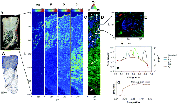
The authors regret that Fig. 3D, 5F and 7 were shown incorrectly in the original article. The correct versions of Fig. 3D, 5F and 7 are shown below.
image file: d1an90083h-f3.tif
Fig. 3 Full profile biopsy specimen of the wound of patient A, 3 days after a single application of the AgNP-containing dressing, analysed by SR-μXRF and SR-XANES. (A) Histological image of the tissue (adjacent slice). (B) Picture of the analysed slice. The scanned area locates in the white frame. (C) Maps of signal intensities (cps) of Ag (log scale), P, S and Cl (linear scales); pixel size 2 μm. (D) Ternary RGB plot of the same elemental maps. The arrows indicate: *stratum corneum; **stratum spinosum; ***stratum basale; ****fragments of epidermis extending in the granulation tissue; #granulation tissue; +reticular dermis; °hypodermis. (E) Zoom RGB map of the region located in the frame (independent acquisition, pixel size 2 μm), the arrow indicates the region selected for SR-μXANES analysis; (F) average SR-μXRF spectrum of the whole zoom map area; (G) average SR-μXANES spectrum in the selected region (dotted line) overlapped to its best LCF function (solid line).

image file: d1an90083h-f5.tif
Fig. 5 Full profile biopsy specimen of the wound of patient A, 12 days after a single application of the AgNP-containing dressing (complete healing), analysed by SR-μXRF. (A) Histological image of the tissue (adjacent slice), with zoom images of the regenerated epidermis (B) and an arterial blood vessel (C). (D) Picture of the analysed slice. The scanned area locates in the white frame. (E) Maps of signal intensities (cps) for Ag (logarithmic scale), P, S and Cl (linear scale); pixel size 2 μm. (F) Ternary RGB plot of the same elemental maps. The symbols indicate: *epidermis; **papillary dermis; ***reticular dermis; ****hypodermis; #arterial vessel. (G) and (H): two consecutive zoom maps of the region located in the white frame, independent acquisitions with pixel size 1 μm and 0.5 μm, respectively. (I) Average μXRF spectrum of the whole zoom map area H.

image file: d1an90083h-f7.tif
Fig. 7 Maps of Ag, Zn, P and C distribution in the skin profile from patient C after 15 days of treatment with repeated applications of the AgNP-containing dressing. Data obtained by LA-ICP-MS with pixel size 24 × 30 μm; signal intensity in cps with linear scale. The arrows indicate: *vein; **artery.

The Royal Society of Chemistry apologises for these errors and any consequent inconvenience to authors and readers.


This journal is © The Royal Society of Chemistry 2021
Click here to see how this site uses Cookies. View our privacy policy here.