
 Open Access Article
 This Open Access Article is licensed under a Creative Commons Attribution-Non Commercial 3.0 Unported Licence
New synthetic procedure for the antiviral sulfonate carbosilane dendrimer G2-S16 and its fluorescein-labelled derivative for biological studies†
Carlos Gutiérrez-Ulloa‡
ab,
Cornelia E. Peña-González‡abcd,
Andrea Barrios-Gumielabcd,
Rafael Ceña-Díezce,
M. Jesús Serramía-Loberae,
M. Ángeles Muñoz-Fernández *ce,
F. Javier de la Mataabcd,
Javier Sánchez-Nieves*abcd and
Rafael Gómez*abcd
*ce,
F. Javier de la Mataabcd,
Javier Sánchez-Nieves*abcd and
Rafael Gómez*abcd
aDpto. de Química Orgánica y Química Inorgánica, Universidad de Alcalá (UAH), Campus Universitario, E-28871 Alcalá de Henares, Madrid, Spain. E-mail: javier.sancheznieves@uah.es; rafael.gomez@uah.es
bInstituto de Investigación Química “Andrés M. del Río” (IQAR), Universidad de Alcalá (UAH), Spain
cNetworking Research Centre on Bioengineering, Biomaterials and Nanomedicine (CIBER-BBN), Spain
dInstituto Ramón y Cajal de Investigación Sanitaria, IRYCIS, Madrid, Spain
eImmunology Section, Head Immuno-Biology Molecular Laboratory, Gregorio Marañón University General Hospital (HGUGM), Gregorio Marañón Health Research Institute (IiSGM), Spanish HIV HGM BioBank, C/Dr Esquerdo 46, 28007 Madrid, Spain. E-mail: mmunoz.hgugm@gmail.com
First published on 27th May 2020
Abstract
The anionic carbosilane (CBS) dendrimer with sulfonate groups G2-S16 is a promising compound for the preparation of a microbicide gel to prevent HIV infection. However, until now its synthesis required aggressive conditions. Hence, a reliable synthetic procedure is very important to face GMP conditions and clinical trials. In this study, G2-S16 has been prepared by a new approach that involves the addition of an amine-terminated dendrimer to ethenesulfonyl fluoride (C2H3SO3F, ESF) and then transformation to the sulfonate dendrimer by treatment with a base. This strategy also makes feasible the synthesis of a labelled sulfonate dendrimer (G2-S16-FITC) to be used as a molecular probe for in vivo experiments. Interestingly, G2-S16-FITC enters into human peripheral blood mononuclear cells (PBMCs).
Introduction
HIV infection continues to be a very important global health problem. The main cause of this infection is unprotected sex, among other reasons because of cultural/social prejudices. The development of a topical microbicide should be very attractive to reduce this serious health risk. Therefore, multivalent macromolecules functionalized with anionic groups have been developed.The polyanionic surface of these systems interacts with virus capsid proteins or cell receptors blocking the infection. One type of these macromolecules are dendrimers, which are hyperbranched molecules of well-defined structure, monodisperse and with a multivalent surface due to their step-by-step growing.1 Regarding antiviral activity, anionic dendrimers have also been explored.2–5 The sulfonate derivative SPL7013 (polylysine framework) was shown to be a potent inhibitor against some HIV-1 strains, and reached phase III clinical trials.5 However, it was not successful because of the lack of activity against R5-HIV-1 strains and because it produced epithelial inflammation, which favour HIV-1 infection.6
We have deeply studied other family of dendrimers, carbosilane (CBS) dendrimers, which framework is formed by low polar C–Si bonds.7,8 Comparison of different anionic groups (phosphonate, carboxylate and sulfonate), topologies (dendrimers, dendrons, nanoparticles), dendrimer cores, and type of functionalization9–12 allowed us to select a second generation dendrimer containing sixteen sulfonate groups G2-S16 (Scheme 1)12 as a promising active principle for the development of a topical microbicide. This dendrimer showed broad antiviral spectrum even in the presence of semen,13 and its safety as topical vaginal microbicide in vitro and in vivo14–17 have been addressed. The gate for sexual transmission of HIV-1 is the cervical and foreskin epithelia and, hence, toxicology of dendrimer in this area is mandatory. For this analysis, the presence of a fluorophore moiety in the molecule is needed. However, the aggressive synthetic conditions to obtain G2-S16 (120 °C for 48 h) has made impossible the introduction of such fragment following the actual synthetic protocol.12 Moreover, this reaction presents reproducibility problems.
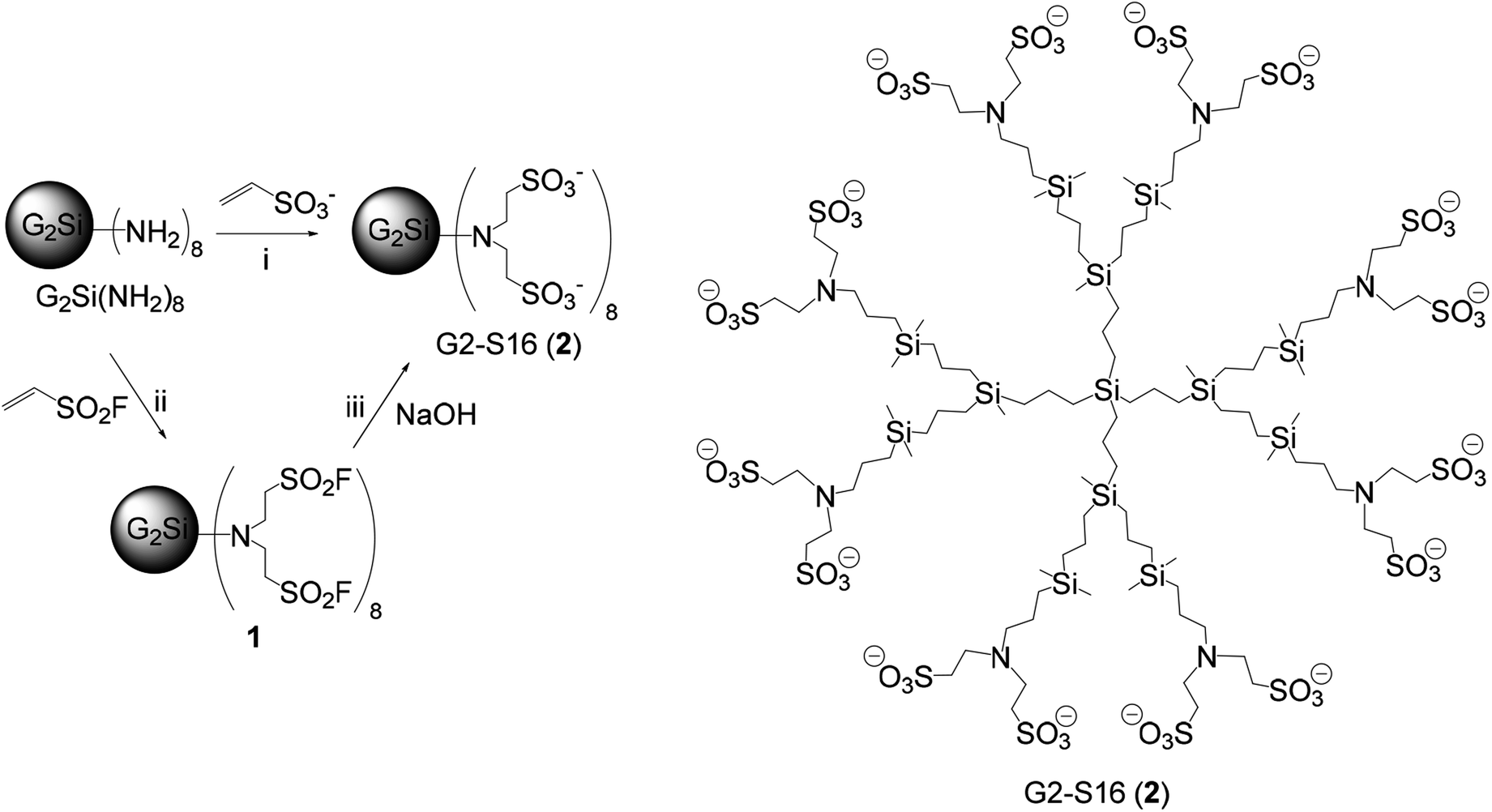 | ||
| Scheme 1 Synthesis and drawing of anionic sulfonate dendrimer G2-S16 (2) (sodium cations are omitted for clarity). (i) C2H3SO3Na, 120 °C, 48 h; (ii) ESF, R.T., 12 h; (iii) NaOH, R.T., 4 h. | ||
In view of these drawbacks, we explored the preparation of similar sulfonate dendrimers employing more selective reactions as thiol–ene addition reactions, which successfully led to anionic dendrimers with high yields in much softer reaction conditions.18 This methodology allowed also obtaining labelled dendrimers for in vitro and in vivo assays.19 Sulfonate dendrimers synthesized by these procedures were tested as antivirals although the analogous of G2-S16 (the so called G2-STE16) produced irritation and inflammation of vaginal epithelium, in contrast to the non-inflammatory behaviour of G2-S16.17 Hence, the necessity of finding an alternative procedure to the actual protocol for G2-S16 becomes of paramount importance. Additionally, the new procedure has to enable the labelling of this dendrimer for complete functional analysis through the design of in vivo experiments.
Nevertheless the problems described above, Michael-type addition of a sulfonate precursor to the dendrimer covered with amine groups G2Si(NH2)8 is the best synthetic choice. Ethenesulfonyl fluoride (C2H3SO3F, ESF) is a strong Michael acceptor20 and the –SO2F moiety presents low reactivity and high stability, making this S–F bond available for subsequent controlled reactivity.21 Thus, these facts make this compound also attractive as building block for dendrimer chemistry.22 Herein we report that the use of ESF as reagent has given us the access to G2-S16 through a less aggressive protocol and to the labelling of this type of compound with a fluorophore (named as G2-S16-FITC). Preliminary toxicity and cell internalization assays with G2-S16-FITC has been carried out.
Results and discussion
Synthesis of anionic dendrimer G2-S16
Sulfonate dendrimer G2-S16 was previously prepared in our laboratories by the reaction of an amine dendrimer (G2Si(NH2)8) with excess vinyl sulfonate at 120 °C for 48 h (Scheme 1). Due to the notable different solubilities of the starting derivatives and harsh reaction conditions, total addition of two vinyl sulfonate equivalents to the terminal amine function is difficult to quantify, since mono- and di-substitution present very similar NMR data. Hence, G2-S16 can present incompletely functionalized branches on its structure.12 Additionally, this procedure lacks of reproducibility for amounts over 200 mg of compound, observing also a remarkable yield reduction (from 75% to yields below 20%).The first step of the new synthetic approach described in this work is the reaction of the amine dendrimer G2Si(NH2)8 with C2H3SO3F (ESF) in THF at room temperature leading to the fluorosulfone derivative G2Si(SO3F)16 (1, Scheme 1 and Fig. S1†) as pale yellow oil in high yields (90%). The slight excess of ESF is removed under vacuum, making purification very simple. Further treatment of 1 with excess NaOH at R.T. led to the desired sulfonate dendrimer G2-S16 (G2Si(SO3−)16, 2, Scheme 1), which was obtained as white solid in high yield.
NMR spectroscopy confirmed the formation of dendrimer 1. The starting CH2N methylene in compound G2Si(NH2)8 was shifted from δ 2.61 (1H NMR) and δ 45.5 (13C NMR) to δ 2.51 (1H NMR) and δ 57.5 (13C NMR) in compound 1. Additionally, the new chain NCH2CH2S were located at δ 3.09 (1H NMR) and δ 47.8 (13C NMR) (NCH2) and at δ 3.54 (1H NMR) and δ 49.3 (13C NMR) (CH2S). Moreover, for this last resonance was observed the coupling with the fluorine atom. The fluorine atom was detected at δ 54.1 in 19F NMR spectroscopy. Regarding compound G2-S16 (2), analytical and characterization data correspond with those described previously (see experimental section).12
Synthesis of labelled anionic dendrimer G2-S16-FITC
To introduce a chromophore probe (fluorescein) to the structure of the anionic sulfonate dendrimer, first, one amine group of dendrimer G2Si(NH2)8 was protected with trifluoroacetic anhydride to give G2Si(NHTFA)(NH2)7 (3, Scheme 2 and Fig. 1). It is important to note that this process of heterofunctionalization cannot lead to monodisperse dendrimers, although in the 1H NMR spectra integral values fit well to monosubstitution. This situation is inherent to any heterofunctionalization of spherical dendrimers containing the same type of groups. This is due to the fact that the steric or electronic situation of the starting functional groups is not modified by the change of only one group, mainly for groups located further away from the modified unit. Next, remaining amine groups were reacted with ESF, as described for 1, forming G2Si(NHTFA)(SO3F)14 (4, Fig. 1). Deprotection of the amine group and generation of anionic sulfonate groups were done simultaneously by addition of excess NaOH, leading to compound G2Si(NH2)(SO3−)14 (5, Fig. 1). The solubility of 5 was very different from its parent dendrimer 4, moving from organic solvents like halogenated to water. This dendrimer was used without further purification for next transformation. Finally, the reaction of 5 with FITC afforded the labelled anionic dendrimer G2Si(NH-FITC)(SO3−)14 (G2-S16-FITC, 6, Fig. 1). Kaiser's test was very useful to check the success of this reaction, being negative due to the transformation of the primary amine function. After purification by ultrafiltration, dendrimer 6 was obtained as orange solid, soluble in water and with good yield. | ||
| Scheme 2 Synthesis of the anionic sulfonate dendrimer labelled with fluorescein G2-S16-FITC (6). (i) Trifluoroacetic anhydride (TFA), K2CO3; (ii) ESF, R.T., 12 h; (iii) NaOH, R.T., 4 h; (iv) FITC, R.T., 12 h. | ||
 | ||
| Fig. 1 Structure of dendrimers 3–6. The circle highlights the additional function for labelling of the dendrimer. | ||
Compounds 3–6 were characterized by NMR and elemental analysis (Fig. S3–S7†). The main data confirming these transformations are discussed next. The protected methylene group –CH2NHC(O)– in dendrimer 3 was observed in 1H NMR spectroscopy at δ 3.26 and in 13C NMR spectroscopy at δ 42.8, remaining the other resonances of the methylene groups –CH2NH2 at δ 2.60 (1H NMR) and δ 45.5 (13C NMR). The carbonyl group of 3 was detected at δ 165.3 in the 13C NMR spectrum and the fluorine nuclei of the CF3 moiety at δ −75.8 in 19F NMR spectroscopy. In compound 4, modification of the outer amine groups with ESF gave, in NMR spectroscopy, resonances for the new peripheral groups similar to those described for compound 1. The methylene group of the –CH2NHC(O) moiety was overlapped with resonances of peripheral –CH2SO2 groups at δ 3.54 (1H NMR) and was observed at δ 42.8 in the 13C NMR spectrum. In dendrimer 5, broad resonances were observed in the 1H NMR spectrum (D2O) due to the hydrophobic character of the CBS framework. Both types of outermost methylene groups of –CH2CH2SO3− were overlapped at δ 2.94 and the internal methylene CH2N was observed at δ 2.46. Qualitative Kaiser's test was positive, confirming the presence of primary amine functions. Compound 5 was not purified and was used as it for transformation to G2-S16-FITC (6). As commented above, the qualitative Kaiser's test for 6 was negative, meaning absence of primary amine functions. 1H NMR spectrum showed resonances expected for CBS framework and external –CH2CH2SO3− groups, together with new resonances belonging to fluorescein unity (δ 6.5–8.0). These signals were very broad, again due to the hydrophobic character of this fragment. DOSY 1H NMR spectroscopy was also helpful, since one diffusion coefficient was observed for all these resonances (Fig. S5†). Regarding 13C NMR spectrum, the new resonances belonging of the fluorescein group were observed (δ 100–150, Fig. S6†). As in the 1H NMR spectrum, resonances for CBS framework and external –CH2CH2SO3− groups were similar to those of compound G2-S16 (2). IR spectroscopy (Fig. S8†) showed the disappearance of the stretching band corresponding to the isocyanate group from fluorescein (at ca. 2000 cm−1). On the other hand, a stretching band corresponding to the carbonyl group belonging to the fluorescein fragment was detected (at ca. 1750 cm−1). Moreover, in the UV spectrum of this dendrimer (Fig. S9†), a broad band centered at about 500 nm was observed due to the presence of fluorescein.23
Toxicity assays
Peripheral blood mononuclear cells (PBMC) belong to the immune system, playing a key role to fight bacterial and viral infections. These cells are target for HIV and have been chosen for toxicity and cellular uptake assays of the anionic dendrimers here described. The biocompatibility of G2-S16 (2) and G2-S16-FITC (6) in PBMCs was evaluated by an MTT assay (Fig. S10†). G2-S16 and G2-S16-FITC dendrimers concentrations with viability above 80% were regarded as non-toxic (in comparison with control). G2-S16 was not toxic at 50 μM. This value is the same than previously reported, meaning that the synthetic procedure does not alter toxicity.15 Presence of the labelling moiety reduces viability, being G2-S16-FITC not toxic at 2 μM, and close to 5 μM.Cellular uptake assays
To determine whether G2-S16-FITC enter into human PBMCs and can be visualized in a biological environment, confocal microscopy assays were performed (Fig. 2). Non-toxic concentration (2 μM) was used to treat the PBMCs at different times. This experiment was analysed after 2 h and 24 h of treatment. Cellular uptake of G2-S16-FITC by PBMCs was observed at both times, although clearly higher signal was detected after 24 h. As can be seen in Fig. 2 (arrows a, b, c), G2-S16-FITC was able to penetrate in different types of PBMC as macrophages, CD4 T and CD8 T lymphocytes. | ||
| Fig. 2 Confocal microscopy images of ability of the polyanionic CBS dendrimer G2-S16-FITC (6) to enter into PBMCs. (I) G2-S16-FITC dendrimer co-located with nuclei of treated cells labelled with DAPI; (II) anti-human CD4-APC co-located with G2-S16-FITC dendrimer; (III) represents co-localization of G2-S16-FITC dendrimer, CD4 and nuclei of treated cells labelled with DAPI. Arrows point different cell types (a) macrophage; (b) CD4 T lymphocyte; and (c) CD8 T lymphocyte. | ||
Conclusions
In conclusion, ESF is an adequate reagent for the preparation of the antiviral anionic sulfonate dendrimer G2-S16, removing an important hurdle in the synthesis of this derivative. With this reagent, G2-S16 can be obtained in much softer conditions with a reproducible protocol for higher amounts than the previously published procedure. Moreover, with the use of this reagent and employing protection-deprotection strategy of an amine function, a labelled derivative G2-S16-FITC (6) with a chromophore has been also affordable.The use of labelled G2-S16-FITC dendrimer for internalization assays with PBMC justifies its synthesis. Analysis by confocal clearly showed that different human cells as macrophages, CD4 T and CD8 T lymphocytes uptake our G2-S16 dendrimer. Hence, the new anionic dendrimer G2-S16-FITC here designed can be proposed for further in vitro and in vivo assays to test biodistribution of this promising antiviral dendrimer. This will help to prove the mechanism of action of this compound facilitating its use as active principle of a topical microbicide that could be tested in phase I of clinical trials.
Conflicts of interest
There are no conflicts of interest to declare.Acknowledgements
This work has been supported by grants from CTQ2017-86224-P (MINECO), PCI2019-103715 (M-ERA.NET Call 2018, European Commission), SBPLY/17/180501/000358 (JCCM), Consortiums NANODENDMED II-CM ref B2017/BMD-3703 and IMMUNOTHERCAN-CM B2017/BMD-3733 (CAM) to UAH. CIBER-BBN as an initiative funded by VI National R-D-i Plan 2008–2011, Iniciativa Ingenio 2010, Consolider Program, CIBER Actions and financed by the ISC III with assistance from the European Regional Development Fund (FEDER). A. B.-G. acknowledges MINECO for a fellowship. This work has been (partially) supported by grants from RD16/0025/0019, projects as part of Strategy Action in Health, Plan Nacional de Investigación Científica, Desarrollo e Innovación Tecnológica (2013–2016) and cofinanced by ISC III (Subdirección General de Evaluación) and FEDER, RETIC PT17/0015/0042, Fondo de Investigacion Sanitaria (FIS) (grant number PI16/01863) and EPIICAL project. This article/publication is based upon work from COST Action CA 17140 “Cancer Nanomedicine from the Bench to the Bedside” supported by COST (European Cooperation in Science and Technology).References
- A. J. Perisé-Barrios, D. Sepúlveda-Crespo, D. Shcharbin, B. Rasines, R. Gómez, G. Kajnert-Maculewicz, M. Bryszewska, F. J. de la Mata and M. A. Muñoz-Fernández, RSC Nanoscience & Nanotechnology, Soft Nanoparticles for Biomedical Applications, 2014, vol. 34, pp. 246–279 Search PubMed.
- M. Witvrouw, V. Fikkert, W. Pluymers, B. Matthews, K. Mardel, D. Schols, J. Raff, Z. Debyser, E. D. Clercq, G. Holan and C. Pannecouque, Mol. Pharmacol., 2000, 58, 1100–1108 CrossRef CAS.
- J. Rojo and R. Delgado, Antiinfect. Agents Med. Chem., 2007, 6, 151–174 CrossRef CAS.
- D. Tyssen, S. A. Henderson, A. Johnson, J. Sterjovski, K. Moore, J. La, M. Zanin, S. Sonza, P. Karellas, M. P. Giannis, G. Krippner, S. Wesselingh, T. McCarthy, P. R. Gorry, P. A. Ramsland, R. Cone, J. R. A. Paull, G. R. Lewis and G. Tachedjian, PLoS One, 2010, 5, e12309 CrossRef PubMed.
- R. Rupp, S. L. Rosenthal and L. R. Stanberry, Int. J. Nanomed., 2007, 2, 561–566 CAS.
- A. B. Moscicki, R. Kaul, Y. Ma, M. E. Scott, I. I. Daud, E. A. Bukusi, S. Shiboski, A. Rebbapragada, S. Huibner and C. R. Cohen, J. Acquired Immune Defic. Syndr., 2012, 59, 134–140 CrossRef CAS PubMed.
- A. W. van der Made and P. W. N. M. van Leeuwen, J. Chem. Soc., Chem. Commun., 1992, 1400–1401 RSC.
- P. Ortega, J. Sánchez-Nieves, J. Cano, R. Gómez and F. J. de la Mata, Dendrimer Chemistry: Synthetic Approaches Towards Complex Architectures, The Royal Society of Chemistry, 2020, vol. 5, pp. 120–151 Search PubMed.
- E. Arnáiz, E. Vacas-Córdoba, M. Galán, M. Pion, R. Gómez, M. A. Muñoz-Fernández and F. J. de la Mata, J. Polym. Sci., Part A: Polym. Chem., 2014, 52, 1099–1112 CrossRef.
- C. Guerrero-Beltrán, R. Ceña-Díez, D. Sepúlveda-Crespo, F. J. de la Mata, R. Gómez, M. Leal, M. A. Muñoz-Fernández and J. L. Jiménez, Nanoscale, 2017, 9, 17263–17273 RSC.
- C. E. Peña-González, P. García-Broncano, M. F. Ottaviani, M. Cangiotti, A. Fattori, M. Hierro-Oliva, M. L. González-Martín, J. Pérez-Serrano, R. Gómez, M. A. Muñoz-Fernández, J. Sánchez-Nieves and F. J. de la Mata, Chem.–Eur. J., 2016, 22, 2987–2999 CrossRef PubMed.
- B. Rasines, J. Sánchez-Nieves, M. Maiolo, M. Maly, L. Chonco, J. L. Jiménez, M. A. Muñoz-Fernández, F. J. de la Mata and R. Gómez, Dalton Trans., 2012, 41, 12733–12748 RSC.
- R. Ceña-Díez, P. García-Broncano, F. J. de la Mata, R. Gómez and M. A. Muñoz-Fernández, Int. J. Nanomed., 2016, 11, 2443–2450 CrossRef PubMed.
- E. Vacas-Córdoba, E. Arnáiz, M. Relloso, C. Sánchez-Torres, F. García, L. Pérez-Alvarez, R. Gómez, F. J. de la Mata, M. Pion and M. A. Muñoz-Fernández, AIDS, 2013, 27, 1219–1229 CrossRef PubMed.
- L. Chonco, M. Pion, E. Vacas, B. Rasines, M. Maly, M. J. Serramía, L. López-Fernádez, F. J. de la Mata, S. Álvarez, R. Gómez and M. A. Muñoz-Fernández, J. Control. Release, 2012, 161, 949–958 CrossRef CAS PubMed.
- D. Sepúlveda-Crespo, M. J. Serramía, A. M. Tager, V. Vrbanac, R. Gómez, F. J. de la Mata, J. L. Jiménez and M. A. Muñoz-Fernández, Nanomedicine, 2015, 11, 1299–1308 CrossRef PubMed.
- R. Ceña-Díez, P. García-Broncano, F. J. de la Mata, R. Gómez, S. Resino and M. Á. Muñoz-Fernández, Nanoscale, 2017, 9, 9732–9742 RSC.
- M. Galán, J. S. Rodríguez, J. L. Jiménez, M. Relloso, M. Maly, F. J. de la Mata, M. A. Muñoz-Fernández and R. Gómez, Org. Biomol. Chem., 2014, 12, 3222–3237 RSC.
- M. Galán, E. Fuentes-Paniagua, F. J. de la Mata and R. Gómez, Organometallics, 2014, 33, 3977–3989 CrossRef.
- R. D. B. J. Krutak, W. H. Moore and J. A. Hyatt, J. Org. Chem., 1979, 44, 3847–3858 CrossRef.
- J. Dong, L. Krasnova, M. G. Finn and K. B. Sharpless, Angew. Chem., Int. Ed., 2014, 53, 9430–9448 CrossRef CAS PubMed.
- X. Zhang, B. Moku, J. Leng, K. P. Rakesh and H.-L. Qin, Eur. J. Org. Chem., 2019, 1763–1769 CrossRef CAS.
- E. Fuentes-Paniagua, M. J. Serramía, J. Sánchez-Nieves, S. Álvarez, M. A. Muñoz-Fernández, R. Gómez and F. J. de la Mata, Eur. Polym. J., 2015, 71, 61–72 CrossRef CAS.
Footnotes |
| † Electronic supplementary information (ESI) available: Experimental section, drawing of dendrimer structures and selected spectra and confocal image. See DOI: 10.1039/d0ra03448g |
| ‡ Both authors contribute equally to this work. |
| This journal is © The Royal Society of Chemistry 2020 |