
 Open Access Article
 This Open Access Article is licensed under a Creative Commons Attribution-Non Commercial 3.0 Unported Licence
Forensic float glass fragment analysis using single-pulse laser ablation inductively coupled plasma time of flight mass spectrometry
Pascal
Becker
 a,
Christoph
Neff
a,
Sabine
Hess
b,
Peter
Weis
c and
Detlef
Günther
*a
a,
Christoph
Neff
a,
Sabine
Hess
b,
Peter
Weis
c and
Detlef
Günther
*a
aDepartment of Chemistry and Applied Biosciences, Laboratory of Inorganic Chemistry, ETH Zurich, Vladimir-Prelog-Weg 1, 8093 Zürich, Switzerland. E-mail: detlef.guenther@sl.ethz.ch
bZurich Forensic Science Institute, Zeughausstrasse 11, 8004 Zurich, Switzerland
cFederal Criminal Police Office, Forensic Science Institute, KT42 Inorganic Materials and Micro Traces, Coatings, Äppelallee 45, 65203 Wiesbaden, Germany
First published on 23rd July 2020
Abstract
The most discriminating method that is currently applied in routine forensic elemental analysis of glass is laser ablation in combination with quadrupole or sector field based inductively coupled plasma mass spectrometry (LA-ICPMS) following the standard method ASTM E2927-16E1. Due to the sequential measurement method, sample size constraints of approximately 400 μm × 200 μm × 100 μm with six individual measurements have been reported to provide optimum parameters for glass discrimination. The necessary sample size is given by the spot size, number of laser pulses per replicate and number of replicate measurements. In this study, a single-pulse laser ablation inductively coupled plasma time of flight mass spectrometry (Single-Pulse LA-ICP-TOFMS) method was developed and applied for the matching and mismatching of forensic float glass evidence. 110 laser pulses with an ablation spot diameter of 90 μm were applied to float glass fragments of a size below 400 μm in diameter. 18 elements were quantified from each individual laser pulse in order to compare the corresponding concentrations between various glass fragments. In contrast to previous work, a modified 5-sigma criterion was used to successfully match fragments from the same source and mismatch fragments from differing sources. The data set generated within this study demonstrates new capabilities when using quasi simultaneous signal detection and reduced the necessary sample volume from 400 μm × 200 μm × 100 μm to 100 μm × 100 μm × 33 μm, which corresponds to a reduction in sample material from 20 μg to 0.8 μg. The proof of concept is shown for the application of a single-pulse based method for glass fragment analysis in a forensic context, which would allow the measurement of smaller fragments than previously possible.
Introduction
Float glass fragments are common pieces of evidence encountered in many different crimes such as vandalism, traffic accidents or burglaries.1,2 Fragments of the size of 0.1 mm to several mm in diameter are often recovered in the criminals' clothing or shoes after breaking a window.3 Determining that a glass fragment found within a suspect's clothing may originate from the broken glass found at the crime scene can be a strong evidence to link the suspect to the crime scene.4,5 The most commonly analysed property of glass fragments for forensic analysis is the refractive index (RI).6 Refractive indices of float glass samples range from 1.51 to 1.53, which results in a limited discrimination power with the currently available instrumental methods.3 Elemental analysis of glass fragments allows for comparison of multiple variables, resulting in higher discrimination power compared to RI analysis.4,5 Elemental analysis techniques that have been employed for forensic glass fragment analysis include X-ray fluorescence (XRF), Laser Induced Breakdown Spectrometry (LIBS), inductively coupled plasma optical emission spectrometry (ICPOES) and inductively coupled plasma mass spectrometry (ICPMS).7,8 Out of these methods, ICPMS coupled to laser ablation (LA-ICPMS) has shown the highest discrimination power while simultaneously requiring low sample amounts.9Analysis of glass samples using LA-ICPMS has been studied since the 1990's but gained further popularity in a forensic context due to the “natural isotopes and trace elements in criminalistics and environmental forensics” (NITE-CRIME) initiative, with the goal of using trace-elements and isotope ratios in forensic sciences.2,10–12 As a result of this project, a standard method for LA-ICPMS has been developed which is now used routinely in forensic institutes around the world.2,13 The method includes quantification of approximately 18 elements (typically Li, Na, Mg, Al, K, Ca, Ti, Mn, Fe, Rb, Sr, Zr, Ba, La, Ce, Nd, Hf and Pb) and the comparison of their concentrations. The standard method requires at least 3, optimally 6, measurements of each glass fragment, using an ablation spot size of 50–100 μm in diameter. A constant ablation time of 50–60 seconds at a laser repetition rate of 10 Hz is applied. This results in a required sample volume of approximately 400 μm × 200 μm × 100 μm per glass fragment. However, the majority of samples retrieved are below that size, making it impossible to measure them according to the standard method.
Additionally, due to the low number of measurements, many statistical models fail in dealing with the multidimensional data acquired by quantifying multiple elements due to the so called “Curse of Dimensionality”, described by Bellman in 1961.14 It describes the increase of distance between data points in “nearest neighbour” statistical approaches when the dimensionality of the data increases. This results in data points being too far away from each other to be clustered when the sample size is too low. As such the amount of required data points often increases exponentially with every additional dimension, which is a general challenge in applied analytical methods. Therefore, there is an interest in methods that can provide similar discrimination power with less sample material and a larger number of measurements.
Quadrupole, sector field and multi-collector instruments rely on sequential measurements to cover the entire mass spectrum. This results in a spectral acquisition time that is dependent on the amount of measured isotopes. An increase in the number of isotopes will increase the measurement time per spectral acquisition, resulting in longer ablation times and increasing sample amount requirements. ICP-TOFMS instruments allow for quasi-simultaneous measurements of the entire mass range, which opens the possibility to quantify more elements at once.15 This allows for further discrimination factors between samples and could possibly increase the discrimination power of the method. When the standard method was developed, first generation ICP-TOFMS instruments showed rather low sensitivity and were not suited for forensic glass examinations. Recent instrumental developments have allowed ICP-TOFMS-instruments to reach sensitivities which are comparable to quadrupole instruments and were therefore reconsidered to extend the range of applications.16,17
Single-pulse laser ablation was first described for ICP-TOFMS in 2001 by Leach et al.18 by resolving the signal of each individual laser pulse in the transient signal. Every laser pulse was quantified individually, increasing the amount of information per ablated material significantly. This increases the amount of measurements from one for 600 laser pulses to one for every laser pulse. With the development of ICP Time-of-Flight Mass Spectrometry (ICP-TOFMS), it is possible to measure the entire mass spectrum at a frequency of more than 30![[thin space (1/6-em)]](https://www.rsc.org/images/entities/char_2009.gif) 000 Hz.19 The quasi-simultaneous detection of all ICPMS accessible elements results in a representative measurement of the ion cloud for every data point. A fast washout of the ablated aerosol is required in Single-Pulse-LA-ICPMS to increase the signal-to-noise ratio and thus lower the limits of detection. In this work, a fast aerosol washout is achieved using a low dispersion tube cell as described by Wang et al. in combination with ICP-TOFMS.20 With the use of this low-dispersion LA cell, it is possible to measure 99% of the total signal within 5–100 ms (5–90 μm spot diameter).
000 Hz.19 The quasi-simultaneous detection of all ICPMS accessible elements results in a representative measurement of the ion cloud for every data point. A fast washout of the ablated aerosol is required in Single-Pulse-LA-ICPMS to increase the signal-to-noise ratio and thus lower the limits of detection. In this work, a fast aerosol washout is achieved using a low dispersion tube cell as described by Wang et al. in combination with ICP-TOFMS.20 With the use of this low-dispersion LA cell, it is possible to measure 99% of the total signal within 5–100 ms (5–90 μm spot diameter).
In this study, a reduced sample consumption method was developed and evaluated to match forensic glass fragments by means of Single-Pulse-LA-ICP-TOFMS. It relies on the fast and “quasi” simultaneous detection of single laser ablation events using an ICP-TOFMS and results in a reduction of required sample amount. This sampling strategy allows the analysis of significantly smaller glass fragments compared to the standard method currently used. Additionally, it increases the amount of individual measurements which opens the possibilities to utilize different statistical methods.
The method proposed in this study was used to quantify a wide range of elements covering concentrations from 1 to 100000 μg g−1. The major goal was focused on matching glass fragments from the same source, while mismatching fragments from different sources through comparison of the element concentrations.
Experimental
Instrumentation
All experiments were carried out with an ArF excimer laser (193 nm, GeoLas C, Lambda Physik, Goettingen, Germany) coupled to an ICP-TOFMS system (icpTOF2R, TOFWERK AG, Thun, Switzerland). Argon gas (99.996% PanGas AG, Dagmersellen, Switzerland) was used to sustain the plasma. Operating conditions are given in Table 1.| Laser ablation | |
| Repetition rate | 2 Hz |
| Laser fluence | ∼15 J cm−2 |
| Spot size | 90 μm |
| Carrier gas (Ar) | 0.75–0.85 L min−1 |
| Carrier gas (He) | 1.2–1.3 L min−1 |
| Number of pulses per spot | 110 |
|
|
| ICPMS | |
| RF power | 1550 W |
| Cooling gas (Ar) | 16 L min−1 |
| Auxiliary gas (Ar) | 0.8 L min−1 |
| Reaction cell (H2) | 2 ml min−1 |
| Sampling depth | 5.5–6 mm |
| Time resolution | 10 ms |
An in-house built modified low dispersion ablation cell, based on the tube cell design,21 was used with mixed argon and helium (99.999%, PanGas AG, Dagmersellen, Switzerland) gas flows to carry the aerosol into the ICP.17,20
The ICP-TOFMS instrument was operated in reaction cell mode, using hydrogen (99.9999%, PanGas AG, Dagmersellen, Switzerland) as reaction gas.22
Samples
Larger float glass fragments (about 2 cm in diameter), which were seized from 10 different criminal investigations within the greater Zurich area as reference materials, were provided by the Zurich Forensic Science Institute, Switzerland. Each fragment was placed in an individual plastic bag and further broken with a pair of pliers to obtain smaller fragments with a size of 0.1–0.5 mm in diameter. For each of the 10 glass sources, 10 smaller fragments were randomly selected and placed on double sided Scotch tape, which was attached to a microscope slide.Analysis procedure
The 10 smaller fragments were mounted on a 3D-movable stage at a time along with the standard reference materials FGS 1, FGS 2, NIST SRM 610 and NIST SRM 612. NIST SRM 610 was used for tuning of the instrument for high sensitivity of 238U, a ratio of 1.0–1.1, and an oxide ratio
  The float glass standard FGS 2 was used as external standard, FGS 1 was used as a quality control sample and NIST SRM 612 served as a standard to quantify elements not certified in FGS 2.23 The measurement series is schematized in Fig. 1, where 110 individually recorded laser pulses represent one measurement.
 | ||
| Fig. 1 Schematic of the measurement series used in this work. A symmetric approach was used to correct for instrumental drift. Samples refer to individual fragments of the same origin while x refers to the amount of times each sample was measured. | ||
Data analysis
The signals of the first 10 laser pulses of every measurement were not considered for data analysis due to surface effects and surface impurities affecting the quantification. The signals of the remaining 100 laser pulses were individually integrated and quantification was carried out according to Longerich et al.,24 where each integrated signal of one laser pulse was considered to be equal to one measurement. 28Si was chosen as internal standard, assuming a SiO2-concentration of 72% in the unknown glass fragments as suggested by the official standard procedure.13 Afterwards, the median of 100 laser pulses was taken for further calculations. The median was chosen over the mean due to more robustness towards outliers. This was possible because the integrated signals showed a normal distribution, which was confirmed with a Shapiro–Wilk test,25 and the amount of data points was sufficient to show a distribution. Calculated concentrations that were lower than 3.3 times the limit of detection (LOD) were set to 0 for further statistical discrimination procedures. LODs were calculated based on Poisson counting statistics for a “well-known” blank and a 95% confidence level, based on Currie.26Glass fragment matching
The matching of glass fragments was carried out according to the currently established standard method by Dorn et al.5 Glass fragments were compared pairwise to each other for each element individually and the concentration of an element was compared for both fragments. The higher of the two determined concentrations was defined as the base concentration and the matching interval was calculated. This was carried out by subtracting the relative standard deviation (RSD) multiplied by 5 and defined as the threshold concentration. If the element concentration of the other fragment lied within the interval of threshold concentration to base concentration, the two fragments were considered “a match” for that element.4,5 If the two fragments were matches for every element measured, they are considered indistinguishable. If the fragments differed in one or more elements, they were considered to be of different origins.Results and discussion
Development of a single-pulse based method
With the presented method, a sample area of roughly 100 μm × 100 μm and a sample depth of 33 μm are required, assuming an ablation rate of 0.3 μm per pulse.27 At a density of 2.5 g cm−3 for float glass,28 this corresponds to approximately 0.8 μg of material. The standard method requires a volume of 400 μm × 200 μm × 100 μm, which would correspond to approximately 20 μg of material. Despite the significant reduction in the mass ablated, all sample discriminating elements of float glass, except Li, can be quantified.13 This is shown by the limits of detection (LOD) for the single-pulse based method (Fig. 2). However, the sensitivity of ICP-TOFMS is not sufficient to determine Li as a trace element, as observed by Borovinskaya et al. previously.16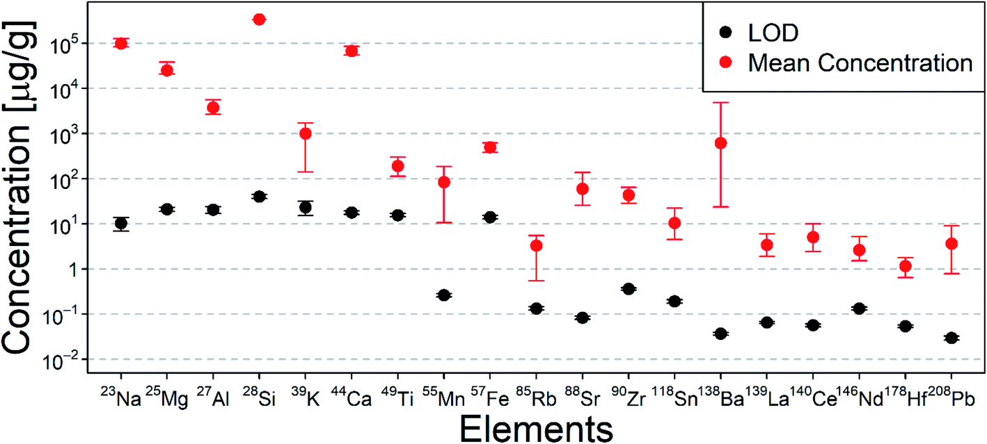 | ||
| Fig. 2 Limits of detection (LOD) for single-pulse laser ablation compared to the concentration ranges determined in all the investigated glass fragments. Black dots represent average LODs based on FGS 2 measurements. Red dots and bars signify the average, lowest and highest individual concentrations in all measured glass fragments. | ||
Measuring FGS 2, the LODs were calculated and compared to the mean concentrations measured in the float glass fragments. It is worth noting, that a notch filter was applied at m/z 23 and 28 to prevent saturation of the detector from Na and Si, which are the main elements in such glasses. This results in higher LODs for low mass elements than one would expect, since it also affects nearby masses.
Measurements consisting of 110 pulses per sampling position were carried out and 100 individual pulses were quantified as a larger number of data points resulted in better reproducibility of the quantitative results. The 10 pulses at the beginning of the analysis were not considered to avoid analysis being affected by surface effects, caused by impurities, crater formation and uneven surfaces (Fig. 3). Furthermore, the uneven shape of glass fragments is suspected to cause local gas turbulences within the ablation cell which could result in incomplete or non-stoichiometric aerosol transport. This becomes evident since a change in signal duration and signal shape was observed for certain samples with uneven surfaces (Fig. 4).
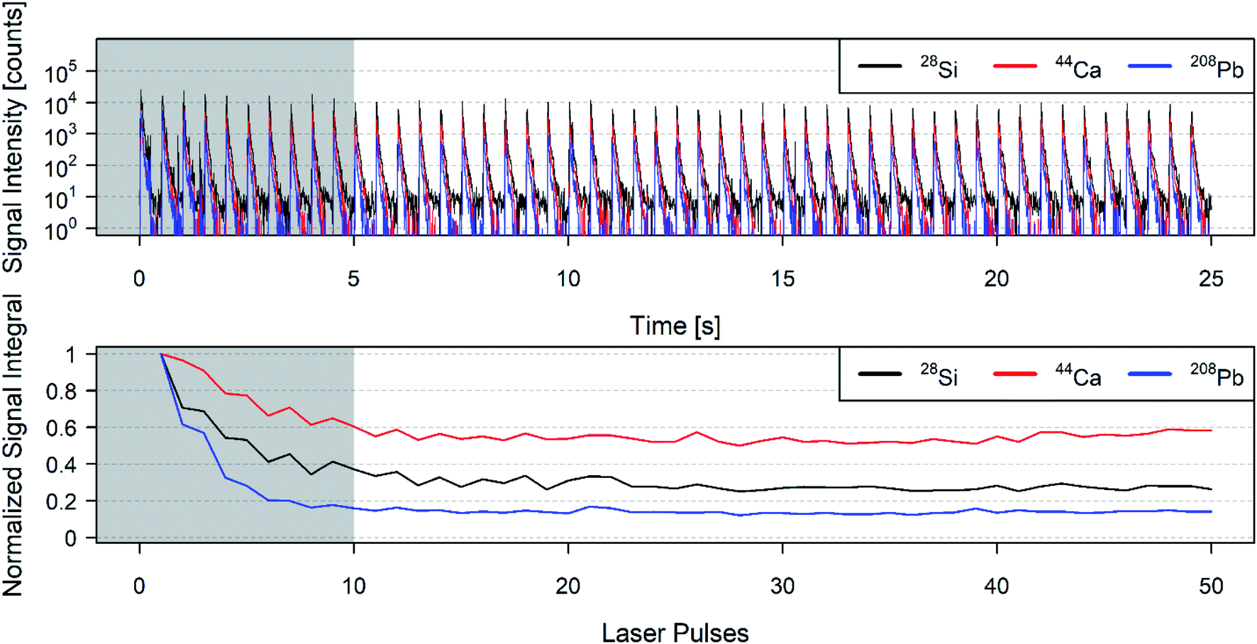 | ||
| Fig. 3 Transient signal of the first 50 laser pulses for 28Si, 44Ca and 208Pb (top) for a 90 μm ablation spot and corresponding individual normalized signal integrals normalized to maximum of a NIST612 measurement (bottom). A clear difference can be seen between the first 10 pulses, signified by the grey area, and the next 40 pulses with respect to signal intensity and signal ratio. | ||
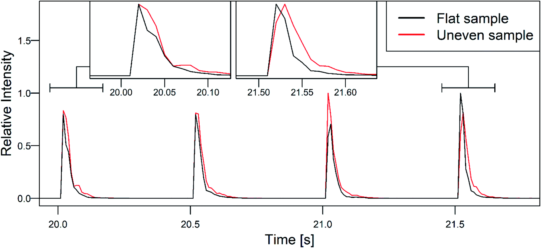 | ||
| Fig. 4 Difference in 28Si signal shape and duration for a flat (external standard) and an uneven sample (glass fragment) for 4 representative laser pulses. Signals are normalized to the maximum signal of that transient signal. An increase in washout time is observed, as well as a more irregular peak shape. A close up of 2 selected peaks is shown at the top. | ||
Matching of glass fragments
The proof of concept study included 10 different glass fragments each from a total of 10 different float glass sources. All fragments were measured using the above described procedure and the required elements for float glass provenance were quantified (except Li). Due to the limited sample size, it was not possible to test other statistical methods. Therefore, a 5-sigma criterion with fixed RSDs was chosen as a first proof of concept.RSDs for the quality control (FGS1 in this case) measurements were calculated by taking the RSDs over all medians for each element. RSDs for samples were determined by calculating the RSDs of the medians of all 10 fragments per glass source and taking the mean RSDs over all 10 glass sources for every element. Optimal RSDs were determined by optimizing the value for every element for the lowest achievable amount of false positives, identifying 2 different fragments as a match, and negatives, identifying 2 equal fragments as a mismatch (see Table 2).
| Element | RSD quality control | RSD samples | RSD optimal | RSD standard method |
|---|---|---|---|---|
| 23Na | 0.04 | 0.05 | 0.05 | 0.03 |
| 25Mg | 0.04 | 0.04 | 0.05 | 0.03 |
| 27Al | 0.06 | 0.06 | 0.06 | 0.03 |
| 39K | 0.07 | 0.07 | 0.09 | 0.03 |
| 44Ca | 0.04 | 0.04 | 0.04 | 0.03 |
| 49Ti | 0.08 | 0.04 | 0.04 | 0.04 |
| 55Mn | 0.03 | 0.04 | 0.05 | 0.04 |
| 57Fe | 0.04 | 0.04 | 0.05 | 0.03 |
| 85Rb | 0.05 | 0.06 | 0.07 | 0.04 |
| 88Sr | 0.05 | 0.04 | 0.03 | 0.04 |
| 90Zr | 0.05 | 0.05 | 0.05 | 0.04 |
| 118Sn | 0.05 | 0.09 | 0.07 | 0.03 |
| 138Ba | 0.06 | 0.08 | 0.09 | 0.04 |
| 139La | 0.05 | 0.05 | 0.04 | 0.04 |
| 140Ce | 0.05 | 0.06 | 0.06 | 0.04 |
| 146Nd | 0.06 | 0.06 | 0.05 | 0.05 |
| 178Hf | 0.05 | 0.08 | 0.07 | 0.03 |
| 208Pb | 0.05 | 0.13 | 0.12 | 0.05 |
The quantification of the quality control standard showed an increase in the RSD between the individual laser pulses compared to the standard method. This increase in the RSD when comparing the single-pulse method to the commonly applied low spatial resolution method found in the standard method was expected due to the approximately 25 times lower amount of sampled material entering the plasma and the significantly shorter integration times.
The RSDs for samples were found to be higher than the RSDs for the quality control. This can be partially explained by a higher inhomogeneity of trace elements in real samples in comparison to specifically produced glass standards. Another possible explanation is fractionation induced by gas turbulences caused by the uneven surfaces found in natural glass fragments. A detailed knowledge about the origin of the increased RSDs will be required since the currently achievable precision makes it difficult to directly apply the currently established matching criteria of the standard method. However, the next goal is to develop a statistical method that reduces false negatives and false positives.
With 10 different sample origins and 10 fragments each, a total of 100 fragments were compared. This resulted in a total of 450 possible comparisons of matching fragments and 4500 possible comparisons of mismatching fragments. The rate of false negatives is thus the amount of false mismatches divided by the total amount of comparisons of matching fragments, whereas the rate of false positives is the amount of false matches divided by the amount of comparisons of mismatching fragments. The three different groups of RSDs can then be compared as matching criteria (Table 3).
| Matching criterion | 5 × RSDQC | 5 × RSDsamples | 5 × RSDoptimal | Standard method |
|---|---|---|---|---|
| False negatives | 17.1% | 4.00% | 0.89% | 1.04% |
| False positives | 0.00% | 0.73% | 0.37% | 0.11% |
Using the RSDs of a quality control (RSDQC) underestimates the concentration intervals significantly, resulting in a very high rate of false negatives. Using instead the average RSDs of all the measured real samples (RSDsamples), effects caused by the interaction of the carrier gas and the shape of the individual fragment can be compensated, which results in an acceptable range of false negatives closer to the standard method. Optimizing the interval for each element to reach the lowest possible amount of false negatives while maintaining a low amount of false positives, by adapting the RSD in increments, allows for a low rate of 0.89% false negatives. This is an optimized criterion for the specific data set, and would not show the same success for a different data set, however it can be used as a proof of concept and an indication of what can be possible when using further improved statistical methods.
The increase of measured data points from 6 to more than 100 would allow working with distributions rather than absolute values for statistical evaluation. This allows for matching criteria based on elemental distributions, which would work better in a high dimensional space. It is possible to further increase the amount of data points by adding a second ablation spot to each fragment, which would double the required sample area but also the amount of data points. The proposed method in this work offers adaptability to the shape of the sample and for example, working around limitations in sample thickness but not in sample area. Doing so may decrease the amount of false negatives and false positives, as precision increases with amount of measured sample material.
Conclusion
This proof of concept study presented here resulted in a new method for glass fragment provenance studies when using single-pulse LA-ICP-TOFMS. The optimization was focused on reducing the sample size requirements of currently applied methods. Estimating an ablation rate of 0.3 μm per pulse, 110 single laser pulse ablation events result in a sample thickness requirement of 33 μm. Compared to the 100 μm that is required for the standard method, this reduces the required sample thickness by a factor of approximately 3. Using a 90 μm spot diameter for single-pulse ablations, an estimated area of 100 μm × 100 μm will be required for analysis. Comparing this sample area to the previously reported 400 μm × 200 μm area required in the standard method application adds another improvement by a factor of 8, resulting in a total reduction of required sample material by more than 1 order of magnitude. This corresponds to a reduction from 20 μg to 0.8 μg of sampling material. As a proof of concept, this method allows a decrease in required sample material whilst increasing the amount of data points. The proposed method offers an extension to the already existing standard method for smaller glass fragments, where the standard method cannot be applied. However, development of new statistical methods is required to handle the different data format, which will require much larger data sets than the data currently collected. In addition, it is important to understand the sources of washout phenomena and the effect on fractionation. The data acquisition scheme shown here will also require development of new statistical methods to handle the different data format that is acquired compared to the standard method.Conflicts of interest
There are no conflicts to declare.Acknowledgements
The authors would like to acknowledge the help of the Zurich Forensic Science Institute, who provided glass fragments from real cases which made this project possible. A special thanks goes out to Dr Christian Bogdal from the Zurich Forensic Science Institute, who gave valuable feedback on the manuscript and the project. We would also like to thank ETH Zurich for funding this project.References
- R. J. Watling, B. F. Lynch and D. Herring, J. Anal. At. Spectrom., 1997, 12, 195–203 RSC.
- C. Latkoczy, D. Günther, G. J. Q. van der Peijl, J. Buscaglia, J. R. Watling, J. R. Almirall, J. A. Hoogewerff, M. Dücking, R. D. Koons and A. Dobney, et al. , J. Forensic Sci., 2005, 50, 1–15 CrossRef.
- J. Thornton, Interpretation of physical aspects of glass evidence, Taylor & Francis, 2001 Search PubMed.
- P. Weis, M. Dücking, P. Watzke, S. Menges and S. Becker, J. Anal. At. Spectrom., 2011, 26, 1273–1284 RSC.
- H. Dorn, D. E. Ruddell, A. Heydon and B. D. Burton, Can. Soc. Forensic Sci. J., 2015, 48, 85–96 CrossRef.
- R. Koons and J. Buscaglia, J. Forensic Sci., 1999, 44, 496–503 CAS.
- C. M. Bridge, J. Powell, K. L. Steele and M. E. Sigman, Spectrochim. Acta, Part B, 2007, 62, 1419–1425 CrossRef.
- J. A. Buscaglia, Anal. Chim. Acta, 1994, 288, 17–24 CrossRef CAS.
- B. E. Naes, S. Umpierrez, S. Ryland, C. Barnett and J. R. Almirall, Spectrochim. Acta, Part B, 2008, 63, 1145–1150 CrossRef.
- L. Moenke-Blankenburg, T. Schumann, D. Günther, H. M. Kuss and M. Paul, J. Anal. At. Spectrom., 1992, 7, 251–254 RSC.
- A. Raith, J. Godfrey and R. C. Hutton, Fresenius' J. Anal. Chem., 1996, 354, 163 CrossRef CAS.
- M. D. Norman, W. L. Griffin, N. J. Pearson, M. O. Garcia and S. O'Reilly, J. Anal. At. Spectrom., 1998, 13, 477 RSC.
- Standard Test Method for Determination of Trace Elements in Soda-Lime Glass Samples Using Laser Ablation Inductively Coupled Plasma Mass Spectrometry for Forensic Comparisons 1, ASTM International, 2013 Search PubMed.
- R. Bellman, Adaptive Control Processes: A Guided Tour, Princeton, NJ, 1961 Search PubMed.
- P. P. Mahoney, S. J. Ray and G. M. Hieftje, Appl. Spectrosc., 1997, 51, 16A–28A CrossRef CAS.
- O. Borovinskaya, B. Hattendorf, M. Tanner, S. Gschwind and D. Günther, J. Anal. At. Spectrom., 2013, 28, 226–233 RSC.
- M. Burger, A. Gundlach-Graham, S. Allner, G. Schwarz, H. A. O. Wang, L. Gyr, S. Burgener, B. Hattendorf, D. Grolimund and D. Günther, Anal. Chem., 2015, 87, 8259–8267 CrossRef CAS PubMed.
- A. M. Leach and G. M. Hieftje, Anal. Chem., 2001, 73, 2959–2967 CrossRef CAS PubMed.
- M. Burger, G. Schwarz, A. Gundlach-Graham, D. Käser, B. Hattendorf and D. Günther, J. Anal. At. Spectrom., 2017, 32, 1946–1959 RSC.
- H. A. O. Wang, D. Grolimund, C. Giesen, C. N. Borca, J. R. H. Shaw-Stewart, B. Bodenmiller and D. Günther, Anal. Chem., 2013, 85, 10107–10116 CrossRef CAS PubMed.
- C. Neff, J. Koch, M. Burger, G. Schwarz and D. Günther, Modified low aerosol dispersion ablation cell for 1000 Hz LA-ICP-TOFMS imaging, manuscript in preparation.
- B. Hattendorf, Ion molecule reactions for the suppression of spectral interferences in elemental analysis by inductively coupled plasma mass spectrometry, PhD thesis, ETH Zurich, 2002 Search PubMed.
- K. P. Jochum, U. Weis, B. Stoll, D. Kuzmin, Q. Yang, I. Raczek, D. E. Jacob, A. Stracke, K. Birbaum, D. A. Frick, D. Günther and J. Enzweiler, Geostand. Geoanal. Res., 2011, 35, 397–429 CrossRef CAS.
- H. P. Longerich, S. E. Jackson and D. Günther, J. Anal. At. Spectrom., 1996, 11, 899–904 RSC.
- S. S. Shapiro and M. B. Wilk, Biometrika, 1965, 52, 591–611 CrossRef.
- L. A. Currie, Anal. Chem., 1968, 40, 586–593 CrossRef CAS.
- I. Horn, M. Guillong and D. Günther, Appl. Surf. Sci., 2001, 182, 91–102 CrossRef CAS.
- T. P. Seward III and T. Vascott, High Temperature Glass Melt Property Database for Process Modeling, Wiley, Westerville, Ohio, 2005 Search PubMed.
| This journal is © The Royal Society of Chemistry 2020 |