
 Open Access Article
 This Open Access Article is licensed under a
 Creative Commons Attribution 3.0 Unported Licence
Correction and removal of expression of concern: Controllable 2H-to-1T′ phase transition in few-layer MoTe2
Yuan
Tan
a,
Fang
Luo
b,
Mengjian
Zhu
*b,
Xiaolong
Xu
cd,
Yu
Ye
cd,
Bing
Li
e,
Guang
Wang
a,
Wei
Luo
a,
Xiaoming
Zheng
f,
Nannan
Wu
a,
Yayun
Yu
a,
Shiqiao
Qin
b and
Xue-Ao
Zhang
*a
aCollege of Arts and Science, National University of Defense Technology, Changsha, China. E-mail: xazhang@nudt.edu.cn
bCollege of Advanced Interdisciplinary Studies, National University of Defense Technology, Changsha, China. E-mail: mengjian.zhu@outlook.com
cState Key Lab for Artificial Microstructure & Mesoscopic Physics, School of Physics, Peking University, Beijing, China
dCollaborative Innovation Center of Quantum Matter, Beijing 100871, China
eCollege of Aerospace Science, National University of Defense Technology, Changsha, China
fHunan Key Laboratory of Super-microstructure and Ultrafast Process, College of Physics and Electronics, Central South University, Changsha, China
First published on 28th November 2019
Abstract
Removal of expression of concern for ‘Controllable 2H-to-1T′ phase transition in few-layer MoTe2’ by Yuan Tan et al., Nanoscale, 2018, 10, 19964–19971.
The following article ‘Controllable 2H-to-1T′ phase transition in few-layer MoTe2’ by Yuan Tan, Fang Luo, Mengjian Zhu, Xiaolong Xu, Yu Ye, Bing Li, Guang Wang, Wei Luo, Xiaoming Zheng, Nannan Wu, Yayun Yu, Shiqiao Qin and Xue-Ao Zhang has been published in Nanoscale. The article reports the laser irradiation-induced phase transition of few-layer MoTe2 from the 2H to 1T′ phase.
Nanoscale published an expression of concern in order to alert our readers to the fact that we were unable to confirm the accuracy of the presented data based on the Raman spectra published in this article.
We have now received new information from authors concerning that investigation that reads as follows:
In the sentence beginning “Instead, the Raman spectrum...” on page 19966 of the published article, the reference to ‘Fig. 1c’ should instead read ‘Fig. 1e’.
Moreover, the Raman spectrum of the laser-irradiated few-layer MoTe2 shown in Fig. 1e in fact exhibits three Raman peaks at 125 cm−1, 140 cm−1 and 268 cm−1. All three peaks are representative Ag modes of 1T′-MoTe2. The third peak between 260 cm−1 and 270 cm−1 is rather weak and not able to be clearly seen in Fig. 1e. In order to clearly show the third Raman peak at 268 cm−1, we re-plotted the Raman spectrum from Fig. 1e between 50 cm−1 and 1000 cm−1, as shown in Fig. 1a below.
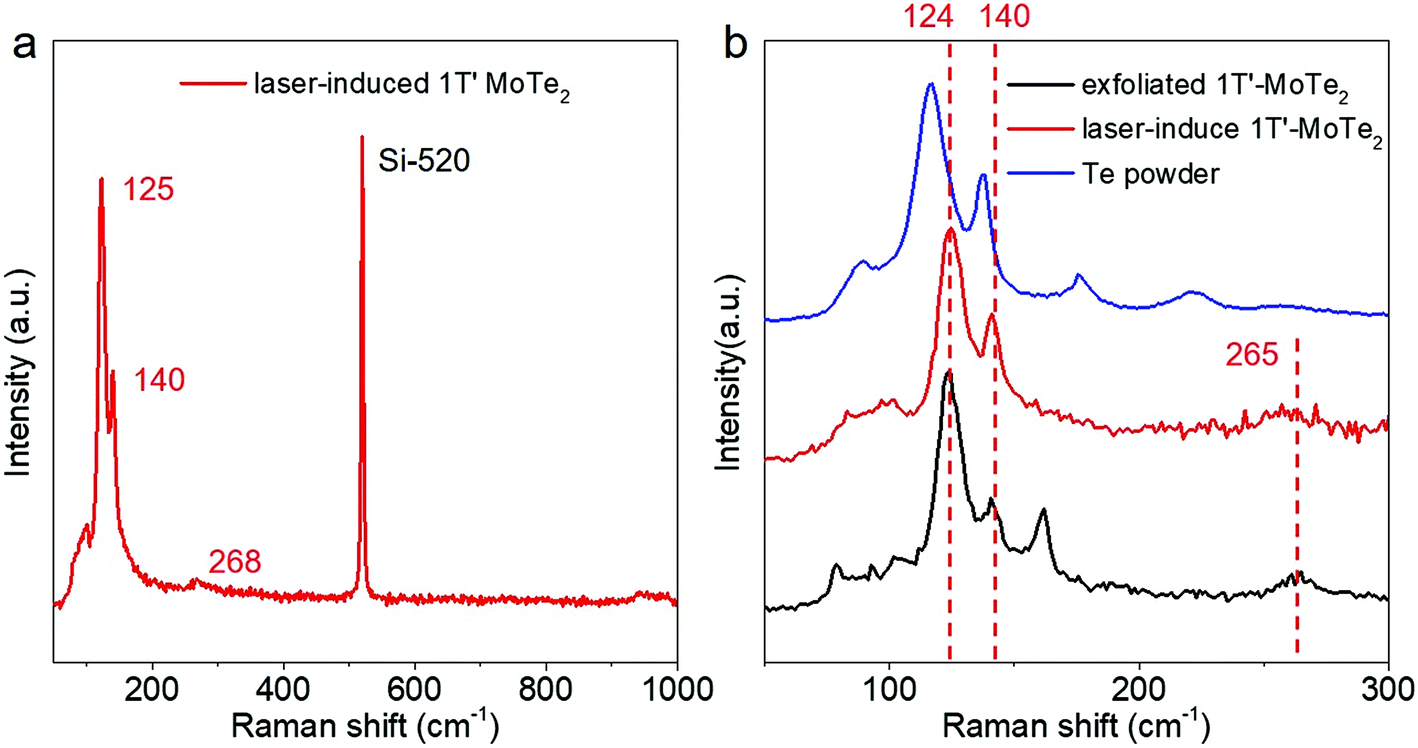 | ||
| Fig. 1 Raman spectra of laser-induced 1T′-MoTe2. (a) Raman spectrum of laser-induced 1T′-MoTe2 re-plotted from Fig. 1e in the original article, showing three active Raman Ag modes at 125 cm−1, 140 cm−1 and 268 cm−1. (b) Raman spectra of mechanically exfoliated 1T′-MoTe2 (black), laser-induced 1T′-MoTe2 (red) and Te powder (blue). The red dashed lines highlight the three active Raman modes of laser-induced 1T′-MoTe2. | ||
In order to exclude the possibility of decomposed Te from MoTe2 due to laser heating, we carried out additional laser irradiation experiments and compared the Raman spectra of Te powder, laser-induced 1T′-MoTe2 and mechanically exfoliated 1T′-MoTe2, as shown in Fig. 1b below. We can see that the laser-induced 1T′-MoTe2 exhibits three active Raman Ag modes at 125 cm−1, 140 cm−1 and 265 cm−1, which are consistent with mechanically exfoliated 1T′-MoTe2 from its bulk crystal (purchased from HQ graphene company). However, Te powder exhibits deviated peaks at 116 cm−1 and 137 cm−1.
To further confirm the reproducibility of the Raman spectrum of laser-induced 1T′-MoTe2 in different samples, we also carried out an additional laser patterning experiment. Few-layer 2H-MoTe2 was mechanically exfoliated on a Si substrate with 290 nm SiO2 on top, as shown in Fig. 2a below. The nature of 2H-MoTe2 was characterized by Raman spectroscopy using a lower laser power of 1.3 mW, with two pronounced Raman peaks exhibited at ∼171 cm−1 and ∼232 cm−1, as shown in Fig. 2c. Following this, we applied laser irradiation to pattern the 2H-MoTe2 with a higher laser power of 17.7 mW and with a laser patterning time of 104 min. The optical image of MoTe2 after laser patterning is shown in Fig. 2b. After laser patterning, we again characterized the MoTe2 using a lower laser power of 1.3 mW. Fig. 2d shows the Raman spectrum, which clearly exhibits three pronounced, highly reproducible Raman peaks at ∼122 cm−1, ∼140 cm−1 and ∼269 cm−1, corresponding to the three Ag modes of 1T’-MoTe2. The third peak, at 269 cm−1, is rather weak compared with the other two Raman peaks, but it is very clear after magnification, as shown in Fig. 2e.
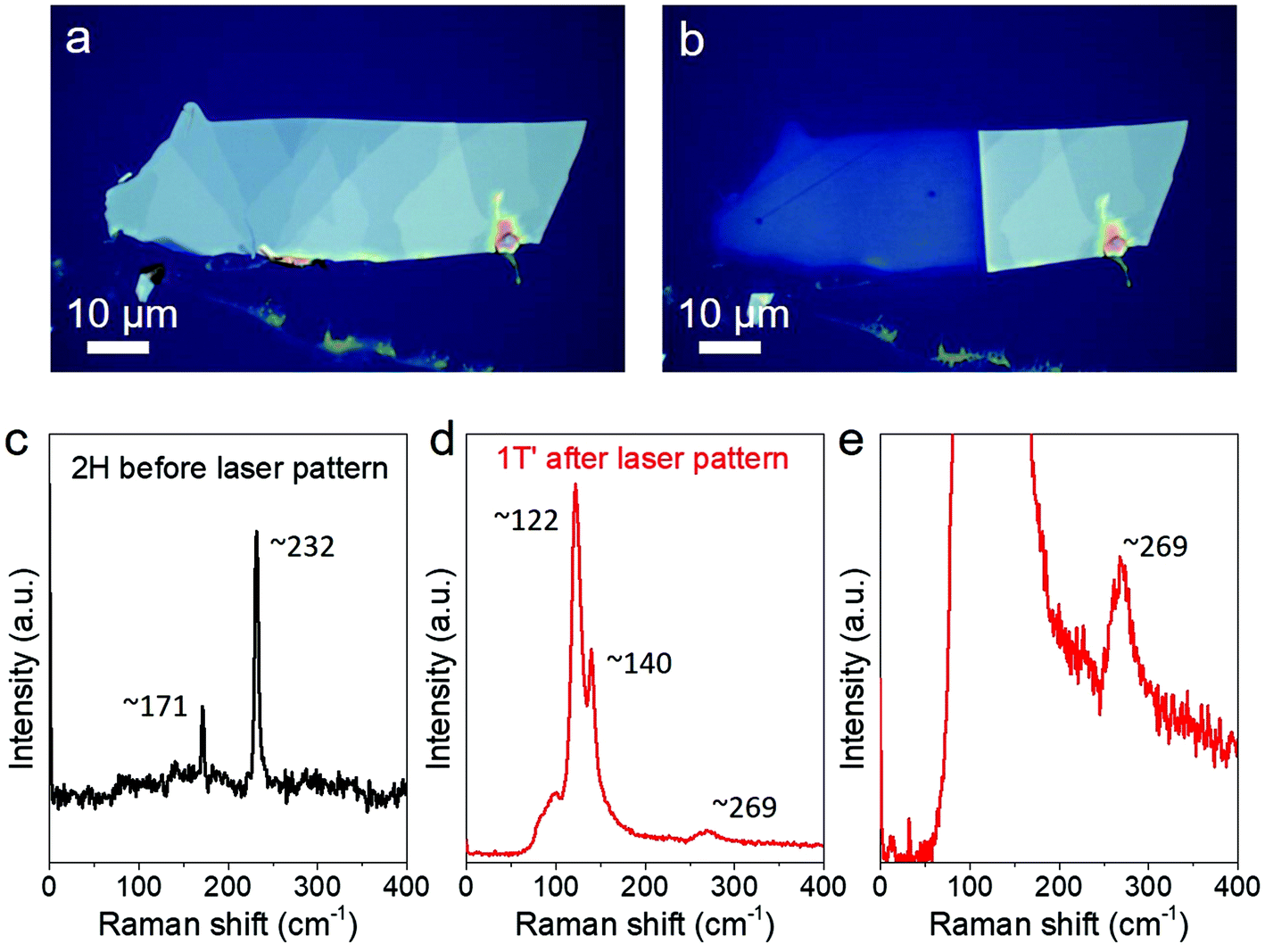 | ||
| Fig. 2 Optical images of MoTe2 (a) before and (b) after laser patterning. Raman spectra of MoTe2 (c) before and (d) after laser patterning. (e) Magnified Raman spectrum of laser irradiated MoTe2, clearly showing the third Raman peak at 269 cm−1. | ||
It was much clearer after we plotted the Raman mapping of intensity, as shown in Fig. 3 below. The Raman mappings defined by 171 cm−1 (width 10 cm−1) and 232 cm−1 (width 20 cm−1) show that the unpatterned area is still 2H-MoTe2 (bright) and the laser patterned area is dark. When we plotted the Raman mappings defined by 122 cm−1 (width 20 cm−1), 140 cm−1 (width 10 cm−1) and 269 cm−1 (width 20 cm−1), the laser patterned area became bright, which means that it changed to 1T′-MoTe2 after laser irradiation.
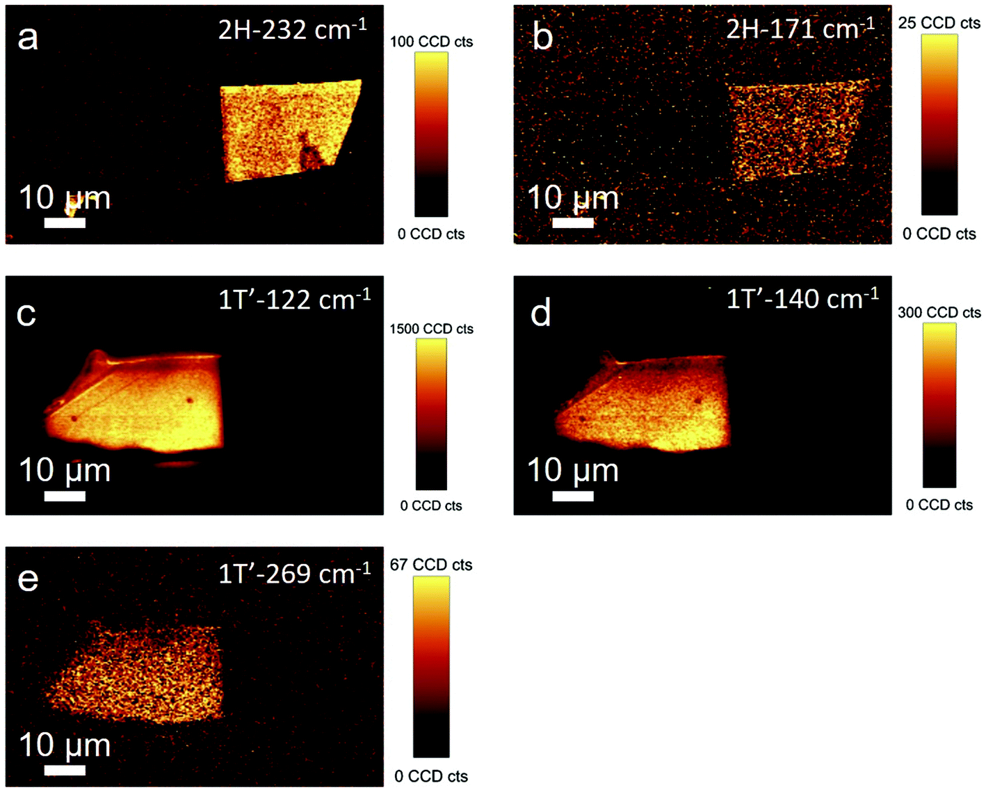 | ||
| Fig. 3 Raman mappings of intensity of laser-patterned MoTe2. The Raman mappings of intensity defined by (a) 232 cm−1 (width 20 cm−1) and (b) 171 cm−1 (width 10 cm−1) show the bright unpatterned 2H-MoTe2. The Raman mappings of intensity defined by (c) 122 cm−1 (width 20 cm−1), (d) 140 cm−1 (width 10 cm−1) and (e) 269 cm−1 (width 20 cm−1) show the bright patterned 1T′-MoTe2. | ||
To further confirm the laser-induced 2H-to-1T′ phase transition in MoTe2, we also carried out angle-dependent second harmonic generation (SHG) measurements. Fig. 4 below shows the optical images of few-layer 2H-MoTe2 before (a) and after (b) laser irradiation (power: 15 mW, time: 10 min). The Raman spectrum of laser-irradiated MoTe2 shows convincing Raman modes of 1T′-MoTe2 at 125 cm−1, 140 cm−1 and 265 cm−1, as shown in Fig. 4c. Additionally, the polarization dependences of the SHGs were characterized separately by measuring their parallel components. Because of the D13h space group in pristine 2H-MoTe2, an expected six-fold pattern was obtained, as shown in Fig. 4d. After laser irradiation, the polarization-dependence of MoTe2's SHG changes significantly, presenting a two-lobe pattern, as shown in Fig. 4e. The important contrast between the SHGs’ polarization dependences in the MoTe2 crystal before and after laser irradiation could be attributed to the phase transition between them.
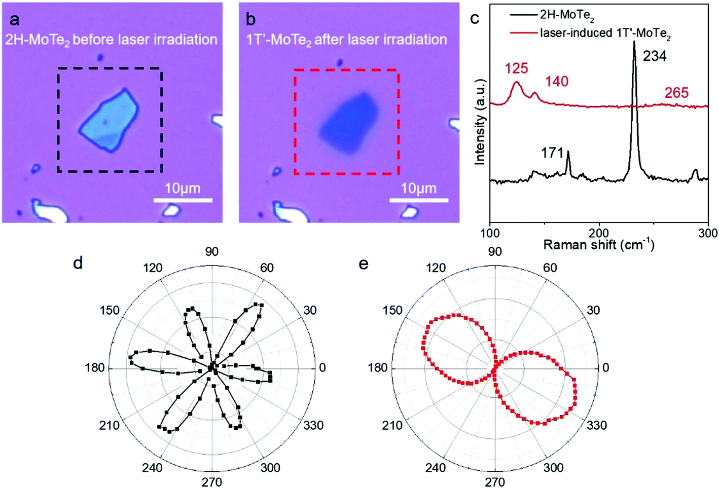 | ||
| Fig. 4 Polarization-dependent SHG of 2H-MoTe2 and 1T′-MoTe2. Optical images of few-layer 2H-MoTe2 (a) before and (b) after laser irradiation. (c) Raman spectra of 2H-MoTe2 and laser-induced 1T′-MoTe2. Polar plots of the SHG intensities from the few-layer MoTe2 (d) before and (e) after laser irradiation. | ||
In view of the above information, Nanoscale is officially removing its expression of concern.
The Royal Society of Chemistry apologises for these errors and any consequent inconvenience to authors and readers.
Michaela Mühlberg
14th November 2019
Managing Editor, Nanoscale
| This journal is © The Royal Society of Chemistry 2019 |