
Ambient mass spectrometry for the molecular diagnosis of lung cancer
Haiyan
Lu
 a,
Hua
Zhang
a,
Yiping
Wei
*b and
Huanwen
Chen
*c
a,
Hua
Zhang
a,
Yiping
Wei
*b and
Huanwen
Chen
*c
aState Key Laboratory of Inorganic Synthesis and Preparative Chemistry, College of Chemistry, Jilin University, Changchun 130012, P. R. China
bDepartment of Cardiothoracic Surgery to Second Affiliated Hospital of Nanchang University, Nanchang 330006, P. R. China
cJiangxi Key Laboratory for Mass Spectrometry and Instrumentation, East China University of Technology, Nanchang 330013, P. R. China. E-mail: chw8868@gmail.com
First published on 27th November 2019
Abstract
Lung cancer is one of the most common malignancies and the leading cause of cancer-related death worldwide. Among the technologies suitable for the rapid and accurate molecular diagnosis of lung cancer, ambient mass spectrometry (AMS) has gained increasing interest as it allows the direct profiling of molecular information from various biological samples (e.g., tissue, serum, urine and sputum) in real-time and with minimal or no sample pretreatment. This minireview summarizes the applications of AMS in lung cancer studies (including tissue molecular identification, the discovery of potential biomarkers, and surgical margin assessment), and discusses the challenges and perspectives of AMS in the clinical precision molecular diagnosis of lung cancer.
 Haiyan Lu | Haiyan Lu is a Ph.D student in Chemistry from Jilin University. Under the supervision of Prof. Huanwen Chen, her major efforts are being spent to develop novel ambient mass spectrometry approaches for cancer research (including tissue identification, discovery of potential biomarkers, tumor molecular margin assessment, and integrative omics study for improved accuracy of cancer differentiation). |
 Hua Zhang | Hua Zhang received his Ph.D in Chemistry from Jilin University in Jul. 2019 under the supervision of Prof. Huanwen Chen, with 29 peer-reviewed papers published in refereed journals. Currently, he continues the development and application of mass spectrometry in bioanalytical chemistry, drug discovery as a post doctor in the group of Prof. Lingjun Li, University of Wisconsin-Madison. |
 Yiping Wei | Yiping Wei earned his Bachelor degree in Medicine, Gannan Medical College, Jiangxi, China. He received his Ph.D in Cardiothoracic Surgery form Sun Yat-sen University in 2008. He is currently an associate Professor at The Second Affiliated Hospital of Nanchang University. His research interests include mass spectrometric analysis in thoracic tumor diagnosis, and the application of metabolomics in discriminating the margin of non-small cell lung cancer. |
 Huanwen Chen | Huanwen Chen received his Ph.D at Jilin University, China (2001). He was a postdoc in the group of Prof. Graham Cooks at Purdue University (2003–2005), and a Simon Fellow hosted by Prof. Renato Zenobi at ETH Zurich (2006–2008). He is a full professor of Analytical Chemistry at East China University of Technology (2008–present). He is the founder and Director of the Jiangxi Key Laboratory for Mass Spectrometry and Instrumentation, with more than 200 peer-reviewed papers, 50 patents, and 10 awards. Research carried out by his team is focused on the fundamentals, instrumentation, and applications of ambient mass spectrometry. |
Introduction
Lung cancer is one of the most common malignancies and the leading cause of cancer-related death in the world,1–3 and non-small cell lung cancer (NSCLC) accounts for 80–85% of all cases of lung cancer.4 Currently, the main methods used for the diagnosis of lung cancer include chest radiography, computerized tomography (CT), bronchoscopy, endoscopic ultrasound (EUS), and endobronchial ultrasound (EBUS).5,6 Surgical excision is widely acknowledged as the gold-standard approach to early-stage NSCLC. Nevertheless, emerging evidence suggests that the current oncosurgical techniques are frequently inadequate. For example, for 30% of stage I NSCLC patients treated with surgery there was the possibility of recurrence,4 and the recurrence rate of NSCLC patients with negative margins confirmed by routine pathological diagnosis was 12.7–13.79%.7 This indicates that the tumor margin determined by conventional histologic evaluation may not provide actual information about the malignant potential of the surgery margin.4 Undoubtedly, re-excision increases the risk of surgery for patients; thus, the development of novel techniques or devices for accurate molecular margin assessment across cancer surgeries is highly critical to the improvement of overall patient survival.Owing to the advantages of real-time and direct profiling of molecular information from various biological samples (e.g., tissue, serum, urine and sputum) with minimal or no sample pretreatment,8 AMS has been increasingly applied to the rapid and accurate molecular diagnosis of cancers.9–11 Although the widely acknowledged method for cancer diagnosis continues to be histopathology, molecular analysis has gained more attention in recent years. This is largely because molecular analysis provides more information that may be unavailable in histopathology,12 and offers a new opportunity to incorporate cancer-related biomarkers into clinical decision-making.13 Comprehensive reviews of AMS for the molecular diagnosis of cancer and surgical margin evaluation have been reported previously.9,10 Herein, this minireview summarizes the applications of AMS for the molecular diagnosis of lung cancer, including tissue molecular identification, the discovery of potential biomarkers, surgical margin assessment, and discusses the challenges and perspectives of AMS in the clinical precision molecular diagnosis of lung cancer.
The applications of AMS in lung cancer
Generally, clinical specimens, including tissue as well as biofluid samples (such as serum, urine and sputum), are widely used for cancer studies. In this section, the applications of AMS techniques in lung cancer from 2005 to date are summarized and discussed.Direct molecular characterization of lung cancer tissue
The tissue specimen is one of the most common samples for clinical cancer study. Tissue assessment and diagnosis play a critical role in the surgical excision of solid cancers for surgical margin evaluation.13 Traditionally, “frozen-section histology” is the gold-standard method for the accurate determination of the tumor margin,14 but it determines the features of tissues on the basis of cell morphology, with potentially unavoidable shortcomings as follows: (1) The cancerous tissue that has not been morphologically expressed could be misrecognized as normal tissue; (2) the results as interpreted by pathologists are inevitably subjective.15 Furthermore, the gold-standard method for tissue diagnosis is time-consuming (∼30 min), and postoperative final pathologic evaluation can take several days, which may delay decision making during diagnostic and therapeutic procedures.13,16 Therefore, the development of novel methods for the in situ, molecular real-time assessment of tumor margins is presently in urgent demand. Chemical analysis by AMS may be an alternative to frozen-section histology,17 because AMS differentiates the characteristics of tissue samples at the molecular level. The output of the complex multivariate analysis results of mass spectrometric and clinical data can be accomplished by advanced algorithms and software, allowing the subjective factor interference to be minimized. In short, the advantages of AMS promote its applications in the molecular diagnosis of lung cancer, showing the accelerating trend of AMS for lung cancer studies in both research lab and clinical diagnosis applications.In surface desorption atmospheric pressure chemical ionization mass spectrometry (DAPCI-MS) (Fig. 1A), a corona discharge is used for the generation of primary ions (such as hydronium ions, protonated reagents and radical water cations) via ion–molecule reactions. Being accelerated by the electric field, the primary ions are directed to impact the sample surface for analyte desorption and chemical ionization, then the generated analytes ions are introduced into the mass analyser through the ion introduction system for MS analysis.18,19 In previous studies, DAPCI-MS has been demonstrated to have high sensitivity and chemical selectivity for the direct detection of analytes on the complex surfaces, showing features such as high ionization and desorption efficiency for various analytes on heterogeneous surface samples. Therefore, DAPCI-MS has been used for the direct analysis of human squamous cell lung carcinoma tissue surface.20 The results indicated that DAPCI-MS could achieve accurate molecular diagnosis of squamous cell carcinoma lung cancer. With the combination of statistical analysis and the imaging data processing method, DAPCI-MS enables mass imaging with highly characteristic fragments recorded in multistage tandem mass spectrometry experiments,21 providing a useful tool for the visualization and differentiation of tumors without false positives as demonstrated by analysis of phosphatidylcholine and sphingomyelin profiles in tissue samples.20
 | ||
| Fig. 1 Schematic diagrams of AMS techniques in the direct molecular characterization of lung cancer tissues. (A) DAPCI-MSI (reproduced from ref. 20 with permission from the Royal Society of Chemistry, copyright [2017]). (B) REIMS (reproduced from ref. 29 with permission from Future Science Group, copyright [2019]). (C) MasSpec Pen (reproduced from ref. 29 with permission from Future Science Group, copyright [2019]). | ||
Air flow-assisted desorption electrospray ionization (AFADESI)22,23 is used for the creation and manipulation of ambient ions derived from large objects and employs a high flow-rate air flow to improve the efficiency of ion collection and remote transportation, demonstrating advantages in operation over the classic version of DESI24,25 for MS imaging of large bulk samples. For example, an in situ metabolomics method based on AFADESI-MS imaging (AFADESI-MSI), which could spatially explore the change in the global metabolites in tissues, was applied in the analysis of lung cancer tissue samples, and discovered that metabolites (including amino acids, choline, peptides, carnitine and lipids) could act as potential histopathological diagnosis biomarkers of lung cancer.26 More recently, AFADESI-MSI was used to accurately classify non-small cell lung cancer (NSCLC) pathology by molecular mapping of EGFR mutation spatial distribution.27
The combination of surgical and AMS techniques offers a new possibility for in situ molecular characterization of cancerous tissue during surgery. Since thermal evaporation of tissues is widely employed in surgery (i.e., electrosurgery and laser surgery), it was sensible to develop dedicated surgical instruments for the surgical process.28 For instance, rapid evaporative ionization mass spectrometry (REIMS) (Fig. 1B) is a technique that allows near-real-time characterization of tissue samples by analysis of the aerosol (“smoke”) released during electrosurgical dissection,16 and is commonly termed “iKnife” in the surgical application. In REIMS, gaseous ions from tissues are generated through thermal ablation surgical methods (electrosurgery and infrared laser surgery) and transmitted through a Venturi gas jet pump for MS analysis.9 REIMS has achieved the separation of different histologically specific data points of lung tumors with the help of linear discriminant analysis (LDA), this further confirmed that the identification of different histologically lung tumors by REIMS is feasible.16 Nevertheless, while REIMS is capable of rapid in vivo analysis, it is limited insofar as it requires electrosurgery.12 On the other hand, a large amount of heat is involved in the operation/ionization sampling process of REIMS, and the degradation of biological analytes is difficult to avoid, which might cause some information to be lost and eventually introduce uncertainties into the results.
For the purposes of tissue diagnosis, an automated and biocompatible handle mass spectrometry device (“MasSpec Pen”) (Fig. 1C) was developed, which allows direct, real-time tissue sampling and volume-controlled extraction of molecules from tissue samples through a discrete water droplet. The device contains three major components: (1) a syringe pump programmed to deliver water with a certain volume to the sampling probe; (2) small diameter polytetrafluoroethylene tubing conduits, which are integrated to fast two-way pinch valves for controlled solvent transport from the pump to the tissue and from the tissue to the mass spectrometer; (3) a hand-held pen-sized probe for direct sampling of biological tissues. The MasSpec Pen is utilized for the rapid, real-time and nondestructive molecular diagnosis of lung cancer tissues with high sensitivity (97.9%), specificity (95.7%), and accuracy (96.8%), and potential biomarkers including (such as PI (36![[thin space (1/6-em)]](https://www.rsc.org/images/entities/char_2009.gif) :1), PG (36:2), PG (34:1) as well as FA (18:1) have been obtained. The reported performance indicates that the MasSpec Pen can potentially be used as a clinical and intraoperative technology for in vivo cancer diagnosis.13 However, the current configuration of MasSpec Pen devices makes the carryover effect a possible factor in imposing interferences on the analytical results, especially for applications where the molecular margin of mutated tissue must be accurately identified.
:1), PG (36:2), PG (34:1) as well as FA (18:1) have been obtained. The reported performance indicates that the MasSpec Pen can potentially be used as a clinical and intraoperative technology for in vivo cancer diagnosis.13 However, the current configuration of MasSpec Pen devices makes the carryover effect a possible factor in imposing interferences on the analytical results, especially for applications where the molecular margin of mutated tissue must be accurately identified.
To minimize the carryover effects, tissue samples can be individually sampled offline rather than online, in vivo. For example, tissue spray ionization mass spectrometry (TSI-MS)30 provides highly specific molecular information for separated tissue samples. In TSI-MS, a Taylor cone was formed when the charged solvent was applied across the sample surface, enabling the chemicals mainly on the tissue surface to be extracted and ionized for direct MS analysis. TSI-MS has been performed for the direct characterization of chemical compounds (e.g., amino acids, hormones, fatty acids, anesthetics, and phospholipids) from various biological tissues including the brain, liver, kidney, adrenal gland, stomach, and spinal cord. Until now, combined with statistical analysis, TSI-MS has achieved the differential analysis of human kidney tumors and adjacent normal tissue from normal tissue,30 human brain tumors,31 and endometriotic tissues of different foci.32,33 Human lung squamous cell carcinoma was also recognized by TSI-MS via the help of a Monte Carlo sampling partial least squares linear discriminant analysis (PLS-LDA), and the accuracy of cancer identification reached 94.42%.17 Practically, in TSI-MS, due to the use of a small volume of solvent, the electrospray is intermittent and can only last several seconds. This causes the signal intensity levels to be recorded with tremendous variations, featuring disadvantages for analyte identification by collision-induced dissociation (CID). Furthermore, the analytes distributed underneath the tissue surface are difficult to be extracted for ionization/detection due to the limited time and solvent employed for TSI-MS.
Optimally, the analytes distributed in a defined minimally-available volume of 3-D tissue could be extracted for ionization/detection with highly stable levels for a relatively long time (e.g., ∼1 min). Therefore, another strategy termed as internal extractive electrospray ionization mass spectrometry (iEESI-MS)34,35 was developed for the straightforward MS analysis of whole-volume tissue samples without mashing/grinding the sample or matrix clean-up. In iEESI-MS, a fused silica capillary is inserted into the middle of the bulk sample (volume ∼1 mm3). The sample is then placed in front of the inlet of the mass spectrometer at a distance of 2–6 mm. Extractive solutions (e.g., methanol/water) biased with high voltage (± 2–6 kV) are directly injected into the bulk sample through the capillary with a certain flow rate (0.1–2 μL min−1). The analytes inside of the tissue are extracted and carried by an extractive solution to form electrospray under the electric field. Based on the advantages of high speed, sensitivity and specificity, low sample consumption as well as simple operations, iEESI-MS has successfully realized the rapid differential analysis of lung cancer tissue and normal tissue.35,36
Traditionally, metabolomics, lipidomics, and proteomics data are collected separately using differential individual tissue samples by multiple analytical techniques,37–39 resulting in the requirement that various bioinformatics tools are often employed to link the corresponding omics data to the rest of the information for better outcomes.40 However, integrating multilayers of omics information from different samples may smear the molecular profile of the individual sample, even lead to misinterpreted conclusions due to systematic bias caused by the nonhomologous samples. In general, it is easy to understand that systematic bias might be rooted in instrumental analysis due to the non-identical instrumental conditions/responses, particularly among the results of metabolomics, lipidomics, and proteomics analyses obtained using conventional omics approaches (such as liquid chromatography mass spectrometry (LC-MS) and gas chromatography mass spectrometry (GC-MS)) because different loci of tissue samples are analysed.26,37 However, systematic bias may be contributed by the samples used for metabolomics, lipidomics, and proteomics for a given study, because the specific sample for metabolomics data production usually cannot be used for other omics studies. Theoretically, maintaining the instrumental conditions constant and recording different omics data using the same piece of tissue sample should improve the accuracy and consistency of the analytical results. Therefore, it is of interest to establish an analytical strategy that could provide maximal molecular information from a single sample with an identical instrumental setup for more integrated system biological studies. Recently, an example of the sequential detection of lipids, small metabolites and proteins in one tissue for improved molecular differentiation accuracy of lung cancer has been confirmed using iEESI-MS, with low sample consumption (ca. 2.0 mg) and minimal systematic errors (Fig. 2).41 These examples demonstrated that AMS techniques play an important role in the direct molecular characterization of lung cancer tissue samples.
 | ||
| Fig. 2 Schematic diagram of iEESI-MS in the sequential detection of lipids, small metabolites and proteins in one tissue for the improved molecular differentiation accuracy of lung cancer tissues. | ||
Although the above-mentioned AMS technologies (such as DAPCI-MS, AFADESI-MSI, REIMS, MasSpec Pen, TSI-MS and iEESI-MS) have been applied in the molecular diagnosis of lung cancer tissues, the clinical specimens still need to be taken back to the laboratory for analysis due to the limitations of the techniques themselves and the mass spectrometer.42 So far, both REIMS16 and DESI-MS43 have realized in vivo molecular diagnoses during human cancer surgery. Since REIMS differentiates tissue through the analysis of aerosol (“smoke”) released during electrosurgical dissection, rigorous surgery conditions might limit the wide clinical application of the REIMS technique. DESI-MS gives results in a few minutes but is applicable to any surgical method and with no alteration in procedures. The MasSpec Pen is a nondestructive molecular diagnosis method and shows great potential in the molecular diagnosis of cancerous tissue, but is currently only suitable for the analysis of tissue samples. Comparing TSI-MS with iEESI-MS, the electrospray could only last for several seconds in TSI-MS,44 while the stable electrospray could last for even several hours in iEESI-MS. Additionally, iEESI-MS could be used for the direct analysis of biofliuds samples (such as urine and blood).45,46 Of course, a potential issue with iEESI-MS is that cells or smaller pieces of tissues may fall off during the analysis and contaminate the instrument when a large amount of tissue sample is continuously analysed over a long time. In future work, the performance of iEESI-MS will continue to be improved for the direct analysis of more types of real samples, including tissues, fluids, cells.41
Direct molecular characterization of lung cancer biofluids
Besides tissue samples, biofluid samples such as urine and serum are also widely used for lung cancer studies. For instance, desorption electrospray ionization mass spectrometry (DESI-MS) was used to provide metabolomics data on urine for the differential analysis of lung cancer and healthy mice, the result indicated that DESI-MS is a promising avenue for biomarker discovery in other biofluid samples.24 Polycyclic aromatic hydrocarbons (PAHs) are widespread and persistent chemical pollutants known to be carcinogenic, and accurate quantification of the human exposure to PAHs is important for health risk assessment.47 Urinary 1-hydroxypyrene (1-OHP, the main metabolite of pyrene) is recognized as a reliable biomarker for human exposure to PAHs.48 The iEESI-MS technique was originally proposed for the direct molecular analysis of bulk samples with a certain volume, such as plant/animal/human tissue samples. In fact, analytes in fluid samples can be selectively enriched on solid substrate to form an artificial bulk sample, which is then analyzed by iEESI-MS. This strategy has obtained successful application, for example, the quantification of 1-OHP in the raw urine of lung cancer patients was successfully achieved using magnetic solid-phase extraction coupled with internal extractive electrospray ionization mass spectrometry (MSPE-iEESI-MS) (Fig. 3A), suggesting that MSPE-iEESI-MS is an alternative strategy for high throughput quantitative detection of urinary 1-OHP for health risk assessment of PAHs exposure.45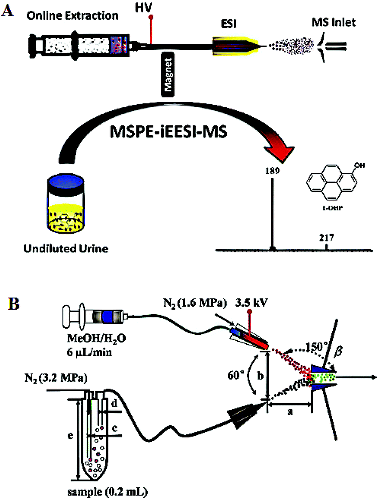 | ||
| Fig. 3 Schematic diagrams of AMS techniques in the direct molecular characterization of lung cancer biofluids. (A) Quantification of 1-OHP in undiluted human urine samples using MSPE-iEESI-MS (reproduced from ref. 45 with permission from Elsevier B. V., copyright [2016]). (B) Direct analysis of sputum samples using ND-EESI-MS (reprinted with permission from ref. 61. Copyright (2018) Japan Society of Analytical Chemistry). | ||
Despite serum and urine being widely applied in the study of various diseases,49–51 bronchoalveolar lavage fluid is in close interaction with lung tissue and avoids the contaminants from saliva,52 which might be a more representative sample for determining lung status, but it is invasively available with a bronchoscope. In addition to serum, urine and bronchoalveolar lavage fluid, sputum is a noninvasively accessible biofluid sample that contains bronchial epithelial cells, and the alteration of molecular information derived from sputum is likely to reveal the tumor-related changes.6,53 Hence sputum cytology has been usually applied in the detection of lung cancer.53,54 However, compared with sputum cytology, the molecular analysis of sputum could detect the cells containing tumor-associated molecular alterations that occur in microscopically normal-appearing epithelia, providing a promising method for the detection of lung cancer.55
Conventional chromatographic approaches such as LC-MS and GC-MS have been employed for the molecular analysis of sputum,56,57 but these methods involve tedious sample pretreatments (e.g., gradation, extraction, enrichment, separation), which affect the analytical efficiency and throughput of a large number of real samples. Fortunately, this was dramatically improved by AMS techniques. For example, neutral desorption extractive electrospray ionization mass spectrometry (ND-EESI-MS),58–60 which combines atmospheric pressure desorption sampling by a gentle stream of air or gas, followed by the neutral molecules being transported to an extractive electrospray ionization (EESI) source for ionization, showing obvious advantages in the rapid analysis of complex viscous samples (such as oil, honey and sputum).42 ND-EESI-MS was applied to the interrogation of sputum samples from NSCLC patients (Fig. 3B), and the results showed that ND-EESI-MS combined with principal component analysis (PCA) is helpful for the discovery of potential biomarkers of NSCLC, and the method has great potential for improving the rate of early detection of NSCLC.6,61 Overall, these examples further confirmed that any type of biological samples including tissue, serum, urine, sputum could be directly analyzed using versatile AMS techniques.
Conclusions and perspectives
Due to minimal/no sample pretreatment, AMS increases the simplicity and throughput of MS analysis. Numerous AMS techniques allow the direct profiling of molecular information from various biological samples (e.g., tissue, serum, urine, sputum and exhaled breath samples62) in real-time, showing promising potential in the molecular analysis of lung cancer. So far, the applications of AMS in lung cancer research include rapid molecular differential analysis, the discovery of potential biomarkers and the assessment of molecular tumor margins, but few efforts have been made to achieve the quantitative analysis of targeted analytes.In general, analytes on surfaces can be quantified by AMS such as DESI-MS, ND-EESI-MS, etc. with the results in μg cm−2.60,63 Classically, quality assessment was reported using bulk analyte concentration (e.g., in μg kg−1 units). Importantly, the surface concentration can significantly differ from the bulk-phase concentration for the real-word solid samples.35 Consequently, the surface concentration cannot be reliably used to evaluate the quality of a bulk sample. Unfortunately, the bulk molecular concentration in AMS can normally be derived only for solution samples,64,65 while the bulk molecular concentration in AMS for solid samples is confronted with great challenges. To promote the application of AMS for the real-world problems, as a model demonstration, iEESI-MS achieved the quantitative determination of six types of β-agonists in pork tissue samples without sample pretreatment,66 providing consistent analytical results validated by GC-MS and LC-MS, with the accuracy of 92–105%. The work with iEESI-MS showed the first example of AMS for the quantitative analysis of real-world tissue in bulk concentration (i.e., in μg kg−1 units) with satisfactory accuracy, offering advantages for the direct quantification of analytes in tissue samples for the improved accuracy of lung cancer diagnosis. Many challenges still exist in the rapid and accurate molecular diagnosis of lung cancer for AMS. For instance, the development of intraoperative high-precision mini mass spectrometers with results being immediately available, rigorous validation studies of the results yielded by different AMS techniques and various machine learning algorithms, etc.
Conflicts of interest
The authors declare no competing financial interest.Acknowledgements
This work was supported by the National Natural Science Foundation of China (No. 21765001), Program for Changjiang Scholars and Innovative Research Team in University (PCSIRT) (No. IRT_17R20) and 111 Project (No. D17006).References
- S. A. Hayes, S. Haefliger, B. Harris, N. Pavlakis, S. J. Clarke, M. P. Molloy and V. M. Howell, J. Breath Res., 2016, 10, 034001 CrossRef PubMed.
- J. Brabender, J. M. Park, R. Metzger, P. M. Schneider, R. V. Lord, A. H. Holscher, K. D. Danenberg and O. V. Danenberg, Ann. Surg., 2002, 235, 440–443 CrossRef PubMed.
- R. L. Siegel, K. D. Miller and A. Jemal, CA Cancer J. Clin., 2019, 69, 7–34 CrossRef PubMed.
- T. F. Lv, J. W. Zou, H. B. Liu, Q. Shen, Z. F. Lu, X. J. Zhou, X. N. Wang and Y. Song, Oncotarget, 2017, 8, 40643–40653 Search PubMed.
- D. R. Aberle, A. M. Adams, C. D. Berg, W. C. Black, J. D. Clapp, R. M. Fagerstrom, I. F. Gareen, C. Gatsonis, P. M. Marcus, J. D. Sicks and T. Natl, Lung Screening Trial Res, N. Engl. J. Med., 2011, 365, 395–409 CrossRef PubMed.
- J. Y. Zhang, J. J. Xu, H. Y. Lu, J. H. Ding, D. L. Yu, P. H. Li, J. W. Xiong, X. X. Liu, H. W. Chen and Y. P. Wei, Oncotarget, 2016, 7, 63158–63165 Search PubMed.
- Z. W. Fang, J. X. He, W. Q. Fang, L. L. Ruan and F. Fang, Heart, Lung Circ., 2016, 25, 392–397 CrossRef PubMed.
- A. Venter, M. Nefliu and R. G. Cooks, TrAC, Trends Anal. Chem., 2008, 27, 284–290 CrossRef CAS.
- D. R. Ifa and L. S. Eberlin, Clin. Chem., 2016, 62, 111–123 CrossRef CAS PubMed.
- Z. Takats, N. Strittmatter and J. S. McKenzie, Adv. Cancer Res., 2017, 134, 231–256 CrossRef CAS PubMed.
- M. Woolman and A. Zarrine-Afsar, Analyst, 2018, 143, 2717–2722 RSC.
- H. M. Brown, V. Pirro and R. G. Cooks, Clin. Chem., 2018, 64, 628–630 CrossRef CAS PubMed.
- J. L. Zhang, J. Rector, J. Q. Lin, J. H. Young, M. Sans, N. Katta, N. Giese, W. Yu, C. Nagi, J. Suliburk, J. Liu, A. Bensussan, R. J. DeHoog, K. Y. Garza, B. Ludolph, A. G. Sorace, A. Syed, A. Zahedivash, T. E. Milner and L. S. Eberlin, Sci. Transl. Med., 2017, 9, eaan3968 CrossRef PubMed.
- W. B. Yun, M. Kalra, J. Michaelson, J. Kirz, S. J. Lewis, W. X. Cong, Q. S. Yang and W. Ge, in Spie Optical Engineering + Applications, 2016 Search PubMed.
- R. E. Nakhleh, Arch. Pathol. Lab. Med., 2011, 135, 1394–1397 CrossRef PubMed.
- J. Balog, L. Sasi-Szabo, J. Kinross, M. R. Lewis, L. J. Muirhead, K. Veselkov, R. Mirnezami, B. Dezso, L. Damjanovich, A. Darzi, J. K. Nicholson and Z. Takats, Sci. Transl. Med., 2013, 5, 194ra93 Search PubMed.
- Y. P. Wei, L. R. Chen, W. Zhou, K. Chingin, Y. Z. Ouyang, T. G. Zhu, H. Wen, J. H. Ding, J. J. Xu and H. W. Chen, Sci. Rep., 2015, 5, 10077 CrossRef CAS PubMed.
- M. Li, B. Jia, L. Y. Ding, F. Hong, Y. Z. Ouyang, R. Chen, S. M. Zhou, H. W. Chen and X. Fang, J. Mass Spectrom., 2013, 48, 1042–1049 CrossRef CAS PubMed.
- H. Zhang, A. Bibi, H. Y. Lu, J. Han and H. W. Chen, J. Mass Spectrom., 2017, 52, 526–533 CrossRef CAS PubMed.
- Y. Z. Ouyang, J. W. Liu, B. H. Nie, N. P. Dong, X. Chen, L. F. Chen and Y. P. Wei, RSC Adv., 2017, 7, 56044–56053 RSC.
- S. P. Yang, H. W. Chen, Y. L. Yang, B. Hu, X. Zhang, Y. F. Zhou, L. L. Zhang and H. W. Gu, Chin. J. Anal. Chem., 2009, 37, 315–318 CAS.
- J. M. He, F. Tang, Z. G. Luo, Y. Chen, J. Xu, R. P. Zhang, X. H. Wang and Z. Abliz, Rapid Commun. Mass Spectrom., 2011, 25, 843–850 CrossRef CAS PubMed.
- Z. G. Luo, J. M. He, Y. Chen, J. J. He, T. Gong, F. Tang, X. H. Wang, R. P. Zhang, L. Huang, L. F. Zhang, H. N. Lv, S. G. Ma, Z. D. Fu, X. G. Chen, S. S. Yu and Z. Abliz, Anal. Chem., 2013, 85, 2977–2982 CrossRef CAS PubMed.
- R. G. Cooks, Z. Ouyang, Z. Takats and J. M. Wiseman, Science, 2006, 311, 1566–1570 CrossRef CAS PubMed.
- M. W. F. Nielen, H. Hooijerink, P. Zomer and J. G. J. Mol, TrAC, Trends Anal. Chem., 2011, 30, 165–180 CrossRef CAS.
- T. G. Li, J. J. He, X. X. Mao, Y. Bi, Z. G. Luo, C. G. Guo, F. Tang, X. Xu, X. H. Wang, M. R. Wang, J. Chen and Z. Abliz, Sci. Rep., 2015, 5, 14089 CrossRef CAS PubMed.
- M. Zhang, J. M. He, T. G. Li, H. X. Hu, X. F. Li, H. Xing, J. Wang, F. Yang, Q. F. Ma, B. Liu, C. H. Tang, Z. Abliz and X. Q. Liu, Front. Oncol., 2019, 9, 804 CrossRef PubMed.
- K. C. Schafer, J. Denes, K. Albrecht, T. Szaniszlo, J. Balog, R. Skoumal, M. Katona, M. Toth, L. Balogh and Z. Takats, Angew. Chem., Int. Ed., 2009, 48, 8240–8242 CrossRef PubMed.
- L. Haenel, M. Kwiatkowski, L. Heikaus and H. Schlueter, Future Sci. OA, 2019, 5, FSO373 CrossRef PubMed.
- J. J. Liu, R. G. Cooks and Z. Ouyang, Anal. Chem., 2011, 83, 9221–9225 CrossRef CAS PubMed.
- A. Kononikhin, E. Zhvansky, V. Shurkhay, I. Popov, D. Bormotov, Y. Kostyukevich, S. Karchugina, M. Indeykina, A. Bugrova, N. Starodubtseva, A. Potapov and E. Nikolaev, Anal. Bioanal. Chem., 2015, 407, 7797–7805 CrossRef CAS PubMed.
- V. V. Chagovets, Z. H. Wang, A. S. Kononikhin, N. L. Starodubtseva, A. Borisova, D. Salimova, I. A. Popov, A. V. Kozachenko, K. Chingin, H. W. Chen, V. E. Frankevich, L. V. Adamyan and G. T. Sukhikh, Sci. Rep., 2017, 7, 2546 CrossRef PubMed.
- V. Chagovets, Z. H. Wang, A. Kononikhin, N. Starodubtseva, A. Borisova, D. Salimova, I. Popov, A. Kozachenko, K. Chingin, H. W. Chen, V. Frankevich, L. Adamyan and G. Sukhikh, J. Am. Soc. Mass Spectrom., 2018, 29, 323–330 CrossRef CAS PubMed.
- H. Zhang, K. Chingin, L. Zhu and H. W. Chen, Anal. Chem., 2015, 87, 2878–2883 CrossRef CAS PubMed.
- H. Zhang, H. W. Gu, F. Y. Yan, N. N. Wang, Y. P. Wei, J. J. Xu and H. W. Chen, Sci. Rep., 2013, 3, 2495 CrossRef PubMed.
- H. Y. Lu, J. Y. Zhang, W. Zhou, Y. P. Wei and H. W. Chen, Chin. J. Anal. Chem., 2016, 44, 329–334 CAS.
- E. J. Want, P. Masson, F. Michopoulos, I. D. Wilson, G. Theodoridis, R. S. Plumb, J. Shockcor, N. Loftus, H. Elaine and J. K. Nicholson, Nat. Protoc., 2013, 8, 17–32 CrossRef CAS PubMed.
- M. Yuan, S. B. Breitkopf, X. M. Yang and J. M. Asara, Nat. Protoc., 2012, 7, 872–881 CrossRef CAS PubMed.
- W. J. Griffiths and Y. Q. Wang, Chem. Soc. Rev., 2009, 38, 1882–1896 RSC.
- R. C. De Vos, S. Moco, A. Lommen, J. J. Keurentjes, R. J. Bino and R. D. Hall, Nat. Protoc., 2007, 2, 778–791 CrossRef CAS PubMed.
- H. Y. Lu, H. Zhang, K. Chingin, Y. P. Wei, J. Q. Xu, M. F. Ke, K. K. Huang, S. H. Feng and H. W. Chen, Anal. Chem., 2019, 91, 10532–10540 CrossRef CAS PubMed.
- H. Y. Lu, H. Zhang, K. Chingin, J. L. Xiong, X. W. Fang and H. W. Chen, TrAC, Trends Anal. Chem., 2018, 107, 99–115 CrossRef CAS.
- V. Pirro, C. M. Alfaro, A. K. Jarmusch, E. M. Hattab, A. A. Cohen-Gadol and R. G. Cooks, Proc. Natl. Acad. Sci. U. S. A., 2017, 114, 6700–6705 CAS.
- J. Y. Zhang, J. J. Xu, Y. Z. Ouyang, J. W. Liu, H. Y. Lu, D. L. Yu, J. H. Peng, J. W. Xiong, H. W. Chen and Y. P. Wei, Sci. Rep., 2017, 7, 3738 CrossRef PubMed.
- H. Zhang, H. Y. Lu, H. C. Huang, J. C. Liu, X. W. Fang, B. F. Yuan, Y. Q. Feng and H. W. Chen, Anal. Chim. Acta, 2016, 926, 72–78 CrossRef CAS PubMed.
- H. Zhang, K. Chingin, J. J. Li, H. Y. Lu, K. K. Huang and H. W. Chen, Anal. Chem., 2018, 90, 12101–12107 CrossRef CAS PubMed.
- N. Li, D. Wu, N. Hu, G. S. Fan, X. T. Li, J. Sun, X. F. Chen, Y. R. Suo, G. L. Li and Y. N. Wu, J. Agric. Food Chem., 2018, 66, 3572–3580 CrossRef CAS PubMed.
- J. N. Hao and B. Yan, Adv. Funct. Mater., 2017, 27, 1603856 CrossRef.
- E. F. Petricoin, A. M. Ardekani, B. A. Hitt, P. J. Levine, V. A. Fusaro, S. M. Steinberg, G. B. Mills, C. Simone, D. A. Fishman, E. C. Kohn and L. A. Liotta, Lancet, 2002, 359, 572–577 CrossRef CAS.
- P. Wurtz, A. J. Kangas, P. Soininen, D. A. Lawlor, G. D. Smith and M. Ala-Korpela, Am. J. Epidemiol., 2017, 186, 1084–1096 CrossRef PubMed.
- E. A. Mathe, A. D. Patterson, M. Haznadar, S. K. Manna, K. W. Krausz, E. D. Bowman, P. G. Shields, J. R. Idle, P. B. Smith, K. Anami, D. G. Kazandjian, E. Hatzakis, F. J. Gonzalez and C. C. Harris, Cancer Res., 2014, 74, 3259–3270 CrossRef CAS PubMed.
- B. Callejon-Leblic, T. Garcia-Barrera, J. Gravalos-Guzman, A. Pereira-Vega and J. L. Gomez-Ariza, J. Proteomics, 2016, 145, 197–206 CrossRef CAS PubMed.
- F. B. Thunnissen, J. Clin. Pathol., 2003, 56, 805–810 CrossRef CAS PubMed.
- B. Lam, S. Y. Lam, M. P. Wong, C. G. C. Ooi, D. Y. T. Fonge, D. C. L. Lam, A. Y. K. Lai, C.-m. Tam, C. B. Y. Pang, M. S. M. Ip and W.-k. Lam, Lung Cancer, 2009, 64, 289–294 CrossRef PubMed.
- N. Anjuman, N. Li, M. Guarnera, S. A. Stass and F. Jiang, Clin. Transl. Med., 2013, 2, 15–15 CrossRef PubMed.
- S. Ma, G. M. Turino and Y. Y. Lin, J. Chromatogr. B: Anal. Technol. Biomed. Life Sci., 2011, 879, 1893–1898 CrossRef CAS PubMed.
- D. M. Cha, D. Cheng, M. M. Liu, Z. R. Zeng, X. W. Hu and W. W. Guan, J. Chromatogr. A, 2009, 1216, 1450–1457 CrossRef CAS PubMed.
- X. Li, B. Hu, J. H. Ding and H. W. Chen, Nat. Protoc., 2011, 6, 1010–1025 CrossRef CAS PubMed.
- J. H. Ding, H. W. Gu, S. P. Yang, M. Li, J. Q. Li and H. W. Chen, Anal. Chem., 2009, 81, 8632–8638 CrossRef CAS PubMed.
- H. W. Chen and R. Zenobi, Nat. Protoc., 2008, 3, 1467–1475 CrossRef CAS PubMed.
- X. F. Gao, Y. P. Xiao and Y. Y. Dai, Anal. Sci., 2018, 34, 1067–1071 CrossRef CAS PubMed.
- D. Garcia-Gomez, P. M. L. Sinues, C. Barrios-Collado, G. Vidal-de-Miguel, M. Gaugg and R. Zenobi, Anal. Chem., 2015, 87, 3087–3093 CrossRef CAS PubMed.
- Z. Takats, J. M. Wiseman, B. Gologan and R. G. Cooks, Science, 2004, 306, 471–473 CrossRef CAS PubMed.
- H. W. Chen, A. Venter and R. G. Cooks, Chem. Commun., 2006, 2042–2044 RSC.
- Y. Tian, M. Yu, J. Chen, C. Liu, J. Shi, H. Chen and G. Jiang, Anal. Chem., 2015, 87, 11962–11966 CrossRef CAS PubMed.
- J. Q. Xu, S. R. Xu, Y. P. Xiao, K. Chingin, H. Y. Lu, R. H. Yan and H. W. Chen, Anal. Chem., 2017, 89, 11252–11258 CrossRef CAS PubMed.
| This journal is © The Royal Society of Chemistry 2020 |