Zinc oxide–black phosphorus composites for ultrasensitive nitrogen dioxide sensing†
Qun
Li
ab,
Yuan
Cen
a,
Jinyu
Huang
a,
Xuejin
Li
a,
Hao
Zhang
a,
Youfu
Geng
a,
Boris I.
Yakobson
c,
Yu
Du
 *a and
Xiaoqing
Tian
 *a
*a
aCollege of Physics and Energy, Shenzhen University, Shenzhen 518060, China. E-mail: duyu@szu.edu.cn; xqtian@szu.edu.cn
bWuhan National Laboratory for Optoelectronics, Huazhong University of Science and Technology, Wuhan 430074, China
cDepartment of Materials Science and NanoEngineering, Department of Chemistry, Smalley Institute for Nanoscale Science and Technology, Rice University, Houston 77005, USA
First published on 16th May 2018
Abstract
Multi-layer black phosphorus (m-BP) has been successfully incorporated in zinc oxide (ZnO) hollow spheres, thus forming hetero-structured ZnO–BP composites; a study performed on their properties reveals that the BP environmental stability can be significantly enhanced by the introduction of ZnO, and the sensors based on ZnO–BP composites exhibit high response, fast response behavior, outstanding selectivity, and ultralow detection limit of 1 ppb towards NO2 gas molecules. The ultrasensitive sensing performance is suggested to be due to the large surface area, excellent carrier mobility, and enhanced charge transfer of ZnO–BP in the presence of BP. Moreover, the mechanism of NO2 sensing with ZnO–BP is confirmed by the calculations obtained from the first-principle studies.
Conceptual insightsPhospohorene, due to its ultrahigh surface–volume ratio, high chemical activity and excellent carrier mobility, has been shown to be promising for gas sensing. However, the fabricated BP sensor suffers from some disadvantages, such as poor environmental stability, low sensitivity and long response–recovery process, due to which it is difficult to use in practical environments or safety monitoring. Multi-layer black phosphorus (m-BP) is successfully incorporated in zinc oxide (ZnO) hollow spheres, thus forming hetero-structured ZnO–BP composites. Interestingly, the introduction of ZnO significantly enhances BP environmental stability, making it a suitable candidate to use in practice. Remarkably, ZnO–BP sensor exhibits higher response, outstanding selectivity, faster response and recovery dynamic process, and an ultralow detection limit of 1 ppb to NO2 in contrast to graphene-, BP- and ZnO-based sensors, which were previously used. This study has provided a route in the search of BP-based materials with excellent properties for use in practice, in particular towards gas sensing. |
Phosphorene, one of the novel elemental 2D materials, exhibits a single layer of phosphorus sheet exfoliated from the bulk black phosphorus (BP); it has recently received extensive attention due to its significant advantages over semi-metallic graphene. These advantages include thickness-dependent direct band gap variation from monolayer to bulk BP in the range of 1.5–0.3 eV and free-carrier mobility (∼1000 cm2 V s−1), which is better than those of other typical 2D semiconductors such as molybdenum disulfide, tungsten diselenide, and boron nitride (BN).1 However, it has been reported that phosphorene can be irreversibly degraded to oxidized phosphorus compounds under ambient conditions.2,3 The proper protection of its surfaces by encapsulating with graphene, BN, oxides or multi-layer black phosphorus (m-BP) can generally improve the structural and environmental stabilities.4–7
Phosphorene, due to its ultrahigh surface–volume ratio, high chemical activity, and excellent carrier mobility, has been demonstrated to be a promising material for gas sensing applications.8–10 A good sensing functionality of phosphorene and doped phosphorene with even better performances than those of graphene and MoS2 was theoretically demonstrated.11 However, to the best of our knowledge and based on few decades of experience in this field, we have found that reports on experimental studies about phosphorene-based sensors are scarce. Recently, Abbas et al. first experimentally explored the chemical sensing of nitrogen dioxide (NO2) gas molecules using field-effect transistors (FET) that were based on m-BP.9 However, the fabricated BP sensor suffered from a few disadvantages: it exhibited low sensitivity and long response–recovery process, and it was not specifically selective to NO2, thus making it difficult to apply in practical items and in safety monitoring systems.
Basically, NO2 is a typical polluting gas with pungent odor; it is generated from the combustion of chemical plants and automotive emissions, and it plays a major role in the formation of acid rain, photochemical smog and ground-level ozone, which are potentially harmful to human health and the ecosystem. The Japanese standards strictly regulated the threshold limit value (TLV) for NO2 to less than 40–60 ppb.12,13 Therefore, the detection of trace levels of NO2 becomes an exceedingly significant task for monitoring environmental pollution. Gas sensors based on metal oxide semiconductors (MOSs) such as In2O3, ZnO, SnO2, Fe2O3, and WO3 are predominant gas detecting devices for domestic, laboratorial and commercial applications, owing to their miniaturized dimensions, availability of easy fabrication methods, low-costs, and considerable chemical, structural and environmental stabilities.14–19 However, in the last decade, the concentration detection limit of MOS sensors was still high, resulting in their threshold and resolution deficiencies to monitor accurately the variations of low concentrations of NO2 in atmosphere.
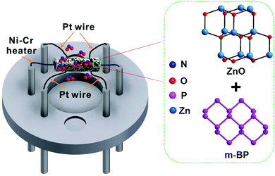
Herein, we illustrate that the incorporation of m-BP nanosheets into ZnO hollow spheres can significantly improve NO2 sensing properties (Fig. 1). The hetero-structured ZnO–BP composites exhibit high response, ultralow detection limit, quick response and recovery characteristics as well as excellent selectivity towards NO2. After incorporation of BP, the detection limit of ZnO–BP sensor decreases to 1 ppb, and the response increases to 20 under 100 ppb NO2, which is more than triple the responses of ZnO and BP sensors. First-principles density functional theory calculations suggest that there is a charge transfer from ZnO to BP via Zn–P bonds in the ZnO–BP heterostructure, which can increase the local chemical affinity of BP and enhance the sensing properties of ZnO–BP towards NO2 molecules. Moreover, the existence of BP increases gas adsorption and diffusion, the surface area-to-volume ratio and carrier mobility. Our results also indicate that the introduction of ZnO can significantly enhance BP environmental stability, making it suitable to use in practice. We believe that this study will benefit further investigations of the fundamental science and applications of phosphorene in the context of chemical sensors.
The morphologies and structures of ZnO and ZnO–BP were characterized with scanning electron microscopy (SEM), transmission electron microscopy (TEM), high angle annular dark-field scanning transmission electron microscopy (HAADF-STEM), high resolution TEM (HRTEM), corresponding selected area electron diffraction (SAED) and atomic force microscopy (AFM). The panoramic SEM image in Fig. 2a shows that the obtained ZnO was composed of numerous monodispersed spheres. The products were hollow in shape with an average diameter of ∼4 μm and thickness of spherical shell of about 400 nm. The external and internal surfaces of the spherical shells composed of abundant interlinked nanoparticles contributed to their rough and porous structure, which increased the specific surface area and the surface defects. The HRTEM image and SAED pattern (Fig. 2b) obtained from a random area on the ZnO hollow spheres indicated that the particles forming the structure were polycrystalline in nature. The lattice inter-planar spacing of 0.28 nm corresponding to the (100) plane of wurtzite ZnO was evidenced. We further investigated the elemental distribution of ZnO hollow spheres using HAADF-STEM (Fig. 2c), where clear peaks for O (red) and Zn (orange) on the whole sphere shells were detected. This profile coupled with composition line-scan (green line) across ZnO verified the hollow shape of the ZnO spheres. Fig. 2d shows the SEM image of the ZnO–BP composites. The morphology of ZnO was maintained even after ultrasonic treatment except that few nanosheets of BP were located on the surface of the hollow spheres. The TEM image shown in Fig. 2e confirmed the presence of 2D BP nanosheets. The 0.218 nm lattice spacing of the fringes was assigned to the (020) plane of orthorhombic BP. The SAED pattern result suggested that these nanosheets are single crystalline in nature.20 AFM of ZnO–BP composites on Si/SiO2 substrate demonstrated the presence of a range of shapes and sizes of phosphorene (Fig. 2f). Height-profiling of nanosheets revealed well-defined edges of 2.95 nm thickness. According to the AFM measurements of BP in a previous study, we can infer that these nanosheets predominantly consist of more than five-layered phosphorene.21
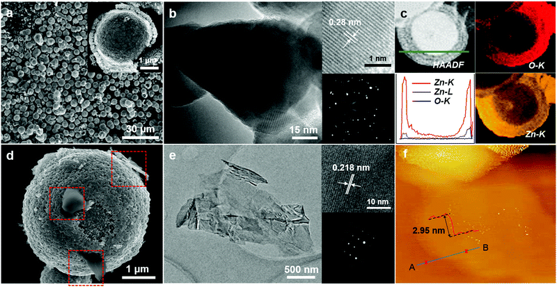 | ||
| Fig. 2 (a–c) SEM, TEM with corresponding SAED and HRTEM, and HAADF-STEM, images of ZnO. (d–f) SEM, HRTEM with SAED, and AFM height profile of m-BP nanosheets images of ZnO–BP. | ||
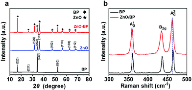
The several peaks of ZnO–BP in X-ray diffraction (XRD) patterns observed at 31.64°, 34.30° and 36.12° (Fig. 3a) are indexed to the diffraction planes (100), (002), and (101) of wurtzite ZnO (JCPDS No. 36-1451). The other diffraction peaks at 16.81°, 26.61° and 52.40° are characteristic peaks for the orthorhombic phase of BP,22 suggesting that the structure of BP remains the same after liquid ultrasonication. Fig. 3b shows the Raman spectra of BP and ZnO–BP composites. Three strong peaks correspond to the out-of-plane vibration mode A1g, in-plane vibration mode B2g and A2g of BP. Noteworthily, ZnO–BP composites exhibit the A2g mode at 462.57 cm−1, which is 1.75 cm−1 lower than that of BP; this is probably due to the stacking-induced anisotropic structure change from bulk to multilayer phosphorene after ultrasonic treatment.3,21 Furthermore, the intensities of Raman peaks related to bulk phosphorus are smaller than those of ZnO–BP, and this is consistent with the results of MoS2–graphene composites, which is attributed to the effects of optical interference.20,23 These results demonstrate the successful synthesis of multilayered BP with anhydrous organic solvents in a sealed-tip ultrasonication system.
To further understand the surface compositions and chemical states of the elements present in ZnO–BP composites, we studied X-ray photoelectron spectra (XPS) of BP, ZnO and ZnO–BP. As shown in Fig. 4a and b, the complete spectra of BP and ZnO–BP confirmed involvement of P, O, C and Zn, P, O, and C elements, respectively. The peak at 284.2 eV corresponded to the adventitious carbon-based contaminants adsorbed on the surfaces. P 2p electron core-level XPS spectra of BP and ZnO–BP composites are shown in Fig. 4c. The two peaks at 130.1 and 131.0 eV were assigned to 2p3/2 and 2p1/2 of P3+, respectively, and they were characteristics of crystalline BP.24,25 Please note that compared to BP, ZnO–BP exhibited a higher XPS signal at 131.0 eV and a distinct lower signal at 134.8 eV, indicating more P3+ and less P5+ in ZnO–BP. It has been reported that the P5+ sub-bands at the higher binding energy are observed due to POx for oxidized phosphorus species, because of the degradation upon exposure to ambient conditions.25–27 The indiscernible signal of P5+ ions in the XPS spectrum of ZnO–BP suggested that the introduction of ZnO hollow spheres can enhance BP environmental stability.26 We also analyzed the time evolution of the P 2p signal, as shown in Fig. S1 (ESI†). The signals of P5+ ions in the XPS spectra of ZnO–BP were still indiscernible after 24, 48, and 96 hours, suggesting good environmental stability of ZnO–BP. In addition, the Zn 2p electron core level XPS spectra of pristine ZnO and ZnO–BP composites are given in Fig. 4d; the results indicated two symmetric peaks at 1022.77 and 1045.88 eV, which were attributed to 2p3/2 and 2p1/2, respectively, of Zn2+ in the pure ZnO hollow spheres. For the ZnO–BP composites, the Zn 2p peaks shifted to high binding energy, evidently after introduction of BP. The fact that the charge density of ZnO decreased, whereas that of BP increased, implied the existence of a charge transfer process from ZnO to BP in the ZnO–BP composites.
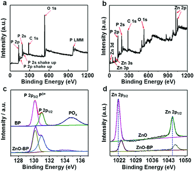 | ||
| Fig. 4 (a and b) Survey XPS spectra of BP and ZnO–BP, respectively. (c) P 2p spectra of BP and ZnO–BP. (d) Zn 2p spectra of ZnO and ZnO–BP. | ||
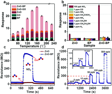
The responses of ZnO–BP composites and different reference materials towards 100 ppb NO2 were measured at various operating temperatures. As shown in Fig. 5a, the maximum response of the sensor based on ZnO–BP was 20 at 160 °C, and the response of ZnO sensor reached 5.2 at 140 °C. Clearly, the ZnO–BP sensor exhibited more than triple the response of that of the ZnO sensor toward NO2 gas. Furthermore, no noticeable responses were generated from the sensors based on BP, GO, and ZnO–GO materials when they were exposed to 100 ppb NO2. From the view of the practical application, a sensor should present a rather high selectivity. Therefore, the cross-sensitivities to 1 ppm oxidizing gases (SO2) and reducing gases (H2, CO, NH3, CH3COCH3, CH3CH2OH, and CH3OH) were also measured for the BP, ZnO and ZnO–BP sensors at 160 °C (Fig. 5b). It was seen that the ZnO–BP sensor exhibited the largest response to 100 ppb NO2 compared with those to 1 ppm of seven other gases, although the concentrations of others were much higher. Meanwhile, relatively lower responses to the interferential gases were observed for the composites than those for pure ZnO; this indicated that the response of ZnO to interferential gases was suppressed after its combination with BP. Due to an enhanced response to NO2 and the decreased responses to the other interferential gases, the as-prepared ZnO–BP composites exhibited an excellent selectivity to NO2. It is well-known that the response and recovery times are also important parameters for evaluating the properties of a gas sensor. The transient characteristics of the sensors based on ZnO–BP and ZnO toward 100 ppb NO2 were investigated at 160 °C (Fig. 5c). The ZnO–BP sensor demonstrated a rapid response, where the response and recovery times were about 9 and 15 s (Fig. S2, ESI†), respectively. Comparatively, the response and recovery times of ZnO were approximately 14 and 109 s, respectively. The ZnO–BP sensor even exhibited higher response–recovery values to NO2 than the sensors based on as-prepared ZnO and previously reported ZnO.28–31 Additionally, after BP decoration, the electrical resistance of ZnO hollow spheres increased, which was probably due to the formation of a heterojunction between p-type BP and n-type ZnO and the generation of an electrical barrier between crystal grains. Fig. 5d shows the transient responses of the ZnO–BP sensor sequentially exposed to 1, 5, 10, 50, 100, and 500 ppb NO2 at 160 °C (Fig. S3, 1–10![[thin space (1/6-em)]](https://www.rsc.org/images/entities/char_2009.gif) 000 ppb, ESI†). The response of the sensor increased with the increasing NO2 concentration. It is worth noting that the present sensor exhibited a clear response of 1.307 to NO2 concentration as low as 1 ppb. At lower NO2 concentrations (1–500 ppb), the response increased rapidly with the increasing NO2 concentration as most of the active sites on the surface of the sensing materials were available for chemical or physical interactions, whereas at higher gas concentrations, the sensor sensitivity gradually saturated because of lack of adequate active sites for further NO2 adsorption. The Japanese standards strictly regulated the threshold limit value (TLV) for NO2 to less than 40–60 ppb. Therefore, the ZnO–BP sensor can be suitable for the detection of low concentrations of NO2 for monitoring environmental pollution (Fig. S4, ESI†). In addition, for 100 ppb NO2, seven reversible cycles of the response curve maintained their initial value without a clear decrease, proving that the sensor exhibited good repeatability (Fig. S5, ESI†).
000 ppb, ESI†). The response of the sensor increased with the increasing NO2 concentration. It is worth noting that the present sensor exhibited a clear response of 1.307 to NO2 concentration as low as 1 ppb. At lower NO2 concentrations (1–500 ppb), the response increased rapidly with the increasing NO2 concentration as most of the active sites on the surface of the sensing materials were available for chemical or physical interactions, whereas at higher gas concentrations, the sensor sensitivity gradually saturated because of lack of adequate active sites for further NO2 adsorption. The Japanese standards strictly regulated the threshold limit value (TLV) for NO2 to less than 40–60 ppb. Therefore, the ZnO–BP sensor can be suitable for the detection of low concentrations of NO2 for monitoring environmental pollution (Fig. S4, ESI†). In addition, for 100 ppb NO2, seven reversible cycles of the response curve maintained their initial value without a clear decrease, proving that the sensor exhibited good repeatability (Fig. S5, ESI†).
Black phosphorene is a layered material similar to graphene. For graphene, good sensing properties have already been illustrated by both theoretical and experimental investigations.32,33 Therefore, a comparison of NO2 sensing performances using ZnO–BP composites as sensing materials and other reported graphene-based, BP-based and ZnO-based sensing materials is presented in Table 1. The ZnO–BP composite sensor displays much lower detection limit, faster response and recovery behavior and higher sensitivity compared to other reported materials.9,34–43
| Samples | Detection limit [ppb] | Response time [s] | Recovery time [s] | Response | Ref. |
|---|---|---|---|---|---|
| a Where ΔG = Ggas − G0; Ggas and G0 are the instantaneous conductances of the sensor detected under target gas exposure and before exposure to target gas, respectively. | |||||
| BP FETs | 5 | ∼280–350 | 130 | 2.9% (5 ppb, ΔG/G0)a | 9 |
| Suspended BP | 25![[thin space (1/6-em)]](https://www.rsc.org/images/entities/char_2009.gif) 000 000 |
∼20 | >20 | 45% (200 ppm, ΔR/R0) | 34 |
| PNSs | 20 | — | — | 190% (20 ppb, ΔG/G0) | 35 |
| RGO | 5000 | 3600 | 600 | 12% (5 ppm, ΔR/Ra) | 36 |
| RGO | 1000 | 420 (5 ppm) | 1680 (5 ppm) | 11.5% (5 ppm, ΔR/Ra) | 37 |
| ZnO | 10 | 270 | 240 | 37% (10 ppb, ΔR/Ra) | 38 |
| ZnO/RGO | 5000 | 96 (50 ppm) | 1552 (50 ppm) | <2% (5 ppm, ΔR/Ra) | 39 |
| ZnO/m-SWCNTs | 2500 | 208 | >208 | ∼52% (2.5 ppm, ΔR/Ra) | 40 |
| ZnO NR's | 1000 | 48 | 180 | 41% (1 ppm, ΔR/Ra) | 41 |
| ZnO BNW's | 1000 | 26 | 162 | 7.5% (1 ppm, ΔR/Ra) | 41 |
| RGO/SnO2 | 500 | 135 (5 ppm) | 200 (5 ppm) | 3.31 (5 ppm, Rg/Ra) | 42 |
| RGO/Cu2O | 64 | — | 500 (2 ppm) | 67.8% (2 ppm, ΔG/G0) | 43 |
| ZnO–BP | 1 | 2 | 16 | 130.7% (1 ppb, ΔR/Ra) | This study |
The possible reasons for the improved gas sensing properties of the ZnO–BP composite sensor are discussed. First, the introduction of BP could improve the electron transfer rate due its excellent carrier mobility, leading to fast response and recovery behavior to the target gas. Second, the large surface area derived from the existence of BP may promote gas adsorption and diffusion on the active surface. Third, the Schottky barrier around the interface of p–n junction can result in the specific migration of charge from ZnO to BP, as proved by XPS data. BP acting as a charge mediator may facilitate the charge transfer from ZnO to NO2 molecules, similar to the observations for WO3–GO composites reported elsewhere.44,45 Therefore, the significant enhancement in charge transfer, excellent carrier mobility and the promoted gas adsorption and diffusion due to the presence of BP account for the superior sensing performance of ZnO–BP composites, which exhibit high response, ultralow detection limit, fast response and excellent selectivity.
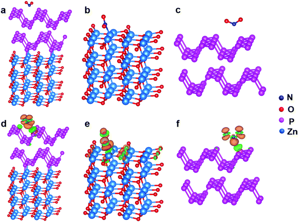
To understand the mechanism of the sensing properties at the atomic level, we performed first-principle calculations based on the density functional theory (DFT). Theoretical calculations were performed using the Quantum Espresso package,46 employing the GGA–PBE exchange correlation functional.47 The non-local van der Waals dispersion (vdW) corrections according to the vdW-DF2 procedure were taken into account.48,49 The geometry optimization was considered to be converged when the residual force on each atom was smaller than 0.01 eV Å−1. The calculated structures for NO2 adsorptions on ZnO–BP, ZnO and BP are shown in Fig. 6a, b and c, respectively, where NO2 molecules physisorb on ZnO–BP and BP and chemisorb on ZnO. The adsorption energy of NO2 molecules is calculated by the following formula:
| Ead = −(Etotal − Esubstrate − nEmolecule)/n | (1) |
| Δρ = ρtotal − ρmolecule − ρsubstrate | (2) |
Conclusions
In summary, ZnO–BP composites have been successfully prepared by the ultrasonic treatment of liquid dispersion of BP in the presence of ZnO hollow spheres synthesized by the microwave-assisted hydrothermal method. Interestingly, the introduction of ZnO significantly enhances BP environmental stability, making it a suitable candidate to use in practice. Remarkably, ZnO–BP sensor exhibits higher response, outstanding selectivity, faster response and recovery dynamic process, and an ultralow detection limit of 1 ppb to NO2 in contrast to graphene-, BP- and ZnO-based sensors, which were previously used. These characteristics are suggested to arise from significantly enhanced charge transfer as well as the promoted gas adsorption and diffusion due to the presence of BP. This study has provided a route in the search of BP-based materials with excellent gas-sensing properties, in particular towards NO2.Conflicts of interest
There are no conflicts to declare.Acknowledgements
The research is supported by Natural Science Foundation of Guangdong (2016A030313059), Science and Technology plan project of Guangdong (2017A010101018), Science and Technology plan project of Shenzhen (JCYJ20160307111047701, JCYJ20160226192754225, JCYJ20160307145209361, JCYJ20160307142444674).References
- P. Lu, A. Carvalho, J. Wu, H. W. Liu, E. S. Tok, A. H. C. Neto, B. Özyilmaz and C. H. Sow, Adv. Mater., 2016, 28, 4090 CrossRef PubMed.
- J. O. Island, G. A. Steele, H. S. J. V. D. Zant and A. Castellanos-Gomez, 2D Mater., 2015, 2, 011002 CrossRef.
- A. Castellanos-Gomez, L. Vicarelli, E. Prada, J. O. Island, K. L. Narasimha-Acharya, S. I. Blanter, D. J. Groenendijk, M. Buscema, G. A. Steele, J. V. Alvarez, H. W. Zandbergen, J. J. Palacios and H. S. Zant, 2D Mater., 2014, 1, 025001 CrossRef.
- N. Gillgren, D. Wickramaratne, Y. M. Shi, T. Espiritu, J. W. Yang, J. Hu, J. Wei, X. Liu, Z. Q. Mao, K. Watanabe, T. Taniguchi, M. Bockrath, Y. Barlas, R. K. Lake and C. N. Lau, 2D Mater., 2014, 2, 011001 CrossRef.
- Y. Q. Cai, G. Zhang and Y.-W. Zhang, J. Phys. Chem. C, 2015, 119, 13929 Search PubMed.
- X. Luo, Y. Rahbarihagh, J. C. M. Hwang, H. Liu, Y. C. Du and P. D. Ye, IEEE Electron Device Lett., 2014, 35, 1314 CrossRef.
- Y. Q. Cai, G. Zhang and Y.-W. Zhang, Sci. Rep., 2014, 4, 6677 CrossRef PubMed.
- Y. Q. Cai, Q. Q. Ke, G. Zhang and Y.-W. Zhang, J. Phys. Chem. C, 2015, 119, 3102 Search PubMed.
- A. N. Abbas, B. L. Liu, L. Chen, Y. Q. Ma, S. Cong, N. Aroonyadet, M. Köpf, T. Nilges and C. W. Zhou, ACS Nano, 2015, 9, 5618 CrossRef PubMed.
- J. Xu, L. Chen, Y.-W. Dai, Q. Cao, Q. Q. Sun, S. J. Ding, H. Zhu and D. W. Zhang, ACS Appl. Mater. Interfaces, 2017, 9, 1602246 Search PubMed.
- L. Z. Kou, T. Frauenheim and C. F. Chen, J. Phys. Chem. Lett., 2014, 5, 2675 CrossRef PubMed.
- X. Wang, J. Su, H. Chen, G.-D. Li, Z. F. Shi, H. F. Zou and X. X. Zou, ACS Appl. Mater. Interfaces, 2017, 9, 16335 Search PubMed.
- A. Maity and S. B. Majumder, Sens. Actuators, B, 2015, 206, 423 CrossRef.
- H. M. Fahad, H. Shiraki, M. Amani, C. C. Zhang, V. S. Hebbar, W. Gao, H. Ota, M. Hettick, D. Klriya, Y.-Z. Chen, Y.-L. Chueh and A. Javey, Sci. Adv., 2017, 3, 1602557 CrossRef PubMed.
- X. H. Liu, T. T. Ma, N. Pinna and J. Zhang, Adv. Funct. Mater., 2017, 27, 1702168 CrossRef.
- H.-R. Kim, A. Haensch, I.-D. Kim, N. Barsan, U. Weimar and J.-H. Lee, Adv. Funct. Mater., 2011, 21, 4456 CrossRef.
- D. Buso, M. Post, C. Cantalini, P. Mulvaney and A. Martucci, Adv. Funct. Mater., 2010, 18, 3843 CrossRef.
- Q. Li, Y. Du, X. J. Li, G. Y. Lu, G. W. Y. Wang, Y. F. Geng, Z. Q. Liang and X. Q. Tian, Sens. Actuators, B, 2016, 235, 39 CrossRef.
- M. T. Kelly, J. K. M. Chun and A. B. Bocarsly, Nature, 1996, 382, 214 CrossRef.
- C. G. Lee, H. G. Yan, L. E. Brus, T. F. Heinz, J. Hone and S. Ryu, ACS Nano, 2010, 4, 2695 CrossRef PubMed.
- W. L. Lu, H. Y. Nan, J. H. Hong, Y. M. Chen, C. Zhu, Z. Liang, X. Y. Ma, Z. H. Ni, C. H. Jin and Z. Zhang, Nano Res., 2014, 7, 853 CrossRef.
- L. Ge, X. Y. Jing, J. Wang, J. Wang, S. Jamil, Q. Liu, F. C. Liu and M. L. Zhang, J. Mater. Chem., 2011, 21, 10750 RSC.
- Y. H. M. S. T. Maruyam, Solid State Commun., 1982, 44, 877 CrossRef.
- A. Brown and S. Rundqvist, Acta Crystallogr., 2010, 19, 684 Search PubMed.
- J. D. Wood, S. A. Wells, D. Jariwala, K. S. Chen, E. Cho, V. K. Sangwan, X. Liu, L. J. Lauhon, T. J. Marks and M. C. Hersam, Nano Lett., 2014, 14, 6964 CrossRef PubMed.
- A. Ziletti, A. Carvalho, D. K. Campbell, D. F. Coker and A. Castro Neto, Phys. Rev. Lett., 2015, 114, 046801 CrossRef PubMed.
- H. U. Lee, S. C. Lee, J. Won, B. C. Son, S. Choi, Y. Kim, S. Y. Park, H.-S. Kim, Y.-C. Lee and J. Lee, Sci. Rep., 2015, 5, 8691 CrossRef PubMed.
- H. W. Kim, J. K. Yong, A. Mirzaei, S. Y. Kang, M. S. Choi, J. H. Bang and S. K. Sang, Sens. Actuators, B, 2017, 249, 590 CrossRef.
- J. Liu, S. Li, B. Zhang, Y. Xiao, Y. Gao, Q. Y. Yang, Y. L. Wang and G. Y. Lu, Sens. Actuators, B, 2017, 249, 715 CrossRef.
- J. K. Yong, A. Mirzaei, S. Y. Kang, M. S. Choi, J. H. Bang, S. K. Sang and H. W. Kim, Appl. Surf. Sci., 2017, 413, 242 CrossRef.
- R. Zhang, W. Pang, Z. H. Feng, X. J. Chen, Y. Chen, Q. Zhang, H. Zhang, C. L. Sun, J. J. Yang and D. H. Zhang, Sens. Actuators, B, 2017, 238, 357 CrossRef.
- O. Leenaerts, B. Partoens and F. M. Peeters, Phys. Rev. B: Condens. Matter Mater. Phys., 2007, 77, 125416 CrossRef.
- A. Miyamoto, Y. Kuwaki, T. Sano, K. Hatakeyama, A. Quitain, M. Sasaki and T. Kida, ACS Omega, 2017, 2, 2994 CrossRef.
- G. Lee, S. Kim, S. Jung, S. Jang and J. Kim, Sens. Actuators, B, 2017, 250, 569 CrossRef.
- S. M. Cui, H. H. Pu, S. A. Wells, Z. H. Wen, S. Mao, J. B. Chang, M. C. Hersam and J. H. Chen, Nat. Commun., 2015, 6, 8632 CrossRef PubMed.
- J. D. Fowler, M. J. Allen, V. C. Tung, Y. Yang, R. B. Kaner and B. H. Weiller, ACS Nano, 2009, 3, 301 CrossRef PubMed.
- P.-G. Su and H.-C. Shieh, Sens. Actuators, B, 2014, 190, 865 CrossRef.
- E. Oh, H. Y. Choi, S.-H. Jung, J. C. Kim, K.-H. Lee, S.-W. Kang, J. Kin and S.-H. Jeong, Sens. Actuators, B, 2009, 141, 239 CrossRef.
- N. Kumar, A. K. Srivastava, H. S. Patel, B. K. Gupta and G. D. Varma, Eur. J. Inorg. Chem., 2015, 1912 CrossRef.
- X. Li, J. Wang, D. Xie, J. L. Xu, Y. Xia and L. Xiang, Mater. Lett., 2017, 206, 18 CrossRef.
- Y. H. Navale, S. T. Navale, N. S. Ramgir, F. J. Stadler, S. K. Gupta, D. K. Aswal and V. B. Patil, Sens. Actuators, B, 2017, 251, 551 CrossRef.
- H. Zhang, J. C. Feng, T. Fei, S. Liu and T. Zhang, Sens. Actuators, B, 2014, 190, 472 CrossRef.
- S. Z. Deng, V. Tjoa, H. M. Fan, H. R. Tan, D. C. Sayle, M. Olivo, S. Mhaisalkar, J. Wei and C. H. Sow, J. Am. Chem. Soc., 2012, 134, 4905 CrossRef PubMed.
- X. Geng, J. J. You, J. Wang and C. Wang, Mater. Chem. Phys., 2017, 191, 114 CrossRef.
- X. Q. An, J. C. Yu, Y. Wang, Y. M. Hu, X. L. Yu and G. J. Zhang, J. Mater. Chem., 2012, 22, 8525 RSC.
- P. Giannozzi, S. Baroni, N. Bonini, M. Calandra, R. Car, C. Cavazzoni, D. Ceresoli, G. L. Chiarotti, M. Cococcioni, I. Dabo, A. D. Corso, S. de Gironcoli, S. Fabris, G. Fratesi, R. Gebauer, U. Gerstmann, C. Gougoussis, A. Kokalj, M. Lazzeri, L. Samos-Martin, N. Marzari, F. Mauri, R. Mazzarello, S. Paolini, A. Pasquarello, L. Paulatto, C. Sbraccia, S. Scandolo, G. Sclauzero, A. Seitsonen, A. Smogunov, P. Umari and R. M. Wentzcovitch, J. Phys.: Condens. Matter, 2009, 21, 395502 CrossRef PubMed.
- J. P. Perdew, K. Burke and M. Ernzerhof, Phys. Rev. Lett., 1996, 77, 3865 CrossRef PubMed.
- M. Dion, H. Rydberg, E. Schroder, D. C. Langreth and B. I. Lundqvist, Phys. Rev. Lett., 2004, 92, 246401 CrossRef PubMed.
- K. Lee, É. D. Murray, L. Kong, B. I. Lundqvist and D. C. Langreth, Phys. Rev. B: Condens. Matter Mater. Phys., 2010, 82, 081101 CrossRef.
Footnote |
| † Electronic supplementary information (ESI) available. See DOI: 10.1039/c8nh00052b |
| This journal is © The Royal Society of Chemistry 2018 |

