
 Open Access Article
 This Open Access Article is licensed under a
 Creative Commons Attribution 3.0 Unported Licence
Diameter-controlled Cu nanoparticles on saponite and preparation of film by using spontaneous phase separation†
Masaya Miyagawaa,
Akane Shibusawaa,
Kaho Maedaa,
Akiyoshi Tashirob,
Toshiki Sugaib and
Hideki Tanaka*a
aDepartment of Applied Chemistry, Faculty of Science and Engineering, Chuo University, 1-13-27, Kasuga, Bunkyo-ku, Tokyo, Japan. E-mail: htanaka@kc.chuo-u.ac.jp
bDepartment of Chemistry, Faculty of Science, Toho University, 2-2-1, Miyama, Funabashi-shi, Chiba, Japan
First published on 30th August 2017
Abstract
Cu nanoparticles have attracted much attention due to their optical, catalytic, and electrical properties. Their syntheses, however, have required hazardous reducing agents. In addition, they are easily oxidized, which destroys their properties. It has also not been possible to control the diameter and the dispersibility for further applications in functional materials. To solve these problems, we have recently synthesized oxide-free Cu nanoparticles adsorbed on kaolinite, a non-dispersive layered clay, by environmentally friendly photoreduction. Focusing on the layer charge of the clay, we report diameter- and dispersibility-controlled Cu nanoparticles on saponite, a dispersive clay, in the present study. The obtained oxide-free Cu nanoparticles were stable under N2 atmosphere, while they exhibited a fast back reaction to Cu2+ upon exposure to fresh air. The diameter of the Cu nanoparticles could be controlled simply with [Cu2+], because Cu2+ was adsorbed on saponite before the photoreduction. The dispersibility of the Cu nanoparticles was controlled without changing the diameter using the size of saponite aggregates: these were stably dispersed at low [Cu2+], and flocculated at high [Cu2+]. The flocculates could be collected by filtration and the obtained viscous paste was useful for preparing a film with the optical properties of the Cu nanoparticles without aggregation. Therefore, the present study revealed that combining the adsorption of Cu2+ on saponite with photoreduction is a novel and environmentally friendly synthetic method for controlling the diameter and the dispersibility of Cu nanoparticles, and it has the potential to further their applications in functional materials.
Introduction
As an alternative to precious metals such as Pt, Au, and Rh, Cu nanoparticles (NPs) have attracted much attention due to their large reserves.1–4 Their optical, catalytic, and electrical properties have recently been energetically investigated.3–11 However, the Cu NPs have generally been synthesized by a chemical reduction method, which requires hazardous reducing agents such as sodium borohydride3,5,8 and hydrazine.2 While reduction with a safer agent has also been investigated,9,12 extensive efforts have been required to optimize multiple conditions such as reaction temperature and reducing agent concentration. In contrast, photoreduction has shown potential since the 21st century due to its environmentally friendly conditions.13–16In most of the previous studies, the metal NPs have been stabilized by protection with polymers14,17 or surface modifiers such as carboxylates18,19 and thiolates.20,21 In contrast, we have previously reported protectant-free Cu NPs adsorbed on kaolinite, a non-exfoliating layered clay mineral, by the photoreduction method.22 In this synthetic method, ethanol has been used as a sacrificial reagent instead of hazardous reducing agents, and the obtained NPs have been composed of pure Cu without oxides, which was notable because Cu NPs are generally easily oxidized to Cu2O and CuO.2,23–25 It is true that oxide-free Cu NPs have been reported previously, in which the Cu NPs are protected with SiO2 (ref. 26) or oleic acid,27 but it has been reported that the protection decreases or destroys the reactivity of the NPs.28,29 In addition, multi-step procedures including heat treatment and centrifugation have also been indispensable in removing excess protectants and hazardous reducing agents, which lowers the yield of the Cu NPs. From these viewpoints, our photoreduction method in combination with clay is promising because the protectant-free pure Cu NPs are obtained without further procedures by using a mild sacrificial reagent.
Unlike kaolinite with non-swelling properties and hence poor dispersibility, smectite clay minerals consist of negatively charged ultra-thin layers and interlayer cations for charge compensation. Because they swell in water, they are easily exfoliated, and the layers, so called nanosheets, are obtained as colloidal solutions. Despite numerous studies on layered clay minerals involving pillaring30,31 and hybridization with cationic molecules including dyes and metal complexes,32–34 however, there have been few studies on hybridization with metal NPs, and these have mainly been limited to Au and Ag NPs synthesized by chemical reduction.35–37 In addition, their diameter control on clay has not been achieved, even though the diameter strongly affects the reactivity of the Cu NPs. The diameter of metal NPs can be affected by various parameters, for instance reaction temperature,38 reaction time,39 metal salts,40 the ratio of ligand to metal salt,41 and the presence of templates such as dendrimers.42 However, no significant change in the diameter has been achieved simply by changing [Cu2+] on an adsorbent, for example graphene oxide.9 This problem may originate from uncontrollable particle growth in solutions without protectants. Hence, we have assumed that growth on an adsorbent would be key to controlling the diameter of the Cu NPs. In this regard, the negatively charged nanosheets of the smectite clay appear to be a promising adsorbent for Cu NPs, because they can effectively adsorb metal cations.43,44 In this case, nucleation and growth should occur on the adsorbent and not in the solution. In other words, the growth of the reduced Cu seeds is expected to be suppressed on clay, resulting in diameter control of the Cu NPs with [Cu2+]. This strategy could not have been implemented in the previous study using kaolinite22 because this does not adsorb Cu2+, and particle growth of the Cu NPs has occurred in the solution before adsorption, resulting in a relatively large diameter (80 nm).
As well as diameter control, dispersibility control of colloidal NPs has also been important in their extraction, selective dispersion, and recycling.45–48 However, contrary to the dispersibility of the polymer-grafted NPs, that of the adsorbed NPs has scarcely been investigated, even though they may become a promising material due to their high reactivity originating from the absence of protectants. In this regard, hybridization of the Cu NPs with clay can be a first experimental example because the dispersibility of the Cu NPs should be explained by that of the adsorbent, clay nanosheets. In this case, the Cu NPs can be collected as clay-hybridized flocculates and be further applied to NP films. Furthermore, because the Cu NPs are expected to be separated from each other by the clay nanosheets, the film can be obtained without aggregation of the Cu NPs, which has potential for optical materials.
Even though combination of the environmentally friendly photoreduction method and the layered clay minerals has not yet been studied, it will provide novel viewpoints with respect to both protectant-free metal NPs and inorganic layered materials. In the present study, we have synthesized highly reactive oxide-free Cu NPs on saponite (Sapo) by the photoreduction method. We have controlled the diameter and the dispersibility of the Cu NPs by changing [Cu2+] and [Sapo]. By using flocculation and following conventional filtration, we have developed a novel preparation method for a composite film of Cu NPs, which were not aggregated due to isolation by the clay matrix.
Experimental
Copper nanoparticles were synthesized by a photoreduction method similar to that reported previously.22 Copper(II) acetate monohydrate (purity > 99.0%) and ethanol (purity > 99.5%) were purchased from Wako Pure Chemical Industries. Synthetic saponite was purchased from Kunimine Ind. Co., and its elemental composition and cation exchange capacity (CEC) were determined to be (Na0.25Mg0.07)(Mg2.98Al0.01)(Si3.6Al0.4)O10(OH)2 and 80 meq. per 100 g, respectively.49 Sapo was dispersed in Milli-Q water, and 5 mL of copper acetate aqueous solution and 1 mL of ethanol were added to the dispersion drop by drop with stirring. The total volume of the mixed solution was adjusted to 10 mL by adding water. The solution was irradiated with UV light from a Hg lamp (Hamamatsu Photonics, L9588-01A) for 2 h with stirring. To prepare the Cu NP film, a small amount of the Cu NP paste obtained by filtration of flocculates was cast on a glass plate, sandwiched with another glass plate, and dried under N2 atmosphere.UV-vis-NIR spectra were measured with a UV-vis-NIR spectrometer (Shimadzu, UV-3600) using a rectangular quartz cuvette having an optical length of 10 mm. The photoirradiated solution was dropped on a glass plate under N2 atmosphere and dried in vacuum. X-ray diffraction (XRD) measurement of the obtained powder sample was performed with an X-ray diffractometer (Rigaku, Smartlab) with CuKα radiation (0.154 nm). For transmission electron microscopy (TEM) measurement, the photoirradiated solution was diluted to avoid charge-up originating from the insulative saponite and dropped on a collodion-coated copper grid under N2 atmosphere. TEM images of the dried grid were observed with a transmission electron microscope (FEI Company, Tecnai G2 F20 S-TWIN), operated at 120 kV, and with a field-emission scanning electron microscope (Hitachi High-Technologies, S-5500) operated at 25 kV. Dynamic light scattering (DLS) measurement of the photoirradiated solution was performed with a particle size analyser (Otsuka Electronics Co., Ltd., ELSZ-2000ZS).
Results and discussion
Oxide-free Cu NPs adsorbed on Sapo
When the mixed solution containing 0.5 mmol L−1 of Cu2+ and 0.5 g L−1 of Sapo was irradiated with UV light for 2 h, a bright red solution was obtained, as shown in Fig. 1(a). This color was characteristic of surface plasmon resonance (SPR) of Cu NPs,50 suggesting that Cu2+ was reduced to Cu0 by UV irradiation. The photoirradiated solution (we call this sample {0.5![[thin space (1/6-em)]](https://www.rsc.org/images/entities/char_2009.gif) :0.5} of Cu–Sapo, hereafter) was not separated into two phases but was stably dispersed. No large flocculates were observed with the naked eye, and the solution appeared homogeneous.
:0.5} of Cu–Sapo, hereafter) was not separated into two phases but was stably dispersed. No large flocculates were observed with the naked eye, and the solution appeared homogeneous.
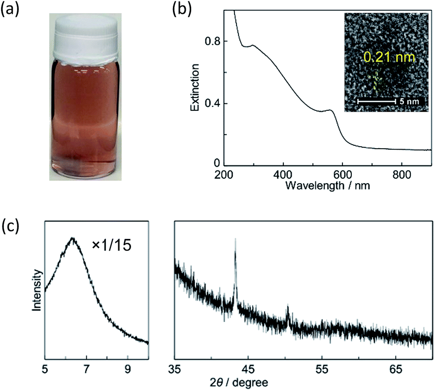 | ||
| Fig. 1 (a) A photograph of the photoirradiated solution, (b) a UV-vis-NIR extinction spectrum and a high-resolution TEM image of the Cu NP (inset), and (c) an XRD pattern of {0.5:0.5}. | ||
Fig. 1(b) shows a UV-vis-NIR extinction spectrum after UV irradiation. A band due to SPR was observed around 560 nm, which originated from spherical Cu NPs.5,51 We note that the band of Cu2+ appearing around 780 nm before UV irradiation, which is shown in Fig. S1(a),† completely disappeared. Also, the spectrum before UV irradiation was almost consistent with the sum of the spectra of the pure copper acetate solution and the Sapo dispersions shown in Fig. S1(b and c†) respectively, suggesting that adsorption on Sapo did not induce a drastic change in the electronic state of Cu2+. Fig. 2(a) and S2† show TEM images of the dried sample at high and low magnifications, respectively. The spherical Cu NPs were observed without aggregation. From the histogram shown in Fig. S3(a),† the average diameter was determined to be 13 ± 2 nm, where the uncertainty represents one standard deviation. It should be noted that the blurring of the Cu NPs originates from the adsorbent, Sapo, which is insulative to induce charge-up. Similar results have also been reported in previous studies on clay-hybridized metal NPs.52,53
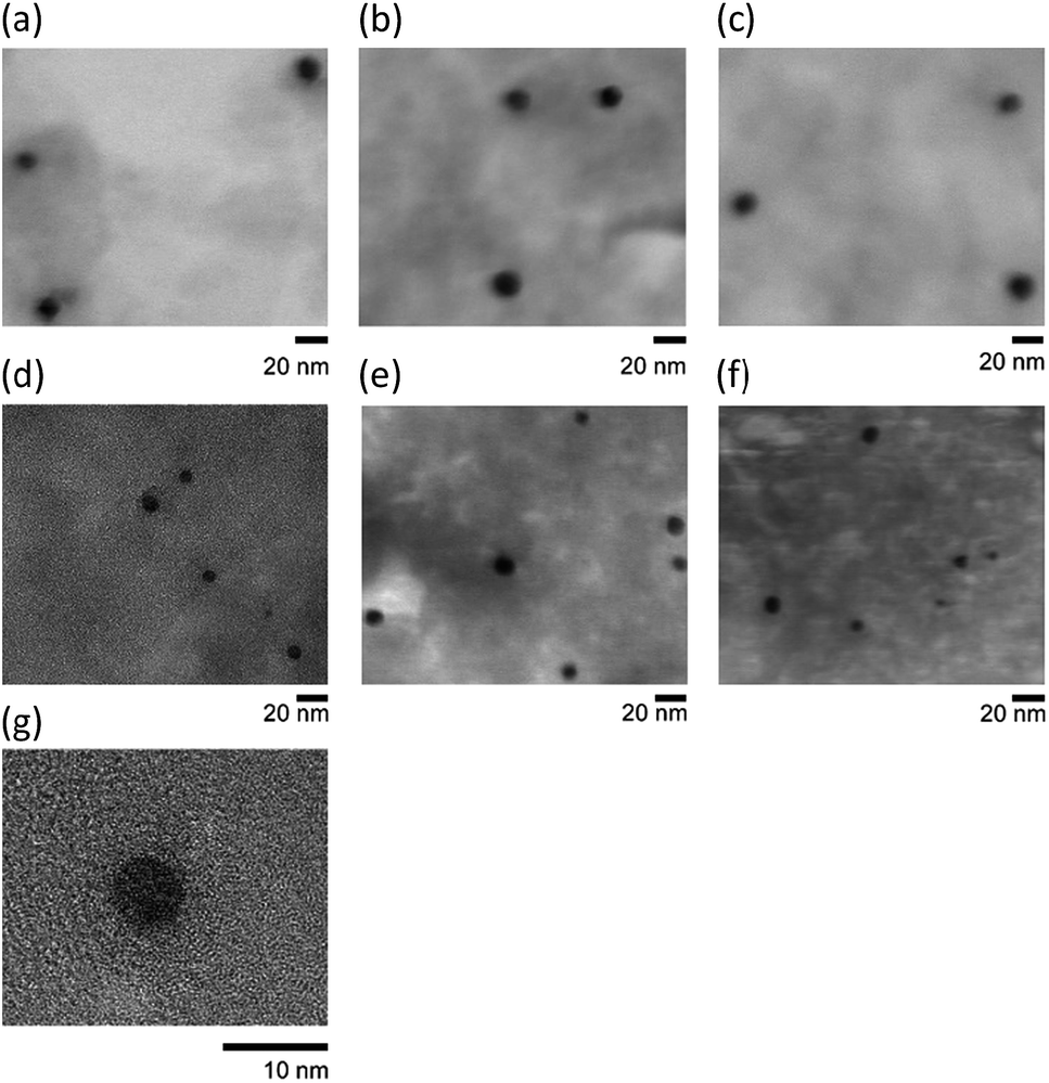 | ||
| Fig. 2 TEM images of (a) {0.5:0.5}, (b) {1:1}, (c) {2:2}, (d) {0.2:0.5}, (e) {0.4:1}, (f) {0.8:2}, and (g) {0.1:0.5}. | ||
By comparison with a control experiment, where large Cu particles with sub-mm sizes were obtained in the absence of Sapo, as reported previously,22 it was found that the Cu NPs were adsorbed on Sapo and their particle growth was suppressed. In addition, while the photoirradiated solution was stable under N2 atmosphere, the solution turned pale light blue from bright red on exposure to fresh air, and copper acetate was detected with UV-vis-NIR spectroscopy, as in our previous studies.4,22,50 It is noted that the bright red color faded away in 10 seconds, which was much faster than the color change in the Cu NPs protected by polyvinylpyrrolidone (20 min).50 We assume that this rapid decomposition of the Cu NPs on Sapo can be ascribed to the absence of protectants. Therefore, it was found that spherical Cu NPs were obtained on Sapo by UV irradiation and that their decomposition occurred quickly, which was probably due to the use of adsorbent rather than protectant.
A high-resolution TEM image is shown in the inset of Fig. 1(b). Crystal planes were observed with a fringe spacing of 0.21 nm, which corresponded to the (111) plane of the fcc phase of metal copper according to JCPDS file no. 4-0836. Thus, it was found that the obtained NPs were composed of pure Cu. This result contrasts with our previous study, in which a core–shell structure composed of Cu-core and Cu2O-shell was obtained when carbon nanotubes were used as the adsorbent.23 In other words, it was found that protectant-free Cu NPs were synthesized without oxides when Sapo was used as the adsorbent.
The absence of Cu2O and CuO was further confirmed by XRD measurement, as shown in Fig. 1(c). Two peaks were observed at 43.3° and 50.4°, corresponding to the (111) and (200) planes of crystalline Cu metal, respectively. No peaks for Cu2O and CuO were observed at 36.4° and 35.5°, respectively (JCPDS files no. 5-0067 and 48-1548). Thus, it was found that Cu2+ was reduced completely to Cu0 by UV irradiation. In addition, while the peak of the (001) plane of Sapo was observed at 6.4°, other strong peaks such as those at 53.2° and 60.7°, which were assigned to the (150) and (310) planes, and the (060) and (330) planes, respectively, were not observed,54 indicating that the layer structure of Sapo was destroyed. Because these peaks were also absent in the XRD pattern before UV irradiation, it was suggested that Sapo aggregated randomly when the copper acetate aqueous solution was added to the Sapo dispersion. The formation of the Sapo aggregates was further supported by DLS measurement. The average particle size after UV irradiation was found to be 560 nm, which was much larger than that of the single nanosheet, 33 nm,55 indicating that Sapo aggregated microscopically in the solution rather than during the drying process for XRD sample preparation. Therefore, we concluded that Sapo supported the production of oxide- and protectant-free Cu NPs and also aggregated microscopically. The aggregation of Sapo is plausible because excess Cu2+ was added, which will be described later in relation to the CEC of Sapo.
Diameter control of Cu NPs
As well as {0.5:0.5}, Cu–Sapo was synthesized with various [Cu2+] and [Sapo], and the TEM images are shown in Fig. 2. The corresponding histograms are shown in Fig. S3.† The average diameters of the Cu NPs are summarized in Table 1. The diameters were 13 ± 3 nm in both {1:1} and {2:2}, which were the same as that in {0.5:0.5} within the error range. Similar results were obtained in {0.4:1} and {0.8:2}, whose diameters were the same as that in {0.2:0.5}, 8 ± 2 nm. Hence, it was found that the diameters did not change significantly if the ratio of [Cu2+] to [Sapo] was constant. In contrast, the diameters decreased from 13 ± 2 nm in {0.5:0.5} to 8 ± 2 and 6 ± 2 nm in {0.2:0.5} and {0.1:0.5}, respectively, indicating that the diameter of the Cu NPs could be controlled simply with [Cu2+]. Thus, the adsorption of Cu2+ on Sapo before photoreduction was found to be effective in controlling the diameter of the Cu NPs. In all cases, the SPR bands of the Cu NPs were observed, as shown in Fig. S4.†
| Sample | Diameter/nm |
|---|---|
| {0.1:0.1} |
6 ± 2 |
| {0.2:0.5} |
8 ± 2 |
| {0.4:1} |
8 ± 2 |
| {0.8:2} |
8 ± 2 |
| {0.5:0.5} |
13 ± 2 |
| {1:1} |
13 ± 2 |
| {2:2} |
13 ± 2 |
Since the microscopic aggregation of Sapo was induced by the added Cu2+ spontaneously adsorbing on Sapo, we expected the diameter of the Cu NPs to be changed by the drip rate of the copper acetate aqueous solution during sample preparation. When the drip rate was changed from 0.21 mL min−1 to 1.4 mL min−1, the average diameter of the Cu NPs in {1:1} increased from 13 ± 3 nm to 20 ± 2 nm. A similar result was also obtained in the case of {2:2}, where the diameter increased from 13 ± 3 to 24 ± 6 nm with a fast drip rate. TEM images and histograms of {1:1} and {2:2} with the fast drip rate are shown in Fig. S5(a and b) and S3(a and b),† respectively. The increase in the diameter of the Cu NPs with the fast drip rate implies two possibilities: one is that larger Sapo aggregates containing more Cu2+ were formed with the fast drip rate, facilitating particle growth when Cu2+ was reduced by UV irradiation. The other possibility is that non-adsorbing Cu2+ was confined to the Sapo aggregates with the fast drip rate.
The formation of the large Sapo aggregates in the former scenario originates from the assumption that Sapo nanosheets aggregate more intensively with the fast drip rate, which was confirmed by the DLS measurement. The size of the Sapo aggregates with the fast drip rate was twice as large as that with the slow drip rate, and more Cu2+ was contained in each Sapo aggregate to increase the diameter of the Cu NPs. In contrast, if the latter is true, the amount of free Cu2+ in the solution should decrease with the fast drip rate. To clarify this, the solution was centrifuged before UV irradiation. Fig. S6† shows bands of Cu2+ in the UV-vis-NIR absorption spectra of the supernatant in {2:2} prepared with slow and fast drip rates. Because no difference was found between the two spectra, the latter scenario was improbable, and the former was more plausible. Therefore, we concluded that the diameter of the Cu NPs could be controlled not only with [Cu2+] but also with the drip rate during sample preparation.
Dispersibility control of Cu NPs by microscopic aggregation of Sapo
Unlike {0.5:0.5} of Cu–Sapo, {2:2} was separated into two phases after keeping the solution unstirred for 1 day. We then examined the effects of [Cu2+] and [Sapo] on the dispersibility of Cu–Sapo, as shown in Fig. 3. It was found that Cu–Sapo was stably dispersed regardless of [Sapo] at [Cu2+] = 1 mmol L−1, while it flocculated spontaneously at [Sapo] = 2 g L−1 when [Cu2+] was relatively high. In {2:2}, {2:3}, and {2:4}, the upper layer was totally colorless and transparent, while the lower layer was red. As expected, the SPR band of the Cu NPs was not observed in the UV-vis-NIR absorption spectrum of the upper layer, as shown in Fig. S7.† Thus, Cu–Sapo was found to be flocculated. It should be noted that the flocculates were not precipitates, because they were transiently stable when the phase-separated solution was stirred. In other words, the stirred solution could be handled as a stable dispersion, because the flocculation took more than 1 h.
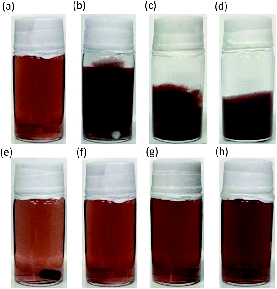 | ||
| Fig. 3 Photographs of (a) {1:2}, (b) {2:2}, (c) {3:2}, (d) {4:2}, (e) {1:1}, (f) {1:2}, (g) {1:4}, and (h) {1:6}. | ||
Since the average diameter of the Cu NPs in {2:2} was the same as that in {0.5:0.5}, the dispersibility of the Cu NPs could be controlled without changing their average diameter. In addition, because large flocculates were observed with the naked eye, the Sapo aggregates were expected to be relatively large. Indeed, the DLS measurement revealed that the size of {2:2} was larger than 10 μm. We note that the size was much larger than that of {0.5:0.5} at 560 nm. Therefore, we concluded that the dispersibility of the Cu NPs was controlled by the size of the Sapo aggregates without changing the average diameter of the Cu NPs.
Fig. 4 shows a dispersion-state phase diagram of Cu–Sapo. At [Cu2+] ≤1 mmol L−1, Cu–Sapo was stably dispersed regardless of [Sapo]. In contrast, it was mostly flocculated at [Cu2+] ≥2 mmol L−1. The fact that Cu–Sapo was not flocculated regardless of [Sapo] at [Cu2+] ≤1 mmol L−1 seems rather peculiar, because the Cu NPs were dispersed in the presence of negatively charged Sapo nanosheets and cations for charge compensation, which should be regarded as a salt to induce the flocculation of general colloidal NPs. We assume that the adsorption on Sapo enabled the Cu NPs to be stably dispersed even at high [Sapo]. In other words, the aggregation of Sapo was induced by the Cu NPs. In addition, the DLS measurement revealed that the Sapo aggregates were relatively small at low [Cu2+], suggesting that the extent of aggregation of Sapo was low, resulting in the stable dispersion of Cu–Sapo. In contrast, larger Sapo aggregates were formed at high [Cu2+], followed by the flocculation of Cu–Sapo, probably because the presence of more Cu NPs caused the Sapo nanosheets to aggregate enough to cause macroscopic flocculation. Also, because Cu–Sapo was stably dispersed in {2:0.5} and {2:1} but flocculated in {2:2}, it was suggested that the more nanosheets there were, the larger the aggregates became, resulting in macroscopic flocculation at high [Sapo]. Therefore, we concluded that the dispersibility of Cu–Sapo was controlled predominantly by [Cu2+] without changing the diameter of the Cu NPs, and the adsorption of the Cu NPs on Sapo enabled Cu–Sapo to be stably dispersed even at high [Sapo] if [Cu2+] was low.
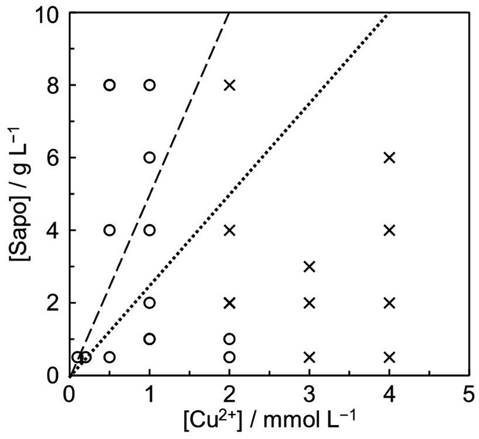 | ||
| Fig. 4 Dispersion-state phase diagram of Cu–Sapo. Symbols ○ and × represent ‘dispersed’ and ‘flocculated’, respectively, when the photoirradiated solution was kept unstirred for 1 day. The dotted and broken lines represent 100% and 50% of Cu2+ against the CEC of Sapo. | ||
Preparation of Cu–Sapo film from the flocculates
To prepare the Cu–Sapo film, the flocculates were filtered, as shown in Fig. S8(a).† Because the Cu NPs were obtained not as a precipitate but as transiently dispersed flocculates, filtration was needed to concentrate the Cu–Sapo, contrary to the electroless deposition reported previously.56,57 The paste was so viscous that it did not drop even when it was turned upside down, as shown in Fig. 5(a). The collection rate of Cu–Sapo by filtration was 88 wt%, suggesting that the Cu NPs could be easily collected as flocculates. In contrast, the filtrate was colorless and transparent, as shown in Fig. S8(b).† Because no SPR band was observed in the UV-vis-NIR absorption spectrum of the filtrate shown in Fig. S9,† it was found that the Cu–Sapo flocculates could be concentrated efficiently as a paste.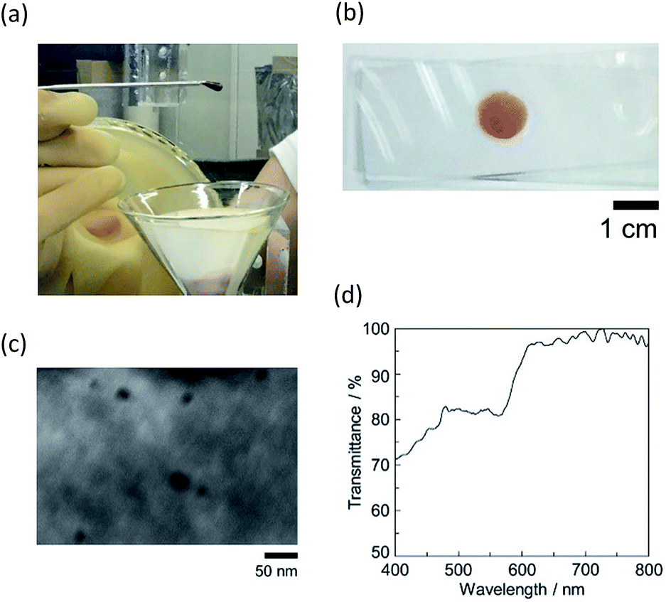 | ||
| Fig. 5 Photographs of (a) the Cu–Sapo paste turned upside down, prepared from the flocculates of {2:2} and (b) the film after drying, (c) a TEM image of the film, and (d) the visible transmission spectrum of the film. | ||
Fig. 5(b) shows a Cu–Sapo film obtained from the paste in {2:2}, whose preparation using the paste is described in the Experimental section. The film was bright red, like the flocculates, suggesting that the Cu NPs were not aggregated in the clay matrix. Indeed, no aggregation of Cu NPs with a similar diameter to those before filtration was observed in the TEM image prepared by casting a small amount of the paste on a grid followed by drying under N2 atmosphere, which is shown in Fig. 5(c). Fig. 5(d) shows a transmission spectrum of the central part of the film. Because no shoulder peak was observed on the long-wavelength side of the SPR band at 560 nm, it was confirmed that the Cu NPs were not aggregated in the film. In addition, the SPR band was not shifted from that measured for the solution. This fact also confirms that non-aggregated Cu NPs with the same diameter are distributed in the film. All these facts should originate from the isolation of the Cu NPs with the clay matrix. Thus, it was found that the optical properties of the Cu NPs were preserved in the photoinactive clay film. Furthermore, the content of the Cu NPs in the film could be changed easily from 1.6 wt% in {2:8} to 34 wt% in {4:0.5}. These values were calculated based on the facts that Cu2+ was completely reduced to Cu0, and that all the Cu NPs and Sapo aggregates were contained in the residue, both of which are supported by Fig. S7 and S9.† Compared to general polymer-grafted NPs, the collection of Cu–Sapo and the preparation of the film by flocculation appears to be a simple method because the dispersibility of Cu–Sapo was controlled easily by [Cu2+].
It should be noted that flocculation played an important role in preparing the film because the paste obtained by filtration was viscous. For comparison, when the same experiment was performed with a stable dispersion, such as {1:1}, the dripped dispersion was sandwiched over the whole glass, and the size of the film could not be controlled. In addition, since the dispersion could not be concentrated by filtration, the obtained film was almost transparent. In other words, the Cu–Sapo film could not be prepared substantially. Thus, it was found that the spontaneous phase separation of the Cu–Sapo solution was useful in preparing the film. In addition, the bright red film could be redispersed in water, because Cu–Sapo existed as flocculates rather than precipitates. This characteristic is based on the hydrophilicity of Sapo. Therefore, we concluded that the spontaneous phase separation of Cu–Sapo was useful in collecting the Cu NPs efficiently on a macroscopic scale as a paste or a film, where the Cu NPs were isolated in the clay matrix without aggregation. We also believe that filtration and the subsequent simple preparation method of the Cu–Sapo film are useful both in restoring the Cu NPs and in applying them in optical and catalytic materials.
Relationship between the dispersibility of Cu–Sapo and CEC of Sapo
One may wonder about the effect of the added amount of Cu2+ on the CEC of Sapo, which represents the maximum amount of cations that can be adsorbed. This was more than 100% in most of the Cu–Sapo syntheses. For example, the added amount of Cu2+ was 250% of the CEC of Sapo in {0.5:0.5}. In other words, 40% of the Cu2+ was adsorbed on Sapo, while the remaining 60% was dissolved in the solution. Indeed, when {0.5:0.5} was kept unstirred before UV irradiation, the solution separated into two phases, as shown in Fig. S10.† Thus, it was found that large Cu2+-adsorbed Sapo aggregates were formed, resulting in flocculation. The DLS measurement further confirmed this finding, where the size of the Sapo aggregates before UV irradiation was larger than 10 μm. As well as the flocculates, the supernatant was also investigated. Fig. S11† shows a UV-vis-NIR absorption spectrum of the supernatant before UV irradiation. A weak band was observed around 800 nm with an intensity of 0.005. Based on the facts that a band of Cu2+ was observed at 780 nm with an intensity of 0.010 for the pure copper acetate solution without Sapo, as shown in Fig. S1(a),† and 40% of the Cu2+ was adsorbed in the presence of Sapo, the obtained intensity was consistent with the estimated value. The bathochromic shift of the band is ascribed to a change in the molecular interaction of Cu2+ due to partial adsorption on Sapo.
Because of the excess amount of Cu2+ in {0.5:0.5}, one may suspect that Cu nanocrystals are formed not only on Sapo but also in solution. In the latter case, secondary particles should be formed, which were not observed in the TEM image. In addition, because the size distribution of the Cu NPs was relatively monodisperse, it was suggested that photo-reduced Cu2+ was adsorbed on Sapo before overcoming the threshold for nucleation, which was consumed during the growth of the existing seeds produced from Cu2+ on Sapo by the initial UV irradiation. Furthermore, because Sapo becomes negatively charged again by the reduction of Cu2+ on Sapo, Cu2+ in the solution is assumed to be subsequently adsorbed on Sapo and should be consumed during the growth of the existing seeds on Sapo. The photoreduction mechanism of Cu2+ was discussed kinetically in detail in our previous study.22
Conclusions
In the present study, Cu nanoparticles were synthesized on saponite by an environmentally friendly photoreduction method. The obtained Cu nanoparticles contained no oxides and were stable under N2 atmosphere. The diameter could be easily controlled from 6 to 24 nm with [Cu2+] and the drip rate of copper acetate solution. In addition, the dispersibility of the Cu nanoparticles was controlled mainly with [Cu2+] without changing the diameter of the Cu nanoparticles, and the size of the saponite aggregates was found to play an important role. The Cu nanoparticles could be collected from the flocculates by filtration, and the film was prepared from the obtained paste, where the Cu nanoparticles were distributed in the clay matrix without aggregation. By combining the above results, we established a synthetic method for protectant-free pure Cu nanoparticles, where not only the diameter but also the dispersibility could be controlled, and which has the potential to further applications in functional materials.Conflicts of interest
There are no conflicts to declare.Acknowledgements
This work was supported by MEXT KAKENHI Grant Number 25248004 and by JSPS KAKENHI Grant Number 25410028. This work was also supported in part by the Supported Program for Strategic Research Foundation at Private Universities (2012–2016), Futaba Memorial Foundation, Shorai Foundation for Science and Technology, the Ogasawara Foundation for the Promotion of Science & Engineering, and Foundation Advanced Technology Institute.Notes and references
- R. Reske, H. Mistry, F. Behafarid, B. R. Cuenya and P. Strasser, J. Am. Chem. Soc., 2014, 136, 6978 CrossRef CAS PubMed.
- D. Talukdar, G. Das, S. Thakur, N. Karak and A. Thakur, Catal. Commun., 2015, 59, 238 CrossRef CAS.
- B. J. Borah, S. J. Borah, L. Saikia and D. K. Dutta, Catal. Sci. Technol., 2014, 4, 1047 CAS.
- M. Miyagawa, M. Yonemura and H. Tanaka, Chem. Phys. Lett., 2016, 665, 95 CrossRef CAS.
- I. Lisiecki, F. Billoudet and M. P. Pileni, J. Phys. Chem., 1996, 100, 4160 CrossRef CAS.
- G. H. Chan, J. Zhao, E. M. Hicks, G. C. Schatz and R. P. Van Duyne, Nano Lett., 2007, 7, 1947 CrossRef CAS.
- S. Xu, B. Man, S. Jiang, J. Wang, J. Wei, S. Xu, H. Liu, S. Gao, H. Liu, Z. Li, H. Li and H. Qiu, ACS Appl. Mater. Interfaces, 2015, 7, 10977 CAS.
- B. J. Borah, D. Dutta, P. P. Saikia, N. C. Barua and D. K. Dutta, Green Chem., 2011, 13, 3453 RSC.
- I. Shown, H.-C. Hsu, Y.-C. Chang, C.-H. Lin, P. K. Roy, A. Ganguly, C.-H. Wang, J.-K. Chang, C.-I. Wu, L.-C. Chen and K.-H. Chen, Nano Lett., 2014, 14, 6097 CrossRef CAS PubMed.
- N. N. Jason, W. Shen and W. Cheng, ACS Appl. Mater. Interfaces, 2015, 7, 16760 CAS.
- W.-H. Chung, H.-J. Hwang and H.-S. Kim, Thin Solid Films, 2015, 580, 61 CrossRef CAS.
- N. Kumar and L. S. B. Upadhyay, Appl. Surf. Sci., 2016, 385, 225 CrossRef CAS.
- R. Jin, Y. W. Cao, C. A. Mirkin, K. L. Kelly, G. C. Schatz and J. G. Zheng, Science, 2001, 294, 1901 CrossRef CAS PubMed.
- Y. Zhai, J. S. DuChene, Y.-C. Wang, J. Qiu, A. C. Johnston-Peck, B. You, W. Guo, B. DiCiaccio, K. Qian, E. W. Zhao, F. Ooi, D. Hu, D. Su, E. A. Stach, Z. Zhu and W. D. Wei, Nat. Mater., 2016, 15, 889 CrossRef CAS PubMed.
- H. Murayama, N. Hashimoto and H. Tanaka, Chem. Phys. Lett., 2009, 482, 291 CrossRef CAS.
- T. Nakato and T. Kasai, Mater. Lett., 2011, 65, 3402 CrossRef CAS.
- H. Murayama, N. Hashimoto and H. Tanaka, Chem. Phys. Lett., 2009, 482, 291 CrossRef CAS.
- M. Wu, D. N. Congreve, M. W. B. Wilson, J. Jean, N. Geva, M. Welborn, T. V. Voorhis, V. Bulović, M. G. Bawendi and M. A. Baldo, Nat. Photonics, 2016, 10, 31 CrossRef CAS.
- J. Deka, A. Paul and A. Chattopadhyay, Nanoscale, 2010, 2, 1405 RSC.
- N. Nishida, Y. Kojima and H. Tanaka, Chem. Lett., 2014, 43, 1227 CrossRef CAS.
- N. R. Jana, L. Gearheart and C. J. Murphy, Langmuir, 2001, 17, 6782 CrossRef CAS.
- M. Miyagawa, T. Maeda, R. Tokuda, A. Shibusawa, T. Aoki, K. Okumura and H. Tanaka, RSC Adv., 2016, 6, 104560 RSC.
- N. Nishida, A. Miyashita, T. Tsukuda and H. Tanaka, Chem. Lett., 2013, 42, 168 CrossRef CAS.
- Y. Kobayashi and T. Sakuraba, Colloids Surf., A, 2008, 317, 756 CrossRef CAS.
- H.-J. Hwang, S.-J. Joo and H.-S. Kim, ACS Appl. Mater. Interfaces, 2015, 7, 25413 CAS.
- S. Shiomi, M. Kawamori, S. Yagi and E. Matsubara, J. Colloid Interface Sci., 2015, 460, 47 CrossRef CAS PubMed.
- S. Jeong, S. H. Lee, Y. Jo, S. S. Lee, Y.-H. Seo, B. W. Ahn, G. Kim, G.-E. Jang, J.-U. Park, B.-H. Ryu and Y. Choi, J. Mater. Chem. C, 2013, 1, 2704 RSC.
- C. Kim, B. L. Suh, H. Yun, J. Kim and H. Lee, ACS Catal., 2017, 7, 2294 CrossRef CAS.
- M. Cargnello, C. Chen, B. T. Diroll, V. V. T. Doan-Nguyen, R. J. Gorte and C. B. Murray, J. Am. Chem. Soc., 2015, 137, 6906 CrossRef CAS PubMed.
- R. Sasai, R. Watanabe and T. Yamada, Appl. Clay Sci., 2014, 93–94, 72 CrossRef CAS.
- S. Hamidouche, O. Bouras, F. Zermane, B. Cheknane, M. Houari, J. Debord, M. Harel, J.-C. Bollinger and M. Baudu, Chem. Eng. J., 2015, 279, 964 CrossRef CAS.
- D. Tokieda, T. Tsukamoto, Y. Ishida, H. Ichihara, T. Shimada and S. Takagi, J. Photochem. Photobiol., A, 2017, 339, 67 CrossRef CAS.
- H. Sato, K. Tamura, K. Ohara and S. Nagaoka, New J. Chem., 2014, 38, 132 RSC.
- M. Colilla, M. Darder, P. Aranda and E. Ruiz-Hitzky, Chem. Mater., 2005, 17, 708 CrossRef CAS.
- L. Zhu, S. Letaief, Y. Liu, F. Gervais and C. Detellier, Appl. Clay Sci., 2009, 43, 439 CrossRef CAS.
- R. Zhang, M. Hummelgård and H. Olin, Langmuir, 2010, 26, 5823 CrossRef CAS PubMed.
- R. Patakfalvi, A. Oszkó and I. Dékány, Colloids Surf., A, 2003, 220, 45 CrossRef CAS.
- B. K. Park, S. Jeong, D. Kim, J. Moon, S. Lim and J. S. Kim, J. Colloid Interface Sci., 2007, 311, 417 CrossRef CAS PubMed.
- A. Umar, J. Lee, J. Dey and S.-M. Choi, Chem. Mater., 2016, 28, 4962 CrossRef CAS.
- A. Andrieux-Ledier, B. Tremblay and A. Courty, J. Phys. Chem. C, 2013, 117, 14850 CAS.
- S. Chen and K. Kimura, Chem. Lett., 1999, 28, 1169 CrossRef.
- M. Zhao, L. Sun and R. M. Crooks, J. Am. Chem. Soc., 1998, 120, 4877 CrossRef CAS.
- O. Abollino, M. Aceto, M. Malandrino, C. Sarzanini and E. Mentasti, Water Res., 2003, 37, 1619 CrossRef CAS PubMed.
- D. Malferrari, M. F. Brigatti, A. Laurora, L. Medici and S. Pini, J. Therm. Anal. Calorim., 2006, 86, 365 CrossRef CAS.
- S. Chen and K. Kimura, Langmuir, 1999, 15, 1075 CrossRef CAS.
- S. Srivastava, P. Agarwal and L. A. Archer, Langmuir, 2012, 28, 6276 CrossRef CAS PubMed.
- R. Mangal, P. Nath, M. Tikekar and L. A. Archer, Langmuir, 2016, 32, 10621 CrossRef CAS PubMed.
- R. Mangal, S. Srivastava and L. A. Archer, Nat. Commun., 2015, 6, 7198 CrossRef PubMed.
- K. Okamoto, K. Tamura, M. Takahashi and A. Yamagishi, Colloids Surf., A, 2000, 169, 241 CrossRef CAS.
- N. Nishida, A. Miyashita, N. Hashimoto, H. Murayama and H. Tanaka, Eur. Phys. J. D, 2011, 63, 307 CrossRef CAS.
- I. Lisiecki and M. P. Pileni, J. Phys. Chem., 1995, 99, 5077 CrossRef CAS.
- K. K. K. Datta, M. Eswaramoorthy and C. N. R. Rao, J. Mater. Chem., 2007, 17, 613 RSC.
- Á. Mastalir, G. Szöllösi, Z. Király and Z. Rázga, Appl. Clay Sci., 2002, 22, 9 CrossRef.
- H. P. Cahoon, Am. Mineral., 1954, 39, 222 CAS.
- R. Miyawaki, T. Sano, F. Ohashi, M. Suzuki, T. Kogure, T. Okumura, J. Kameda, T. Umezome, T. Sato, D. Chino, K. Hiroyama, H. Yamada, K. Tamura, K. Morimoto, S. Uehara and T. Yatta, J. Clay Sci. Soc. Jpn., 2010, 48, 158 CAS.
- M. D. Susman, Y. Feldman, A. Vaskevich and I. Rubinstein, Chem. Mater., 2012, 24, 2501 CrossRef CAS.
- F. Muench, L. Sun, T. Kottakkat, M. Antoni, S. Schaefer, U. Kunz, L. Molina-Luna, M. Duerrschnabel, H.-J. Kleebe, S. Ayata, C. Roth and W. Ensinger, ACS Appl. Mater. Interfaces, 2017, 9, 771 CAS.
Footnote |
| † Electronic supplementary information (ESI) available. See DOI: 10.1039/c7ra08659h |
| This journal is © The Royal Society of Chemistry 2017 |