
 Open Access Article
 This Open Access Article is licensed under a
 Creative Commons Attribution 3.0 Unported Licence
Simultaneous patterning of proteins and cells through bioconjugation with photoreactable phospholipid polymers†
Ji-Hun Seoab and
Kazuhiko Ishihara *a
*a
aDepartment of Materials Engineering, School of Engineering, The University of Tokyo, Tokyo 113-8656, Japan. E-mail: ishihara@mpc.t.u-tokyo.ac.jp
bDepartment of Materials Science and Engineering, Korea University, Seoul 02841, Korea
First published on 21st August 2017
Abstract
Photoreactable bioconjugated macromolecules were synthesized via atom transfer radical polymerization (ATRP), using an N-hydroxysuccinimide ester functional end group with the anti-biofouling 2-methacryloyloxyethyl phosphorylcholine (MPC) units and photoreactable 2-(4-azidobenzamido)ethyl methacrylate units. The polymer was then conjugated with bovine serum albumin (BSA), and the simultaneous patterning of BSA and L-929 fibroblast cells was prepared by a simple coating and irradiation (254 nm UV) process.
Patterned protein arrays have many potential utilities in the field of biomedical engineering and cell biology. Because of the specific functionality of protein molecules, protein microarrays are now being widely used in many engineering fields as a means of delivering controlled cellular growth, for enzymatic assays, and for multiple analyte immunoassays, amongst others.1 Patterned living cell arrays have also been reported on extensively over the past few decades. Because cells are known to regulate their physiological activity by sensing the microenvironment, cells cultured on a controlled surface are able to provide well-defined information relating to the applied stimulations.2 For these reasons, patterned cell arrays are now being used in many biomedical research fields such as stem cell engineering, cell biology, and drug screening, amongst others.3–5
Recently, multi-functional patterned surfaces composed of biomolecules and cells have gained a significant amount of attention because of their potential utility in high-throughput bioassays, as a co-culture system for various cells, for real-time analysis of cellular responses, and multi-functional drug screening systems as well as other uses. Various patterning technologies have been utilized to achieve well-defined protein and cell patterning.6–8 These are mainly chemical methods based on nitroarylazide photochemistry or photo-induced silane chemistry, etc. Although the availability of a multi-functional patterned surface has been widely demonstrated in many biomedical fields, their successful implementation often requires a sophisticated patterning process. For instance, chemical immobilization, represented by the photo-lithographic method is one of the methods most widely used to develop multi-functional patterning of biomolecules and living cells.9–11 To this end, a multi-step chemical coating process, including repeated grafting and removal processes has been adopted by introducing photolabile groups on the grafting surface. This is laborious, so development of a facile and effective method capable of simultaneously patterning biomolecules and cells with a minimal number of steps is highly required in this field.
Physical adsorption, represented by imprinting method of protein molecules, is one of the efficient methods used to reduce the number of steps in the overall patterning process, because the process of surface functionalization through chemical modification that has been used for protein immobilization increases the complexity of the method. Although the imprinting method is the fastest method used to develop a patterned protein surface, physically adsorbed proteins are often replaced by other proteins (referred to as the Vroman effect) when the downstream cell patterning process is conducted.12,13 Furthermore, the conformational stability of proteins physically adsorbed on the solid surface tends to be lower compared to proteins conjugated to the surface using hydrophilic polymers.14,15 In order to overcome these drawbacks of the chemical and physical patterning processes used for protein patterning, an approach using direct photo-immobilization of proteins through conjugation with a photo-induced cross-linkable phospholipid polymer is proposed here. Cell membrane-inspired 2-methacryloyloxyethyl phosphorylcholine (MPC) polymers are known to prevent protein adsorption and cell adhesion.16,17
Moreover, these MPC polymers are known to preserve the conformation of proteins when two macromolecules are conjugated to each other.18–20 Therefore, it is anticipated that a protein conjugated using a photoinduced crosslinkable MPC polymer can be directly immobilized on the surface by one-step photoirradiation, and the co-immobilized MPC polymer can prevent the disruption of the patterning induced by non-specific protein adsorption. Fig. 1 shows the overall synthetic procedure used for bioconjugation of the photoreactable MPC polymer. In this study, fluorescein isothiocyanate (FITC)-labeled bovine serum albumin (F-BSA) was chosen as a model protein in order to confirm the feasibility of the proposed strategy. Because the molecular weight of BSA is 66 kDa, it was necessary for the molecular weight of the MPC polymer to be below 66 kDa, so that the immobilized BSA could not become entangled with the higher molecular weight crosslinked long chain MPC polymers. To this end, N-hydroxysuccinimide (NHS) ester-end functional atom transfer radical polymerization (ATRP) initiator was used to prepare the photo-reactable MPC polymer.21 NHS-ester end-functional ATRP initiator is a well-known method used for the synthesis of various (meth)acrylic polymers including MPC that are capable of being conjugated with lysine residues on the surface of protein molecules.22 As a surface immobilization group, photoreactable 2-(4-azidobenzamido)ethylmethacrylate (AzMA) was synthesized (Fig. S1†) and then copolymerized with MPC via the ATRP method. Fig. 2(a) shows the kinetic plot of the ATRP reaction. The approximately linear plot of ln([Mo]/[M]) indicates that there was almost a constant concentration (living radical) of the radical species for the duration of the polymerization process. Fig. 2(b) and (c) show the result of the molecular weight trace measured by size exclusion chromatography (SEC). As is clearly shown, the unimodal chromatogram continuously moved toward the higher molecular weight region through the consumption of monomers. As a result, the overall molecular weight increased linearly with the monomer conversion, and its polydispersity (PD) value were maintained around at 1.2. The final molecular weight of the synthesized copolymer was 42.7 k, with a PD value of 1.21. Although these results were indicative of a well-controlled ATRP reaction, it was necessary to confirm uniform growth of the polymer chain had occurred along with a constant MPC and AzMA composition. As shown in Fig. 2(d), the composition of the randomly growing copolymer chain was almost constantly maintained at the in-feed ratio for the duration of the polymerization process. Although the polarities of the two different monomers were quite different, the two monomers homogeneously participated in the polymerization process in the co-solvent resulting in the formation of a uniform random copolymer structure. The finally synthesized copolymer contained 9.94% AzMA, which is almost same as the feed-in ratio of 10%. Successful synthesis of the end-functional, photoreactable phospholipid copolymer (PMAz) was confirmed using 1H-NMR and FT-IR (Fig. S2 and S3†).
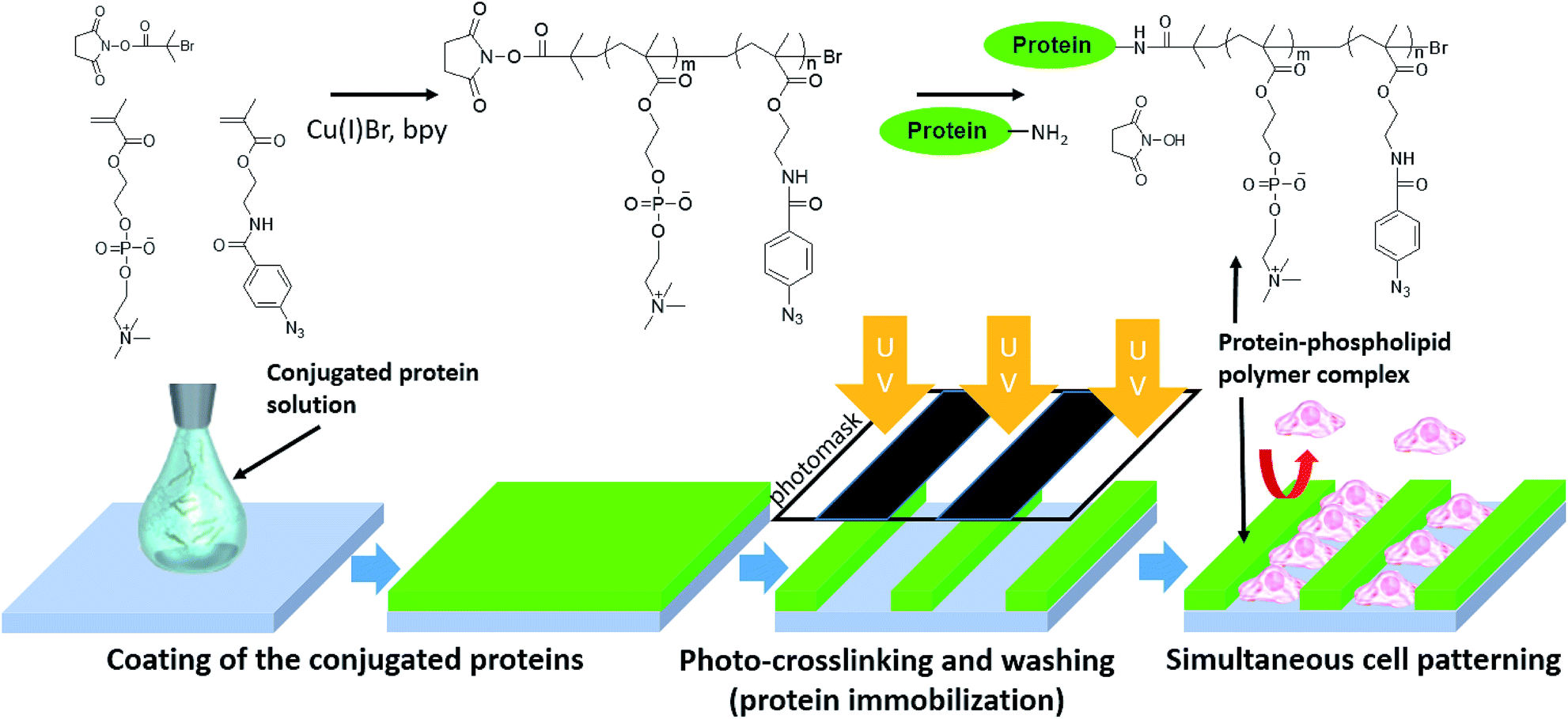 | ||
| Fig. 1 Overall concept of preparing simultaneous patterning of conjugated protein and cells by using a photoreactable MPC polymer. | ||
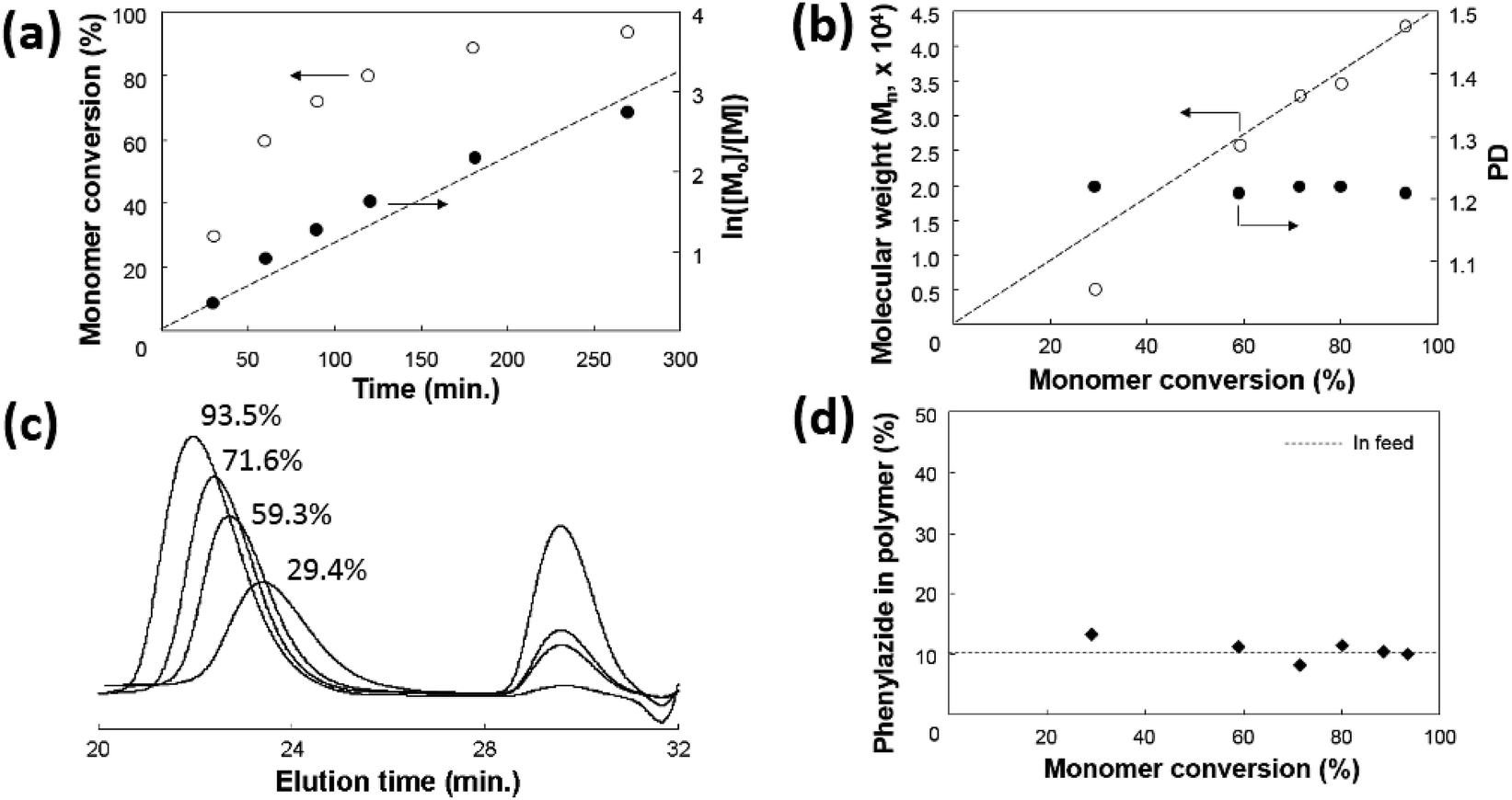 | ||
Fig. 2 (a) Kinetic analysis, (b) molecular weight changes, (c) SEC trace for the monomer conversions of 29.4, 59.3, 71.6, and 93.5%, (d) phenylazide contents of the polymer during the ATRP process. [Initiator]![[thin space (1/6-em)]](https://www.rsc.org/images/entities/char_2009.gif) :[Cu(I)Br]:[bpy]:[MPC]:[AzMA] = 1:1:2:90:10. :[Cu(I)Br]:[bpy]:[MPC]:[AzMA] = 1:1:2:90:10. | ||
Bioconjugation of PMAz with F-BSA was then conducted in order to prepare photoreactable F-BSA (F-BSA–PMAz). Fig. 3 shows the result of sodium dodecyl sulfate polyacrylamide gel electrophoresis (SDS-PAGE) of the conjugated F-BSA. The random copolymer composed of MPC and AzMA without the NHS-ester end-functional group was also synthesized as a control polymer, and mixed with F-BSA. As shown in Fig. 3, no significant conjugation band was observed when F-BSA was mixed with the random copolymer. In contrast, PMAz-mixed F-BSA showed a distinctive high molecular weight band. The characteristic broad band in the higher molecular weight region is very well known phenomenon when protein molecules are conjugated with the synthesized polymers because of the distributed molecular weight of the synthesized polymers.23 Although several of the ε-amine groups from the lysine residues in BSA (4–5 Lys) were already conjugated with FITC,24 the unconjugated free amine could still be successfully conjugated with PMAz because of the large amount of lysine residues present (∼35 Lys) on BSA.
 | ||
| Fig. 3 SDS-PAGE of (a) F-BSA, (b) PMAz, (c) F-BSA + PMAz without end functionality, and (d) F-BSA + PMAz. | ||
An aqueous solution of the conjugated BSA (0.1 wt%) was then coated onto polypropylene (PP) discs (10 mm diameter), and air-dried in a clean environment. Following this, the coated PP disc was covered with a micro-patterned photomask, and irradiated with 254 nm UV light (300 mW cm−2) for 10 s. After thorough washing with phosphate buffered saline (PBS), the PP surface was observed under a fluorescence microscope. Fig. 4 shows a photograph of the patterned photomask and the resulting fluorescence images. As is clearly shown, F-BSA was patterned on the PP surface only where UV light irradiation occurred through the photomask. This result indicates that the phenylazide groups present on the BSA protein can immobilize the protein to the PP surface. The phenylazide group is known to form an amine linkage by attacking arbitrary hydrocarbons present on organic substrates or polymers when it is exposed to the appropriate UV light (254 nm).25 For this reason, phenylazide, or similar photoreactable group-containing polymers have been widely used as a negative photoresist material to develop patterned polymer surfaces on organic substrates.26–28 In the present study, we confirmed that a similar procedure could be used for the simple patterning of a protein molecule onto an organic substrate.
 | ||
| Fig. 4 Two different types of micropatterning, (a) stripe and (a′) rectangle-type photomask, and the following patterned F-BSA (b), (b′), and L929 fibroblasts (c), (c′). Bar: 200 μm. | ||
The protein-patterned surface was then brought into contact with L-929 mouse fibroblast cells in order to confirm the feasibility of achieving simultaneous patterning of proteins and cells. Cell adhesion on material surfaces is generally mediated through a protein layer found on the outside of cells, referred to as the extracellular matrix (ECM). This matrix contains a variety of adhesive proteins that mediate attachment to a variety of different matrices, as well as to other cells. The ECM proteins are derived from biofluids, including the artificial cell culture medium, as well as from cell secretion. Inhibition of both ECM sources is therefore an effective way to prevent cell adhesion and downstream activation and aggregation. This mechanism has been adapted in order to prevent bacterial cell adhesion and biofilm formation during the infection process on material surfaces.29,30 Because the anti-biofouling MPC polymer chains coexist with the BSA molecules, ECM formation on the BSA-patterned area was anticipated to be effectively prevented. Fig. 4 shows the patterned cells on the F-BSA-immobilized surface. As is clearly shown, cells adhere to the surface where the F-BSA is not immobilized even after the fully proliferated state. This result indicates that the formation of ECM on the F-BSA-immobilized surface is effectively prevented, and that the ECM was only formed on the exposed PP surface, where the MPC polymer was not immobilized, resulting in the induction of significant cell adhesion.
The simultaneous patterning of proteins and cells has many potential utilities in the field of biomedical engineering. However, the multi-step process required for the sequential patterning of protein and cells often requires sophisticated surface treatment, including the repeated passivating-immobilization process. In the present study, immobilization of protein molecules was conducted along with the surface passivation process using a simple UV light-irradiation process, and the simultaneous patterning of proteins and cells was achieved. Because cell functions have been known to greatly depend on the microenvironment of adhering cells, this result is expected to contribute to the fundamental studies of cell biology. For instance, cellular interactions between different types of cells are very important to understand many biological responses such as tissue regeneration, immune responses, and cancer cell behaviour. If specific protein or peptides that are able to induce specific cell adhesion are conjugated with PMAz and patterned on the surface, the different types of cells are expected to be alternatively patterned on the surface in well-defined manner. In any event, the feasibility of photoreactable bioconjugation system on the development of multi-functional biointerface was successfully confirmed in this research.
Conflicts of interest
There are no conflicts to declare.References
- P. Wu, D. Castner and D. J. Grainger, J. Biomater. Sci., Polym. Ed., 2008, 19, 725–753 CrossRef CAS PubMed.
- C. S. Chen, M. Mrksich, G. M. Whitesides and D. E. Ingber, Science, 1997, 276, 1425–1428 CrossRef CAS PubMed.
- T. Konry, S. Sarkar, P. Sabhachandani and N. Cohen, Annu. Rev. Biomed. Eng., 2016, 18, 259–284 CrossRef CAS PubMed.
- N. M. Rodriguez, R. A. Desai, B. Trappmann, B. M. Baker and C. S. Chen, Langmuir, 2014, 30, 1327–1335 CrossRef CAS PubMed.
- A. A. Popova, S. M. Schillo, K. Demir, E. Ueda, A. Nesterov-Mueller and P. A. Levkin, Adv. Mater., 2015, 27, 5217–5222 CrossRef CAS PubMed.
- P. Y. Wang, H. Thissen and P. Kingshott, Acta Biomater., 2016, 45, 31–59 CrossRef CAS PubMed.
- S. Khan and G. A. Newaz, J. Biomed. Mater. Res., Part A, 2010, 93, 1209–1224 CrossRef PubMed.
- D. Falconnet, G. Csucs, H. M. Grandin and M. Textor, Biomaterials, 2006, 27, 3044–3063 CrossRef CAS PubMed.
- W. He, C. P. Halberstadt and K. E. Gonsalves, Biomaterials, 2004, 25, 2055–2063 CrossRef CAS PubMed.
- S. H. Lee, J. J. Moon and J. L. West, Biomaterials, 2008, 29, 2962–2968 CrossRef CAS PubMed.
- M. Kim, J. C. Choi, H. R. Jung, J. S. Katz, M. G. Kim and J. Doh, Langmuir, 2010, 26, 12112–12118 CrossRef CAS PubMed.
- S. Y. Jung, S. M. Lim, F. Albertorio, G. Kim, M. C. Gurau, R. D. Yang, M. A. Holden and P. S. Cremer, J. Am. Chem. Soc., 2003, 125, 12782–12786 CrossRef CAS PubMed.
- M. Zhang and T. A. Horbett, J. Biomed. Mater. Res., Part A, 2009, 89, 791–803 CrossRef PubMed.
- J.-H. Seo, R. Matsuno, Y. Lee, T. Konno, M. Takai and K. Ishihara, Acta Biomater., 2010, 7, 1477–1484 CrossRef PubMed.
- P. Roach, D. Farrar and C. C. Perry, J. Am. Chem. Soc., 2005, 127, 8168–8173 CrossRef CAS PubMed.
- K. Ishihara, N. P. Ziats, B. P. Tierney, N. Nakabayashi and J. M. Anderson, J. Biomed. Mater. Res., 1991, 25, 1397–1407 CrossRef CAS PubMed.
- K. Ishihara, H. Nomura, T. Mihara, K. Kurita, Y. Iwasaki and N. Nakabayashi, J. Biomed. Mater. Res., 1998, 39, 323–330 CrossRef CAS PubMed.
- S. Sakaki, N. Nakabayashi and K. Ishihara, J. Biomed. Mater. Res., 1999, 47, 523–528 CrossRef CAS PubMed.
- D. Miyamoto, J. Watanabe and K. Ishihara, Biomaterials, 2004, 25, 71–76 CrossRef CAS PubMed.
- K. Ishihara, M. Mu, T. Konno, Y. Inoue and K. Fukazawa, J. Biomater. Sci., Polym. Ed., 2017, 28, 884–899 CrossRef CAS PubMed.
- E. J. Lobb, I. Ma, N. C. Billingham, S. P. Armes and A. L. Lewis, J. Am. Chem. Soc., 2001, 123, 7913–7914 CrossRef CAS PubMed.
- A. L. Lewis, US Pat., US8048408 B2, 2002.
- D. Bontempo, K. L. Heredia, B. A. Fish and H. D. Maynard, J. Am. Chem. Soc., 2004, 126, 15372–15373 CrossRef CAS PubMed.
- L. O. Andersson, A. Rehnstrom and D. L. Eaker, Eur. J. Biochem., 1971, 20, 371–380 CrossRef CAS PubMed.
- L. Renaudie, C. Le Narvor, E. Lepleux and P. Roger, Biomacromolecules, 2007, 8, 679–685 CrossRef CAS PubMed.
- K. Fukazawa, A. Nakao, M. Maeda and K. Ishihara, ACS Appl. Mater. Interfaces, 2016, 8, 24994–24998 CAS.
- K. Fukazawa and K. Ishihara, ACS Appl. Mater. Interfaces, 2013, 5, 6832–6836 CAS.
- X. Lin, K. Fukazawa and K. Ishihara, ACS Appl. Mater. Interfaces, 2015, 7, 17489–17498 CAS.
- N. P. Boks, H. J. Kaper, W. Norde, M. C. van der Mei and H. J. Busscher, J. Colloid Interface Sci., 2009, 331, 60–64 CrossRef CAS PubMed.
- D. Pavithra and M. Doble, Biomed. Mater., 2008, 3, 034003–034015 CrossRef CAS PubMed.
Footnote |
| † Electronic supplementary information (ESI) available: Synthetic procedure of photoreactable monomer and the polymer, bioconjugation, and the overall patterning process with 1H-NMR and FT-IR data. See DOI: 10.1039/c7ra07389e |
| This journal is © The Royal Society of Chemistry 2017 |