Deformable and wearable carbon nanotube microwire-based sensors for ultrasensitive monitoring of strain, pressure and torsion†
Jian
Zhou
*a,
Xuezhu
Xu
a,
Hu
Yu
ab and
Gilles
Lubineau
*a
aKing Abdullah University of Science and Technology (KAUST), Physical Sciences and Engineering Division, COHMAS Laboratory, Thuwal 23955-6900, Saudi Arabia. E-mail: jian.zhou@kaust.edu.sa; gilles.lubineau@kaust.edu.sa; Tel: +966(12)8082983
bShanghai Jiao Tong University, School of Mechanical Engineering, State Key Laboratory of Mechanical Systems and Vibration, 800 Dongchuan Road, Minhang District, Shanghai, 200240, P.R. China
First published on 2nd December 2016
Abstract
Human joints have the ability to recover their mechanical functions after moderate dislocation. This remarkable capability inspired us to develop a “bone-skin-like” mechanosensor that can detect multiple mechanical deformations after recovery from electrical disconnection. To create this sensor, we embedded a low-strength, wet-spun single-walled carbon nanotube wire in polydimethylsiloxane. When various mechanical stimuli are applied, the wire gets fragmented and its resistance increases dramatically (from 360 Ω to practically infinity) in a reversible, recoverable manner even after the electrical failure/disconnection. The sensor is sensitive enough (a gauge factor of 105 at 15% uniaxial strain, a pressure sensitivity of 105 MPa−1 at 0.9 MPa pressure and a torsion sensitivity of 860 at a twisting angle of 60°) to be used for accurate sensing of a variety of deformation modes, suggesting a wide range of applications in wearable and deformable mechanical sensors.
1. Introduction
Wearable mechanical sensors capture and monitor various human activities.1–4 Conventional mechanical gauges made from metallic foils or semiconductors tolerate small deformations (<5%),4,5 and are easily damaged when removed from measured targets, making their reuse impossible. Commercially produced mechanical sensors are usually designed to sense only one deformation mode, such as uniaxial strain, pressure or torsion. When it is necessary to capture two or more deformation modes, the design of the sensor is often complicated and sophisticated.3,5–9Nanomaterials, such as metal nanoparticles, metal nanowires, carbon nanotubes (CNTs) and graphene-based materials, are flexible when deposited into thin films. They also offer superior electrical properties,10–13 suggesting that they can be used as smart mechanical sensors.1,2,14,15 Pang and coworkers developed a mechanical sensor based on interlocked conductive nanofibers. This sensor measures three loading modes (pressure, shear and torsion) with high sensitivity. However, its working range is limited to 5% strain.7 Gold nanowire coated soft substrates could resolve various complex forces including pressing, bending, torsion and acoustic vibrations. The sensing mechanism is ascribed to pressing force-dependent contact between nanowires and interdigitated electrode arrays. However, the gauge factor drops when strain increases, showing large sensitivity deviations in the range of the strain measurement.16,17 Graphene-based composite fibers with “compression ring” architecture can increase a sensor's operational strain while providing excellent stretching-, bending- and twisting-sensitive properties. However, the architecture of the sensor is very complex and its gauge factor (GF) to some mechanical stimuli is low (GF = 1.5 at 200% strain).6 A skin-like pressure and strain sensor based on CNT films on polydimethylsiloxane (PDMS) was developed by Lipomi and coworkers. The pressure sensitivity (PS = 0.22 MPa at 0.9 MPa) and the gauge factor (GF = 3.4 at 150%) of this sensor are extremely low due to the high initial resistance (100 kΩ).3 Moreover, this sensor and other previously described sensors cannot recover their resistance at large mechanical deformation (after electrical disconnection). Developing sensors with such resistance recoverability and high sensitivity under mechanical deformation remains a challenge. Thus, the conventional mechanical sensors need to be replaced by next generation smart sensors which have features of detection of multi-mode deformation, highly sensitive measurement, operation in a large strain regime and excellent durability and reusability.
Here, we develop a mechanical sensor from a low-strength, wet-spun SWCNT wire embedded in PDMS. A network of randomly distributed SWCNTs in the wire enables us to achieve high sensitivity to stretching, pressing and torsion. Moreover, our sensor has a unique ability to recover its electrical resistance after electrical disconnection due to large deformation. The sensor's response was highly repeatable and reproducible up to 5000 cycles with excellent resistance recoverability. These characteristics make our sensor a good candidate as a “smart” mechanosensor that detects various human motions.
2. Experimental
2.1. Materials
SWCNTs functionalized with 2.7% carboxyl groups were purchased from Cheap Tubes, Inc., with over 90 wt% purity and containing more than 5 wt% of MWCNT. The true density of these SWCNTs was 2.1 g cm−3. Methanesulfonic acid (MeSO3H) and SYLGARD 184 PDMS were purchased from Sigma Aldrich.2.2. Preparation of SWCNT dope
A 2 wt% SWCNT dope was prepared by adding 0.2 g of SWCNTs into 9.8 g of MeSO3H and stirred for 2 min, followed by bath sonication using a Branson 8510 sonicator (250 W) (Thomas Scientific) for 60 min. Then the mixture was further stirred for 24 hours, followed by passing through a 30 μm syringe filter (Pall corporation) to remove aggregates.2.3. Wet spinning of CNT wires
The SWCNT ink was loaded into a 5 ml syringe and spun into a water bath through stainless steel needles with different gauges (23 G and 34 G). The flow rate of the ink was controlled to 58 and 5 μl min−1 by using a Fusion 200 syringe pump (Chemyx. Inc.) with the 23 G and 34 G needles, respectively. The SWCNT wires or meshes were spun onto sticky PDMS substrates (5.3 g, 10 × 10 cm), which were made by mixing the PDMS base and a curing agent with a ratio of 2.5 to 1 and cured at 70 degrees for 12 min.The wet-spinning of SWCNT wires was controlled by using a MD-P-821 autodrop x − y − z stepping motor (accuracy: 1 μm) (Microdrop Technologies). The nozzle was immersed in water and the distance between the nozzle and the substrate was controlled by the z stepping motor to 3 mm. The setup of the wet-spinning system is shown in Fig. S2a.† The wire spinning speed, υw, is determined by  , where υs (μl min−1) is the ink-dosing speed, D(μm) is the nozzle diameter, υm is the substrate movement speed, which is kept constant at 10 mm s−1. To generate continuous SWCNT wires, υw (10.8 mm s−1) was kept slightly higher than that of υm.
After spinning, the wires were dried at 70 °C overnight to remove residual water. Then, two ends of the SWCNT wires were connected with copper wires and painted with silver epoxy. The SWCNT wires were sealed with 5.3 g of PDMS precursor, which was cured at 70 °C for 2 h. The effective length of the wires between the silver paste was about 4 ± 0.2 cm. In this study, 60 μm-SWCNT and 32 μm-SWCNT represent SWCNT wires spun using 23 G and 34 G needles, respectively (Table S1†). For comparison, encapsulation of carbon fiber samples (diameter: 7 μm) was prepared by the same method.
2.4. Characterization
Scanning electron microscopy (SEM) on SWCNT wires was performed using a Quanta 3D (FEI Company). The setup for electromechanical testing of the specimen is shown in Fig. S2a.† The loading/unloading of the sample is controlled by using a 5944 Instron machine. The dog-bone specimen was glued to two metal plates that were clamped by the Instron machine. The distance between the metal plates was 4 mm, which is also the effective length of the sample. The change in electrical resistance in the wires was monitored by using an U1252B digital multimeter. Generally, the stretching process was applied to each sample under 50% strain with a speed of 0.4 mm min−1. Then, a cyclic stretching/relaxing program was applied to the wire with the maximum strains of 30% and 5% at each cycle. The resistance data were recorded every 1 s during the test. Stretching/relaxing of the SWCNT wires in PDMS was captured by using a commercial video camera.To measure the strain sensitivity of our sensor, we define a gauge factor (GF) as: GF = (ΔR/R0)/ε, where R0 is the initial resistance, ΔR/R0 is the relative change in resistance and ε is the applied strain. Pressure sensing is performed by loading/unloading compression cyclic tests using a 1.25 mm-radius pressure head at 0.1 mm min−1 (inset in Fig. 4b) on a 60 μm SWCNT sample. The maximum displacement of the pressure head was 0.3 mm. The resistance was recorded with an U1252B digital multimeter. The pressure sensitivity, PS, is defined as PS = (ΔR/R0)/P, where P is the applied pressure. Cyclic twisting/untwisting torsion motion was controlled by using a motor at a speed of 18 °s−1 with a maximum twisting angle of 180° (inset in Fig. 4e). The torsion sensitivity, TS, is defined as TS = (ΔR/R0)/θ, where θ is the twisting angle in rads. Bending/unbending motion was controlled by the 5944 Instron machine with a rate of 10 mm min−1. The initial and final distances between the samples are set at 54 mm and 27 mm, respectively.
3. Results and discussion
3.1. SWCNT-microwire-based strain sensors
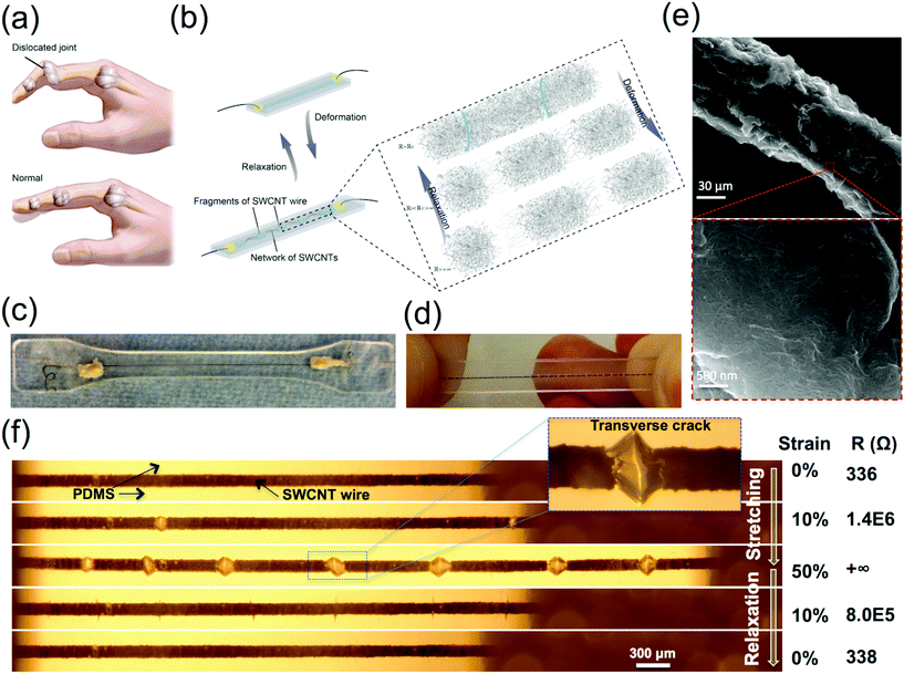
Fig. 1 shows the bone-skin-like structure inspired approach for realizing a resistance recoverable mechanical sensor. Finger joints have remarkable capability to recover mechanical function after moderate dislocation (Fig. 1a). This characteristic inspired us to develop a mechanical sensor that uses a low-strength SWCNT wire as the “bone” and an elastic PDMS substrate as the “skin”. We used a fragile SWCNT wire that could be easily fragmented during stretching. Disconnection and reconnection of SWCNT networks in cracks formed after stretching lead to sensing and resistance recovery (inset to Fig. 1b). Fig. S1† presents a detailed schematic illustration of the key steps in fabricating a SWCNT-wire-based strain sensor (see the Experimental section and ESI† for details). We used a mild acid, methanesulfonic acid (CH3SO3H), to disperse the SWCNTs. The dispersion of 2 wt% SWCNT/CH3SO3H was extruded from a movable nozzle into a coagulation water bath. CH3SO3H was then extracted from the dispersion with water. By controlling the motor that moved the injection nozzle along a predetermined track, we could obtain arrays of SWCNT wires on a PDMS substrate as shown in Fig. S1.† The wet-spinning process we introduced here is actually a very continuous drawing process to form the microwires. It is advantageous compared with the inkjet or screen printing process, which normally need low concentration inks.18–20 After removing the water and drying the sample overnight, we connected copper wires to the SWCNT wires with silver epoxy. Then we poured a second layer of the PDMS precursor onto the sample and cured it at 70 °C to encapsulate the SWCNT wires. We used a dogbone die to cut the samples to obtain a standard dog-bone-shaped specimen (Fig. 1c). Fig. 1d shows that by stretching the sample, the wire in the PDMS substrate fragments into many pieces, which is resulting from the porous structure of the SWCNT wire with randomly distributed SWCNT networks (Fig. 1e). This randomly oriented structure results in low-strength SWCNT assemblies (Fig. S3†) that facilitate breakage of the fragile wire in PDMS.Fig. 1f shows the specimen in Fig. 1c being stretched to 50% strain. The deformation of the SWCNT wires is recorded with a video camera. The crack density of the samples at the same strain is quite consistent as observed from our experiment. The average crack density of the 60 μm-SWCNT wire is 1.8 ± 0.2 mm−1 at 50% strain, as tested from at least 5 samples. This result indicates that the formation of the cracks is repeatable for different 60 μm-SWCNT wires, the reproducibility of the cracks is a prerequisite for the reproducibility of these sensors. We observed that the wire is initially well bonded to the surrounding PDMS. As the strain increases, the wire breaks sequentially into fragments. The multiplication of cracks is called fragmentation, which results in a quasi-random network of transverse cracks.21,22 The resistance of the sample increases up to a non-measurable value (that we call practically infinity) on our measuring instrument, but the resistance can be recovered to its initial value after relaxation (Fig. 1e and S2†). Fig. 1e shows the initiation, progression and closing of transverse cracks during loading/unloading. It shows that the recoverability of resistance comes from the closure of the transverse cracks.
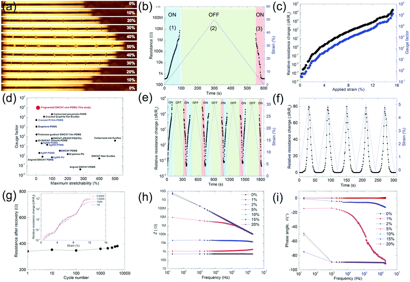
The response observed during the first fragmentation process and during subsequent loading–unloading cycles differed significantly. Fig. 2 demonstrates the strain-sensing behavior of fragmented SWCNT wires in PDMS. The optical images in Fig. 2a corresponding to each level of strain show the microstructural changes of the wire. Generally, new cracks were not created as demonstrated in Fig. 2a. In detail, Fig. 2b shows that changes in resistance can be divided into three regimes. (1) The strain sensing regime: 0 < ε < εc, where εc is the critical strain, where resistance (R) increases to infinity. In this regime R increases from 336 Ω to 10 MΩ before the resistance increases to infinity. The circuit is in the “ON” state; we do not observe an increase in the number of cracks because the wire has already been fragmented. Only the average opening distance of the cracks (Lc) increases with strain, which leads to the increase in the resistance. Even though large gaps can be seen at a relatively high strain (ε = 15%) in this regime, the resistance of the wire does not go to infinity (R = 10 MΩ). This observation suggests that there are SWCNT networks in the cracks that are connected to the fragments nearby. (2) The open circuit regime: ε > εc, the SWCNTs are completely disconnected from each other. We measured the average crack opening distance (Lc) to be longer than 50 μm, which is longer than the length of the SWCNTs (5–30 μm). The Lc of the SWCNT wire is long enough to result in an “OFF” circuit. (3) Resistance recovery regime: the resistance of the SWCNT wire recovers to its initial value after relaxation and the circuit recovers to the “ON” state. The Lc between the fragments reduces as the applied strain decreases. In the completely relaxed state, cracks are no longer visible. In this regime, PDMS obviously plays an important role in the recovery of the electrical resistance.
We then plot ΔR/R0 and the gauge factor (GF) with respect to strain as shown in Fig. 2c. The operational strain range and GF are dramatically improved when compared with those of self-standing SWCNT fibers (Fig. S3d,† GF = 1 at a maximum strain of 1.1%). The dramatic change in resistance (ΔR/R0 = 5 × 10⁴) in the 60 μm-SWCNT wire enables us to obtain a GF of 105 at ε = 15%. Fig. 2d and Table S2† show that our GF value is the highest value compared with the GF values recently reported for CNT, graphene, metal and other strain sensors at this strain level.1,3,4,6,11,23–29 We further tested the reproducibility of the strain sensor with repeated stretching/relaxation cycles. Fig. 2e presents five cycles of a strain to be 30%. The ΔR/R0 increased with almost complete reversibility even after the electrical disconnection. The conversion of “ON–OFF” states occurred at almost the same strain in all cycles. The sensor also features high sensitivity as it can detect subtle changes as small as 0.1% as shown in Fig. 2f. Fig. 2g shows resistance after recovery from 10% strain over the course of 5000 cycles of stretching. The resistance increased by 10% during the 5000 cycles. The sensing performance at 1, 100, 1000 and 5000 cycles remains nearly unchanged, demonstrating the excellent durability and repeatability of the strain sensor (inset in Fig. 2g). To examine the response time of our sensors to the strain rate, the output resistance signals were compared with the dynamic cyclic loading/unloading strain inputs at 0.1, 0.8, 2.4, 4 and 8 mm min−1 (Fig. S5†). Note that the response of the sensor could be extremely fast and resistance-recoverable.
The strain-sensing mechanism is captured by electrical impedance spectroscopy (EIS). EIS provides a clear understanding of an equivalent circuit in terms of the components of the complex impedance. In addition, it allows insights into the electron transport mechanism occurring across the bulk and interfaces.30,31Fig. 2h and i show the dependency of the modulus of the complex impedance (Z) and the phase angle (θ), respectively, on frequency. The dependence of Z on frequency is nearly constant at low applied strain (<2%). These observations suggest that the conduction mechanism in a sample at low strain is dominated by the resistive behavior of the SWCNT wire. However, the electrical response of the sensor at higher strains is dependent on frequency. At low applied strain, electron transport through the nanotube network is the primary connection–disconnection mechanism (electron transport mechanism). Inter-network electron tunnelling in the interface between PDMS and the SWCNT wire becomes dominant at higher strains, when total disconnection between nearby fragments occurs. This becomes more clear when looking at the difference in the phase angle, θ, at different strains (Fig. 2i). At larger strain (>5%), the capacitive part largely increases with frequency. This comes from the unconnected SWCNT networks that act as capacitors rather than resistors.
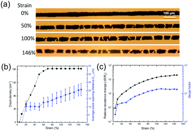
Although the GF in the results was high, the maximum stretchability of the sensor was limited to 15%. To obtain higher stretchability while maintaining a continuous resistance change in the operational range of the strain, we need to increase the crack density in the SWCNT wires. Fig. 3a shows that by reducing the diameter of the wire from 60 to 32 μm, we were able to obtain a higher crack density in the wire. Before the critical strain (60%), the crack density increases with the applied strain. After reaching the critical strain at 60%, the crack density remains constant, while Lc increases with the applied strain (Fig. 3b). The PDMS substrate breaks at 146% due to the concentration of stress near the cracks, suggesting that the stretchability of the sensor can be dramatically increased by using narrower SWCNT wires. However, the narrower wire had a small ΔR/R0 and a low GF of 15 at 146% (Fig. 3c). These characteristics can be ascribed to the high initial resistance 32 μm-SWCNT wire (10 ± 0.8 kΩ), which is more than one order of magnitude higher than that of the 60 μm-SWCNT wire (338 Ω) and to the maximum Lc, which is less than 25 μm, indicating that SWCNT networks remain in a percolated state. The fragmentation strategy can be modified to tune the GF and stretchability of the sensors. It can be realized by adjusting the crack density by using wires with different diameters and choosing substrates with different stretchability.
3.2. The sensor's response to pressure, torsion and bending
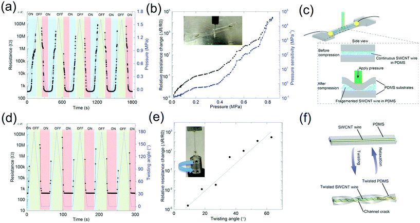
We then monitored the response of the sensor pressure, torsion and bending to help us clarify the sensitivity and selectivity of the sensor to different deformation modes (Fig. 4). We performed multiple cycle tests by repeated loading/unloading using a 1.25 mm-radius pressure head. Fig. 4a shows the ability of the sensor to recover its resistance after the circuit is broken. At high pressure (typically 1.1 to 1.5 MPa), the electrical circuit shifts to the OFF state when the SWCNT wire breaks. After the pressure is released, the resistance recovers almost to its initial value. The sensor exhibits a wide change in resistance (340 Ω to 10 MΩ), which is similar to the resistance recoverability of an in-plane unidirectional strain sensor (Fig. 2). Upon pressing, the Poisson effect induces in-plane stretching of the wire. This results in extremely high pressure sensitivity (up to 4 × 10⁵ MPa−1 at 0.9 MPa). The attained sensitivity is several orders of magnitude higher than the recently reported high-performance piezoelectric pressure sensors.2 We believe that the pressure-sensing mechanism before circuit breakage is due to induced fragmentation of the SWCNT wire in the upper PDMS layer, followed by disconnection of the SWCNT networks in the transverse cracks, as show in Fig. 4c, suggesting that SWCNT wires in PDMS can be used as highly sensitive pressure sensors for realization of e-skins, human–machine interfaces and health monitoring.1,2We also twisted the sensor while measuring the electrical resistance with respect to the twisting angle, ϕ. Fig. 4d and e clearly indicate that there are two regimes during twisting: one at 0° ≤ ϕ ≤ 60°, where there is a dramatic increase in resistance after 360 Ω to tens of MΩ. An average torsion sensitivity of 860 was obtained at 60°. The second regime is at ϕ > 60°, where the resistance of the wire increases to infinity because the SWCNT wire breaks due to over twisting. We note that the twisted sample exhibits resistance recoverability after untwisting (Fig. 4d and f). We ascribe this response to the reconnection of SWCNT networks after relaxation of the PDMS substrate.
Finally, we performed a bending test as shown in Fig. S7.† The bending angle, θ, is defined in the inset of Fig. S7.† The test reveals that the samples have stable electrical properties before and after fragmentation. The samples with cracks have a ΔR/R0 of less than 0.01 at a bending angle of 120°. We ascribe this low sensitivity to the fact that the SWCNT wire in our design is in the neutral axis of the sample. The bending sensitivity could be dramatically improved by engineering the thickness of the PDMS substrate.
3.3. Application in human motion detection
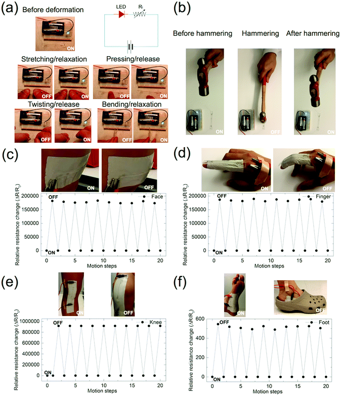
We demonstrated the resistance recoverability of our sensors by wiring a light-emitting diode (LED) with the sensor. Before any mechanical deformation, the SWCNT wire resistance was low enough to light the LED at 3 V, as shown in Fig. 5a. The sensor exhibited a good resistance recoverability after large mechanical deformation (stretching, pressing, twisting and bending), demonstrating that the light intensity of the LED had negligible degradation after the sensor was released from the deformations. Extreme mechanical deformation was then applied to the sensor by stretching to 50 or 100% of strain. The LED then turned off, confirming that the SWCNT wire fragmented and its conductive pathway disconnected under large strains. After relaxation from 50 or 100% strain, the LED automatically relits, demonstrating that the open circuit reconnected due to automatic recovery of the resistance after relaxation of the strain (Fig. S7†). We further applied an extreme impact with a hammer (Fig. 5b). Upon impact of the hammer, the LED turned off. It readily relit as soon as we took the hammer away from the sample. These results show that our sensors are different from conventional sensors, which cannot restore their sensing functions after electrical disconnection by large deformation. For example, the electrical properties of carbon fibers embedded in PDMS are not recoverable after relaxation of the substrate from stretching beyond its strain limit (Fig. S4†).We attribute the remarkable resistance recoverability of our sensors after large deformation beyond the elongation or pressure at break of the SWCNT wire to the fact that (1) the SWCNT wire has good adhesion with PDMS due to the excellent capillary surface of the SWCNT. By immersing SWCNT wires in the PDMS precursor around, PDMS firmly attaches to the SWCNT wire. (2) After the fragmentation of the wires, high density of the single SWCNTs and SWCNT bundles will be formed on the surface of the crack slips, the inherent self-recovery ability of PDMS can bring the SWCNT networks back to ensure a good electrical contact. (3) The low stiffness of the SWCNT wire makes it easy to fragment and recover.
We also demonstrate machine/human interface interactions by using our sensors, as shown in Fig. 5c–f. We attached the sensors to human skin with adhesive tape at different locations of the body (face, finger, knee and foot). Our sensor demonstrated reproducible ON/OFF switching with large deformation. Fig. 5c shows the ΔR/R0 response of a sensor attached to a face. ΔR/R0 increases to almost infinity with expansion of the facial muscle and decreases again by the opposite movement of the facial muscle. Each expansion of the muscle results in electrical disconnection of the wire. As a consequence, each part of the facial muscle movement can be detected in a repeatable manner. When attached to a finger, the sensor is able to detect the finger's bending motion (Fig. 5d). When our sensor is attached to a knee or foot, it can be used as a pedometer that accurately records the number of steps during walking (Fig. 5e and f). The pedometer mechanism of our sensor lies in the stretching/relaxation and pressing/relaxation of the sensor on the knee and foot, respectively. In the basic motion of the human body, stretches and contractions are as large as 55%, which exceeds the limits of conventional metallic gauges (5%).4 The resistance recovery feature of our sensor enables us to use the sensor in deformable electronics. The sensor also has an additional functionality of recovery after large mechanical deformation. Our sensor can also be used as a deformable mechanosensor with resistance recoverable properties exceeding the wire deformation limit (<5%). Moreover, the capabilities of the sensor are not limited to switch devices and large strain of human motion detection. The sensor can also be used for ultralow strain detection for high sensitivity devices such as human belly movement and wrist pulse detection (Fig. S8†).
4. Conclusion
Although the sensor configuration we present here is very simple, we have shown a route to realize mechanical stimuli detection by using a sensor based on wet-spun SWCNT wires embedded in a PDMS elastomer. The sensor has extremely high sensitivity to various mechanical stimuli, including stretching, pressing and torsion. More importantly, it has an additional function of resistance recoverability after electrical disconnection induced by large deformation. The sensor is a promising alternative to conventional mechanical sensors combining an easy fabrication process, multi-mode deformation detection, high sensitivity, excellent durability and reusability. Our research suggests that our sensors can find a wide range of applications in wearable and deformable mechanosensors for human activity monitoring.Acknowledgements
The research described in this paper was supported by the King Abdullah University of Science and Technology (KAUST) baseline research funding. The authors are grateful to KAUST for its support.References
- M. Amjadi, K. Kyung, I. Park and M. Sitti, Adv. Funct. Mater., 2016, 26, 1678 CrossRef CAS.
- T. Q. Trung and N. Lee, Adv. Mater., 2016, 28, 4338–4372 CrossRef CAS PubMed.
- D. J. Lipomi, M. Vosgueritchian, B. C. K. Tee, S. L. Hellstrom, J. A. Lee, C. H. Fox and Z. N. Bao, Nat. Nanotechnol., 2011, 6, 788–792 CrossRef CAS PubMed.
- T. Yamada, Y. Hayamizu, Y. Yamamoto, Y. Yomogida, A. Izadi-Najafabadi, D. N. Futaba and K. Hata, Nat. Nanotechnol., 2011, 6, 296–301 CrossRef CAS PubMed.
- J. Park, I. You, S. Shin and U. Jeong, ChemPhysChem, 2015, 16, 1155–1163 CrossRef CAS PubMed.
- Y. Cheng, R. Wang, J. Sun and L. Gao, Adv. Mater., 2015, 27, 7365–7371 CrossRef CAS PubMed.
- C. Pang, G. Y. Lee, T. I. Kim, S. M. Kim, H. N. Kim, S. H. Ahn and K. Y. Suh, Nat. Mater., 2012, 11, 795–801 CrossRef CAS PubMed.
- J. Ge, L. Sun, F. R. Zhang, Y. Zhang, L. A. Shi, H. Y. Zhao, H. W. Zhu, H. L. Jiang and S. H. Yu, Adv. Mater., 2016, 28, 722–728 CrossRef CAS PubMed.
- S. Park, H. Kim, M. Vosgueritchian, S. Cheon, H. Kim, J. H. Koo, T. R. Kim, S. Lee, G. Schwartz, H. Chang and Z. A. Bao, Adv. Mater., 2014, 26, 7324–7332 CrossRef CAS PubMed.
- J. Lee, S. Kim, J. Lee, D. Yang, B. C. Park, S. Ryu and I. Park, Nanoscale, 2014, 6, 11932–11939 RSC.
- M. Amjadi, A. Pichitpajongkit, S. Lee, S. Ryu and I. Park, ACS Nano, 2014, 8, 5154–5163 CrossRef CAS PubMed.
- B. Xie, Y. L. Liu, Y. T. Ding, Q. S. Zheng and Z. P. Xu, Soft Matter, 2011, 7, 10039–10047 RSC.
- J. H. Wu, J. F. Zang, A. R. Rathmell, X. H. Zhao and B. J. Wiley, Nano Lett., 2013, 13, 2381–2386 CrossRef CAS PubMed.
- Q. Cao and J. A. Rogers, Adv. Mater., 2009, 21, 29–53 CrossRef CAS.
- W. Obitayo and T. Liu, J. Sens., 2012, 2012, 652438 Search PubMed.
- S. Gong, W. Schwalb, Y. W. Wang, Y. Chen, Y. Tang, J. Si, B. Shirinzadeh and W. L. Cheng, Nat. Commun., 2014, 5, 3132 Search PubMed.
- S. Gong, D. T. H. Lai, B. Su, K. J. Si, Z. Ma, L. W. Yap, P. Z. Guo and W. L. Cheng, Adv. Electron. Mater., 2015, 1, 1400063 CrossRef.
- J. Zhou, E. Q. Li, R. Li, X. Xu, I. Aguilar Ventura, A. Moussawi, D. Anjum, M. N. Hedhili, D. Smilgies, G. Lubineau and S. T. Thoroddsen, J. Mater. Chem. C, 2015, 3, 2528–2538 RSC.
- J. Zhou, M. Mulle, Y. Zhang, X. Xu, E. Q. Li, F. Han, S. T. Thoroddsen and G. Lubineau, J. Mater. Chem. C, 2016, 4, 1238–1249 RSC.
- K. Chen, W. Gao, S. Emaminejad, D. Kiriya, H. Ota, H. Y. Y. Nyein, K. Takei and A. Javey, Adv. Mater., 2016, 28, 4397–4414 CrossRef CAS PubMed.
- A. Arteiro, G. Catalanotti, A. R. Melro, P. Linde and P. P. Camanho, Compos. Struct., 2014, 116, 827–840 CrossRef.
- A. Arteiro, G. Catalanotti, A. R. Melro, P. Linde and P. P. Camanho, Composites, Part A, 2015, 79, 127–137 CrossRef CAS.
- Z. Y. Liu, D. P. Qi, P. Z. Guo, Y. Liu, B. W. Zhu, H. Yang, Y. Q. Liu, B. Li, C. G. Zhang, J. C. Yu, B. Liedberg and X. D. Chen, Adv. Mater., 2015, 27, 6230–6237 CrossRef CAS PubMed.
- J. Seo, T. J. Lee, C. Lim, S. Lee, C. Rui, D. Ann, S. B. Lee and H. Lee, Small, 2015, 11, 2990–2994 CrossRef CAS PubMed.
- E. Roh, B. U. Hwang, D. Kim, B. Y. Kim and N. E. Lee, ACS Nano, 2015, 9, 6252–6261 CrossRef CAS PubMed.
- R. Rahimi, M. Ochoa, W. Y. Yu and B. Ziaie, ACS Appl. Mater. Interfaces, 2015, 7, 4463–4470 CAS.
- K. K. Kim, S. Hong, H. M. Cho, J. Lee, Y. D. Suh, J. Ham and S. H. Ko, Nano Lett., 2015, 15, 5240–5247 CrossRef CAS PubMed.
- Y. R. Jeong, H. Park, S. W. Jin, S. Y. Hong, S. S. Lee and J. S. Ha, Adv. Funct. Mater., 2015, 25, 4228–4236 CrossRef CAS.
- S. Kang, A. R. Jones, J. S. Moore, S. R. White and N. R. Sottos, Adv. Funct. Mater., 2014, 24, 2947–2956 CrossRef CAS.
- C. S. S. Sangeeth, A. Wan and C. A. Nijhuis, J. Am. Chem. Soc., 2014, 136, 11134–11144 CrossRef CAS PubMed.
- I. A. Ventura, J. Zhou and G. Lubineau, Nanoscale Res. Lett., 2015, 10, 485 CrossRef PubMed.
Footnote |
| † Electronic supplementary information (ESI) available. See DOI: 10.1039/C6NR08096K |
| This journal is © The Royal Society of Chemistry 2017 |

