
 Open Access Article
 This Open Access Article is licensed under a
 Creative Commons Attribution 3.0 Unported Licence
Correction: Infrared nonlinear optical properties of lithium-containing diamond-like semiconductors Li2ZnGeSe4 and Li2ZnSnSe4
Ashley
Weiland
a,
Jian-Han
Zhang
ab,
Daniel J.
Clark
c,
Jacilynn A.
Brant
ad,
Charles W.
Sinagra
III
a,
Yong Soo
Kim
ce,
Joon I.
Jang
cf and
Jennifer A.
Aitken
*a
aDepartment of Chemistry and Biochemistry, Duquesne University, Pittsburgh, PA 15282, USA. E-mail: aitkenj@duq.edu
bSanming University, School of Resources and Chemical Engineering, Sanming, Fujian 365004, China
cDepartment of Physics, Applied Physics and Astronomy, Binghamton University, Binghamton, NY 13902, USA
dDepartment of Chemistry and Physics, Nova Southeastern University, Fort Lauderdale, FL 33304, USA
eDepartment of Physics and Energy Harvest-Storage Research Center, University of Ulsan, Ulsan 44610, South Korea
fDepartment of Physics, Sogang University, Seoul 04107, South Korea
First published on 20th July 2017
Abstract
Correction for ‘Infrared nonlinear optical properties of lithium-containing diamond-like semiconductors Li2ZnGeSe4 and Li2ZnSnSe4’ by Jian-Han Zhang et al., Dalton Trans., 2015, 44, 11212–11222.
The list of authors and affiliations for this correction is given above; this represents a change from the original published article.
In the original paper, we reported that both compounds, Li2ZnGeSe4 and Li2ZnSnSe4, displayed optical bandgaps around 1.8 eV, as estimated from UV/Vis/NIR optical diffuse reflectance spectroscopy. However, optical bandgaps of 1.8 eV are not consistent with the optical images of the orange-red and light red crystals of Li2ZnGeSe4 and Li2ZnSnSe4, respectively, as displayed in Fig. 1 of that paper. After the publication of this work, we continued our research on the cadmium analogs, Li2CdGeSe4 and Li2CdSnSe4.1 With these compounds, we also noted a slight discrepancy between the observed color of fresh samples and the optical bandgaps later obtained from UV/Vis/NIR optical diffuse reflectance spectroscopy. Additionally, we noted a difference in the visually observed color with time. This prompted us to study the diffuse reflectance as a function of exposure time to ambient conditions. We noticed that fresh samples that were immediately measured displayed wider optical bandgaps than those that had been exposed to ambient conditions for one week (see ESI in ref. 1). While fresh samples of Li2CdGeSe4 and Li2CdSnSe4 displayed optical bandgaps of ∼2.5 and 2.2 eV, respectively, the same samples that were exposed to ambient conditions for a period of one week displayed optical bandgaps of ∼1.8 eV. Measurements taken a few hours after exposure already showed some shift in the band edge to lower energy. Once the samples displayed optical bandgaps of 1.8 eV, the absorption edges remained constant over time. We concluded that the crystals react with air and/or moisture resulting in a surface degradation, which is responsible for the color change in the materials. The color change is observed visually, while the effect on the optical absorption edge can be observed spectroscopically. This surface degradation majorly alters the near-gap absorption behavior of these materials. Knowing this, we reinvestigated the zinc-containing compounds and observed a similar phenomenon.
 | ||
| Fig. 1 UV/Vis/NIR data converted to absorption for Li2ZnGeSe4 (left) and Li2ZnSnSe4 (right). Data collected for fresh samples are represented by the solid traces and the data collected after exposure to ambient conditions for one week are shown as the dotted traces. The marked difference between solid and dotted traces is due to surface degradation of the samples. | ||
Fig. 1 displays the optical diffuse reflectance spectra converted to absorption, via the Kubelka–Munk2 equation, for Li2ZnGeSe4 and Li2ZnSnSe4. The solid traces represent the data taken immediately on fresh samples, while the dotted traces display the data collected after one week of exposure to ambient conditions. As is the case with the cadmium-containing compounds, fresh samples of Li2ZnGeSe4 and Li2ZnSnSe4 show wider optical bandgaps than those exposed to ambient conditions for one week. Therefore, we conclude that Fig. 6 of the original paper, displaying optical bandgaps of 1.8 eV represents the spectra of surface-degraded samples. While X-ray powder diffraction patterns of both fresh samples and those exposed to ambient conditions for one week are identical and representative of phase-pure materials, the optical absorption edges for the fresh samples are not indicative of phase-pure materials, as Fig. 1 suggests that some surface degradation probably take place immediately for these samples. The fresh Li2ZnGeSe4 sample shows an absorption edge with a variably changing slope over a wide range of energy, ∼3.2 eV–1.8 eV, while the fresh Li2ZnSnSe4 sample shows an absorption edge with significant Urbach3 tailing from ∼2.0–1.75 eV. To correct the original paper, we now propose that the fundamental bandgaps are ∼2.5 and 2.0 eV for Li2ZnGeSe4 and Li2ZnSnSe4, respectively. We obtain these values by extrapolation of the onset of the absorption edge to the baseline as displayed in Fig. 2.
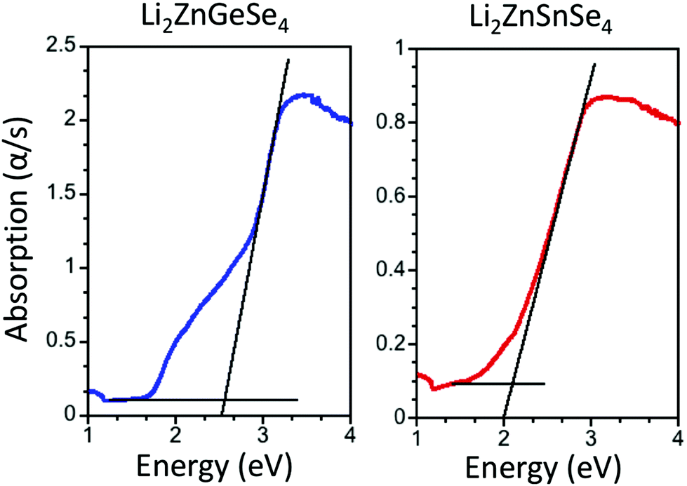 | ||
| Fig. 2 UV/Vis/NIR data converted to absorption for fresh samples of Li2ZnGeSe4 (left) and Li2ZnSnSe4 (right). The black lines show the extrapolations of the onsets of the absorption edges to the baselines that were used to estimate the optical bandgaps of ∼2.5 and ∼2.0 eV for Li2ZnGeSe4 and Li2ZnSnSe4, respectively. | ||
In the original paper, we stated that the title compounds have wide optical transparency windows, ≳65%, from 0.7 to 25 μm. Using the new optical absorption edge data, we constructed a new figure to display the optical transparency windows of the title materials. Therefore, Fig. 3 replaces Fig. 5 of the original paper. While the new data do not alter our statement regarding the breadth of the optical transparency window, the percent transparency is different. We now conclude that both compounds display a wide optical transparency, ≳60%, from 0.7 to 25 μm.
 | ||
| Fig. 3 Optical transparency windows for Li2ZnGeSe4 (top) and Li2ZnSnSe4 (bottom). The windows are constructed by stitching together the UV/Vis/NIR diffuse reflectance spectra (left) and the attenuated total reflectance (ATR) FT-IR spectra (right) converted to transmittance. This figure replaces Fig. 5 of the original paper. | ||
We propose that the degradation of these samples only takes place on the surface of the crystallites because the X-ray powder diffraction patterns of the fresh and exposed samples are identical. This is likely because X-ray powder diffraction is a bulk analysis method and the degradation product on the surface is likely not crystalline. Yet the surface degradation has a significant effect on the optical diffuse reflectance, which is a more surface-sensitive technique. Importantly, we do not believe that this degradation has played a role in the nonlinear optical (NLO) properties that we reported (particularly the second-order NLO susceptibility and the phase-matching range, where both of them were determined far below the bandgap of the materials), since the samples were prepared for NLO measurements using fresh samples. After these samples were sieved into discrete particle size ranges, they were sealed in evacuated fused-silica tubes where they were protected from the environment.
The Royal Society of Chemistry apologises for these errors and any consequent inconvenience to authors and readers.
References
- J.-H. Zhang, D. J. Clark, A. Weiland, S. S. Stoyko, Y. S. Kim, J. I. Jang and J. A. Aitken, Inorg. Chem. Front., 2016 10.1039/c7qi00004a , in press.
- P. Kubelka and F. Munk, Z. Tech. Phys., 1931, 12, 593–601 Search PubMed.
- F. Urbach, Phys. Rev., 1953, 92, 1324 CrossRef CAS.
| This journal is © The Royal Society of Chemistry 2017 |