
 Open Access Article
 This Open Access Article is licensed under a Creative Commons Attribution-Non Commercial 3.0 Unported Licence
Passive microrheology in the effective time domain: analyzing time dependent colloidal dispersions†
Bhavna M.
Vyas
ab,
Ashish V.
Orpe
*a,
Manish
Kaushal‡
b and
Yogesh M.
Joshi
 *b
*b
aChemical Engineering Division, National Chemical Laboratory, Pune 411 008, India. E-mail: av.orpe@ncl.res.in
bDepartment of Chemical Engineering, Indian Institute of Technology Kanpur, Kanpur 208016, India. E-mail: joshi@iitk.ac.in
First published on 30th August 2016
Abstract
We studied the aging dynamics of an aqueous suspension of LAPONITE®, a model time dependent soft glassy material, using a passive microrheology technique. This system is known to undergo physical aging during which its microstructure evolves progressively to explore lower free energy states. Optical microscopy is used to monitor the motion of micron-sized tracer probes embedded in a sample kept between two glass plates. The mean square displacements (MSD) obtained from the motion of the tracer particles show a systematic change from a purely diffusive behavior at short aging times to a subdiffusive behavior as the material ages. Interestingly, the MSDs at all the aging times as well as different LAPONITE® concentrations superpose remarkably to show a time–aging time master curve when the system is transformed from the real time domain to the effective time domain, which is obtained by rescaling the material clock to account for the age dependent relaxation time. The transformation of the master curve from the effective time domain to the real time domain leads to the prediction of the MSD in real time over a span of 5 decades when the measured data at individual aging times are only over 2 decades. Since the MSD obtained from microrheology is proportional to the creep compliance of a material, by using the Boltzmann superposition principle along with the convolution relation in the effective time domain, we predict the stress relaxation behavior of the system in real time. This work shows that the effective time approach applied to microrheology facilitates the prediction of long time creep and relaxation dynamics of a time dependent soft material by carrying out short time experiments at different aging times.
I. Introduction
Time dependent soft materials are thermodynamically out of equilibrium. Their microstructure evolves as a function of time by undergoing reorganization or physical aging to take the system to a progressively lower free energy state.1 There are many industrially important materials that fall in this category such as highly concentrated suspensions and emulsions, industrial (including agrochemical and pharmaceutical) pastes, colloidal gels, various types of paste-like food products, etc. The physical aging that takes place in these materials can sometimes be extremely sluggish and continues over very large timescales. This may affect the behavior of a product stored over long durations. Furthermore, in these materials structural build-up takes place over different length scales. Consequently, their rheological behavior is also dependent on length scales, whose knowledge is important when such materials are used in miniature systems such as microfluidic devices, micro-reactors, etc. Over the past two decades, microrheological techniques have been developed, wherein by monitoring/controlling the motion of submicron size tracer particles in soft materials, rheological properties at very small length scales can be obtained.2–7 In this work, we studied an aqueous suspension of LAPONITE®, a model time dependent soft material, and obtain its rheological behavior over microscopic length scales using passive microrheology. More importantly, we propose an effective time methodology in conjunction with microrheology to obtain very long time rheological behavior by carrying out a few tests at short times.Microstructural evolution to attain progressively lower free energy states as a function of time is a very natural response of materials that are thermal in nature and have been kinetically constrained from achieving the thermodynamic equilibrium state. Soft materials that fall in this category have been termed as soft glassy materials in the literature.8 The mesoscopic elements that constitute these materials are structurally arrested in energy wells formed due to their interactions with neighboring elements. Since the well depths that arrest the mesoscopic elements are significantly larger than the thermal energy, a material as a whole is unable to achieve the thermodynamic equilibrium state.9 These elements undergo microscopic dynamics during structural rearrangements and on an average sink in their own well or a new well with a deeper depth as a function of time. By virtue of the distribution of energy well depths there exists a distribution of relaxation times. As various elements age with time, the distribution shifts to slower relaxation modes. Owing to time dependency, these materials do not obey time translational invariance, and consequently the fundamental principles of linear viscoelasticity are not applicable to them.1 Joshi and coworkers,10–12 through a series of papers, proposed that the concept of time translational invariance can be applied to these materials by transforming their behavior from the real time domain to an effective time domain. In an effective time domain, time is normalized by the mean relaxation time so that the relaxation dynamics become invariant of aging time. The work by Joshi and coworkers did not simply validate the Boltzmann superposition principle and time temperature superposition principle for soft glassy materials, but also validated the convolution relation that relates two response functions, namely modulus and compliance, with each other.11,12 Very importantly, this methodology allows the use of either the short time creep or stress relaxation behavior of a time dependent material to obtain a long time prediction of the other. However, in order to apply this methodology, aging should affect only the mean value of the relaxation times and not the shape of their distribution.13 While the above work by Joshi and coworkers has been carried out using bulk rheology measurements, it is quite pertinent to assess whether similar analysis can be extended to measurements carried out at a microscopic level. The need for analysis at a microscopic level is specifically important in spatially inhomogeneous systems wherein the bulk rheology can only provide average measurements, thus, precluding rich behavior that may occur at a local level, and which may not match the bulk measurements.
One of the well-known techniques to carry out microscopic measurements in a sample is particle tracking microrheology.3 This involves tracking the motion of micron-sized tracer particles suspended in a sample, which are driven by inherent thermal fluctuations (of the order of kBT).14–17 Since the motion is governed by the state of a system and not driven by any active external force, this technique is also termed as passive microrheology. It should be noted that the suspension system needs to be soft enough to allow the motion of the tracer particles only on the basis of kBT for a system. The motion of the tracer particles can be detected using microscopy, dynamic light scattering (DLS) or diffusive wave spectroscopy (DWS). The detected centers of the tracer particles, using standard particle tracking algorithms,18 are used to obtain individual particle displacements, and the ensemble average over all the tracer particles provides the mean square displacement 〈r2(tlag)〉 as a function of lag time (tlag), which is defined as the time elapsed since mean square displacement measurements have started. Using the Langevin equation for diffusing micron sized particles with radius a having a spherical shape, the unilateral Laplace transform [for any arbitrary function: 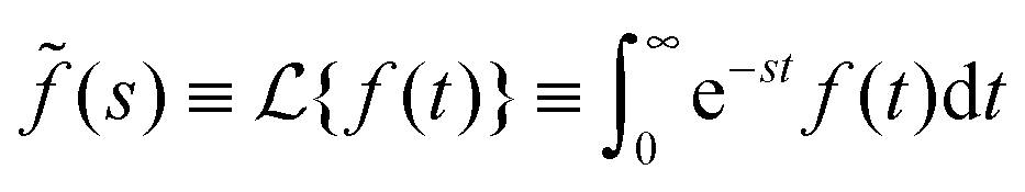 , where s is the Laplace frequency] of the complex shear modulus [
![[G with combining tilde]](https://www.rsc.org/images/entities/i_char_0047_0303.gif) (s)] can be related to the Laplace transform of 〈r2(t)〉 as:3,19,20
(s)] can be related to the Laplace transform of 〈r2(t)〉 as:3,19,20
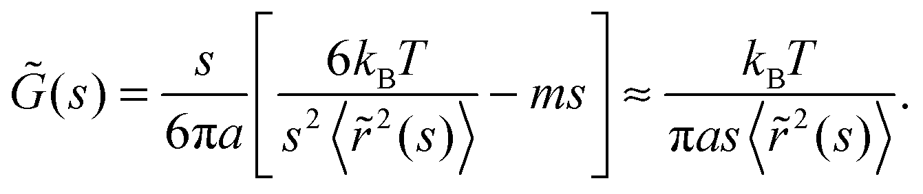 | (1) |
r(s)] is related to (s) as: G(s) = sr(s),3,19,20 while the creep compliance (J(t)) is related to the stress relaxation modulus as:21 | (2) |
 | (3) |
Various paste-like systems such as Carbopol suspensions,24 gellan gum,25 peptide dispersions,26 hectorite clay dispersions,27etc. have been studied using microrheology techniques. However, in this paper, we concentrate only on the microrheology studies of the time dependent materials that are relevant to the present work. Houghton et al.27 used passive microrheology to study the gelation of aging hectorite clay suspensions. The results showed increasing inhomogeneity in the samples with time as exhibited by the anisotropy in the curves for the mean square displacements in different spatial directions. A precise gel point was obtained by Larsen and Furst28 for a time dependent physical (peptide) gel and a concentration dependent chemical (polyacrylamide) gel. The mean square displacements of the probe particles were separated into two master curves using suitable shift factors: one before the gel point and one after the gel point. The shift factors for both, pre- and post-gel point regimes, exhibited divergence at a unique time (for a physical gel) and concentration (for a chemical gel) which represented the critical state. Interestingly, at the gel point, the critical relaxation exponent and the dynamic scaling exponents showed remarkable agreement with the predictions from the theories for fractal polymers. The kinetics of peptide self-assembly was studied using microrheology by Savin and Doyle.26 An increase in the self-assembly of the peptide led to a subdiffusive behavior exhibited by the probe mean square displacements, suggestive of a gel formation. The gel point was determined by the congruency of the frequency dependence of the viscous and elastic moduli29 as calculated from the mean square displacements. It was also observed that the pH of the solution has a profound effect (almost hundred-fold) on the gelation time, even at a very low concentration of peptide. The mechanism of gelation, though, was found to be independent of pH.
Quite recently, a few groups have used the microrheology technique to study the aging behavior for LAPONITE® suspensions, which is a subject matter of this work. Abou and Gallet30 carried out an experimental study to check the validity of the fluctuation–dissipation theorem for an aging LAPONITE® suspension. Their measurements revealed a non-monotonic variation of the effective temperature with increasing aging times, with the maximum corresponding to about twice the batch temperature. The observed behavior was attributed to the possible evolution of the relaxation time distribution expected in a system with evolving heterogeneity. However, subsequent work by Jabbari-Farouji31 showed that the effective temperature remains constant and does not differ from the bath temperature over a much wider range of aging times, thus, implying no violation of the fluctuation–dissipation for such non-equilibrium systems. The reasons for the qualitative differences between these and the measurements made by About and Gallet30 are, however, not quite clear, except perhaps the use of different concentrations of LAPONITE® in each study. The measurements made by Jabbari-Farouji31 also revealed that the overall viscoelasticity in the system arises from two contributions, viz. the strong frequency dependent response at high frequencies and a more elastic response (weakly dependent on frequency) during the aging.
Oppong et al.32 studied the gelation of 1 weight% LAPONITE® suspension using a microrheology technique with 0.5 micron radius microspheres as the probe particles. The magnitude of the viscous and elastic moduli obtained from the mean square displacements of the probe particles increased with the aging time. At short aging times, the viscous modulus was found to be larger than the elastic modulus indicating a viscous behavior while at longer times the trend was reversed, suggestive of a solid-like structure. In agreement with the theory of Winter–Chambon,29 the elastic and viscous moduli showed identical dependence on frequency at the gel point. Qualitatively similar results were obtained from the measurements of bulk rheology, although the gel point was found to occur much earlier than that obtained from microscopic measurements. This difference in the gel point obtained from two measurements was attributed to the difference in the length scale at which the rheology is probed. While the bulk rheology being probed at a larger length scale considers a material to be a critical gel, the probe particles in the same sample are relatively free to move due to several vacant regions, thus perceiving the same sample as a fluid on a microscopic scale. With more aging time, the aggregates continue to grow and eventually the fluid regions are diminished completely, which leads to freezing of the probe particles, and hence perceiving the sample as a gel at a later time on a microscopic scale. The gel time, thus, shows an inverse relationship with the length scale being probed. This aspect was further explored by Rich et al.,33 who found that the gel time increases by an order of magnitude for nearly one-fourth reduction in the probe particle size. The observed behavior suggests that the heterogeneity in the system changes with aging time, i.e. the sample microstructure evolves with aging time causing a continual increase in the solid-like behavior. The detailed microstructure as studied by the authors in terms of the heterogeneity ratio and correlation between successive displacements provides evidences of microstructural confinement. For different LAPONITE® concentrations, the authors identified a probe size–concentration superposition for the gelation time data. The scaling exponents were found to be dependent on probe sizes and LAPONITE® concentration.
We believe that since time dependent materials do not obey time translational invariance, microrheological analysis poses difficulties. Particularly, eqn (1)–(3) are applicable only for a small enough lag time for which the time dependency can be neglected and therefore time translational invariance can be assumed to be valid. However, the consideration of very small lag times does not allow generation of substantial amount of data. This problem can be partly addressed by extending the effective time domain framework, which has been developed for bulk rheological analysis, to microrheology. In this work, we develop such framework for microrheological analysis by considering an example of aging (time dependent) aqueous suspension of LAPONITE®.
II. Material, sample preparation and experimental protocol
LAPONITE® is an inorganic synthetic clay with primary particles having a disk-like shape with diameter 25 nm and thickness 0.92 nm. In an aqueous medium with pH around 10, its edge is positively charged while the faces are negatively charged. Soon after the incorporation of LAPONITE® in water beyond 1 weight% under vigorous stirring, it forms a clear suspension, whose viscoelastic properties show spectacular evolution as a function of time. Eventually it leads to the formation of a soft solid that sustains its weight against gravity. The microstructure of the suspension that is responsible for the time dependent evolution of its viscoelastic properties (physical aging) is proposed to be due to a combination of attractive as well as repulsive interactions among the particles.34,35In a typical sample preparation protocol, dry LAPONITE® XLG powder (dried over 4 hours at 120 °C) is gradually added to deionized, distilled water having pH 10 (using NaOH) containing 4 mM NaCl to obtain a suspension. The resulting mixture is stirred vigorously for about 30 min using a turrax drive and then passed through a microfilter (0.45 μm mesh, PRAMA® Nylon – non-sterile) to break the aggregates. This represents the initial state of a suspension (tw = 0) for every experiment performed. Following filtration, fluorescent polystyrene micro-particles of 1 μm diameter and density 1.05 g cc−1 are randomly dispersed in the suspension via rapid (∼15 s) vortex mixing. The fluorescent particle concentration is just enough to visualize sufficient number of particles for better statistics. In this work, we prepare suspensions using three LAPONITE® concentrations, 1.8, 2 and 2.2 weight%, all containing 4 mM NaCl. Hereafter, we shall represent the suspensions only by the LAPONITE® concentration without mentioning the salt concentration.
For the bulk rheology experiments, a freshly prepared filtered LAPONITE® suspension is introduced in the Couette cell (a concentric cylinder geometry with an outer diameter of 18.08 mm and a gap of 1 mm) of an Anton Paar MCR 301 rheometer. A suspension is then subjected to successive frequency sweeps (of 0.628–10 rad s−1) with a stress magnitude of 0.1 Pa. A thin layer of low viscosity silicon oil is applied on the free surface of a sample in order to prevent evaporation of water over the duration of the experiment.
For the microrheological experiments a freshly prepared filtered sample is placed in a glass slide having a small rectangular cavity (volume ∼ 0.045 cm3) in the center, which is then sealed using a cover slip. The fluorescent microspheres in the cavity are imaged using an inverted fluorescence microscope (Carl Zeiss) equipped with a 40× objective to give a spatial resolution of 0.323 microns per pixel. The images are taken in the center of the cavity to prevent any end wall effects. The fluorescent particle concentration used ensures about 50 particles in the microscope’s field of view. A sequence of images is acquired up to a duration of 60 s lag time, at varying aging times (tw) from the same sample position, using a camera operated at 20 frames per second. The duration of image acquisition is very small to expect any significant changes in the suspension properties during the course of measurements. All the measurements were made at 25 °C. Each such experiment was repeated three times using a fresh sample of LAPONITE® and the data shown in the figures represent the average over the three experiments.
The image sequence at all the aging times (from each experiment) is analyzed using standard particle tracking algorithms18 to obtain the microsphere particle trajectories from the identified particle centers in each image. The particle trajectories are further analyzed to obtain the mean square displacements defined as 〈r2(τ′)〉 = 〈|x(t + τ′) − x(t)|2 + |y(t + τ′) − y(t)|2〉, where t is the arbitrary time, τ′ is the lag time between two particle positions on a trajectory and (x,y) represents the two-dimensional spatial field for particle displacements. The angle brackets 〈·〉 indicate the mean over all the particles in an image window. Each such mean value, for a given lag time, is further averaged over three experiments and the figures represent these average values.
III. Results and discussion
We first discuss the linear viscoelastic behavior over macroscopic (bulk) length-scales, which is obtained by subjecting a time dependent suspension sample to successive cyclic frequency sweeps in a rheometer. In Fig. 1(a) we plot the time evolution of G′ and G′′ of a freshly prepared 2 weight% system for ω = 10 rad s−1. It can be seen that in the limit of small times G′, which is smaller than G′′, evolves at a faster rate and eventually crosses G′′. Beyond the crossover point G′ continues to increase while G′′ shows a plateau. In Fig. 1(b) we plot G′ and G′′ as a function of ω for tw = 1800 s, which shows G′ to be a weak function of ω and to be greater than G′′ throughout over the explored frequency window. The observed rheological behavior very clearly indicates the soft glassy (pasty) nature of the studied LAPONITE® suspension. | ||
| Fig. 1 The elastic (G′, filled symbols and full line) and viscous (G′′, open symbols and dashed line) moduli are plotted as a function of (a) tw for ω = 10 rad s−1 and (b) ω for tw = 1800 s. The symbols represent the experimental data while the lines represent the prediction from the microrheology experiments. | ||
The dynamics over microscopic length-scales are obtained by the mean square displacement of the tracer particles. The variation of the ensemble mean square displacements 〈r2(t − tw)〉 with experimental time (t − tw) is shown for several aging (or aggregation) times tw in Fig. 2. For all the aging times, 〈r2(t − tw)〉 increases with t − tw. However, the increase in 〈r2(t − tw)〉 gets slower with an increase in the aging time, which is suggestive of a progressive enhancement in the constrained dynamics, which arises due to restricted motion of the tracer particles indicating a cage-like surrounding caused by the microstructure build-up in the system.
 | ||
| Fig. 2 MSD(〈(r(t − tw))2〉 = (kBT/πa)J(t − tw)) is plotted as a function of t − tw for different tw for 2 weight% suspension. The symbols represent the experimental data while the solid lines represent prediction using eqn (10). The inset shows the transformed data (shown in main figure) in the effective time domain using eqn (8) for α = 0.077 ± 0.0013, which demonstrates time–aging time superposition, and thus validates the effective time-translational invariance. | ||
The distributions of the displacements of individual particles are shown in Fig. 3 for the aging times reported in Fig. 2. The displacements are obtained over a duration of 1 second for each aging time. The motion of particles is isotropic and hence the distributions are shown only for the displacements in one spatial (x) direction. For tw = 15 min or earlier (not shown), the distribution is Gaussian in nature as indicated by the solid line. The Gaussian distribution essentially represents a homogeneous system, devoid of any microstructure, allowing tracer particles to exhibit a random (purely diffusive) motion. The distribution progressively deviates from a Gaussian as tw increases. This deviation represents a gradual enhancement in subdiffusive dynamics caused by microstructure evolution, leading to spatially variant tracer particle motion. The spatial variation arises due to particles located in different regions sampling different local behavior, which is dependent on the specific microstructure in the system. For very large values of tw, the distribution is extremely narrow, which suggests that most of the tracer particles are in an immobilized state or are localized for the entire experimental time duration and the system is in a structurally arrested state.
 | ||
| Fig. 3 Probability distribution curves of displacements for different tw for a 2 weight% suspension. The solid lines represent the Gaussian fit to the experimental data shown in symbols. The data are shown only for x-direction displacements as the y-direction displacements exhibit similar behavior. | ||
The deviation of the displacement distribution from the Gaussian shape can be quantified in terms of the non-Gaussian parameter, N, defined as:24
 | (4) |
 | ||
| Fig. 4 Non-Gaussian parameter N plotted as a function of lag time (t − tw) for different aging times. | ||
The behavior observed in Fig. 2 and 3 is corroborated by the sample trajectories obtained at different aging times shown in Fig. 5. Every trajectory is recorded over an experimental time of 60 seconds. As expected, the trajectory at tw = 15 minutes shows significant particle movement, exploring a much larger space, in a homogeneous (liquid-like) state of the system. At subsequent aging times, with progressive evolution of the microstructure in the system, the movement of the tracer particle is significantly reduced, which is almost of the order of particle size for tw = 50 minutes and even smaller at further aging times (not shown).
 | ||
| Fig. 5 Sample trajectories recorded over 60 s for different aging times (tw = 15, 20, 30, 40, 45, and 50 min) for 2 weight% suspension. | ||
The above discussion provides reasonable signatures of an evolving microstructure within the static LAPONITE® suspension system with increasing aging times, which are in agreement with the previous microrheology studies on time evolving systems.28,32,33,36 The data from the experiments, though conducted separately at each tw, provide a clear picture of a continual, monotonic evolution of the suspension microstructure over time leading to its eventual structural arrest. Fig. 2 also depicts an important difference between the behavior of the MSD for materials that are at thermodynamic equilibrium and those materials that undergo time evolution. In the former, the MSD is only a function of lag time (t − tw): 〈r2〉 = 〈r2(t − tw)〉, while, in the latter, the MSD is also dependent on the time at which the system is explored (tw, in the present case it is the same as aging time) in addition to that of lag time:
| 〈r2〉 = 〈r2(t − tw,tw)〉 | (5) |
Through a series of papers, Joshi and coworkers proposed the effective time domain approach10–12,41 in order to accommodate the dependence of rheological response functions (in this case creep compliance through MSD) on tw. On one hand, such an approach validates effective time translational invariance and allows the representation of the Boltzmann superposition principle in the conventional form, but in the effective time domain.11,12 On the other hand, such an approach allows the prediction of very long time rheological behavior from short time experiments.10 In the subsequent paragraphs, we try and extend the effective time domain approach to the microrheology of an aging LAPONITE® suspension, an example of a time dependent material.
It is well known that the viscoelastic response of a material depends primarily on the spectrum of relaxation time scales. In glassy materials, including that of soft glassy materials, it is usually observed that during physical aging, all the relaxation modes evolve with time in such a way that the shape of relaxation time spectrum is preserved. Under such conditions, based on the work of Hopkins42 and Struik,43 Joshi and coworkers proposed the effective time domain (ξ) as:10–12,42,43
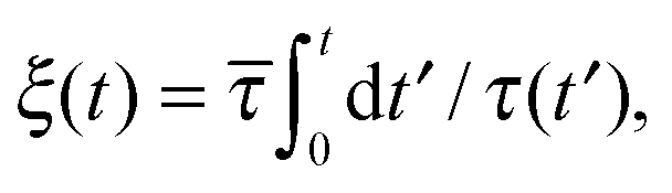 | (6) |
![[small tau, Greek, macron]](https://www.rsc.org/images/entities/i_char_e0d4.gif) ).
).
According to eqn (6), the dependence of relaxation time on the aging time is required to transform aging time dependent MSD data to the effective time domain. Now, the available literature states that the relaxation time of the aqueous LAPONITE® suspension, immediately following its preparation, shows exponential dependence on aging time given by:10,44,45
τ(t′) = τ0![[thin space (1/6-em)]](https://www.rsc.org/images/entities/char_2009.gif) exp(αt′), exp(αt′), | (7) |
= τ0 lead to: | (8) |
The superpositions obtained for all the three LAPONITE® concentrations are plotted in Fig. 6. The shape of the curve does not exhibit any obvious dependence on the LAPONITE® concentration. Since the MSD has been equated with the creep compliance, the very fact that the shape of superposition is preserved over the three decades of timescales suggests that the shape of the relaxation time spectrum remains unchanged by the change in the LAPONITE® concentration. Interestingly, the observed behavior corroborates quite well with that observed by Shahin and Joshi35 for bulk rheological measurements, wherein the shape of a spectrum of the relaxation times was observed to remain unchanged with the change in the LAPONITE® concentration. The values of 〈r2(ξ − ξw)〉 shown in Fig. 6, as expected, are seen to decrease with the increase in the LAPONITE® concentration, suggesting that the mean relaxation time increases with the increase in the LAPONITE® concentration. The values of 〈r2(ξ − ξw)〉, for LAPONITE® concentrations of 1.8 weight% and 2 weight%, are shifted horizontally to overlay on the data for the reference concentration (cL,R = 2.2 weight%) leading to a master superposition [time–aging time–LAPONITE® concentration superposition] as shown in Fig. 7. The horizontal shift factor given by hc = (cL,R)/(cL) is plotted in the inset of Fig. 7. As expected, hc, which is proportional to the inverse of the relaxation time, decreases with the increase in the LAPONITE® concentration (cL). A slight vertical shifting is needed only for the 2.2 weight% suspension in order to get better quality of superposition; the corresponding shift factors (b) are shown in Fig. S3 (ESI†).
 | ||
| Fig. 6 Variation of superimposed MSD(〈r2〉) with the effective time difference (ξ − ξw) for an aqueous LAPONITE® suspension of different concentrations. The values of α are: 0.04 ± 0.0017 (1.8%), 0.077 ± 0.0013 (2%) and 0.105 ± 0.007 (2.2%). A slight vertical shifting is needed for the 2.2 weight% suspension, whose values are given in the ESI;† for the other two concentrations we do not need any vertical shifting. The factor β carries no physical meaning and is simply used to translate the curves horizontally to show the superimpositions more clearly (β = 0.1 for 1.8%, 0.3 for 2.0% and 1 for 2.2%). | ||
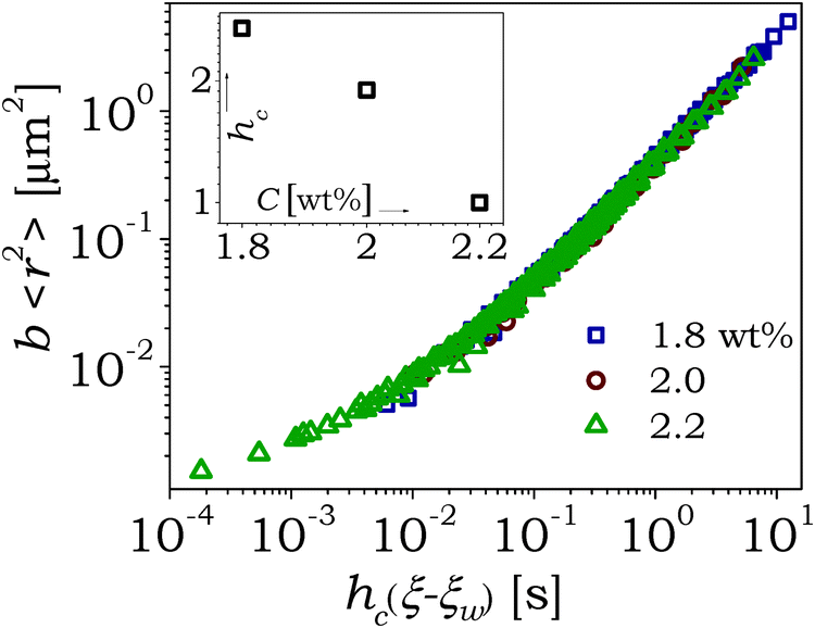 | ||
| Fig. 7 The global master MSD(〈r2〉) curve plotted in the effective time domain, obtained by horizontally shifting the three master curves corresponding to three different LAPONITE® concentrations, shown in Fig. 6. The inset shows the horizontal shift factors (hc = (cL,R)/(cL)) plotted as a function of the LAPONITE® concentration. | ||
The ordinate of Fig. 7 that shows master superposition of 〈r2(ξ − ξw)〉 is also equivalent to (kBT/πa)J(ξ − ξw). With the knowledge of creep compliance in the effective time domain, the stress relaxation modulus, then, can be obtained using the convolution relation given by:
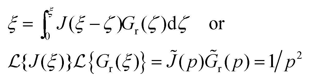 | (9) |
 . We solve eqn (9) numerically by using the scheme proposed by Zhu et al.46 to obtain the relaxation modulus in the effective time domain given by (πa/kBT)Gr(ξ − ξw), which is shown in the inset of Fig. 8. Fig. 8 shows the relaxation modulus associated with an individual LAPONITE® concentration, which is obtained by dividing the abscissa in the inset of Fig. 8 by the horizontal shift factor (hc) plotted in the inset of Fig. 7, which leads to the separation of superpositions associated with each concentration. It can be seen that the predicted relaxation modulus is constant in the limit of very small effective times and decreases with the increase in ξ − ξw, and eventually decays to zero. As expected, the relaxation gets delayed in time with the increase in the LAPONITE® concentration.
 | ||
| Fig. 8 The individual superposition of stress relaxation modulus (πa/kBT)Gr(t) in the effective time domain for the three studied concentrations: from left to right 1.8, 2 and 2.2 weight%, which are obtained from the master superposition of the stress relaxation modulus plotted in the effective time domain (by using global master MSD curve shown in Fig. 7 by solving eqn (9)), shown in the inset. | ||
The global master superposition for 〈r2(ξ − ξw)〉 or (kBT/πa)J(ξ − ξw) shown in Fig. 7 and that of (πa/kBT)Gr(ξ − ξw) shown in Fig. 8 describe the behavior of respective parameters if the experiments were conducted for a system having concentration cL,R in the effective time domain. The creep compliance (or MSD) and stress relaxation data in the effective time domain can be transformed back to the real time domain by simply inverting eqn (8) to give:
| t − tw = ln[(e−αtw − α{ξ − ξw})−1/α] − tw. | (10) |
 | ||
| Fig. 9 The time evolution of the stress relaxation modulus (πa/kBT)Gr(t) is plotted in the real time domain for different tw (from left to right: 15, 20, 30, 40, 45, and 50 min) for a 2 weight% LAPONITE® suspension obtained from eqn (10). | ||
Knowledge of Gr(t) at various tw also allows us to obtain complex modulus G*, as a function of ω by using:
 | (11) |
The present work clearly demonstrates several advantages of carrying out analysis of time dependent soft materials in the effective time domain. The extra time dependence incorporated in eqn (5) states that the values of MSD get affected by aging that occurs during the lag time, when the lag time is of the order of aging time [t − tw = O(tw)] and consequently, eqn (1)–(3) cannot be applied to such time dependent materials. While such restriction affects all the aging times, it critically affects the measurements at the beginning of aging (small tw). However, once MSD experiments have been performed in the limit of smaller lag times (t − tw ≪ tw), whence the eqn (1)–(3) are applicable, expressing the MSD data obtained at different tw in the effective time domain allows a linear viscoelastic representation of the same, which otherwise is not possible. Moreover, since the relaxation time is small at low tw, it facilitates the prediction of long time MSD behavior (and in general, the rheological behavior) over large lag times. Equivalently, the MSD data at large tw facilitates prediction over very small lag times. The present work even goes one step further wherein superposition has also been obtained by accommodating LAPONITE® concentration dependence. Such superpositions, which have been proposed to be valid for bulk rheological measurements, allow the prediction of the MSD over much greater lag times.35 The major requirement for the feasibility of such analysis is a prior knowledge of relaxation time dependence on time, which can be obtained either independently or through the process of obtaining superposition itself. It is also important to note that the applicability of effective time domain expressed by eqn (6) is not only limited to a single relaxation time dependence, but also to various functional forms of τ(t) at different times that can be easily accommodated in eqn (6) as a sum of the integrals.
IV. Conclusion
In the present work, the rheological behavior of time dependent (physically aging) aqueous suspensions of LAPONITE®, a model soft glassy material, is studied using a passive microrheology technique. We employ an optical microscope to probe the thermal motion of micron-sized tracer particles embedded in the sample. The motion of the tracer particles shows purely diffusive behavior at short aging times that progressively changes to subdiffusive behavior as the material ages. We obtain the MSDs for different aging times (tw) and for suspensions of three different LAPONITE® concentrations in such a way that the lag time (t − tw) is very small (t − tw ≪ tw). Within the range of lag times studied, the variation of relaxation time is negligible, and so the MSD can be equated to the creep compliance using standard microrheology analysis. The MSDs obtained at different aging times as well as LAPONITE® concentrations, however, possess different relaxation times. In order to get rid of time dependency, we employ a concept of effective time, wherein the real time is normalized by the time dependent relaxation time to readjust the material clock. Consequently, we transform the MSD data from the real time domain to the effective time domain that results in time–aging time superposition. Interestingly, the time–aging time superpositions for different LAPONITE® concentrations show similar shapes, which eventually leads to a time–aging-time–concentration master curve. The existence of such a master curve, which has also been previously observed, but for the macroscopic rheological behavior of LAPONITE® suspensions, suggests that the aging time as well as the LAPONITE® concentration only affects the mean relaxation time but not the shape of the relaxation time distribution. Since the MSD behavior of tracer particles is qualitatively analogous to the creep compliance, the use of the standard convolution relation of linear viscoelasticity in the effective time domain allows the prediction of the stress relaxation behavior of the suspension. Interestingly the transformation of both creep compliance (MSD) and stress relaxation data from the effective time domain to the real time domain leads to the real time behaviors of the respective parameters at different aging times and LAPONITE® concentrations. Particularly, for the MSD, such transformation allows prediction over larger lag times (over five decades) compared to those incorporated in experimental measurements (around two decades), demonstrating the significant advantage of the proposed approach. Overall, we believe that the effective time domain approach provides an important step forward in analyzing time dependent materials using microrheological techniques.Acknowledgements
The financial support from the Department of Atomic Energy – Science Research Council, Government of India, is greatly acknowledged.References
- Y. M. Joshi, Annu. Rev. Chem. Biomol. Eng., 2014, 5, 181–202 CrossRef CAS PubMed.
- P. Cicuta and A. M. Donald, Soft Matter, 2007, 3, 1449–1455 RSC.
- T. M. Squires and T. G. Mason, Annu. Rev. Fluid Mech., 2010, 42, 413–438 CrossRef.
- A. M. Puertas and T. Voigtmann, J. Phys.: Condens. Matter, 2014, 26, 243101 CrossRef CAS PubMed.
- K. M. Schultz and E. M. Furst, Soft Matter, 2012, 8, 6198–6205 RSC.
- A. A. Abdala, S. Amin, J. H. van Zanten and S. A. Khan, Langmuir, 2015, 31, 3944–3951 CrossRef CAS PubMed.
- T. Moschakis, Curr. Opin. Colloid Interface Sci., 2013, 18, 311–323 CrossRef CAS.
- M. E. Cates and M. R. Evans, Soft and fragile matter, The Institute of Physics Publishing, London, 2000 Search PubMed.
- S. M. Fielding, P. Sollich and M. E. Cates, J. Rheol., 2000, 44, 323–369 CrossRef CAS.
- A. Shahin and Y. M. Joshi, Phys. Rev. Lett., 2011, 106, 038302 CrossRef CAS PubMed.
- M. Kaushal and Y. M. Joshi, Soft Matter, 2014, 10, 1891–1894 RSC.
- M. Kaushal and Y. M. Joshi, Macromolecules, 2014, 47, 8041–8047 CrossRef CAS.
- G. B. McKenna, T. Narita and F. Lequeux, J. Rheol., 2009, 53, 489–516 CrossRef CAS.
- T. G. Mason and D. A. Weitz, Phys. Rev. Lett., 1995, 74, 1250–1253 CrossRef CAS PubMed.
- T. A. Waigh, Rep. Prog. Phys., 2005, 68, 685–742 CrossRef.
- T. A. Waigh, Rep. Prog. Phys., 2005, 68, 685–742 CrossRef.
- J. Liu, M. L. Gardel, K. Kroy, E. Frey, B. D. Hoffman, J. C. Crocker, A. R. Bausch and D. A. Weitz, Phys. Rev. Lett., 2006, 96, 118104 CrossRef CAS PubMed.
- J. C. Crocker and D. G. Grier, J. Colloid Interface Sci., 1996, 179, 298–310 CrossRef CAS.
- X. Jingyuan, V. Virgile and D. Wirtz, Rheol. Acta, 1998, 37, 387–398 CrossRef.
- T. G. Mason, Rheol. Acta, 2000, 39, 371–378 CrossRef CAS.
- J. D. Ferry, Viscoelastic Properties of Polymers, 1980 Search PubMed.
- M. T. Valentine, P. D. Kaplan, D. Thota, J. C. Crocker, T. Gisler, R. K. Prud’homme, M. Beck and D. A. Weitz, Phys. Rev. E: Stat., Nonlinear, Soft Matter Phys., 2001, 64, 061506 CrossRef CAS PubMed.
- D. Weihs, M. A. Teitell and T. G. Mason, Microfluid. Nanofluid., 2007, 3, 227–237 CrossRef CAS.
- F. K. Oppong, P. Coussot and J. R. de Bruyn, Phys. Rev. E: Stat., Nonlinear, Soft Matter Phys., 2008, 78, 021405 CrossRef PubMed.
- M. Caggioni, P. T. Spicer, D. L. Blair, S. E. Lindberg and D. A. Weitz, J. Rheol., 2007, 51, 851–865 CrossRef CAS.
- T. Savin and P. S. Doyle, Soft Matter, 2007, 3, 1194–1202 RSC.
- H. A. Houghton, I. A. Hasnain and A. M. Donald, Eur. Phys. J. E: Soft Matter Biol. Phys., 2008, 25, 119–127 CrossRef CAS PubMed.
- T. H. Larsen and E. M. Furst, Phys. Rev. Lett., 2008, 100, 146001 CrossRef PubMed.
- F. Chambon and H. H. Winter, J. Rheol., 1987, 31, 683–697 CrossRef CAS.
- B. Abou and F. Gallet, Phys. Rev. Lett., 2004, 93, 160603 CrossRef PubMed.
- S. Jabbari-Farouji, D. Mizuno, M. Atakhorrami, F. C. MacKintosh, C. F. Schmidt, E. Eiser, G. H. Wegdam and D. Bonn, Phys. Rev. Lett., 2007, 98, 108302 CrossRef PubMed.
- F. K. Oppong, L. Rubatat, B. J. Frisken, A. E. Bailey and J. R. de Bruyn, Phys. Rev. E: Stat., Nonlinear, Soft Matter Phys., 2006, 73, 041405 CrossRef PubMed.
- J. P. Rich, G. H. McKinley and P. S. Doyle, J. Rheol., 2011, 55, 273–299 CrossRef CAS.
- B. Ruzicka and E. Zaccarelli, Soft Matter, 2011, 7, 1268–1286 RSC.
- A. Shahin and Y. M. Joshi, Langmuir, 2012, 28, 15674–15686 CrossRef CAS PubMed.
- J. P. Rich, J. Lammerding, G. H. McKinley and P. S. Doyle, Soft Matter, 2011, 7, 9933–9943 RSC.
- A. Shahin and Y. M. Joshi, Langmuir, 2012, 28, 5826–5833 CrossRef CAS PubMed.
- M. Cloitre, R. Borrega and L. Leibler, Phys. Rev. Lett., 2000, 85, 4819–4822 CrossRef CAS PubMed.
- A. S. Negi and C. O. Osuji, Phys. Rev. E: Stat., Nonlinear, Soft Matter Phys., 2009, 80, 010404 CrossRef PubMed.
- P. Coussot, H. Tabuteau, X. Chateau, L. Tocquer and G. Ovarlez, J. Rheol., 2006, 50, 975–994 CrossRef CAS.
- R. Gupta, B. Baldewa and Y. M. Joshi, Soft Matter, 2012, 8, 4171–4176 RSC.
- I. L. Hopkins, J. Polym. Sci., 1958, 28, 631–633 CrossRef CAS.
- L. C. E. Struik, Physical Aging in Amorphous Polymers and Other Materials, Elsevier, Houston, 1978 Search PubMed.
- F. Schosseler, S. Kaloun, M. Skouri and J. P. Munch, Phys. Rev. E: Stat., Nonlinear, Soft Matter Phys., 2006, 73, 021401 CrossRef CAS PubMed.
- A. S. Negi and C. O. Osuji, Phys. Rev. E: Stat., Nonlinear, Soft Matter Phys., 2010, 82, 031404 CrossRef PubMed.
- Y. Zhu, L. Sun and H. Xu, J. Appl. Mech., Trans. ASME, 2011, 78, 031002 CrossRef.
- M. Tassieri, F. D. Giudice, E. J. Robertson, N. Jain, B. Fries, R. Wilson, A. Glidle, F. Greco, P. A. Netti, P. L. Maffettone, T. Bicanic and J. M. Cooper, Sci. Rep., 2015, 5, 8831 CrossRef CAS PubMed.
- M. Buchanan, M. Atakhorrami, J. F. Palierne, F. C. MacKintosh and C. F. Schmidt, Phys. Rev. E: Stat., Nonlinear, Soft Matter Phys., 2005, 72, 011504 CrossRef CAS PubMed.
- G. Pesce, A. C. D. Luca, G. Rusciano, P. A. Netti, S. Fusco and A. Sasso, J. Opt. A: Pure Appl. Opt., 2009, 11, 034016 CrossRef.
Footnotes |
| † Electronic supplementary information (ESI) available. See DOI: 10.1039/c6sm00829a |
| ‡ Present address: Laboratoire de Physique des Solides, CNRS, Univ. Paris-Sud, Université Paris-Saclay, 91405 Orsay Cedex, France. |
| This journal is © The Royal Society of Chemistry 2016 |