
Engineering plasmonic Ag/AgCl–polydopamine–carbon nitride composites for enhanced photocatalytic activity based on mussel chemistry†
Wei Lia,
Guoxiang Zhangab,
Wenbo Shenga,
Changchang Donga,
Yu Daia,
Cuihua Lia,
Rongjie Wanga,
Yulin Shia,
Xuhong Guoac and
Xin Jia*a
aSchool of Chemistry and Chemical Engineering, Key Laboratory for Green Processing of Chemical Engineering of Xinjiang Bingtuan, Key Laboratory of Materials-Oriented Chemical Engineering of Xinjiang Uygur Autonomous Region, Engineering Research Center of Materials-Oriented Chemical Engineering of Xinjiang Bintuan, Shihezi University, Shihezi 832003, People's Republic of China. E-mail: jiaxin@shzu.edu.cn
bGansu Dayu Water-Saving Group Co., Ltd, Jiuquan 735000, People's Republic of China
cState Key Laboratory of Chemical Engineering East China University of Science and Technology, Shanghai 200237, People's Republic of China
First published on 31st October 2016
Abstract
The interfacial interaction plays an important role in composites, by affecting the physical and chemical properties of those composites. Herein, we present a simple and versatile approach for the construction of plasmonic Ag/AgCl–polydopamine–CN (SPCN) composites with enhanced photocatalytic properties by mussel chemistry. Dopamine hydrochloride acts as both the reactant and the reducing agent, and the product polydopamine (PDA) serves as the adhesive layer and the electron transfer bridge. The photocatalytic degradation of rhodamine B (RhB) shows that the SPCN50 composite displays excellent photocatalytic activity. A radical trapping experiment indicates that holes are the main oxidative species in the photocatalytic process, and a possible photocatalytic mechanism is proposed. All the results prove that PDA indeed improves the separation efficiency of photogenerated carriers and the photocatalytic activity. This work may provide a green, universal method to synthesize plasmonic photocatalytic composites with high catalytic performance.
1. Introduction
In recent years, due to the serious energy crisis and environmental pollution, semiconductor photocatalysis has been a very hot research topic; it can use solar energy to split water into hydrogen, degrade organic pollutants and reduce carbon dioxide into other fuels without depletion of fossil energy. Since 1972,1 a myriad of photocatalysts have been developed encompassing inorganic materials,2 organic materials,3 organic–metal complexes, and elements.4 One kind of nitrogen-doped carbon material, carbon nitride (CN), has received tremendous attention because of its easy availability, appropriate band positions (Eg = 2.70 eV),5 excellent chemical stability, special optical features, and high thermal stability.6 However, the high recombination rate of its photogenerated carriers and low absorption efficiency above 460 nm in the spectrum of sunlight hinder its further application.7 To solve these problems, many methods have been proposed to improve the photocatalytic performance of CN, such as using sensitizers,8 doping with nonmetal or metal elements,9 introducing various metals as cocatalysts,10 creating a hollow11 or mesoporous structure,5a,12 forming composites with other semiconductors6,13 and so on. For example, on the basis of surface plasmon resonance (SPR) of metal nanoparticles, Xu et al. prepared AgX/CN (X = Br or I) composites which displayed high photocatalytic activities in methyl orange degradation.14 Zhang et al. synthesized a novel efficient Ag@AgCl/CN plasmonic photocatalyst by a rational in situ ion exchange approach.7b Liu et al. used geothermal water as the chlorine source to obtain Ag/AgCl/CN for the photodegradation of methylene blue.15 Yuan et al. produced Ag/AgBr/CN using cetylmethylammonium bromide as the Br source.16 Li et al. investigated the photocatalytic activity of CN coated Ag/AgCl.17 Xu et al. used FeCl3 as the oxidant and Cl source to get Ag/AgCl/CN.18 Xia et al. combined a solvothermal method with dichloromethane as the Cl source to synthesize an Ag/AgCl/CN photocatalyst.19 However, although these methods can enhance the properties of CN to some extent, the present CN-based photocatalysts do not meet the demands of practical applications because of poor interactions between the host and the guest resulting in the exfoliation of the guest and secondary environmental pollution. Therefore, it is still necessary to develop a reasonable strategy to further enhance the photocatalytic properties of CN-based photocatalysts without disturbing the structure of CN.Polydopamine (PDA), a black insoluble biopolymer produced by autoxidative polymerization of dopamine (DA), is widely known as a versatile bioinspired functional system for various applications, and incorporates abundant catechol and amine functional groups. During oxidative polymerization, PDA will spontaneously form a coating layer atop nearly any substance present in the reaction medium through the strong binding affinity of its catechol functional groups.20 Moreover, DA and PDA can reduce noble metallic salts into metallic nanoparticles.21 Feng and co-workers synthesized monodisperse core/shell structural Ag@PDA, based on the redox reaction of PDA toward Ag+ ions, for the photocatalytic degradation of neutral red.21b Our group recently synthesized PDA/Au nanoparticles to prepare a N-doped carbon–Au catalyst.21a In addition, PDA has good biocompatibility, conductivity and stability. Park's group reported on the capability of PDA as an electron gate as well as a versatile adhesive for mimicking natural photosynthesis, and demonstrated that PDA can accelerate the rate of photoinduced electron transfer.22 Nam et al. prepared PDA-based organic phototransistors with high photosensitivity and photo-controlled switching properties under light illumination.23 Recently, our group prepared controllable PDA-based Ag@AgCl (P-Ag/AgCl) photocatalyst composites with high activity and stability using dopamine hydrochloride as the Cl source and reductant.24 Inspired by these studies, we envisaged that PDA, as an adhesive layer and electron acceptor, could enhance the stability and photocatalytic activity of CN. To the best of our knowledge, the use of PDA as an adhesive layer to anchor Ag/AgCl on CN has not been reported previously.
In this work, we present a simple and versatile approach for the construction of plasmonic Ag/AgCl–PDA–CN (SPCN) composites for enhanced stability and photocatalytic properties by adopting multi-purpose PDA inspired by mussel chemistry. The composites were characterized by scanning electron microscopy (SEM), transmission electron microscopy (TEM), X-ray diffraction (XRD), Fourier-transform infrared (FTIR) spectroscopy, thermogravimetric analysis (TGA) and UV-visible diffuse reflectance spectroscopy (UV-vis DRS). The composites exhibit better photocatalytic activity than P-Ag/AgCl, CN and Ag/AgCl–CN toward the photodegradation of rhodamine B (RhB). Meanwhile, based on photoluminescence technology, radical trapping and photoresponse, a possible photocatalytic mechanism is proposed.
2. Experimental
2.1 Materials
Melamine and dopamine hydrochloride (DA) were purchased from Sigma-Aldrich. Silver nitrate (AgNO3, ≥99.0%) was obtained from Shanghai Fine Chemical Materials Research Institution. Poly(vinylpyrrolidone) (PVP) was purchased from Solarbio Company. Concentrated hydrochloric acid (HCl, 36.0–38.0%) and sodium hydroxide (NaOH, 98.0%) were obtained from Guangzhou Chemical Reagent Co., China. All other reagents were used as obtained.2.2 Synthesis of CN
Carbon nitride (CN) was synthesized by thermal oxidation etching according to a procedure reported previously.25 In detail, melamine (C3H6N6, 99.0%) was heated at 550 °C for 4 h in the air with a ramp rate of 2.3 °C min−1. The resultant yellow agglomerates were milled into powder in a mortar. Then, the bulk product was placed in an open ceramic container and heated at 500 °C for 2 h with a ramp rate of 5 °C min−1 to obtain CN.2.3 Synthesis of SPCN composites
In a typical experiment, a given amount of CN was placed in 55 mL of deionized water and ultrasonicated for 30 min. Then DA (0.18 g) and PVP (0.27 g) were dissolved in the above mixture, and the pH was adjusted to 7.8 using 1 M NaOH. Subsequently, 5 mL of AgNO3 aqueous solution (3.5 wt%) was injected into the solution and it was vigorously stirred for 30 min. The SPCN composites were obtained by centrifugation, and were washed three times with water. The composition of SPCN was achieved by adjusting the amount of CN. The as-obtained products were denoted as SPCNx (where x indicates the intial addition amount of CN). For comparison, P-Ag/AgCl and Ag/AgCl–CN were also synthesized. P-Ag/AgCl was prepared according to our previous experiments.24 Ag/AgCl–CN was prepared in a similar way to SPCN but without DA.2.4 Characterization
UV-vis DRS was performed on a TU-1901 UV-vis spectrophotometer, equipped with an integrating sphere, and BaSO4 was used as a reference. XRD patterns at wide (10–90°) angles were collected using a Bruker D8 advanced X-ray diffractometer with Cu Kα irradiation at 40 kV and 40 mA. X-ray photoelectron spectroscopy (XPS) was performed using an Axis Ultra spectrometer with a monochromatized Al Kα X-ray as the excitation source (225 W). TGA was conducted with a TGA/DTA (differential thermal analysis) system (SDT Q600, America) over the range from room temperature to 800 °C with an air flow of 10 mL min−1. FTIR spectroscopy was recorded on a Nicolet AVATAR 360 IR spectrometer. The morphology was investigated using a Hitachi scanning electron microscope. The photoresponsive activity was evaluated with CHI760D measurements using a three-electrode test cell at room temperature.2.5 Photocatalytic activity
The photocatalytic activities were evaluated by the photocatalytic degradation of the dyes rhodamine 6G (Rh 6G) or RhB in aqueous solutions under visible light irradiation using a 300 W Xe lamp with a 420 nm cut-off filter. The light intensity was 197 mW cm−2. For the photocatalytic experiments, 5 mg of the photocatalyst was dispersed in an aqueous solution of dye (50 mL, 5 mg L−1). Before irradiation, each suspension was magnetically stirred for 60 min to reach adsorption–desorption equilibrium in the dark. Upon irradiation, 2.5 mL aliquots were collected at certain time intervals and then centrifuged to remove the photocatalysts. Then the concentration of dyes was recorded using a UV-vis spectrophotometer. To investigate the active species generated in the photocatalytic degradation process, free radical capture experiments were carried out to test for hydroxyl radicals (˙OH), electrons (e−) and holes (h+) using isopropanol (IPA), potassium dichromate (K2Cr2O7) and ethylenediamine tetraacetic acid disodium salt (EDTA-2Na), respectively.To investigate the transition of photoinduced electrons of CN and SPCN50, a standard three-electrode cell with a working electrode, a carbon rod as the counter electrode and a standard calomel electrode (SEC) as the reference electrode was used. The electrolyte solution used was 0.1 M Na2SO4. The working electrode was prepared as follows: 2 mg of as-prepared photocatalyst and 10 μL of polytetrafluoroethylene (PTFE) (1 wt%) were suspended in 1 mL ethanol/water mixture and ultrasonicated for 30 min. Then 100 μL of the mixture was coated onto 1 cm × 1 cm carbon fiber paper. The electrode was obtained after the solvent was evaporated.
3. Results and discussion
The fabrication procedure for the SPCN photocatalysts is shown in Scheme 1. DA and PVP were mixed with ultrasonically dispersed CN, and the pH value of the reaction solution was adjusted to 7.8. After immediate addition of AgNO3, Ag/AgCl nanoparticles were deposited on the CN via a one-pot method inspired by dopamine chemistry at room temperature. As the AgNO3 solution was injected into the mixture, AgCl precipitates were formed between Ag+ and Cl− from the DA. Meanwhile, a redox reaction occurred between Ag+ and DA, resulting in the formation of Ag and PDA. PDA, as a strong adhesive, anchored Ag/AgCl on the CN through hydrogen bonding and π–π stacking interactions, enhancing the interaction between Ag/AgCl and CN. | ||
| Scheme 1 Schematic illustration and reaction mechanism for the preparation of SPCN composites. | ||
In order to confirm the CN content and the thermostability of the SPCN composites, TGA was carried out in an open system in the temperature range from room temperature to 800 °C at a heating rate of 10 °C min−1 under an air atmosphere. The amount of Ag/AgCl loaded on the CN was calculated. As shown in Fig. 1, the decomposition of CN began at 550 °C and was almost complete at 750 °C, which is attributed to the burning of CN. This weight loss region could be seen in the SPCN composite samples. The amount of CN in the SPCN50, SPCN100, SPCN200, SPCN300 and SPCN400 composites was 34.97, 49.31, 66.42, 71.05 and 74.45%, respectively.
 | ||
| Fig. 1 TGA curves of CN and as-prepared SPCN composites. | ||
The XRD patterns of the P-Ag/AgCl, CN, and SPCN composites are shown in Fig. 2. CN gave two consistent peaks at 13.1° and 27.6° corresponding to (0 0 1) and (0 0 2), respectively, which is similar to the result previously reported.25 The diffraction peaks at 27.9, 32.2, 46.2, 54.8, 57.5, 67.4, 74.4, 76.7, and 85.6° were assigned to the (1 1 1), (2 0 0), (2 2 0), (3 1 1), (2 2 2), (4 0 0), (3 3 1), (4 2 0), (4 2 2) planes of the cubic phase of AgCl (JCPDS no. 31-1238), respectively. The diffraction peaks at 38.1, 44.3 and 77.3° could be indexed to the cubic phase of Ag (JCPDS no. 65-2871), which confirmed the formation of Ag/AgCl. P-Ag/AgCl exhibited similar peaks to cubic phase Ag and AgCl, which is in accordance with our previous report.24 For the SPCN composites, the peaks at 27.8° became broader with the increase of CN content, which confirmed the successful preparation of SPCN.
 | ||
| Fig. 2 XRD patterns of CN, P-Ag/AgCl and as-prepared SPCN. | ||
The FTIR spectra of CN, P-Ag/AgCl, CN–Ag/AgCl and SPCN50 are shown in Fig. 3. In the FTIR spectrum of CN, the peak at 808 cm−1 is attributable to the typical breathing mode of triazine units.14,26 The strong band of 1200–1700 cm−1, with the typical peaks at 1236, 1317, 1406, 1541 and 1633 cm−1, corresponds to the stretching vibrations of C–N and C![[double bond, length as m-dash]](https://www.rsc.org/images/entities/char_e001.gif) N. The main peaks of CN clearly appear in the Ag/AgCl–CN and SPCN50 spectra. The pure P-Ag/AgCl spectrum has some characteristic peaks at 1633 cm−1 (aromatic rings, stretching band), 1523 cm−1 (amide bond, N–H, shearing band) and 1406 (C–N, stretching band) which can be ascribed to the nature of PDA.27 However, the typical peaks of PDA do not appear in the SPCN spectrum. This could be due to overlapping CN peaks. In the spectrum of the SPCN composite, the characteristic peaks of CN do not move (red shift or blue shift) after the introduction of P-Ag/AgCl indicating that P-Ag/AgCl does not change the structure of CN.
N. The main peaks of CN clearly appear in the Ag/AgCl–CN and SPCN50 spectra. The pure P-Ag/AgCl spectrum has some characteristic peaks at 1633 cm−1 (aromatic rings, stretching band), 1523 cm−1 (amide bond, N–H, shearing band) and 1406 (C–N, stretching band) which can be ascribed to the nature of PDA.27 However, the typical peaks of PDA do not appear in the SPCN spectrum. This could be due to overlapping CN peaks. In the spectrum of the SPCN composite, the characteristic peaks of CN do not move (red shift or blue shift) after the introduction of P-Ag/AgCl indicating that P-Ag/AgCl does not change the structure of CN.
 | ||
| Fig. 3 FTIR spectra of CN, P-Ag/AgCl, Ag/AgCl–CN and SPCN50. | ||
The morphology of CN and the formation of Ag/AgCl were studied by SEM imaging. Fig. 4 shows the SEM images of P-Ag/AgCl, pure CN and SPCN50. Fig. 4A shows the nanoparticle structure of P-Ag/AgCl. For pure CN (Fig. 4B), large aggregates composed of lamellar structures are observed. By comparing the images with that of pure CN, it is clearly shown in Fig. 4C and D that P-Ag/AgCl particles with cubic morphology are evenly decorated on the CN in the SPCN50 composite. However, for Ag/AgCl–CN without PDA, only some Ag/AgCl particles are observed on the CN (Fig. S1†). Moreover, after ultrasonication for 1 h (shown in Fig. S2†), the amount of P-Ag/AgCl on the CN is higher than that of Ag/AgCl on the CN in the absence of PDA. All these results show that PDA acts as the adhesive layer enhancing the interaction between Ag/AgCl and CN through π–π stacking or hydrogen bonding.
 | ||
| Fig. 4 SEM images of P-Ag/AgCl (A), pure CN (B), SPCN50 (C) and an enlarged view of SPCN50 (D). | ||
XPS was used to characterize the chemical compositions of the as-prepared photocatalysts, as shown in Fig. 5. In Fig. 5A, new Cl and Ag peaks are observed in the spectrum of the SPCN composite when compared to that of CN, confirming the successful decoration of Ag/AgCl on the CN surface. The high resolution XPS spectra of Ag 3d, Cl 2p, C 1s, N 1s, O 1s for SPCN50 are shown in Fig. 5B–F. For Ag 3d (Fig. 5B), two bands can be observed which could be ascribed to Ag 3d5/2 and Ag 3d3/2, respectively. These two bands could be further deconvoluted into four peaks at 367.7, 368.6, 373.8, and 374.5 eV. The peaks at 367.7 and 373.8 eV could be attributed to the Ag+ species of AgCl, and the peaks at 368.6 and 374.5 eV could be assigned to the Ag0 species, which proves the presence of Ag in the SPCN composites.28 In addition, the spectrum of Cl 2p shows two peaks which are consistent with the Cl− state (Fig. 5C).17 Fig. 5D shows five distinct peaks for C 1s at 284.8 (CC), 285.5 (C–C), 286.2 (C–N/C–OH), 288.2 (CN) and 289.2 eV (CO). Fig. 5E shows three peaks detected in the N 1s spectrum which are CN–C (399.5 eV), N–(C)3 (400.3 eV) and C–N–H (401.4 eV).29 The O 1s spectrum shows three peaks (CO 531.9 eV, C–O 532.8 eV and adsorbed H2O 533.9 eV) which indicates the presence of PDA.30 All these spectra confirm the successful preparation of SPCN.
 | ||
| Fig. 5 XPS survey spectra of CN and SPCN50 (A). The high-resolution XPS spectra of: (B) Ag 3d, (C) Cl 2p, (D) C 1s, (E) N 1s, and (F) O 1s for SPCN50. | ||
The photocatalytic activities of the as-obtained composites towards the degradation of dyes under visible light irradiation were evaluated. Firstly, Rh 6G was selected in order to examine the photocatalytic performances of the as-prepared SPCN photocatalysts under visible light irradiation (λ > 420 nm) (Fig. S3†). The initial concentration of the Rh 6G was measured and is denoted C0. The fraction of dye remaining, Y, is denoted as C/C0 where C is the concentration of Rh 6G at the indicated reaction time. As shown in Fig. S2,† 21% degradation of Rh 6G was observed after irradiation for 40 min in the absence of catalysts because Rh 6G is very unstable. Compared to pure CN, all SPCN composites exhibited enhanced photocatalytic activities and the photocatalytic activities increased gradually with increasing proportions of Ag/AgCl in the composites. The SPCN50 composite showed the highest activity.
To further study the photocatalytic activities of the SPCN composites, we selected RhB as the pollutant. The photocatalytic activities of CN, Ag/AgCl–CN, P-Ag/AgCl and commercial P25 were also tested for comparison under the same conditions. As shown in Fig. 6A, for P25, at 30 min, 31.2% of the RhB solution was degraded. The decreased concentration of RhB by P25 is due to the self-sensitization-induced degradation of RhB, as TiO2 only works under UV light. 17, 77.6 and 83.1% of RhB was removed under the same conditions by CN, Ag/AgCl–CN and P-Ag/AgCl, respectively. However, 95.6% of RhB was degraded by the SPCN50 composite after 30 min under visible light irradiation. Fig. 6B illustrates the variations in RhB absorbance with time over SPCN50. The absorbance of RhB obviously decreases with the increase of irradiation time. No new peaks are observed after irradiation for 25 min. In addition, the chemical oxygen demand (COD) values (Fig. S4†) decrease along with the degradation time. The COD values of RhB further confirm the decomposition of RhB rather than its decoloration.
 | ||
| Fig. 6 (A) Photocatalytic degradation of RhB (50 mL, 5 mg L−1) over CN, P25, P-Ag/AgCl, Ag/AgCl–CN and as-prepared SPCN50. (B) UV-visible absorption spectra of RhB dye after the corresponding degradation times over SPCN50. | ||
The stability of the photocatalyst is also an important factor for its practical application. The stability of the product was investigated by cycle degradation of RhB under visible light irradiation. As shown in Fig. 7A, after four cycles of RhB degradation, only a small loss of photocatalytic activity is observed; the photocatalyst still exhibits relatively high photocatalytic activity. The stability of the used samples was examined by XRD patterns (Fig. 7B). The intensity of AgCl decreases a little due to photocorrosion. The results demonstrate that the photocatalyst displays good photocatalytic stability under visible light.
 | ||
| Fig. 7 Recycling tests (A) and XRD patterns (B) of SPCN50 under visible light irradiation. | ||
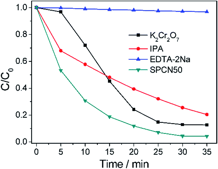
Scavengers of photogenerated holes and electrons were added into the RhB dye solution to study the mechanism of the photocatalytic degradation performance of the SPCN50 composite. Fig. 8 shows the photocatalytic RhB degradation curves of the SPCN50 composite in RhB solution containing scavengers of photogenerated holes (EDTA-2Na), electrons (K2Cr2O7), or hydroxyl radicals (IPA). As shown in Fig. 8, both K2Cr2O7 and IPA were observed to only partially suppress the photodegradation rates as compared to pure SPCN50 without the scavengers. The photodegradation was obviously suppressed with the addition of EDTA-2Na. The results indicate that holes are the main oxidative species in the photocatalytic process.
 | ||
| Fig. 8 Effect of different scavengers on the photocatalytic degradation of RhB (50 mL, 5 mg L−1) over SPCN50. | ||
Photoluminescence (PL) is usually used to investigate the migration, transfer and recombination processes of photoinduced electron–hole pairs in semiconductors. Strong intensity of the generated fluorescence means that the photoinduced electrons and holes are prone to recombination and the lifetimes of the photoinduced electrons are short. However, weak intensity means that the separation efficiency of the photoinduced electrons and holes is high, resulting in longer lifetimes for the photoinduced electrons and holes. Fig. S5† shows PL spectroscopy of CN, Ag/AgCl–CN and SPCN50. For pure CN, a strong luminous broad peak emerges at 420 to 600 nm. Compared to pure CN, Ag/AgCl–CN shows weak intensity for the luminous peak. After compositing with P-Ag/AgCl, the intensity of the luminous broad peak at 420 to 600 nm dramatically weakens, indicating that the presence of P-Ag/AgCl can effectively improve the separation efficiency and the lifetimes of the photogenerated electrons and holes.
The absorbance properties of the as-prepared samples were measured using UV-vis DRS. Fig. 9 displays UV-vis DRS of the P-Ag/AgCl, CN and SPCN composites. The pure CN has an absorption onset of 445 nm, corresponding to the band gap of 2.79 eV. For the P-Ag/AgCl, there is strong absorption intensity in both the UV and visible regions because of the SPR of Ag. In contrast to pure CN with its absorption edge at 445 nm, the absorption intensities of the SPCN composites are stronger in the whole spectrum window of interest, especially in the visible-light region, due to the introduction of P-Ag/AgCl. In addition, the absorption spectra are largely red-shifted, and a broad absorption ranging from 450 to 800 nm is detected. These results indicate that the SPCN composites could be stimulated by visible light (λ > 445 nm), leading to the generation of more electrons and holes. Therefore, the SPCN composites should have favourable visible-light photocatalytic activity.
 | ||
| Fig. 9 UV-vis DRS of P-Ag/AgCl, CN and SPCN composites. | ||
The separation efficiency of the photogenerated electrons and holes can also be supported from data of the photoinduced current densities of the prepared photoelectrodes. Fig. S6† shows the variations of the photogenerated current densities of the photoelectrodes prepared by CN and the SPCN50 composite. As shown in Fig. S6,† the SPCN50 composite generate a much larger photoinduced current density than CN, demonstrating the separation efficiency of the photogenerated carriers.
Based on the above results, a possible photocatalytic mechanism is proposed (Fig. 10). The CN can be excited by visible light, while AgCl cannot. Under visible light irradiation, CN can induce π–π* transitions and transport the electrons from the valence band (VB) to the conduction band (CB). Thus the holes and electrons are generated. As PDA can transport electrons,22,23 the excited electrons on CN can be injected into the CB of AgCl via the PDA layer which reduces the recombination rate of photoinduced holes and electrons. Because of the SPR of Ag, the photoinduced electrons can also inject into the CB of AgCl from Ag. These electrons can react with oxygen to form hydroxyl and superoxide radicals. The radicals and the photoinduced holes are able to oxidize the pollutant, thus producing obvious photocatalytic activity. The introduction of PDA plays an important role in improving the separation of the photogenerated carriers, resulting in enhanced photocatalytic activity.
 | ||
| Fig. 10 An illustration of the charge transfer in the SPCN system under visible light irradiation. | ||
4. Conclusions
In summary, plasmonic Ag/AgCl–PDA–CN (SPCN) composites were prepared by a one-pot method combining mussel chemistry and a precipitation method. PDA can enhance the interfacial interaction between Ag/AgCl and CN. The as-prepared SPCN composites exhibited excellent photocatalytic activity for the degradation of RhB, which is superior to those of pure CN and P-Ag/AgCl under visible light irradiation (λ > 420 nm). The PL and photoresponses indicated that the introduction of PDA efficiently improved the separation of photogenerated electrons and holes and enhanced the photocatalytic activity. Therefore, PDA plays multiple roles, acting as an adhesive layer between the Ag/AgCl and CN, a good electron transfer material and a product of the redox reaction. This work may provide a green, energy-saving and universal method to synthesize photocatalytic composites with high catalytic performance.Acknowledgements
This work was supported by the Natural Science Foundation of China (21264013, 21364010), the Bingtuan Innovation Team in Key Areas (2015BD003) and the Technology Foundation for Selected Overseas Chinese Scholar, the Ministry of Human Resources and Social Security of the People's Republic of China and the Scientific Research Foundation for High Level Talents of Shihezi University (RCZX 201206).Notes and references
- A. Fujishima and K. Honda, Nature, 1972, 238, 37 CrossRef CAS PubMed.
- J. Schneider, M. Matsuoka, M. Takeuchi, J. Zhang, Y. Horiuchi, M. Anpo and D. W. Bahnemann, Chem. Rev., 2014, 114, 9919 CrossRef CAS PubMed.
- X. Wang, K. Maeda, A. Thomas, K. Takanabe, G. Xin, J. M. Carlsson, K. Domen and M. Antonietti, Nat. Mater., 2009, 8, 76 CrossRef CAS PubMed.
- G. Liu, P. Niu and H.-M. Cheng, ChemPhysChem, 2013, 14, 885 CrossRef CAS PubMed.
- (a) H. Li, B. Sun, L. Sui, D. Qian and M. Chen, Phys. Chem. Chem. Phys., 2015, 17, 3309 RSC; (b) L. Yin, Y.-P. Yuan, S.-W. Cao, Z. Zhang and C. Xue, RSC Adv., 2014, 4, 6127 RSC.
- H. Yan and Y. Huang, Chem. Commun., 2011, 47, 4168 RSC.
- (a) B. Chai, T. Peng, J. Mao, K. Li and L. Zan, Phys. Chem. Chem. Phys., 2012, 14, 16745 RSC; (b) S. Zhang, J. Li, X. Wang, Y. Huang, M. Zeng and J. Xu, ACS Appl. Mater. Interfaces, 2014, 6, 22116 CrossRef CAS PubMed.
- J. Xu, Y. Li, S. Peng, G. Lu and S. Li, Phys. Chem. Chem. Phys., 2013, 15, 7657 RSC.
- (a) G. Zhang, M. Zhang, X. Ye, X. Qiu, S. Lin and X. Wang, Adv. Mater., 2014, 26, 805 CrossRef CAS PubMed; (b) G. Dong, K. Zhao and L. Zhang, Chem. Commun., 2012, 48, 6178 RSC; (c) Y. Zhang, T. Mori, J. Ye and M. Antonietti, J. Am. Chem. Soc., 2010, 132, 6294 CrossRef CAS PubMed; (d) J. Li, B. Shen, Z. Hong, B. Lin, B. Gao and Y. Chen, Chem. Commun., 2012, 48, 12017 RSC; (e) X. Wang, X. Chen, A. Thomas, X. Fu and M. Antonietti, Adv. Mater., 2009, 21, 1609 CrossRef CAS.
- (a) N. Cheng, J. Tian, Q. Liu, C. Ge, A. H. Qusti, A. M. Asiri, A. O. Al-Youbi and X. Sun, ACS Appl. Mater. Interfaces, 2013, 5, 6815 CrossRef CAS PubMed; (b) X. Bai, R. Zong, C. Li, D. Liu, Y. Liu and Y. Zhu, Appl. Catal., B, 2014, 147, 82 CrossRef CAS; (c) X. Chen, J. Zhang, X. Fu, M. Antonietti and X. Wang, J. Am. Chem. Soc., 2009, 131, 11658 CrossRef CAS PubMed; (d) J. Yu, K. Wang, W. Xiao and B. Cheng, Phys. Chem. Chem. Phys., 2014, 16, 11492 RSC.
- J. Sun, J. Zhang, M. Zhang, M. Antonietti, X. Fu and X. Wang, Nat. Commun., 2012, 1139 CrossRef.
- (a) X. Wang, K. Maeda, X. Chen, K. Takanabe, K. Domen, Y. Hou, X. Fu and M. Antonietti, J. Am. Chem. Soc., 2009, 131, 1680 CrossRef CAS PubMed; (b) K. K. Han, C. C. Wang, Y. Y. Li, M. M. Wan, Y. Wang and J. H. Zhu, RSC Adv., 2013, 3, 9465 RSC.
- (a) D. Chen, K. Wang, D. Xiang, R. Zong, W. Yao and Y. Zhu, Appl. Catal., B, 2014, 147, 554 CrossRef CAS; (b) J. Zhou, M. Zhang and Y. Zhu, Phys. Chem. Chem. Phys., 2015, 17, 3647 RSC; (c) S. Hu, R. Jin, G. Lu, D. Liu and J. Gui, RSC Adv., 2014, 4, 24863 RSC.
- H. Xu, J. Yan, Y. Xu, Y. Song, H. Li, J. Xia, C. Huang and H. Wan, Appl. Catal., B, 2013, 129, 182 CrossRef CAS.
- X. Yao, X. Liu and X. Hu, ChemCatChem, 2014, 6, 3409 CrossRef CAS.
- Y. Yang, W. Guo, Y. Guo, Y. Zhao, X. Yuan and Y. Guo, J. Hazard. Mater., 2014, 271, 150 CrossRef CAS PubMed.
- S. Kang, Y. Fang, Y. Huang, L.-F. Cui, Y. Wang, H. Qin, Y. Zhang, X. Li and Y. Wang, Appl. Catal., B, 2015, 168–169, 472 CrossRef CAS.
- T. Zhou, Y. Xu, H. Xu, H. Wang, Z. Da, S. Huang, H. Ji and H. Li, Ceram. Int., 2014, 40, 9293 CrossRef CAS.
- Y. Wang, M. Xia, K. Li, X. Shen, T. Muhanmood and F. Wang, Phys. Chem. Chem. Phys., 2016, 18, 27257–27264 RSC.
- (a) H. Lee, S. M. Dellatore, W. M. Miller and P. B. Messersmith, Science, 2007, 318, 426 CrossRef CAS PubMed; (b) X. Jia, W. Sheng, W. Li, Y. Tong, Z. Liu and F. Zhou, ACS Appl. Mater. Interfaces, 2014, 6, 19552 CrossRef CAS PubMed.
- (a) Z. Ma, X. Jia, J. Hu and B. Dai, RSC Adv., 2014, 4, 1853 RSC; (b) J. Feng, P. Zhang, A. Wang, Q. Liao, J. Xi and J. Chen, New J. Chem., 2012, 36, 148 RSC.
- J. H. Kim, M. Lee and C. B. Park, Angew. Chem., Int. Ed., 2014, 53, 6364 CrossRef CAS PubMed.
- H. J. Nam, J. Cha, S. H. Lee, W. J. Yoo and D.-Y. Jung, Chem. Commun., 2014, 50, 1458 RSC.
- W. Li, Z. Ma, G. Bai, J. Hu, X. Guo, B. Dai and X. Jia, Appl. Catal., B, 2015, 174–175, 43 CrossRef CAS.
- P. Niu, L. Zhang, G. Liu and H.-M. Cheng, Adv. Funct. Mater., 2012, 22, 4763 CrossRef CAS.
- S. Kumar, B. Kumar, A. Baruah and V. Shanker, J. Phys. Chem. C, 2013, 117, 26135 CAS.
- (a) P. Das, S. Yuran, J. Yan, P. S. Lee and M. Reches, Chem. Commun., 2015, 51, 5432 RSC; (b) H. Jin, Y. Zhou, W. Huang, Y. Zheng, X. Zhu and D. Yan, Chem. Commun., 2014, 50, 6157 RSC.
- Y. Shen, P. Chen, D. Xiao, C. Chen, M. Zhu, T. Li, W. Ma and M. Liu, Langmuir, 2015, 31, 602 CrossRef CAS PubMed.
- F. He, G. Chen, Y. Yu, S. Hao, Y. Zhou and Y. Zheng, ACS Appl. Mater. Interfaces, 2014, 6, 7171 CAS.
- Y. Ding, L.-T. Weng, M. Yang, Z. Yang, X. Lu, N. Huang and Y. Leng, Langmuir, 2014, 30, 12258 CrossRef CAS PubMed.
Footnote |
| † Electronic supplementary information (ESI) available: SEM, TEM, photocatalytic degradation of Rh 6G, COD, PL spectra and photoresponse. See DOI: 10.1039/c6ra24637k |
| This journal is © The Royal Society of Chemistry 2016 |