
Ultra-long MWCNTs highly oriented in electrospun PVDF/MWCNT composite nanofibers with enhanced β phase
Xia Liuab,
Sixing Xuab,
Xuanlin Kuangab and
Xiaohong Wang*ab
aInstitute of Microelectronics, Tsinghua University, Beijing 100084, P. R. China. E-mail: wxh-ime@tsinghua.edu.cn; Tel: +86-010-62798432
bTsinghua National Laboratory for Information Science and Technology, Tsinghua University, Beijing 100084, P. R. China
First published on 25th October 2016
Abstract
We report aligned polyvinylidene fluoride (PVDF)/multiwalled carbon nanotube (MWCNT) composite nanofibers, in which micrometer-long MWCNTs were highly oriented within the PVDF nanofiber matrix and thus the β-phase formation was enhanced. The PVDF/MWCNT composite nanofibers were successfully fabricated via far-field electrospinning at a low injection rate of 0.1 mL h−1. In this work, we propose a method to eliminate defects and excess amorphous carbon in order to visualize and capture high-resolution images of MWCNTs within the nanofibers using transmission electron microscopy (TEM). The TEM studies show that the MWCNTs were embedded within the nanofiber matrix and oriented parallel to the fiber axis. Moreover, the α- to β-phase transformation was led by both the electrospinning process and electrostatic interactions between the highly oriented MWCNTs and the PVDF chains, and the β-phase content was found to be 86.6% (at 0.1 wt% MWCNT), as verified using wide angle X-ray diffraction patterns.
1. Introduction
Because of their unique structures and surface modifiability, carbon nanotubes (CNTs) have absolute predominance as nanofiller materials and are used to improve the electrical properties of polymers via their small sizes, high surface areas, high aspect ratios, and good crystalline structure.1–3 For instance, CNT/PANI composite nanofibers exhibit high thermoelectric properties when highly oriented polymer backbones are constructed using chemical treatments.4 Uniaxial alignment causes PAN/PVP/MWCNT nanofiber meshes to increase their anisotropic surface resistance by 15.4 times.5 This suggests that CNTs provide good conductivity pathways, as they are oriented preferentially along the fiber axis. G. Wang et al. found that aligned CNTs provide an effective channel for electrical conductance along CNT/PVA composite nanofibers.6 W. A. Yee et al. reported that CNTs promoted the formation of β-phase extended-chain crystallites, resulting in 150% increase in their piezoelectric response.7 Unzipped CNTs can improve the dispersion of pristine CNTs in the PVDF matrix, bringing about high β-phase formation in the casting PVDF nanocomposite.8 Thus, use of CNTs as polymer nanofillers can result in CNT-doped composite polymers with high conductivity and enhanced piezoelectric properties.9Polyvinylidene difluoride (PVDF), with the molecular formula (–CH2–CF2–)n, is a semi-crystalline homopolymer which can crystallize in five different crystallite polymorphs (α, β, γ, δ, and ε phases) depending on processing conditions.10 The most common polymorph is the α-phase, which has a nonpolar TGTG′ conformation. The piezoelectric properties of PVDF are mainly observed due to the polar crystalline phases, such as the β-phase (TTTT conformation) and γ-phase (TTTGTTTG′ conformation). Of these crystalline phases, the β-phase has attracted the widest interest due to its strong polar moment. The polarity of the γ-phase is moderate and lies between those of the α- and β-phases. The unique properties derived from the β-phase of PVDF make it attractive in a wide range of applications including sensors, actuators, energy harvesters, transducers, and portable electronic devices.11–14 Over the past few decades, enormous efforts have been made to increase the β-phase content of PVDF using various processing techniques. Amongst them, two techniques stand out: electrospinning15 and addition of nanofillers16 such as nanoparticles,17 graphene,18 or carbon nanotubes.19 It is well-known that nanofillers act as nucleating agents in the formation of β-phase nanocrystals during initial crystallization. However, each nanofiller follows its own mechanism to achieve nucleation. In the case of nanoparticle nanofillers, the formation of the β-phase is realized through interactions between the ion pole groups in the nanoparticles and the fluorine poles in the PVDF molecules.20 In the case of CNT nanofillers, a large surface area is available for adsorption of the PVDF chains through interactions between the π electrons in the CNTs and the fluorine atoms in the PVDF.21 The key factors that influence the nucleation effect of CNTs in the polymer composites are CNT dispersion, CNT orientation, and interfacial interactions between the CNTs and the polymer matrix.
We chose MWCNTs due to their superior reinforcement properties, as compared to single walled carbon nanotubes (SWCNTs).22,23 Thanks to their rigid structure, MWCNTs coil less and are less prone to aggregation than SWCNTs. Besides that, MWCNTs have high defects, more complex surface morphology, and better surface adhesion, thus to efficiently form the interface with the PVDF polymer. The reinforcement of the CNTs is critically dependent on the nanotube–polymer interfacial interactions. Therefore, their orientations under an electric field may be more uniform than those of SWCNTs. These factors strongly suggest that low-diameter MWCNTs are the optimum material for reinforcing polymer composites. The addition of MWCNTs promotes the α- to β-phase transformation in PVDF by acting as a nucleating agent during the crystallization process and inducing charge accumulation at the interface. In most cases, a significant increase in the piezoelectric response is observed, which is due to the high level of orientation achieved by the MWCNT nanofillers. However, the orientation of nanotubes in highly aligned nanofibers is still rare.24,25 It is still difficult to obtain long, straight MWCNTs in a parallel manner, which limits their applications in device design.26 Since both electrospinning and incorporation of MWCNTs promote the β-phase formation in PVDF, it is indubitably worthwhile to study the MWCNT orientation in electrospun PVDF/MWCNT composite nanofibers.
In this study, we designed highly aligned PVDF/MWCNT composite nanofibers with MWCNT contents from 0 to 1.0 wt%. The composite nanofibers were fabricated by electrospinning at different injection rates without any post processing. We present the experimental results with regard to MWCNT orientation within the electrospun composite nanofibers, as measured using transmission electron microscopy (TEM). The individual crystalline phases of the PVDF/MWCNT composite nanofibers were also determined using curve deconvolution of wide angle X-ray diffraction (WAXD) patterns. The β-phase formation mainly occurs at the interfacial region, where the interactions between PVDF and MWCNTs can induce phase transformations. Furthermore, the reinforcement of MWCNTs can maintain the β-phase.
2. Experimental
2.1 Materials
The polymer used in the experiments is a semi-crystalline PVDF powder (Sigma-Aldrich, St. Louis, MO, USA) with an average molecular weight of 534![[thin space (1/6-em)]](https://www.rsc.org/images/entities/char_2009.gif) 000 g mol−1. N,N-Dimethylacetamide (DMAC) and acetone were purchased from Fisher Scientific. MWCNTs (purity: 95% (Raman), average diameter: 8–10 nm, length: 30 μm) were purchased from Beijing Dk Nanotechnology Co., LTD (China).
000 g mol−1. N,N-Dimethylacetamide (DMAC) and acetone were purchased from Fisher Scientific. MWCNTs (purity: 95% (Raman), average diameter: 8–10 nm, length: 30 μm) were purchased from Beijing Dk Nanotechnology Co., LTD (China).
2.2 Preparation of PVDF/MWCNT solutions and electrospinning
A mixture of DMAC and acetone (4:6 w/w) was used as the solvent. The MWCNTs were mixed and dispersed in the above solvent to obtain solutions with MWCNT w/v ratios of 0.01, 0.05, 0.1, 0.2, 0.5, and 1.0 wt%, respectively. Subsequently, 16 wt% PVDF was dissolved in the solution under stirring for 2 h at 60 °C. Electrospinning was started by loading the solution into a 1.0 mL plastic syringe, tipped with a 25-gauge stainless steel needle. The positive lead from the high voltage supply was connected to the needle, applying a DC bias of around 30 kV. The solution was injected into the needle at various injection rates (0.1 mL h−1 or 2.0 mL h−1) using a syringe pump. The distance between the needle and the grounded collector (a pair of parallel electrodes) was 12 cm.
2.3 Measurements and characteristics
The morphology of the electrospun PVDF/MWCNT composite nanofibers was observed via scanning electron microscopy (SEM, FEI Quanta 450, The Netherlands). All nanofiber samples were gold-coated prior to SEM imaging. Digital images were analyzed using ImageJ software (National Institute of Health). The orientation of MWCNTs in the composite nanofibers was visualized via TEM (JEOL JEM-2010F FasTEM, Japan) using an accelerating voltage of 140 kV. In order to conduct TEM experiments, the nanofibers were directly dropped onto the copper grids. Fast Fourier Transform (FFT) patterns from the TEM images were analyzed to preview the crystallite structure. Differential scanning calorimetry (DSC) tests were conducted to determine the crystalline phase transformations of the nanofiber samples using a TA Instruments Q5000IR at a heating rate of 10 °C min−1. The crystallite structures were then determined using WAXD (D/max2550HB+/PC diffractometer, Rigaku, Japan) with Cu (40 kV, 200 mA) Kα radiation. The samples were scanned from 10° to 40° at a scan rate of 2° min−1. The incident and diffraction X-ray beams were in the same plane, i.e., vertical to the electrospun nanofiber mat.2.4 Strategic steps of TEM measurement
Successive through focus images were recorded with a 0.4 s exposure and a dose rate of 30 electrons per second per Å2. A defocus step size of 0.5 nm was used. It is well-known that secondary radiation events can make PVDF more sensitive to radiation damage. Therefore, care was taken to limit the exposure of the sample to the electron beam before image capture. The wide range of magnifications available in the TEM not only allowed visualization of the contrast within the structure, but also allowed recording of molecular scale features within the composite nanofibers. Since the electron density of the MWCNTs is higher than that of the surrounding PVDF polymer, the nanotubes, which contain hole structures, exhibited a dark concentric tubular type structure that contrasts with the surrounding PVDF matrix.3. Results and discussion
3.1 Morphologies
All of the aforementioned solutions were electrospun and the resulting ultrafine nanofibers were collected and aligned in parallel. Fig. 1a and b show SEM images of aligned electrospun PVDF/MWCNT composite nanofibers containing 0.1 wt% MWCNTs at low and high injection rates, respectively. The nanofibers have regular diameters ranging from 100 to 900 nm. The average diameter of the low-rate sample is about 200 nm smaller than that of the high-rate sample, as shown in the statistical distributions of diameters (Fig. 1c and d). This phenomenon occurs because of the high charge density of the electrified jet, which leads to the formation of uniform, thin nanofibers. Moreover, the decrease in the injection rate can attribute to the proper dispersion of MWCNTs in the polymer matrix because the repulsive forces generated by MWCNTs can alleviate the chain elongation of PVDF macromolecules.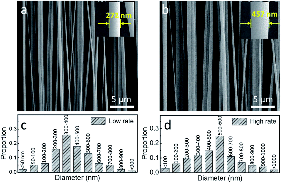 | ||
| Fig. 1 SEM images of highly aligned PVDF/MWCNT composite nanofibers at (a) low and (b) high injection rates. (c and d) The statistical distribution of diameters from (a) and (b), respectively. | ||
3.2 Orientation of MWCNTs
Bright field TEM (BF-TEM) was used to study the distribution of MWCNTs in the composite nanofibers. Two 300 nm-thick PVDF/MWCNT composite nanofibers (at 0.1 wt% MWCNT), electrospun at different injection rates, were chosen for examination at high magnification, as shown in Fig. 2. Initially, the nanotubes are randomly oriented in the electrospinning solution. Eventually, they become oriented along the streamlines of the electrospinning solution due to elongation of the solution jet.27 The reinforcement of the MWCNTs and crystallization of PVDF suppress any relaxation effects, so that both the PVDF chains and MWCNT networks retain largely their oriented conformations. In many regions of the low-rate sample (Fig. 2a), the embedded nanotubes appeared to be highly oriented along the fiber axis. A high-magnification image of the nanofiber (Fig. 2b) shows that the embedded MWCNTs are well dispersed and oriented side by side along the fiber axis. BF-TEM studies have revealed that shear force causes the MWCNTs to self-orient within the polymer matrix during electrospinning, although it is difficult to achieve orientation of MWCNTs using normal mechanical drawing.27 While, in the high-rate sample (Fig. 2c), it is evident that most of the MWCNTs aggregate or bundle within the PVDF matrix (Fig. 2d), although they successfully embed themselves within the matrix. Thus, electrospinning at a low injection rate produces nanotubes that are well dispersed and highly oriented within the polymer nanofibers. | ||
| Fig. 2 (a) Highly oriented MWCNTs in the PVDF/MWCNT composite nanofiber were electrospun at a low injection rate of 0.1 mL h−1 onto a lacey film supported by a copper sample grid. (b) MWCNTs are uniformly dispersed and uniaxially oriented within the composite nanofiber. (c) MWCNTs aggregate into a bundle within the composite nanofiber electrospun at a high injection rate of 2.0 mL h−1. (d) The zoomed-in image of (c) shows that most of the MWCNTs aggregate. | ||
Two TEM images were captured along the suspended section of the same PVDF/MWCNT composite nanofiber prepared at low-rate and combined to provide one composite image (Fig. 3). The length of the nanofiber is more than 15 μm and the single nanotube had at least 8 μm of length (as indicated by the blue arrows). It is clearly presented in the TEM images that the long MWCNTs are straight and uniaxially oriented along the fiber axis, with no curling or buckling. The surface functionalities of the MWCNTs greatly improve their dispersion in the PVDF solution, and hence no MWCNT aggregate was observed. Further study of thicker electrospun nanofibers by TEM also showed elongated MWCNTs.
 | ||
| Fig. 3 A composite picture of two TEM images taken from two parts of the same PVDF/MWCNT composite nanofiber (a: left part; b: right part) prepared at a low injection rate. The MWCNTs are long, straight, and oriented in the PVDF polymer matrix along the fiber axis with no curling or buckling. The length of single straight MWCNT is more than 15 μm. | ||
Thanks to the slow formation of nanofibers, the embedded MWCNTs can be elongated as much as possible. In addition to injection rates, the MWCNT content also plays a crucial role in nanotube orientation. K. Ke et al. reported that the morphologies of composite nanofibers with MWCNT contents of more than 0.5 wt% became irregular and rough, which may be related to the aggregation and bundling of MWCNTs seen here.21 It is reasonable to deduce that the irregularity of the MWCNTs is still responsible for their entanglement. However, when the MWCNT content decreases to less than 0.01 wt%, it becomes extremely difficult to clearly observe the MWCNTs in the nanofibers.
The molecules of the PVDF/MWCNT composite nanofibers were visualized via high resolution TEM (Fig. 4a). Fast Fourier Transform (FFT) data of the image (Fig. 4b and c) produced diffraction patterns by including diffraction occurring in the center and edge of the composite nanofibers, respectively. A clear boundary line can be seen near the edge. The electron diffraction pattern of the edge section (Fig. 4c) is higher than that of the middle section (Fig. 4b), which implies that the MWCNTs contribute to PVDF crystallization. However, we were unable to observe electron diffraction from the MWCNTs embedded with-in the composite nanofibers. This could be due to the reduced sensitivity of the polymer crystallinity to electron radiation, when compared to the carbon layers of the nanotubes.
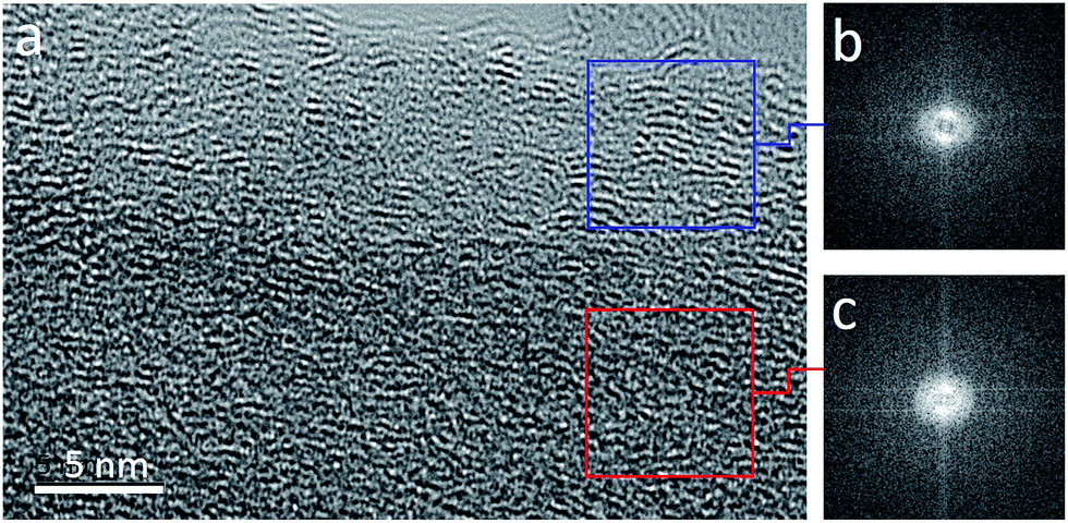 | ||
| Fig. 4 (a) High resolution TEM image of a PVDF/MWCNT composite nanofiber prepared at a low injection rate. Fast Fourier Transforms (FFT) of (b) the center and (c) edge sections of the nanofiber. | ||
3.3 Crystallization characteristics
The DSC testing results of the nanofiber samples are shown in Fig. 5. Comparing to the neat PVDF nanofibers, the DSC thermograms of the PVDF/MWCNT nanofibers present a peak melting temperature shifting toward higher temperatures and narrowing of the melting point features with MWCNT addition, which implies that the β-phase becomes more prevalent in crystalline structure of the composite nanofibers. Since the melting temperature of the β-phase is known to be higher than that of the α-phase.28 These thermograms also prove that a low injection rate induces an increase in the total amount of crystalline β-phase in the composite nanofibers. | ||
| Fig. 5 DSC thermograms of and the PVDF/MWCNT composite nanofibers at low and high injection rates, comparing to the neat nanofibers. | ||
Wide angle X-ray diffraction (WAXD) is a powerful tool to investigate the details of the effect of the MWCNT content on the crystallization of the PVDF/MWCNT composite nanofibers, revealing the crystalline phase distribution of the nanofibers. Fig. 6a and b show WAXD spectra of the composite nanofibers with various MWCNT contents at low and high rates, respectively. Absorption peaks at about 17.7° and 18.3°, which correspond to the α-phase, are present. With regard to the low-rate sample, the α-phase peak intensities from the composite nanofibers with the MWCNT contents of 0.01, 0.05, 0.1, and 0.2 wt% gradually decrease when compared to the neat sample. Significantly, the α-phase content greatly decreases in both samples when the MWCNT content is 0.1 wt%. Another absorption peak at about 20.5° is assigned to the (200)/(110) planes of the β-phase that initially increases (0.01, 0.05, and 0.1 wt%) and then decreases (0.2, 0.5, and 1.0 wt%). When the MWCNT content is over 0.2 wt%, the over-loaded MWCNTs are entangled and prevent the polar-phase nanocrystals from nucleating. The growth of spherulites is hindered by the neighboring molecules, which leads to crystal defects and decreases in crystal size, thereby weakening the intensity of the diffraction peaks.
 | ||
| Fig. 6 WAXD patterns for PVDF/MWCNT composite nanofibers with different MWCNT contents produced at (a) a low injection rate of 0.1 mL h−1 and (b) a high injection rate of 2.0 mL h−1. | ||
Since the conformation of the γ-phase has some overlap with the β- and α-phases, separating the γ-phase from α- and β-phases can be confusing. We analyzed the compositions of all of the crystalline phases of the low-rate sample (0.1 wt% MWCNT), the high-rate sample (0.1 wt% MWCNT), and the neat sample generated at a low injection rate. It is also noted that there is no difference in the crystal structures of neat PVDF nanofibers electrospun at different rates.
The individual α, β, and γ-phases for the low-rate sample, the high-rate sample, and the neat sample were examined using a curve deconvolution technique (Fig. 7), where all of the phases (including the small β-phase peak at 36.1°) were considered. The peaks at 17.7° and 18.3° were assigned to the α-phase and the peaks at 20.5° and 36.1° to the β-phase. The peak at 18.5° was assigned to the γ-phase. The following equations were used to calculate the relative contents of the electroactive non-polar α- and polar β/γ-phases, individually. The total degree of crystallinity (DoC, φtotal), degree of α-crystallinity (φα), degree of β-crystallinity (φβ), degree of γ-crystallinity (φγ), degree of polar-crystallinity (φpolar), β-phase content (Fβ), and γ-phase content (Fγ) were calculated using the following equations:
 | (1) |
 | (2) |
| φpolar = φβ + φγ | (3) |
 | (4) |
 | (5) |
 | ||
| Fig. 7 WAXD patterns of representations of crystalline reflections of the nanofibers: (a) PVDF/MWCNT composite nanofibers electrospun at a low rate of 0.1 mL h−1; (b) PVDF/MWCNT composite nanofibers electrospun at a high rate of 2.0 mL h−1; and (c) neat PVDF nanofibers electrospun at a low rate of 0.1 mL h−1. The inset tables show the individual contents of the crystalline phases (α, β, and γ). | ||
Significantly, the incorporation of MWCNTs and PVDF visibly improves φtotal and φβ (Fig. 7), which directly influence the material properties.30 The increase in crystallinity is observed because MWCNTs can efficiently restrict and order the PVDF chain arrangement (defined as a “molecule movement restriction” effect) due to their superior mechanical strength.31 DoC, one of characteristics of the semi-crystalline polymer, is usually accompanied during the α- to β-phase transformation.32 Consequently, the resulting polar β-phase content of the low-rate sample (i.e., Fβ = 86.6%) was also found to be higher than that of the high-rate sample (i.e., Fβ = 72.8%). In contrast, the β-phase content of the neat PVDF nanofibers was 65.8%. Therefore, it can be expected that the PVDF/MWCNT composite nanofibers would exhibit better piezoelectric responses than the neat PVDF nanofibers, as the piezoelectric property is directly proportional to the DoC and the polar β-phase content.
The polar β- and γ-phase contents were quantitatively characterized as a function of the MWCNT content in the composite nanofibers (Fig. 8), underlining the fact that the β-phase content of the low-rate sample is higher than that of its high-rate counterpart, which is in turn higher than the neat sample. Although the γ-phase can be also obtained from α-phase transformation, the γ-phase content is significantly less than the β-phase content in the low-rate sample. Notably, the γ-phase content of the high-rate sample is higher than in the low-rate sample, which suggests that the higher injection rate assists the transformation of α- to γ-phase.
 | ||
| Fig. 8 (a) Plots of the β or γ-phase contents as a function of the concentration of MWCNTs in PVDF/MWCNT composite nanofibers at low and high rates, respectively. (b) The schematic showing the α-phase → β/γ-phase transformation. | ||
In this study, the enhancement of β-phase nanocrystals within the PVDF nanofiber matrix is mainly achieved through the application of long, straight MWCNTs and the far-field electrospinning technique. The high orientation of MWCNTs along the fiber axis can be attributed to their successful functionalization and optimization of the electrospinning conditions. Several factors are thought to contribute to the high orientation of MWCNTs along the fiber axis. First, the embedded MWCNTs were reoriented towards the fiber axis under the synergistic effect of the electric field and the shear forces generated during rapid stretching of the solution jet during the electrospinning. Second, the enhanced orientation of PVDF chains also contributed to the orientation of the nanotubes due to nanoscale confinement. Third, the MWCNTs have zigzag-structured carbon backbones, which align perfectly with the all-trans TTTT conformation of PVDF in the melted state. The nucleating effect of the MWCNTs induces the β-phase nanocrystal formation and spherical reinforcement. Thus, the cumulative effect of the above factors results in preferential orientation of the long, straight MWCNTs.
Furthermore, this phenomenon arises because the electric field can induce the reorientation of both the PVDF chains and the MWCNTs along the fiber axis. Moreover, the well-oriented PVDF chains can also lead the MWCNTs to reorient towards the fiber axis. Thus, interactions between the PVDF chains and the MWCNTs reinforce the achieved orientation. In addition, the highly oriented MWCNTs increase the local electric field during the electrospinning and in situ poling, resulting in a greater electrostatic force, which in turn results in an increased formation of polar phases compared to the neat PVDF sample. Conversely, the decrease in the polar crystallinity of the high-rate sample, compared to the low-rate sample, is most likely caused by incomplete elongation under the viscoelastic and electrostatic forces.
4. Conclusions
In conclusion, we have demonstrated that long, straight MWCNTs can be embedded within PVDF/MWCNT composite nanofibers. The MWCNTs align parallel to the fiber axis. The composite nanofibers exhibit a higher degree of crystallinity and higher polar phase content (86.6% β-phase and 4.5% γ-phase at 0.1 wt% MWCNT) than the neat PVDF nanofibers. From TEM studies, we found that MWCNTs with tens of micrometers in length could be oriented in the polymer matrix by shear forces during electrospinning, although this orientation is difficult to achieve under normal mechanical drawing.Furthermore, the unique crystallite structure of the PVDF/MWCNT composite nanofibers was achieved via cumulative interactions between the MWCNTs and PVDF chains, which promote the nucleation of highly oriented polar-phase extended-chain crystallites at the interface.
Abbreviations
| TTTT, TGTG′ | (T = trans, G = gauche +, G′ = gauche −) |
| PANI | Polyaniline |
| PAN | Polyacrylonitrile |
| PVP | Polyvinyl pyrrolidone |
| PVA | Polyvinyl alcohol |
Acknowledgements
This work was supported by the grants from the National Natural Science Foundation of China (no. 61474071, 61531166006) and National Basic Research Program (973 Program, no. 2015CB352106).Notes and references
- S. Iijima, Nature, 1991, 354, 56–58 CrossRef CAS.
- P. M. Ajayan, O. Stephan, C. Colliex and D. Trauth, Science, 1994, 265, 1212 CAS.
- M. Musameh, M. R. Notivoli, M. Hickey, I. L. Kyratzis, Y. Gao, C. Huynh and S. C. Hawkins, Adv. Mater., 2011, 23, 906–910 CrossRef CAS PubMed.
- Q. Wang, Q. Yao, J. Chang and L. Chen, J. Mater. Chem., 2012, 22, 17612–17618 RSC.
- L.-Y. Mei, P. Song and Y.-Q. Liu, J. Appl. Polym. Sci., 2015, 132, 41995 CrossRef.
- G. Wang, Z. Tan, X. Liu, S. Chawda, J. S. Koo, V. Samuilov and M. Dudley, Nanotechnology, 2006, 17, 5829–5835 CrossRef CAS.
- W. A. Yee, J. Kong, C. Zhang, T. Liu, M. Kotaki and X. Lu, Polymer, 2012, 53, 5097–5102 CrossRef CAS.
- L. He, G. Xia, J. Sun, Q. Zhao, R. Song and Z. Ma, J. Colloid Interface Sci., 2013, 393, 97–103 CrossRef CAS PubMed.
- H. Yu, T. Huang, M. Lu, M. Mao, Q. Zhang and H. Wang, Nanotechnology, 2013, 24, 405401 CrossRef PubMed.
- A. K. Nandi and L. Mandelkern, J. Polym. Sci., Part B: Polym. Phys., 1991, 29, 1287–1297 CrossRef CAS.
- X. Liu, H. Zhao, Y. Lu, S. Li, L. Lin, Y. Du and X. Wang, Nanoscale, 2016, 8, 7278–7286 RSC.
- L. Persano, C. Dagdeviren, Y. Su, Y. Zhang, S. Girardo, D. Pisignano, Y. Huang and J. A. Rogers, Nat. Commun., 2013, 4, 1633 CrossRef PubMed.
- Y.-K. Fuh, J.-C. Ye, P.-C. Chen, H.-C. Ho and Z.-M. Huang, ACS Appl. Mater. Interfaces, 2015, 7, 16923–16931 CAS.
- R. Yang, Y. Qin, L. Dai and Z. L. Wang, Nat. Nanotechnol., 2009, 4, 34–39 CrossRef CAS PubMed.
- H. Shao, J. Fang, H. Wang and T. Lin, RSC Adv., 2015, 5, 14345–14350 RSC.
- L. Priya and J. P. Jog, J. Polym. Sci., Part B: Polym. Phys., 2002, 40, 1682–1689 CrossRef CAS.
- D. Dhakras, V. Borkar, S. Ogale and J. Jog, Nanoscale, 2012, 4, 752–756 RSC.
- L. Huang, C. Lu, F. Wang and L. Wang, RSC Adv., 2014, 4, 45220–45229 RSC.
- F. Bai, G. Chen, M. Nie and Q. Wang, RSC Adv., 2015, 5, 54171–54174 RSC.
- S. K. Karan, D. Mandal and B. B. Khatua, Nanoscale, 2015, 7, 10655–10666 RSC.
- K. Ke, P. Pötschke, D. Jehnichen, D. Fischer and B. Voit, Polymer, 2014, 55, 611–619 CrossRef CAS.
- A. Baji, Y.-W. Mai, M. Abtahi, S.-C. Wong, Y. Liu and Q. Li, Compos. Sci. Technol., 2013, 88, 1–8 CrossRef CAS.
- M. Cadek, J. N. Coleman, K. P. Ryan, V. Nicolosi, G. Bister, A. Fonseca, J. B. Nagy, K. Szostak, F. Béguin and W. J. Blau, Nano Lett., 2004, 4, 353–356 CrossRef CAS.
- Y. Ahn, J. Y. Lim, S. M. Hong, J. Lee, J. Ha, H. J. Choi and Y. Seo, J. Phys. Chem. C, 2013, 117, 11791–11799 CAS.
- H. Hou, J. J. Ge, J. Zeng, Q. Li, D. H. Reneker, G. Andreas and S. Z. D. Cheng, Chem. Mater., 2005, 17, 967–973 CrossRef CAS.
- P. Zhang, X. Zhao, X. Zhang, Y. Lai, X. Wang, J. Li, G. Wei and Z. Su, ACS Appl. Mater. Interfaces, 2014, 6, 7563–7571 CAS.
- Y. Dror, W. Salalha, R. L. Khalfin, Y. Cohen, A. L. Yarin and E. Zussman, Langmuir, 2003, 19, 7012–7020 CrossRef CAS.
- A. J. Lovinger, Science, 1983, 220, 1115–1121 CAS.
- H. Huo, S. Jiang and L. An, Macromolecules, 2004, 37, 2478–2483 CrossRef CAS.
- S. K. Ghosh, M. M. Alam and D. Mandal, RSC Adv., 2014, 4, 41886–41894 RSC.
- S. Garain, S. Jana, T. K. Sinha and D. Mandal, ACS Appl. Mater. Interfaces, 2016, 8, 4532–4540 CAS.
- S. Manna, S. K. Batabyal and A. K. Nandi, J. Phys. Chem. B, 2006, 110, 12318–12326 CrossRef CAS PubMed.
| This journal is © The Royal Society of Chemistry 2016 |