Integration of TiO2 photoanode and perovskite solar cell for overall solar-driven water splitting

Bo Sunab, Tielin Shia, Zhiyong Liua, Zirong Tanga, Jianxin Zhou*b and Guanglan Liao*a
aState Key Laboratory of Digital Manufacturing Equipment and Technology, Huazhong University of Science and Technology, Wuhan 430074, China. E-mail: guanglan.liao@hust.edu.cn; Tel: +86-27-87793103
bState Key Laboratory of Materials Processing and Die & Mould Technology, Huazhong University of Science and Technology, Wuhan 430074, China. E-mail: zhoujianxin@hust.edu.cn; Tel: +86-27-87558147

Received 29th September 2016 , Accepted 15th November 2016

First published on 15th November 2016


Abstract

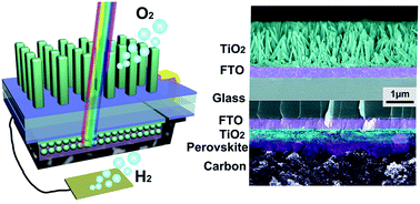
We demonstrate a fully integrated device based on a TiO2 photoanode and perovskite solar cell for overall solar-driven water splitting. The integration substantially broadens the solar absorption spectra and realizes solar water splitting without an external assist. In the integrated configuration, UV light is used by the TiO2 photoanode to generate holes for water oxidation, while the visible light passes through the TiO2 photoanode and irradiates the solar cell. To improve the performance of the integrated device, transmittance of the photoanode is tuned by adjusting the morphology of the TiO2 nanorods, and Sn doping is also introduced to enhance the photoactivity. The solar to hydrogen conversion efficiency is calculated as 1.5%, which is comparable to most of the latest research focused on overall water splitting. Thus, the integrated device provides a promising choice for solar water splitting, considering its high stability under successive illumination.


1. Introduction

In recent decades, the growing energy demands and increasing concern about climate change have caused much attention on clean and renewable energy resource.1 Solar energy is abundant on the earth, and can provide sufficient energy to fulfill humanity's consumption.2 However an efficient method to realize the conversion and storage of the energy is urgently needed, considering the intermittency of sunlight and transportation of energy.3,4 Hydrogen production directly from solar energy driven water splitting has been considered as one of the most promising approaches to solve this problem.5–7

Based on the free energy required to split water, a solar generated photopotential of 1.23 V should be sufficient.8 Due to the kinetic barriers associated with oxidation and reduction reactions, usually 1.7 V or more is necessary.9,10 Theoretically, it can be realized by a single semiconductor with an appropriate band-gap under the illumination of visible light. The conflict between higher photopotential and solar absorption makes it a challenge to design such a S2 system.9 Although, large band-gap semiconductors such as TiO2, ZnO, and WO3 etc. can fulfill this requirement, the consequent low solar absorption strictly limits the efficiency of water splitting. An attractive method is constructing the system consisting of two or more semiconductors to generate sufficient free energy and optimize the solar energy absorption.11–13 In the dual-absorber configuration, four photons are utilized to generate a molecule of H2, which mimics the Z-scheme in nature photosynthesis.5,14 Tremendous efforts have been dedicated to design photoelectrodes based on the nano heterostructure, such as Si/TiO2,15 Si/InGaN,16 Si/WO3 (ref. 17) and ZnO/CdS18 etc. The unassisted overall solar water splitting also has been realized via elaborative design of the photoanode/photocathode of the tandem cell.19–22

As an alternative, the Z-scheme based on photovoltaic/photoelectrode design is always ignored. Due to the low photovoltage of conventional solar cell, the integration of several photovoltaic cells is required to generate sufficient photopotential for water splitting, which substantially increase the complexity of the device to balance the solar absorption of each component.8,23,24 What's exciting is that Sivula et al. demonstrated a tandem cell composed of single dye sensitized solar cell and iron oxide photoanode with a solar-to-hydrogen efficiency of 1.17%.25 Besides, the rapid development of perovskite solar cell (PSC) brings new hope for directly solar water splitting based on the tandem photovoltaic/photoelectrode cell.26 The photovoltage of perovskite solar cell is also adequate. Recently, Gurudayal et al. proposed a perovskite-hematite tandem cells for overall solar driven water splitting.27 The iron nickel oxide and BiVO4 photoanode were also explored to construct efficient integrated tandem cell.28–30 It is meaningful to investigate the integrated device based on TiO2 which is one of the most promising materials for water splitting. Thus, in this paper we proposed an integrated tandem cell based on TiO2 nanorods and the perovskite solar cell. To balance the UV transmittance and photocurrent of the photoanode, the morphology and property of the nanorods is tuned.

2. Experimental

2.1 Perovskite solar cell fabrication

The conductive fluorine doped tin oxide (FTO) glasses (12.5 mm × 25 mm) were etched by diluted hydrochloric acid and zinc powder firstly to form the desired pattern of conductive layer. After ultrasonic cleaning, a compact TiO2 as hole-blocking layer was deposited on it by spin-coating the precursor solution prepared by dissolving titanium isopropoxide in ethanol (0.254 mol L−1) with the addition of HCl (0.02 mol L−1). The spin-coating was conducted at 5000 rpm for 60 s, followed by annealing at 500 °C for 30 min in ambient atmosphere. The mesoporous TiO2 layer was deposited by spin coating using TiO2 paste diluted in ethanol (2[thin space (1/6-em)]:[thin space (1/6-em)]7, weight ratio). After drying, the TiO2 films were also annealed at 500 °C for 30 min in ambient atmosphere.

Lead halide iodide perovskite was deposited on the mesoporous TiO2 layer using the spinning and dipping method. PbI2 solution as precursor was prepared by dissolving PbI2 in N,N-dimethylformamide at 60 °C. Then, 30 μL of PbI2 solution (1.2 mol L−1) was spin-coated at 3000 rpm for 45 s on the pre-heated mesoporous TiO2 layer. After drying at 70 °C for 5 min, the film was dipped into 2-propanol for 1–2s and subsequently dipped in the solution of CH3NH3I in 2-propanol (10 mg mL−1) for 15 min followed by rinsing with 2-propanol. Color of the film changed from yellow to black during the dipping process, indicating the formation of perovskite CH3NH3PbI3. Finally, carbon counter electrode was printed directly on the CH3NH3PbI3 layer by doctor blade method using carbon paste. The cell was encapsulated with PDMS to prevent the moisture.

2.2 TiO2 photoanode fabrication

TiO2 photoanode was prepared by hydrothermal growth. 25 mL concentrated hydrochloric acid (37%) and 25 mL of DI water was mixed in a Teflon-lined stainless steel autoclave. The mixture was stirred for several minutes before adding of 0.8 mL tetrabutyl titanate. Then tin tetrachloride solution was added for the synthesis of Sn-doped TiO2 nanorods with the molar ratio of Sn/Ti = 1%. Subsequently, the cleaned FTO substrate was placed into the Teflon-lined autoclave, which then was sealed and put into electric oven. The reaction temperature and time were set as 180 °C and 6 hours, respectively, for most of the experiments. After that, the autoclave was cooled, and the FTO substrate was taken out and rinsed with DI water. Finally, the sample was annealed at 450 °C for 2 hours in the air to remove the organic reactant and enhance the crystallization of the nanorods. For the synthesis of pristine TiO2 nanorods, the process was all the same, except for the elimination of the Sn precursor.

2.3 Characterization and measurement

To fabricate the integrated device, the as-prepared photoanode was connected to the positive electrode of perovskite solar cell. The negative electrode was jointed to a copper wire which will connect to a Pt slice during water splitting. To enhance the conductivity, the junctions were coated with silver paste. At last, the tandem cell was encapsulated with epoxy, in which only the white TiO2 film was exposed. During the measurement, the working surface area of the photoelectrode was limited to 0.13 cm2. The morphology and element composition of the integrated device were monitored by the field emission scanning electron microscopy (FESEM, Nova NanoSEM 450, FEI) combined with energy dispersive X-ray detector (EDX, Inca X-Max 50). UV-Visible spectrophotometer (UV 2600, Shimadzu) was employed to collect the transmittance spectra of the photoanode. X-ray photoelectron spectroscope (XPS, Axis Ultra DLD, Shimadzu) was used to detect the chemical composition and state of the photoanode and perovskite. The PEC measurements were performed using a electrochemical workstation (AUTOLAB PGSTAT302N, Metrohm) in two-electrode or three-electrode configuration with 1 M KOH (pH = 13.5) electrolyte. Pt slice and Ag/AgCl was used as counter electrode and reference electrode, respectively. A class 3A solar simulator (Oriel 94043A, Newport) with light intensity of 100 mW cm−2 (AM 1.5G) was used as light source, which was calibrated by reference solar cell and meter (Oriel 91150V, Newport). The incident photon-to-electron conversion efficiency (IPCE) was tested under illumination of monochromatic light from a xenon lamp coupled with a monochromator (TLS1509, Zolix).

3. Results and discussion

Fig. 1a schematically illustrates the structure of the integrated device in which the positive electrode of perovskite solar cell is connected to the TiO2 photoanode using copper wire. The negative electrode is connected to Pt sheet acting as photocathode. As we all known, the TiO2 only absorbs the UV light, and visible light can pass through the photoanode and illuminate the solar cell. Performance of the solar cell in the integrated device mainly depends on the transmitted light. Fig. 1b displays the optical images of the integrated device at deferent steps of fabrication process. The morphology of TiO2 photoanode is shown in Fig. 1c. To balance the photocurrent and transmittance of the photoanode, morphology of TiO2 nanorods film is tuned by adjusting the growth time (Fig. S1, ESI). Besides, the Sn-doping is also introduced to enhance the photocatalytic activity. It can be observed that the length and density of the nanorods increases as the time increases no matter for the pristine or Sn-doped nanorods. However, compared to the pristine nanorods, the density of Sn-doped nanorods seems lower. SEM image of the perovskite solar cell is displayed in Fig. 1d. Obviously, TiO2 layer as electron transfer layer is composed of a compact layer and a mesoporous layer, which are 40 nm and 450 nm in thickness, respectively. The thickness of perovskite layer covered on TiO2 is about 500 nm. The carbon counter electrode printed on the perovskite layer is typically about 25 μm (Fig. S2, ESI).
image file: c6ra24247b-f1.tif
Fig. 1 (a) Schematic of the integrated device based on TiO2 nanorods/perovskite solar cell. (b) Optical images of the fabrication process for integrated device. (c) SEM image of the TiO2 nanorods. (d) SEM image of the perovskite solar cell.

Fig. 2 illustrates the element composition of a representative integrated device. The element mappings of Ti, O, and Sn shown in Fig. 2a displays clear boundary at top and bottom of the Sn-doped TiO2 nanorods. Due the low doping level, the signal of Sn in the nanorods is relatively weak. The intense signal ribbon at the bottom can be attributed to the FTO (F[thin space (1/6-em)]:[thin space (1/6-em)]SnO2) layer. Fig. 2b shows the element composition of perovskite solar cell. Obviously, the distribution of C is completely corresponding to the morphology of the solar cell. The signal of C mainly concentrate in carbon layer and perovskite (CH3NH3PbI3) layer, while the weak signal from other layers may be caused by the surface contamination of samples handled under ambient conditions. The element mappings of Pb and I are well agree with the position of perovskite layer. The distribution of Ti, O and Sn shown in Fig. 2b are coincide with the electron transfer layer (TiO2) and conductive layer (FTO). These results directly demonstrate the layered element distribution of the integrated device.


image file: c6ra24247b-f2.tif
Fig. 2 (a) Element mappings of Sn-doped TiO2 nanorods, (b) element mappings of perovskite solar cell.

To further elucidate the surface composition and chemical states of the TiO2 nanorods and perovskite, XPS spectra were collected from the TiO2 photoanode and perovskite solar cell without carbon layer (Fig. S3, ESI). High resolution measurements conducted on TiO2 photoanode are depicted in Fig. 3a–c. The two peaks in Ti 2p spectra centered at the binding energies of 458.8 and 464.6 eV are in accord with the typical values of Ti 2p3/2 and 2p1/2 states for TiO2. The XPS peak at about 530.1 eV corresponds to O 1s state. The two peaks of the spectrum shown in Fig. 3c at about 486 and 496.6 eV can be attributed to Sn 3d5/2 and 3d3/2 states, which further confirms that the main dopant is Sn4+. Quantitative analysis of the spectra reveals that the Sn/Ti molar ratio is about 1% for the Sn-doped TiO2 nanorods. The high resolution XPS spectra of perovskite layer are displayed in Fig. 3d–f. The binding energy of the C 1s (285 eV) was used for the energy calibration, which is also a characteristic peak of perovskite. The two peaks shown in Fig. 3e at about 138.44 and 143.29 eV can be attributed to Pb 4f7/2 and 4f5/2 states, and the two peaks in Fig. 3f at 619.32 eV and 630.77 eV correspond to the I 3d5/2 and 3d3/2 states, respectively. Besides, Ti 2p peaks are not appeared in the spectra of perovskite sample, which confirms the compactness of the perovskite layer.


image file: c6ra24247b-f3.tif
Fig. 3 High resolution XPS spectra collected from the TiO2 photoanode and perovskite layer, (a) Ti 2p, (b) O 1s, (c) Sn 3d, (d) C 1s, (e) Pb 4f, (f) I 3d.

To investigate the remaining spectra for perovskite solar cell, the UV-vis transmittance spectra of pristine TiO2 nanorods and Sn-doped TiO2 nanorods with different growth time (4 to 7 hours) were measured as shown in Fig. 4a. Obviously, all the spectra are dropped suddenly at about 420 nm, which is corresponds to the band-gap of rutile TiO2 (3.0 eV). In the visible region, the transmittance of the nanorods decreases as the growth time increases. Noteworthy is that the nanorods with Sn doping show a higher transmittance compared with the pristine nanorods at same growth time. However as the time extended, the difference is gradually eliminated. After 6 hours growth, the spectra of the nanorods with or without Sn doping are very close. This result is consistent with density of the nanorods (Fig. S1, ESI).


image file: c6ra24247b-f4.tif
Fig. 4 (a) Transmittance of the TiO2 nanorods with different growth parameter. (b) LSV curves of the Sn-doped TiO2 photoanode and perovskite solar cell. (c) LSV curves of the Sn-doped TiO2 photoanode obtained in three-electrode configuration. (d) Applied bias photon-to-current efficiency of the photoanode derived from the LSV curve obtained in two-electrode configuration.

Fig. 4b depicts the line sweep voltammetry (LSV) curves of TiO2 photoanode and the perovskite solar cell. The LSV curves shown in Fig. 4b are obtained at two-electrode configuration. The photocurrent of the photoanode increases as the growth time increases. The photocurrent density of Sn-doped TiO2 nanorods grown for 4 hours is about 0.93 mA cm−2 at 0.7 V vs. Pt, while the value of the nanorods grown for 6 hours is improved to about 1.2 mA cm−2. Obviously, there are three regions in the curve: low photocurrent density region at low bias potential, plateau of photocurrent density at high bias potential, and the increasing photocurrent density region. The photocurrent is closed to zero as the bias potential is lower than 0.2 V vs. Pt because most photogenerated holes and electrons are recombined before holes transfer into the electrolyte. The photocurrent plateau appears as the potential sweeps to high positive direction, since the photocurrent is limited by the number of photogenerated holes. Three-electrode configuration is common used in the reports focused on photoanode or photocathode. However, to simulate the work status of the photoanode in the integrated device, we mainly adopt the two-electrode configuration in this paper. Besides, the value of photocurrent plateau obtained in three-electrode configuration is also close to 1.2 mA cm−2 as shown in Fig. 4c, which further confirms the accuracy of our photoelectrochemical measurements.

The IV curves of the solar cell were tested with correspond TiO2 cover to simulate the tandem work status. The open voltage and short current of the solar cell under Sn-doped TiO2 photoanode grown for 4 and 6 hours is about 0.88 V, 5 mA and 0.87 V, 4.7 mA, respectively. The original open voltage and short current of the solar cell with the illumination of full solar spectrum is about 0.95 V and 8.9 mA (Fig. S4, ESI). In our experiment, the best result was obtained by the Sn-doped nanorods grown for 6 hours. Unless specially stated, the TiO2 photoanode used in the following studies was the Sn-doped nanorods grown for 6 hours. As depicted in Fig. 4b, the working status can be estimated by the crossing point of the curves, which is about 1.2 mA cm−2 at 0.74 V. It is clear that the performance of the integrated device is mainly limited by the photocurrent of the TiO2 nanorods. Besides, the IV characterization of the solar cell after 2 hours successive testing is also supported in Fig. 4b (green curve) to illustrate the photocurrent degradation of the perovskite solar cell.

To further investigate the performance of the integrated device, solar-to-hydrogen conversion efficiency was calculated according to the following equation9

ηSTH = (J × 1.23 × ηF)/Psolar
where J is the working current density (mA cm−2), ηF is the faradaic efficiency which is always assumed as 100%, and PSolar is the light intensity of 100 mW cm−2 (AM 1.5G). For the integrated device based on perovskite solar cell and Sn-doped TiO2 photoanode grown for 6 hours, the solar-to-hydrogen efficiency at the working status without any bias potential is about 1.5%. This value is comparable to most of the latest researches focused on overall solar water splitting.20,31–33 To evaluate the significance of the integration, we further calculated the applied bias photon-to-current efficiency of TiO2 photoanode.34 As shown in Fig. 4d, the top efficiency the photoanode is about 0.7% at 0.55 V vs. Pt, which is also similar to the value derived from the three-electrode measurement (Fig. S5, ESI). All of these results demonstrate that the integration substantially increase the efficiency of solar-driven water splitting.

Time-dependent measurements have been carried out on the best integrated tandem cell to investigate the stability. As shown in Fig. 5a the sample displays highly stable photocurrent density with repeated on/off cycles of illumination. Besides, there is no obvious transient current at the time of light turn on/off, indicating the efficient separation and transport of photoexcited charge carriers.35 It is well known that the stability of perovskite solar cell is poor.36,37 However, in our experiment the stability of the integrated tandem cell is relative acceptable during the water splitting process. As shown in Fig. 5b, photocurrent decay is not very severe over 1 hour test. This can be elucidated by the IV characterization of the solar cell and photoanode shown in Fig. 4b. Although the short current of the solar cell decrease significantly after 2 hours test, the open voltage is well maintained. As the crossing point locates at the photocurrent plateau, working current density does not appear obvious decay. These results suggest that the integrated tandem cell is a promising candidate for solar water splitting.


image file: c6ra24247b-f5.tif
Fig. 5 (a) Operating current density of the integrated device under repeated on/off cycles of illumination. (b) Time-dependent photocurrent density under successive illumination.

To elucidate the photoresponse of each component of this integrated tandem cell, IPCE spectra were measured as depicted in Fig. 6a. The value was calculated using the following equation4

IPCE = (1240J)/(λP)
where P is the incident light power density (mW cm−2) for each wavelength, λ (nm), and J is the measured photocurrent density (mA cm−2) at 0.7 V vs. Pt. It is obvious that the TiO2 nanorods only response to the light below 420 nm which is also in accordance with the UV-vis transmittance spectra shown in Fig. 4a. Obviously, the Sn-doped TiO2 photoanode exhibits superior performance in the ultraviolent region compared with the pristine TiO2 photoanode, which is the main reason for the enhanced photocatalytic activity. The IPCE of perovskite solar cell is above 50% between 400 and 750 nm.


image file: c6ra24247b-f6.tif
Fig. 6 (a) IPCE of perovskite solar cell and the TiO2 photoanode with and without Sn-doping. (b) Photo flux irradiate on each component and the electron flux calculated based on IPCE spectra. The highlight area under electron flux is used to predict the photocurrent density.

Fig. 6b illustrates the photo flux irradiated on each component of the integrated device and the corresponding electron flux. Standard solar flux (AM 1.5G) is incident on the Sn-doped TiO2 photoanode directly. The photon flux incident on the solar cell was calculated by multiplying the standard solar flux with the UV-vis transmittance of the Sn-doped TiO2. Then the electron flux of each component can be obtained by multiplying the incident photon flux with its IPCE value. The maximum photocurrent density of the perovskite solar cell with Sn-doped TiO2 cover was predicted from the electron flux, which is about 4.8 mA cm−2. This value is in coincidence with the short current value shown in Fig. 4b. The calculated photocurrent density of the Sn-doped TiO2 photoanode is about 1.2 mA cm−2, which corresponds to the working current of the integrated tandem cell. Although there is a little overlap of light absorption of the two components, the photocurrent of perovskite solar cell is still adequate. The performance of the integrated device is mainly limited by the photocurrent of the photoanode as the operating point of the integrated device is far away from the maximum power point of the solar cell as shown in Fig. 4b. Broaden the optical absorption of the photoanode appropriately via doping, sensitization or designing light trapping structure,38–42 while allowing sufficient light transmittance for perovskite solar cell would be a promising approach to improve the performance of the integrated device.

4. Conclusions

In this paper, we demonstrate an efficient and stable integrated tandem cell based on PSC and TiO2 photoanode for overall solar-driven water splitting. The structure and fabrication process of the integrated device were clearly displayed. Then the performance of the integrated device was systematically analyzed. To improve the performance of the integrated device, the transmittance of photoanode was tuned by adjust the morphology of the TiO2 nanorods. Sn-doping is also introduced to enhance the photocatalytic activity of the nanorods. Line sweep voltammetry curves of the TiO2 photoanodes and perovskite solar cell reveal that the integrated device based on Sn-doped TiO2 nanorods grown for 6 hours reach the best photocurrent density which is about 1.2 mA cm−2. The solar to hydrogen conversion efficiency is about 1.5%. Besides, the integrated device shows high stability for water splitting despite of poor stability of the PSC under successive illumination. Currently, the performance of the integrated device is still limited by the photocurrent of photoanode. Enhancing the photocatalytic activity of the TiO2 photoanode in UV region and broadening the optical absorption appropriately are potential approaches to further increase the efficiency of the integrated device. Above all, the tandem configuration would be a promising choice for overall solar water splitting as the photoanode and PSC improved continually.

Acknowledgements

The authors acknowledge the financial supports by the National Natural Science Foundation of China (Grant No. 51675209 and 51675210), the Program for Changjiang Scholars and Innovative Research Team in University (Grant No. IRT13017), and the China Postdoctoral Science Foundation (Grant No. 2016M602283).

References

  1. Y. W. Chen, J. D. Prange, S. Duehnen, Y. Park, M. Gunji, C. E. D. Chidsey and P. C. McIntyre, Nat. Mater., 2011, 10, 539–544 CrossRef CAS PubMed.
  2. S. J. Davis, K. Caldeira and H. D. Matthews, Science, 2010, 329, 1330–1333 CrossRef CAS PubMed.
  3. M. J. Kenney, M. Gong, Y. Li, J. Z. Wu, J. Feng, M. Lanza and H. Dai, Science, 2013, 342, 836–840 CrossRef CAS PubMed.
  4. W. B. Huang, X. Y. Wang, Y. L. Xue, Y. Yang and X. Y. Ao, RSC Adv., 2015, 5, 56098–56102 RSC.
  5. Y. Tachibana, L. Vayssieres and J. R. Durrant, Nat. Photonics, 2012, 6, 511–518 CrossRef CAS.
  6. J. Luo, J.-H. Im, M. T. Mayer, M. Schreier, M. K. Nazeeruddin, N.-G. Park, S. D. Tilley, H. J. Fan and M. Graetzel, Science, 2014, 345, 1593–1596 CrossRef CAS PubMed.
  7. J. Y. Gan, X. H. Lu and Y. X. Tong, Nanoscale, 2014, 6, 7142–7164 RSC.
  8. S. Y. Reece, J. A. Hamel, K. Sung, T. D. Jarvi, A. J. Esswein, J. J. H. Pijpers and D. G. Nocera, Science, 2011, 334, 645–648 CrossRef CAS PubMed.
  9. M. G. Walter, E. L. Warren, J. R. McKone, S. W. Boettcher, Q. Mi, E. A. Santori and N. S. Lewis, Chem. Rev., 2010, 110, 6446–6473 CrossRef CAS PubMed.
  10. J. Luo, S. D. Tilley, L. Steier, M. Schreier, M. T. Mayer, H. J. Fan and M. Graetzel, Nano Lett., 2015, 15, 1395–1402 CrossRef CAS PubMed.
  11. Y. T. Song, L. Y. Lin, Y. S. Chen, H. Q. Chen, Z. D. Ni, C. C. Tu and S. S. Yang, RSC Adv., 2016, 6, 49130–49137 RSC.
  12. M. Wang, M. Pyeon, Y. Gonullu, A. Kaouk, S. H. Shen, L. J. Guo and S. Mathur, Nanoscale, 2015, 7, 10094–10100 RSC.
  13. B. Joshi, H. Yoon, H. Kim, M. W. Kim, M. G. Mali, S. S. Al-Deyab and S. S. Yoon, RSC Adv., 2015, 5, 85323–85328 RSC.
  14. M. Gratzel, Nature, 2001, 414, 338–344 CrossRef CAS PubMed.
  15. J. Shi, Y. Hara, C. L. Sun, M. A. Anderson and X. D. Wang, Nano Lett., 2011, 11, 3413–3419 CrossRef CAS PubMed.
  16. Y. J. Hwang, C. H. Wu, C. Hahn, H. E. Jeong and P. Yang, Nano Lett., 2012, 12, 1678–1682 CrossRef CAS PubMed.
  17. R. H. Coridan, K. A. Arpin, B. S. Brunschwig, P. V. Braun and N. S. Lewis, Nano Lett., 2014, 14, 2310–2317 CrossRef CAS PubMed.
  18. Z. Bai, X. Yan, Y. Li, Z. Kang, S. Cao and Y. Zhang, Adv. Energy Mater., 2016, 6, 1501459 CrossRef.
  19. P. Xu, J. Y. Feng, T. Fang, X. Zhao, Z. S. Li and Z. G. Zou, RSC Adv., 2016, 6, 9905–9910 RSC.
  20. C. Liu, J. Tang, H. M. Chen, B. Liu and P. Yang, Nano Lett., 2013, 13, 2989–2992 CrossRef CAS PubMed.
  21. M. S. Prevot and K. Sivula, J. Phys. Chem. C, 2013, 117, 17879–17893 CAS.
  22. C. Ding, W. Qin, N. Wang, G. Liu, Z. Wang, P. Yan, J. Shi and C. Li, Phys. Chem. Chem. Phys., 2014, 16, 15608–15614 RSC.
  23. R. E. Rocheleau, E. L. Miller and A. Misra, Energy Fuels, 1998, 12, 3–10 CrossRef CAS.
  24. F. F. Abdi, L. Han, A. H. M. Smets, M. Zeman, B. Dam and R. van de Krol, Nat. Commun., 2013, 4, 2195 Search PubMed.
  25. J. Brillet, J.-H. Yum, M. Cornuz, T. Hisatomi, R. Solarska, J. Augustynski, M. Graetzel and K. Sivula, Nat. Photonics, 2012, 6, 823–827 CrossRef.
  26. A. R. Bin, M. Yusoff and J. Jang, Chem. Commun., 2016, 52, 5824–5827 RSC.
  27. Gurudayal, D. Sabba, M. H. Kumar, L. H. Wong, J. Barber, M. Graetzel and N. Mathews, Nano Lett., 2015, 15, 3833–3839 CrossRef CAS PubMed.
  28. C. G. Morales-Guio, M. T. Mayer, A. Yella, S. D. Tiley, M. Gratzel and X. L. Hu, J. Am. Chem. Soc., 2015, 137, 9927–9936 CrossRef CAS PubMed.
  29. Y. B. Kuang, Q. X. Jia, H. Nishiyama, T. Yamada, A. Kudo and K. Domen, Adv. Energy Mater., 2016, 6, 1501459 CrossRef.
  30. Y. Qiu, W. Liu, W. Chen, W. Chen, G. Zhou, P. C. Hsu, R. Zhang, Z. Liang, S. Fan, Y. Zhang and Y. Cui, Sci. Adv., 2016, 2, e1501764 Search PubMed.
  31. A. Kargar, J. Khamwannah, C. H. Liu, N. Park, D. Wang, S. A. Dayeh and S. Jin, Nano Energy, 2016, 19, 289–296 CrossRef CAS.
  32. M. R. Shaner, M. T. McDowell, A. Pien, H. A. Atwater and N. S. Lewis, J. Electrochem. Soc., 2016, 163, H261–H264 CrossRef CAS.
  33. M. G. Kibria, F. A. Chowdhury, S. Zhao, B. AlOtaibi, M. L. Trudeau, H. Guo and Z. Mi, Nat. Commun., 2015, 6, 6797 CrossRef CAS PubMed.
  34. I. S. Cho, Z. Chen, A. J. Forman, D. R. Kim, P. M. Rao, T. F. Jaramillo and X. Zheng, Nano Lett., 2011, 11, 4978–4984 CrossRef CAS PubMed.
  35. C. Y. Cummings, F. Marken, L. M. Peter, A. A. Tahir and K. G. U. Wijayantha, Chem. Commun., 2012, 48, 2027–2029 RSC.
  36. X. Xu, Z. Liu, Z. Zuo, M. Zhang, Z. Zhao, Y. Shen, H. Zhou, Q. Chen, Y. Yang and M. Wang, Nano Lett., 2015, 15, 2402–2408 CrossRef CAS PubMed.
  37. W. Nie, J. C. Blancon, A. J. Neukirch, K. Appavoo, H. Tsai, M. Chhowalla, M. A. Alam, M. Y. Sfeir, C. Katan, J. Even, S. Tretiak, J. J. Crochet, G. Gupta and A. D. Mohite, Nat. Commun., 2016, 7, 11574 CrossRef CAS PubMed.
  38. S. Hoang, S. Guo, N. T. Hahn, A. J. Bard and C. B. Mullins, Nano Lett., 2012, 12, 26–32 CrossRef CAS PubMed.
  39. J. Luo, L. Ma, T. He, C. F. Ng, S. Wang, H. Sun and H. J. Fan, J. Phys. Chem. C, 2012, 116, 11956–11963 CAS.
  40. H. Dotan, O. Kfir, E. Sharlin, O. Blank, M. Gross, I. Dumchin, G. Ankonina and A. Rothschild, Nat. Mater., 2013, 12, 158–164 CrossRef CAS PubMed.
  41. Q. Liu, D. Y. Ding, C. Q. Ning and X. W. Wang, RSC Adv., 2015, 5, 95478–95487 RSC.
  42. H. Li, J. Q. Chen, Z. B. Xia and J. H. Xing, J. Mater. Chem. A, 2015, 3, 699–705 CAS.

Footnote

Electronic supplementary information (ESI) available. See DOI: 10.1039/c6ra24247b

This journal is © The Royal Society of Chemistry 2016
Click here to see how this site uses Cookies. View our privacy policy here.