Synthesis of ZnO nano-powders via a novel PVA-assisted freeze-drying process

Bolin Liua, Yangguang Youa, Haifei Zhangb, Hao Wuac, Junsu Jin*a and Hongtao Liu*a
aBeijing Key Laboratory of Membrane Science and Technology, College of Chemical Engineering, Beijing University of Chemical Technology, Beijing 100029, China. E-mail: jinjs@mail.buct.edu.cn; Tel: +86 10 64434788
bDepartment of Chemistry, University of Liverpool, Liverpool L69 7ZD, UK
cChangzhou Institute of Advanced Materials, Beijing University of Chemical Technology, Changzhou 213164, China

Received 28th September 2016 , Accepted 15th November 2016

First published on 15th November 2016


Abstract

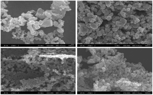
Zinc oxide (ZnO) nano-powders were prepared by a simple PVA-assisted freeze-drying process. Porous materials were firstly prepared by freeze-drying of polyvinyl alcohol (PVA) and zinc nitrate aqueous solutions with different mass ratios, and then calcined to produce ZnO nano-powders directly. PVA was applied as a polymeric carrier and its interaction with zinc nitrate was investigated for its effects on the morphology of the obtained ZnO nano-powders. The PVA/Zn(NO3)2 foams were analyzed by X-ray diffraction (XRD), thermogravimetric analysis (TGA), differential thermal analysis (DTA), and scanning electron microscopy (SEM). The obtained ZnO nano-powders were also characterized using various techniques and were evaluated as photocatalysts for dye degradation.


1. Introduction

Zinc oxide (ZnO) is one of the most attractive n-type semiconductors with excellent performance. Due to the quantum size effect, ZnO nano-materials exhibit special properties and have been widely used in various fields such as photocatalysis, gas sensors, solar cells and so on.1,2 ZnO nanoparticles are mainly prepared by a mechanochemical process, controlled precipitation, the sol–gel method, solvothermal and hydrothermal methods, and a method using an emulsion and microemulsion environment.3 Among these techniques, the sol–gel method was more widely used for offering some particular advantages such as ability to control the composition and morphology of the final ZnO nanoparticles.4 In sol–gel processes, the formation of the densely cross-linked three-dimensional network is based on hydrolysis and condensation of molecular precursors or chelation of zinc cations in solution.5 The polymeric network structure obtained in sol–gel processes is proposed for a steric entrapment role to immobilize the atomic level mixing of reagents in solution. Initiating from this solution, the solid gelation can be obtained after the aging and drying processes, with the zinc precursors trapped and dispersed uniformly. Formation of the network structure and gelation by sol–gel method is essential to the synthesis of ZnO nanoparticles with expected morphology and good particle size distribution, which however, is very time consuming.6–8 Although the so called polyacrylamide gel route and other improved sol–gel methods make the sol–gel processes more time and cost efficient, synthesis of ZnO nanoparticles by sol–gel method is challenged with the use of environmentally unfriendly organic materials and the inability to produce monodispersed powders.9

Freeze-drying, also known as lyophilization, is a process which consists of freezing a solution (aqueous or not) in a cold bath, followed by the sublimation of the frozen solvents (water most of the time) from the solid to the gas state under reduced pressure directly. A porous dried structure is obtained in a way without bringing impurities, where pores are a replica of the solvents crystals.10–13 The freeze-drying process has been widely used in pharmaceutical research, food science and technology as well as material preparation.14,15 In the nanotechnology field, attributing to the replacement of traditional ball-milling, vacuum-drying and heating-drying methods and the combination with traditional precipitation methods, sol–gel processes and emulsion environment, freeze-drying process helps to prepared nanoparticles with smaller size, more regular shape, more uniform distribution and less agglomeration.16 In the materials preparation field, freeze-drying process is applied as both a drying method and a synthetic procedure.17 Owing to its fascinating characteristics and tremendous advantages, the application of freeze-drying process in nano-sized materials preparation has attracted more and more attentions of researchers.18,19

In the present work, ZnO nano-sized powders are obtained by a novel process that simply combines the application of water-soluble polymer with freeze-drying process (Fig. 1). The aqueous solution is prepared in a round plate which can be directly put in the freeze-dryer, by dissolving polyvinyl alcohol (PVA) and zinc nitrate hexahydrate [Zn(NO3)2·6H2O] into deionized water. Then water in the solution is frozen to ice crystal and is sublimated by the freeze-drying process. Removal of the ice crystal results in dried foams that contain PVA as supporting scaffold and Zn(NO3)2 in amorphous phase. Random channels and pores in the dried polymeric foams are introduced from the ice-crystal templates synthesized in the random freezing process. Finally, ZnO nano-powders are obtained after removal of PVA and decomposition of zinc nitrate by calcination in air. Proposed mechanisms for these processes are discussed in relation to the observations, characterization data and previous literature reports. In addition, degradation of Remazol Brilliant Blue R photocatalysed by obtained ZnO nano-powders was performed to evaluate their potential as the photocatalyst.


image file: c6ra24154a-f1.tif
Fig. 1 Schematic representation of the PVA-assisted freeze-drying process to prepare ZnO nano-powders.

2. Experimental

2.1 Materials

Zinc nitrate hexahydrate (Zn(NO3)2·6H2O, analytical-grade, Mw = 297.49 g mol−1) and polyvinyl alcohol (PVA, 97% hydrolyzed, Mw = 16[thin space (1/6-em)]000 g mol−1) were purchased from Aladdin industrial Co., Ltd. and J&K Scientific Co., Ltd respectively. Remazol Brilliant Blue R (dye mass content > 60%, Mw = 626.54 g mol−1) were purchased from Tianjin Neowns Biochem LLC. All reagents were used as received without further purification. Deionized water was used routinely as required.

2.2 Preparation of ZnO nano-powders

In a typical procedure, firstly, PVA (1.0 g) was dissolved in 50 mL of deionized water while stirring in 70 °C water bath. Then, to this aqueous solution (2% wt PVA) that has been cooled to room temperature, Zn(NO3)2·6H2O (0.25 g, 1.0 g, 4.0 g, 8.0 g) was added with PVA/Zn(NO3)2·6H2O mass ratios of 4[thin space (1/6-em)]:[thin space (1/6-em)]1, 1[thin space (1/6-em)]:[thin space (1/6-em)]1, 1[thin space (1/6-em)]:[thin space (1/6-em)]4, 1[thin space (1/6-em)]:[thin space (1/6-em)]8, respectively. After stirring for 15 min, these as-prepared aqueous PVA–Zn(NO3)2 solutions were frozen in a refrigerator (Haier, model BC/BD-102HT) at −18 °C for 6 h. The fully frozen samples were freeze-dried for approximately 10 h using a freeze dryer (Biocoo, model FD-1A-50, T < −50 °C, P < 20 Pa). Finally, the dried PVA–Zn(NO3)2 solid foams obtained by this freeze-drying process was calcined in a furnace (in air) at 550 °C for 4 h with a heating ramp of 4 °C min−1, resulting in the formation of ZnO nano-powders.

2.3 Characterization and measurement

2.3.1 Material characterization. Differential thermal analysis (DTA) and thermogravimetric analysis (TGA) were carried out using a Hitachi STA7300 Thermal Analysis System. Measurements were performed by heating the sample specimens at a rate of 10 °C min−1 to 1000 °C under an air atmosphere for the dried PVA–Zn(NO3)2 solid foam samples. X-ray diffraction data were collected on a Bruker D8 Advance X-ray Diffractometer using Cu Kα radiation. The XRD patterns of the prepared dried foams were collected over 10–70° 2θ at a scan rate of 6 deg min−1, and the synthesized powder XRD patterns were collected over 20–80° 2θ at the same rate. The morphologies of the prepared dried foams and ZnO powders were observed by a JEOL JSM-6701F Field Emission Scanning Electron Microscope (SEM). One drop of the dispersed powders suspension in ethanol was deposited on a SEM stud and allowed to dry. The non-conducting dried foam samples were adhered to the aluminium stubs directly. Samples on the stubs were then coated with gold using a JEOL JFC-1600 Auto Fine Coater before imaging. The obtained powders were also characterized by using a Nicolet Avatar 360 Fourier Transform Infrared Spectrometer (FT-IR) in the range of 4000–400 cm−1 with KBr method.
2.3.2 Photocatalytic evaluation. ZnO nano-powders (3 g L−1) were added into aqueous Remazol Brilliant Blue R solution (100 mg L−1). The degradation was initiated using a 40 W UV lamp light at 365 nm with stirring. The distance from the solution level to the UV lamp was determined to be around 10 cm. The change of Remazol Brilliant Blue R concentration was monitored by a Persee TU-1080 UV-vis spectrophotometer in the room temperature. A 9 mL aliquot of liquid was taken from the Remazol Brilliant Blue R solution per hour and centrifuged for 10 min at 1800 rpm to obtain a clear solution. A 2 mL aliquot of the clear solution was used for UV-vis analysis at 592 nm. All the solution and the precipitated particles were then placed back into the original solution to continue the degradation reaction. To confirm the photocatalytic performance of synthesized ZnO nanoparticles, two comparative degradation trails were also conducted with conditions: (1) no addition of photocatalyst in the dye solution; (2) no light illumination in a black box, as other experimental process maintained the same.

The reusability of this obtained ZnO nano-powders used as photocatalysts was also studied. For the recycling experiments, 0.18 g of the ZnO nano-powders was added to 60 mL of the Remazol Brilliant Blue R solution (100 mg L−1). After 4 h degradation process in a similar way as before, the ZnO powders were centrifuged and separated from the supernatant solutions. The change of Remazol Brilliant Blue R concentration was monitored by the same way before. Without being washed, dried or calcined, the separated ZnO nano-powders was reused for the next cycle directly.

The degradation efficiency (D) of Remazol Brilliant Blue R solution was calculated according to the equation:

D = (C0C)/C0 × 100%
where C0 is the initial absorbance of Remazol Brilliant Blue R solution and C is the absorbance measured per hour.

3. Results and discussion

3.1 Analysis of the freeze-dried PVA–Zn(NO3)2 solid foams

To investigate the crystalline structure of the dried polymeric foams prepared by freeze-drying process, samples with different PVA/Zn(NO3)2·6H2O mass ratios of (a) 4[thin space (1/6-em)]:[thin space (1/6-em)]1, (b) 1[thin space (1/6-em)]:[thin space (1/6-em)]1, (c) 1[thin space (1/6-em)]:[thin space (1/6-em)]4, and (d) 1[thin space (1/6-em)]:[thin space (1/6-em)]8 were investigated by XRD. As shown in Fig. 2, There is a typical broad peak centred on 2θ = 20.10° and one more peak at 2θ = 26.28°, indicating the semi-crystalline nature of the PVA and presence of crystalline and amorphous regions.20–22 The intensity of the peak at 2θ = 26.28° increased with the mass ratio of PVA/Zn(NO3)2·6H2O becoming higher. This behavior may be caused by intermolecular interactions between hydroxyl groups of the polymer and zinc cations from the dissolved nitrates.21,23,24 The apparent reduction of PVA characteristic peak height with increasing content of Zn(NO3)2 indicates decrease of the degree of crystallization of PVA, which can be attributed to the polymer plasticization as observed in the freeze-drying experimental process.25 No peaks can be seen from the pattern for sample (d) with excessive content of Zn(NO3)2.
image file: c6ra24154a-f2.tif
Fig. 2 XRD patterns of polymeric foams with different PVA/Zn(NO3)2·6H2O mass ratios of (a) 4[thin space (1/6-em)]:[thin space (1/6-em)]1, (b) 1[thin space (1/6-em)]:[thin space (1/6-em)]1, (c) 1[thin space (1/6-em)]:[thin space (1/6-em)]4, and (d) 1[thin space (1/6-em)]:[thin space (1/6-em)]8.

The evolution of TGA and DTA thermal curves (Fig. 3) of the dried foams with mass ratio of 1[thin space (1/6-em)]:[thin space (1/6-em)]4 has also been studied. The first stage of weight loss below 150 °C is due to the removal of physisorbed and crystal water. The second stage, registered in the range 140–170 °C, is very interesting, with a visible exothermic effect on DTA curve which corresponds to the redox reaction that takes place between zinc nitrate and PVA.23 This redox reaction results in the oxidation of PVA to carboxylate anions that coordinate to the metal cations forming carboxylate coordination compounds, used as precursor for the desired zinc oxides.26 Observation of the evolved reddish-brown nitrogen oxides as the dried foams were calcined in a furnace can also be a affirmation of this redox reaction.27 The third weight loss occurs with a exothermic peak in the range 250–460 °C corresponds to the thermal decomposition of the Zn(II) carboxylates products formed in the redox reaction, as reported in the literature.28


image file: c6ra24154a-f3.tif
Fig. 3 TGA and DTA thermal curves of the polymeric PVA–Zn(NO3)2 foams with mass ratio of 1[thin space (1/6-em)]:[thin space (1/6-em)]4.

By analyzing the crystalline structure and thermal decomposition of prepared PVA–Zn(NO3)2 polymeric foams, the advantages of this PVA-assisted freeze-drying process can be observed. The stabilization of the cations in the precursor is established not only through the chelating effect on the metallic cations with the functional groups, but also, in major part, through the physical entrapment of the metal ions in the dried polymer network.29,30 As shown in Fig. 4, the synthesized PVA foams with random channels and pores, which are introduced from the ice-crystal templates formed in the freezing process, act as a polymeric carrier contributing to the stabilization and dispersion of the cations.


image file: c6ra24154a-f4.tif
Fig. 4 SEM image of the freeze-dried polymeric foams with PVA/Zn(NO3)2·6H2O mass ratio of 1[thin space (1/6-em)]:[thin space (1/6-em)]4.

In the process of thermal decomposition, a big advantage of the PVA's presence in the system is the carbonaceous residue that results by its thermal decomposition, acting as a surfactant for the oxides particles, thus preventing their aggregation.31 PVA acts not only as a metal-chelating agent thereby inhibiting the segregation of metals during heating,25,29 but also as a reductant in the redox reaction between PVA and Zn(NO3)2 during calcinations of the precursor. Another interesting feature is that the nitrate ions provide an in situ oxidizing environment for the decomposition of PVA. This special reaction between PVA and zinc nitrate actually reduces the heat consumption during the calcining process.32–34

3.2 Characterization of prepared ZnO powders

Phase composition and purity of the as-prepared samples were identified by X-ray diffraction (XRD) and Fourier Transform Infrared Spectroscopy (FT-IR). Fig. 5 shows the XRD pattern of obtained ZnO nano-powders, and FT-IR spectra of the powders samples prepared with different mass ratio of (a) 4[thin space (1/6-em)]:[thin space (1/6-em)]1, (b) 1[thin space (1/6-em)]:[thin space (1/6-em)]1, (c) 1[thin space (1/6-em)]:[thin space (1/6-em)]4, (d) 1[thin space (1/6-em)]:[thin space (1/6-em)]8 is presented in Fig. 6. As shown in Fig. 5, all the reflection peaks in the XRD pattern can be indexed as pure hexagonal wurtzite ZnO with cell parameters a = 3.249 Å and c = 5.206 Å (International Center for Diffraction Data, JCPDS 36-1541).
image file: c6ra24154a-f5.tif
Fig. 5 XRD patterns of ZnO powders prepared with different PVA/Zn(NO3)2·6H2O mass ratios of (a) 4[thin space (1/6-em)]:[thin space (1/6-em)]1, (b) 1[thin space (1/6-em)]:[thin space (1/6-em)]1, (c) 1[thin space (1/6-em)]:[thin space (1/6-em)]4, (d) 1[thin space (1/6-em)]:[thin space (1/6-em)]8.

image file: c6ra24154a-f6.tif
Fig. 6 FT-IR spectra of the ZnO powders samples prepared with PVA/Zn(NO3)2·6H2O mass ratio of (a) 4[thin space (1/6-em)]:[thin space (1/6-em)]1, (b) 1[thin space (1/6-em)]:[thin space (1/6-em)]1, (c) 1[thin space (1/6-em)]:[thin space (1/6-em)]4, (d) 1[thin space (1/6-em)]:[thin space (1/6-em)]8.

As shown in Fig. 6, the FT-IR spectra of the four samples are roughly the same. As each spectrum shows, there are absorption bands corresponding to hydroxyl groups (O–H) between 2900 and 3500 cm−1, which were identified as the physisorbed water. The bands between 500 and 650 cm−1 corresponded to the Zn–O bonds. No impurities, such as Zn(NO3)2, Zn(OH)2, or other organic compounds can be detected from the XRD patterns or FT-IR spectra. And the relative strong and sharp peaks in the XRD pattern confirm that the products are well-crystallized.

The morphology of the ZnO nano-powders prepared with different PVA/Zn(NO3)2·6H2O mass ratios was investigated by SEM techniques, as presented in Fig. 7. The result validates that the products were irregular rounded nano-sized particles. The ZnO particles from sample (c) are more homogenous and smaller in size than the particles from sample (a) and (b), with average particle size of about 80 nm. The image of sample (d) indicates that, ZnO particles were partly agglomerated when the mass ratio is 1[thin space (1/6-em)]:[thin space (1/6-em)]8 with excessive content of Zn(NO3)2.


image file: c6ra24154a-f7.tif
Fig. 7 SEM images of ZnO powder samples prepared with different PVA/Zn(NO3)2·6H2O mass ratios of (a) 4[thin space (1/6-em)]:[thin space (1/6-em)]1, (b) 1[thin space (1/6-em)]:[thin space (1/6-em)]1, (c) 1[thin space (1/6-em)]:[thin space (1/6-em)]4, (d) 1[thin space (1/6-em)]:[thin space (1/6-em)]8 and a control ZnO sample (e) prepared via the conventional vacuum drying process.

In order to compare the conventional drying process with this novel PVA-assisted freeze drying process, a control ZnO powder sample (e) was prepared via a similarly process to Fig. 1 but with conventional vacuum drying (P = 0.01 MPa, T = 70 °C) to replace the freeze drying. The PVA/Zn(NO3)2·6H2O mass ratio of this control sample (e) is 1[thin space (1/6-em)]:[thin space (1/6-em)]4. The SEM image of the control ZnO powder sample is presented as Fig. 7(e). As Fig. 7(e) shown, the ZnO powders obtained by conventional drying process were severely agglomerated. On the other hand, ZnO powders prepared via the novel PVA-assisted freeze drying process under the similar experimental condition (Fig. 7(c)) have better dispersivity.

In the control experiment, PVA gelatin containing Zn(NO3)2 was obtained after the solution was dried by the vacuum drying process. Compared with the gelatin, porous foams synthesized via the freezing drying process can better disperse the Zn source. So in this novel PVA-assisted freeze drying process, freeze drying was applied as both a drying method and a synthetic procedure.

3.3 Photocatalytic performance of obtained ZnO nano-powders

Synthesized ZnO nano-powders were evaluated for dye photodegradation to demonstrate the potential application as the photocatalyst. Remazol Brilliant Blue R was chosen as a model dye because it has been used extensively in the dye industry.35,36 The degradation efficiencies of the dye solution photocatalysed by prepared ZnO nano-powders are shown in Fig. 8.
image file: c6ra24154a-f8.tif
Fig. 8 Degradation efficiencies of dye solution photocatalysed by ZnO nano-powders samples prepared with different PVA/Zn(NO3)2·6H2O mass ratios of (a) 4[thin space (1/6-em)]:[thin space (1/6-em)]1, (b) 1[thin space (1/6-em)]:[thin space (1/6-em)]1, (c) 1[thin space (1/6-em)]:[thin space (1/6-em)]4, (d) 1[thin space (1/6-em)]:[thin space (1/6-em)]8 and dye solution conducted with comparative conditions: (e) no addition of photocatalyst in the dye solution, (f) no light illumination in a black box, as other experimental process maintained the same.

Adsorption of the dye molecules onto the ZnO particles and self-degradation of the dye solution under the UV light illumination were not expected in this photocatalytic degradation test. There was no decrease of the dye solution concentration as presented in Fig. 8(e) and (f), which confirmed the photocatalytic degradation of the dye by ZnO nanoparticles. 70% degradation efficiency was achieved within 1 h reaction time for sample (c), which indicates that ZnO nano-powders synthesized by this novel process have similar photocatalytic performance with the ZnO nanostructure obtain by other methods in the reported literatures,37–39 and that Remazol Brilliant Blue R solution can be degradated efficiently by the synthesized ZnO nanoparticles used as photocatalyst. The degradation efficiency of the dye solution after 4 h photocatalytic reaction time was 98.66%, 97.11%, 96.55% and 96.02% for sample (c, b, d, a), respectively. Compared with other ZnO samples, sample (c) is better dispersed and has smaller size (as observed in SEM images), which can provide more surface active sites for the photocatalytic reaction. This is thought to be the main reason for the superior degradation performance of sample (c).40,41

Reusability is an important feature of heterogeneous photocatalyst materials for their practical application.42 As shown in Fig. 9, although photocatalytic performance of ZnO nano-powders deteriorates with more recycling times, degradation efficiencies still keep over 80% after five-time recycling of ZnO nano-powders sample (c).


image file: c6ra24154a-f9.tif
Fig. 9 Reusability of ZnO nano-powders sample (c) prepared with PVA/Zn(NO3)2·6H2O mass ratio of 1[thin space (1/6-em)]:[thin space (1/6-em)]4 applied as a photocatalyst in dye degradation.

It should be emphasized that, without being washed, dried or calcined, ZnO nano-powders have been reused for ten times in the experiment, and then the degradation efficiency of Remazol Brilliant Blue R solution is still up to 70%. The obtained ZnO photocatalyst in this work possesses superior reusability than other ZnO nanostructure reported43 when photodegradating Remazol Brilliant Blue R. This was thought to be the consequence of the none adsorption effect for the dye molecules, high crystallinity and well-dispersity of the obtained ZnO nano-powders.44 In addition, the natural sedimentation of synthesized ZnO nano-powders in solution was easy to be accomplished, as observed in experimental process. With the excellent photocatalytic performance and high stability observed by the photocatalytic evaluation, ZnO nano-powders synthesized by this PVA-assisted freeze-drying process may have a good potential as photocatalyst to apply in the treatment of wastewaters containing organic dyes.

4. Conclusions

In summary, this paper reports a novel simple PVA-assisted freeze-drying process to synthesize well-dispersed ZnO nano-powders. The effect of different mass ratios of the materials (PVA/Zn(NO3)2·6H2O) on the morphology of ZnO nanoparticles was studied. The large chain molecule of PVA, which included hydroxyl functional groups, dispersed the zinc ions homogeneously by steric entrapment and chelating effect. In the thermal decomposition step, the redox reaction between PVA and nitrates actually provided carbonaceous residues acting as a surfactant for the oxides particles, thus preventing their aggregation. Water in the solution was removed by freeze-drying process, resulting in random channels and pores in the obtained dried foams, which was contributing to the stabilization and dispersion of the zinc cations. Without use of expensive materials and toxic organic solvents, the process initiated from an aqueous solution described here present a simple approach to the formation of ultrafine ZnO nano-powders with excellent photocatalytic performance, especially with convenient and efficient reusability more than five cycles.

Acknowledgements

This research was financially supported by the funds awarded by Department of Science and Technology of Jiangsu Province (No. BY2014052) and National Natural Science Foundation of China (No. 21476008).

References

  1. Y. K. Mishra, G. Modi, V. Cretu, V. Postica, O. Lupan, T. Reimer, I. Paulowicz, V. Hrkac, W. Benecke, L. Kienle and R. Adelung, ACS Appl. Mater. Interfaces, 2015, 7, 14303–14316 CAS .
  2. X. Li, X. Zhang, L. Li, L. Huang, W. Zhang, J. Ye and J. Hong, Mater. Lett., 2016, 175, 122–125 CrossRef CAS .
  3. A. Kołodziejczak-Radzimska and T. Jesionowski, Materials, 2014, 7, 2833–2881 CrossRef .
  4. D. D. Athayde, D. F. Souza, A. M. A. Silva, D. Vasconcelos, E. H. M. Nunes, J. C. Diniz da Costa and W. L. Vasconcelos, Ceram. Int., 2016, 42, 6555–6571 CrossRef CAS .
  5. A. Douy, Int. J. Inorg. Mater., 2001, 3, 699–707 CrossRef CAS .
  6. B. K. Sodipo and A. A. Aziz, J. Magn. Magn. Mater., 2016, 416, 275–291 CrossRef CAS .
  7. G. J. Owens, R. K. Singh, F. Foroutan, M. Alqaysi, C. M. Han, C. Mahapatra, H.-W. Kim and J. C. Knowles, Prog. Mater. Sci., 2016, 77, 1–79 CrossRef CAS .
  8. A. E. Danks, S. R. Hall and Z. Schnepp, Mater. Horiz., 2015, 3, 91–112 RSC .
  9. C. Pan, X. Li, F. Wang and L. Wang, Ceram. Int., 2008, 34, 439–441 CrossRef CAS .
  10. H. Zhang, I. Hussain, M. Brust, M. F. Butler, S. P. Ran nard and A. I. Cooper, Nat. Mater., 2005, 4, 787–793 CrossRef CAS PubMed .
  11. H. Zhang and A. I. Cooper, Adv. Mater., 2007, 19, 1529–1533 CrossRef CAS .
  12. L. Qian and H. Zhang, J. Chem. Technol. Biotechnol., 2011, 86, 172–184 CrossRef CAS .
  13. M. Barrow, A. Eltmimi, A. Ahmed, P. Myers and H. Zhang, J. Mater. Chem., 2012, 22, 11615–11620 RSC .
  14. J. Zhao, Y. He, L. Zhang and K. Lu, J. Alloys Compd., 2016, 678, 36–41 CrossRef CAS .
  15. S. Wanning, R. Süverkrüp and A. Lamprecht, Int. J. Pharm., 2015, 488, 136–153 CrossRef CAS PubMed .
  16. G. Chen and W. Wang, Drying Technol., 2007, 25, 29–35 CrossRef CAS .
  17. B. Wang, W. Zhang, W. Zhang, A. S. Mujumdar and L. Huang, Drying Technol., 2005, 23, 7–32 CrossRef CAS .
  18. V. V. Patil, P. P. Dandekar, V. B. Patravale and B. N. Thorat, Drying Technol., 2010, 28, 624–635 CrossRef CAS .
  19. M. J. Choi, S. Briançon, J. Andrieu, S. G. Min and H. Fessi, Drying Technol., 2004, 22, 335–346 CrossRef CAS .
  20. K. S. Hemalatha, K. Rukmani, N. Suriyamurthy and B. M. Nagabhushana, Mater. Res. Bull., 2014, 51, 438–446 CrossRef CAS .
  21. B. M. A. El-Khair, S. M. Mokhtar, A. Z. Dakroury and M. B. S. Osman, J. Macromol. Sci., Part B: Phys., 1994, 33, 387–395 CrossRef .
  22. R. C. Agrawal, A. Chandra, A. Bhatt and Y. K. Mahipal, New J. Phys., 2008, 10, 2653–2659 CrossRef .
  23. M. Stoia, M. Barbu, M. Ştefănescu, P. Barvinschi and L. Barbu-Tudoran, J. Therm. Anal. Calorim., 2012, 110, 85–92 CrossRef CAS .
  24. S. Rajendran, M. Sivakumar and R. Subadevi, Solid State Ionics, 2004, 167, 335–339 CrossRef CAS .
  25. M. Ari, K. J. Miller, B. A. Marinkovic, P. M. Jardim, R. de Avillez, F. Rizzo and M. A. White, J. Sol-Gel Sci. Technol., 2011, 58, 121–125 CrossRef CAS .
  26. M. Barbu, M. Stoia, P. Barvinschi, L. Barbu-Tudoran and M. Stefanescu, Thermochim. Acta, 2013, 564, 43–50 CrossRef CAS .
  27. M. Stefanescu, M. Barbu, T. Vlase, P. Barvinschi, L. Barbu-Tudoran and M. Stoia, Thermochim. Acta, 2011, 526, 130–136 CrossRef CAS .
  28. M. Stoia, P. Barvinschi and L. Barbu-Tudoran, J. Therm. Anal. Calorim., 2013, 113, 21–30 CrossRef CAS .
  29. A. Sen and P. Pramanik, J. Mater. Synth. Process., 2002, 10, 107–111 CrossRef CAS .
  30. M. A. Gülgün, M. H. Nguyen and W. M. Kriven, J. Am. Ceram. Soc., 1999, 82, 556–560 CrossRef .
  31. M. Stoia, L. B. Tudoran and P. Barvinschi, J. Therm. Anal. Calorim., 2013, 113, 11–19 CrossRef CAS .
  32. S. Kumar Saha, A. Pathak and P. Pramanik, J. Mater. Sci. Lett., 1995, 14, 35–37 CrossRef .
  33. M. Stefanescu, M. Stoia, O. Stefanescu and P. Barvinschi, J. Therm. Anal. Calorim., 2010, 99, 459–464 CrossRef CAS .
  34. M. Stoia, P. Barvinschi, L. B. Tudoran, M. Barbu and M. Stefanescu, J. Therm. Anal. Calorim., 2012, 108, 1033–1039 CrossRef CAS .
  35. V. Yonten, M. Tanyol, N. Yildirim, N. C. Yildirim and M. Ince, Desalin. Water Treat., 2016, 57, 15592–15602 CrossRef CAS .
  36. N. A. Rahmat, A. A. Ali, Salmiati, N. Hussain, M. S. Muhamad, R. A. Kristanti and T. Hadibarata, Water, Air, Soil Pollut., 2016, 227, 1–11 CrossRef CAS .
  37. H. C. Ma, S. Y. Yuan, Y. H. Fu, X. L. Dong, X. F. Zhang, C. Ma and X. X. Zhang, Adv. Mater. Res., 2012, 518, 746–749 CrossRef .
  38. G. L. Yang, Q. Liu, Y. H. Fu, H. C. Ma, C. Ma, X. L. Dong, X. X. Zhang and X. F. Zhang, J. Mater. Res., 2015, 30, 1902–1913 CrossRef CAS .
  39. L. Kashinath, K. Namratha and K. Byrappa, Mater Today, 2016, 3, 74–83 CrossRef .
  40. H. Wang, C. Xie, W. Zhang, S. Cai, Z. Yang and Y. Gui, J. Hazard. Mater., 2007, 141, 645–652 CrossRef CAS PubMed .
  41. S. M. Lam, J. C. Sin, A. Z. Abdullah and A. R. Mohamed, Desalin. Water Treat., 2012, 41, 131–169 CrossRef CAS .
  42. B. Weintraub, Z. Zhou, Y. Li and Y. Deng, Nanoscale, 2010, 2, 1573–1587 RSC .
  43. M. R. Khodadadi, M. E. Olya and A. Naeimi, Korean J. Chem. Eng., 2016, 33, 2018–2026 CrossRef CAS .
  44. M. Kanna, S. Wongnawa, S. Buddee, K. Dilokkhunakul and P. Pinpithak, J. Sol-Gel Sci. Technol., 2010, 53, 162–170 CrossRef CAS .

This journal is © The Royal Society of Chemistry 2016
Click here to see how this site uses Cookies. View our privacy policy here.