
Phase transformations of novel CuxS nanostructures as highly efficient counter electrodes for stable and reproducible quantum dot-sensitized solar cells†
Mallineni Venkata-Haritha‡
,
Chandu V. V. M. Gopi‡,
Young-Seok Lee and
Hee-Je Kim*
School of Electrical Engineering, Pusan National University, Busandaehak-ro 63 beon-gil, Geumjeong-gu, Busan 46241, South Korea. E-mail: heeje@pusan.ac.kr; Fax: +82 51 513 0212; Tel: +82 51 510 2364
First published on 11th October 2016
Abstract
An ideal counter electrode (CE) that is cost-effective and has high electrocatalytic activity, high performance stability, and a simple fabrication process is essential for quantum dot-sensitized solar cells (QDSSCs). We report a facile one-step chemical bath deposition method for the growth of an L-cysteine-dependent CuxS thin film on a fluorine-doped tin oxide substrate, which was used as a highly efficient CE for QDSSCs. SEM images revealed that the surface morphology of CuxS changed from nanoplatelets to nanosheets to nanospheres to nanoflowers based on the concentration of L-cysteine. Also, the concentration of L-cysteine in the formation of CuxS thin films is expected to play an important role in determining the phase of CuxS thin films (Cu2S, Cu1.75S, Cu1.12S and CuS). QDSSCs based on Cu1.12S nanosphere CEs possess a high power conversion efficiency of 5.88%, which is much higher than those of QDSSCs based on Cu2S nanoplatelets (5.10%), Cu1.75S nanosheets (5.32%), CuS nanoflowers (4.88%) and Pt (1.36%) CEs. Electrochemical measurements revealed that a Cu1.12S nanosphere CE has better electrocatalytic activity, which increases the rate of reduction of polysulfide in comparison to those of Cu2S nanoplatelets, Cu1.75S nanosheets, CuS nanoflowers and Pt CEs. A QDSSC also displayed superior stability in an operational state for over 20 h, which is a major challenge for CuxS CEs.
1. Introduction
The continuous growth of the global population and the increasing need for energy have led to greater efforts to exploit solar energy, which will supply abundant energy in the future.1 Dye-sensitized solar cells (DSSCs), which were developed by Grätzel, have displayed outstanding potential as third-generation solar cells owing to their cost-effectiveness, facile fabrication, abundance, and higher conversion efficiency.2,3 However, the photodegradation observed in DSSCs has made researchers try to discover an alternative material with enhanced overall performance and device durability.4,5Quantum dot-sensitized solar cells (QDSSCs) are attractive liquid junction photoelectronic devices and have received much attention for developing next-generation solar cells.6–8 They could replace DSSCs because of the unique properties of quantum dots (QDs). Their intriguing optical properties include the ability to tune their optical energy band gaps,9 hot-carrier transfer,10 high extinction coefficients,11 large intrinsic dipole moments,12 low production costs and simple synthetic procedure.5 In addition, QDs have an admirable theoretical thermodynamic efficiency (44%), which benefits from utilizing the generation of multiple excitons by the impact ionization effect.13
The device structure and working principle of QDSSCs are similar to those of DSSCs. They include a TiO2 photoanode loaded with a wide-band-gap semiconductor, a sensitizer, a polysulfide redox electrolyte (S2−/Sn2−), and a counter electrode (CE).14 Recently, many efforts have been made based on improvements to the structure of the TiO2 photoanode, investigating new QDs, and employing different types of redox couple. A focus on the optimization and design strategy of CEs has had a considerable impact on the entire conversion of cells. The mechanism of the TiO2 electrode comprises the collection of photoexcited electrons injected from the sensitizer (QDs or dye molecules) into TiO2 nanocrystallites to provide a transport bridge between an external circuit and the sensitizer. Various kinds of semiconductor QD sensitizers have been widely researched to improve the overall performance of QDSSCs, such as CdS, CdSe, PbS, CIS, CISe, and CdSexTe1−x.15–20 As well as the QD sensitizer, the CE also plays a vital role as an important part of a solar cell and is responsible for catalysis and the reduction of the oxidized redox species. If the rate of the reduction reaction is unable to increase the rate of generation of electron–hole pairs, collapse of electrical equilibrium occurs in the form of the accumulation of holes in the interior of the cell. The accumulated holes may result in chemical corrosion of the QDs, which significantly affects the stability of the cell. Therefore, the most important function of the CE is its ability to effectively catalyze the reduction of the S2−/Sn2− electrolyte so that the reaction is facilitated as much as possible.21,22 To achieve superior electrocatalytic properties, CEs must have excellent electrical conductivity, high catalytic activity, and a large surface area. In addition, CEs should be chemically stable for a long time, as this avoids corrosion by adsorption of the S2−/Sn2− electrolyte onto the surface of the CE. There have also been some attempts to limit the manufacturing cost of CEs, because conventional CEs consist of expensive noble metals such as Pt and Au, and this could be an obstacle to commercializing QDSSCs in practice. Nevertheless, with the excellent properties of QDs the theoretical thermodynamic efficiency (44%) of a QDSSC is higher than that of a DSSC with conventional dyes, but the currently recorded PCE of QDSSCs is typically around 11.6%, which is still far below the value for DSSCs (14.3%) owing to low photovoltaic performance due to electron loss via charge separation and transfer at the TiO2/electrolyte and QD/electrolyte interfaces.23–25
In DSSCs, the commonly used standard Pt CE still exhibits outstanding electrocatalytic activity for the organic iodide/triiodide (I−/I3−) redox couple electrolyte, with low charge transfer resistance (Rct) at the CE/electrolyte interface.2 The I−/I3− redox couple electrolyte is not chemically compatible with QDSSCs and results in photodegradation and continual photocorrosion problems.26 To overcome this, the sulfide/polysulfide redox system (S2−/Sn2−) has been reported to be an ideal electrolyte that exhibits robust discrimination towards CEs without photocorrosion issues, thereby improving the photovoltaic performance of QDSSCs.27
The performance of Pt CEs in conjunction with polysulfide electrolytes in QDSSCs may suppress surface activity and conductivity in the reduction of Sn2− sulfur species. This leads to a poisoning effect and limits the durability of QDSSCs.28,29 To overcome this problem, researchers have made greater attempts to find an alternative material to Pt and Au CEs for polysulfide solutions. Inexpensive nanostructured metal sulfide materials (CuxS, CoS, NiS, PbS and Cu2ZnSnSe4)30–35 and carbonaceous materials36–38 have been widely studied as promising candidates for superior electrocatalytic activity in QDSSCs. Among all metal sulfides, CuxS has been found to be the most preferred transition metal chalcogenide with low charge transfer resistance, superior electrocatalytic activity and conductivity, and cost-effectiveness. Moreover, the excellent morphology of this CE material can also improve its electrocatalytic performance via a simple fabrication process.30,31 CuxS has excited considerable interest owing to its stoichiometric composition, valence states, complex structures, different unique properties, and different stoichiometric forms. With its different stoichiometries, the applications of copper sulfide include solar cells, photothermal conversion, photocatalysis, sensors, and lithium rechargeable batteries. However, the electrocatalytic properties of CuxS nanosphere thin films as CEs have not been investigated in QDSSCs.
In previous reports on the fabrication of metal sulfides prepared using L-cysteine, the electrocatalytic activity of metal sulfides was not investigated in detail as a function of L-cysteine.39,40 The formation of CuxS nanostructures becomes more intricate and interesting using L-cysteine as a reagent, and thus it deserves further investigation. Here, we successfully fabricated nanoplatelet, nanosheet, nanosphere and nanoflower-structured CuxS thin films on FTO by a facile chemical bath deposition (CBD) process using an optimized concentration of L-cysteine as a reagent, and the thin films were used as CEs for QDSSCs without the need for further treatment, which has not yet been achieved, to the best of our knowledge. As well as the surface morphology, the concentration of L-cysteine greatly affects the stoichiometry of copper sulfides; as a result, Cu2S (hexagonal), Cu1.75S (anilite), Cu1.12S (yarrowite) and CuS (hexagonal) have been obtained. In this study, the CuxS thin film on FTO exhibited many advantages. Firstly, this film exhibited strong adhesion to the FTO substrate owing to in situ growth. Secondly, the preparation of this film reduced the cost and simplified the fabrication process. Finally, the CuxS film can provide more adsorption of electrolytes and electrocatalytic active sites for the reduction of polysulfide electrolytes. We revealed the electrochemical behavior of the thin films and their influence on the performance and efficiency of QDSSCs.
We also observed that the surface morphology and atomic ratio of Cu![[thin space (1/6-em)]](https://www.rsc.org/images/entities/char_2009.gif) :S were influenced by the concentration of L-cysteine. An improved power conversion efficiency of 5.88% was achieved for a QDSSC based on a Cu1.12S nanosphere CE. The efficiency of a Cu1.12S CE based on L-cysteine (5.88%) was superior to those of bare CuS (3.18%)30 Cu2S nanoplatelets (5.10%), Cu1.75S nanosheets (5.32%), CuS nanoflowers (4.88%), and Pt (1.36%) CEs. The present study revealed that the electrocatalytic activity of CuxS is dictated by its composition and the photovoltaic performance of CuxS is also substantially governed by its stoichiometry.
:S were influenced by the concentration of L-cysteine. An improved power conversion efficiency of 5.88% was achieved for a QDSSC based on a Cu1.12S nanosphere CE. The efficiency of a Cu1.12S CE based on L-cysteine (5.88%) was superior to those of bare CuS (3.18%)30 Cu2S nanoplatelets (5.10%), Cu1.75S nanosheets (5.32%), CuS nanoflowers (4.88%), and Pt (1.36%) CEs. The present study revealed that the electrocatalytic activity of CuxS is dictated by its composition and the photovoltaic performance of CuxS is also substantially governed by its stoichiometry.
2. Experimental section
2.1. Materials
All chemicals used in the synthesis of CuxS thin film CEs and photoanodes were purchased from Sigma-Aldrich. They were analytical grade reagents and were used as received without any purification. Copper sulfate pentahydrate (CuSO4·5H2O), thioacetamide (C2H5NS), L-cysteine (C3H7NO2S), cadmium acetate dihydrate [Cd(CH3COO)2·2H2O], zinc acetate dihydrate [Zn(CH3COO)2·2H2O], sodium sulfide (Na2S), sulfur (S), selenium (Se), potassium chloride (KCl), sodium thiosulfate (Na2S2O3), and TiO2 paste (Ti-nanoxide HT/SP) were supplied by Solaronix.2.2. Fabrication of CuxS and Pt CEs on FTO glass
Morphology-controlled L-cysteine-based CuxS thin films were deposited on well-cleaned fluorine-doped tin oxide (FTO) glass substrates with a sheet resistance of 7 Ω cm−2 (Hartford Glass). The CE thin films were applied to the FTO substrates by CBD. Prior to deposition, the FTO glass was ultrasonically cleaned using acetone, ethanol, and DI water for 10 min each. We used copper sulfate pentahydrate (CuSO4·5H2O) and thioacetamide (C2H5NS) as precursors of Cu2+ and S2−, respectively, and L-cysteine was used as a reagent.The deposition solution was prepared with 0.1 M CuSO4·5H2O and different concentrations of L-cysteine (0.005 mM, 0.01 mM, 0.02 mM, and 0.03 mM) dissolved in deionized water. These were stirred vigorously for 30 min to obtain a transparent solution. Under constant stirring, 1 M C2H5NS was added to the above solution and the reaction mixture was vigorously stirred for another 30 min. Ultrasonically cleaned FTO glass samples were carefully dipped vertically into a host cation–anion solution and placed in a hot air oven for 90 min, and deposition was carried out at a temperature of 60 °C. After deposition for 90 min, the samples were rinsed with DI water and ethanol. The CuxS CEs obtained with L-cysteine concentrations of 0.005 mM, 0.01 mM, 0.02 mM, and 0.03 mM were denoted as Cu2S, Cu1.75S, Cu1.12S, and CuS, respectively. To synthesize a Pt CE, well-cleaned FTO glass was coated with commercial platinum paste (Solaronix, Pt-catalyst T/SP) using the doctor blade method and annealed for 10 min at 450 °C in air.
2.3. Fabrication of QDSSCs
FTO substrates were cleaned ultrasonically with acetone, ethanol, and distilled water (DI) for 10 min each. The well-cleaned FTO substrates were coated with commercially available TiO2 paste with a particle size of 20 nm by the doctor blade method with an active area of 0.27 cm2. The samples were annealed at 450 °C for 30 min with a thickness of 7.5 μm.41 TiO2 photoelectrodes were sequentially sensitized with CdS, CdSe QDs and a Mn–ZnSe passivation layer using the successive ionic layer adsorption and reaction (SILAR) method. The photoanodes were immersed in host anion and cation solutions to allow the ions to be adsorbed onto the TiO2 paste and were then rinsed with ethanol and DI water. They were dried under N2 gas between each dipping process.Initially, TiO2 samples were alternately dipped in an aqueous solution of a cationic precursor containing 0.1 M Cd(CH3COO)2·2H2O for 5 min to allow Cd2+ ions to be adsorbed onto TiO2. They were then dipped into an aqueous solution of an anionic precursor, namely, 0.1 M Na2S for 5 min each. The desired CdS was formed by reacting the adsorbed Cd2+ ions with S2−. This two-step dipping process represents one CBD cycle and was repeated for up to 5 cycles. The prepared electrodes were named as TiO2/(CdS)5.
A CdSe layer was sequentially deposited on the as-prepared TiO2/(CdS)5. For the deposition of CdSe, the TiO2/(CdS)5 film was immersed in an aqueous solution of a cationic precursor containing 0.1 M Cd(CH3COO)2·2H2O for 5 min at room temperature and then in a refluxing solution of an anionic precursor, namely, Na2SeSO3 for 5 min at 50 °C. The entire process was repeated for 8 cycles. The as-prepared photoanodes were denoted as TiO2/(CdS)5/(CdSe)8. The solution of Na2SeSO3 was prepared by refluxing 0.2 M Se in an aqueous solution containing 0.4 M Na2SO3 for 4 h at 120 °C. Finally, a passivation layer of Mn–ZnSe was deposited twice on the sensitized samples by the SILAR method by dipping in aqueous solutions of cationic and anionic precursors, namely, 10 mM Mn(CH3COO)2·4H2O mixed with 0.1 M Zn(CH3COO)2·2H2O and 0.1 M Na2S for 1 min each. The final samples were denoted as TiO2/(CdS)5/(CdSe)8/(Mn–ZnSe)2. The passivation layer effectively covered the QDs and prevented corrosion and leakage of electrons from TiO2 to the electrolyte.
The as-prepared TiO2/CdS/CdSe/Mn–ZnSe photoanode and the fabricated CuxS and Pt CEs were inserted between plates of hot-melt sealant sheets (double sheet of SX 1170-60, Solaronix) at 100 °C to seal the solar cell. The internal space around the electrodes was filled by capillary action with a liquid polysulfide redox electrolyte containing 1 M Na2S, 2 M S, and 0.1 M KCl in a mixture of methanol and water in a ratio of 7:3. After filling with the electrolyte, holes were covered with the sealant and glass and then soldered. The cells were left for a few hours for diffusion of the electrolyte in the photoanode, and the cells were then tested under one-sun illumination of 100 mW cm−2.
2.4. Fabrication of symmetric cells
Symmetrical CuxS and Pt CEs were sealed using a 60 μm hot-melt sealant sheet at 100 °C (SX 1170-60, Solaronix). The internal space between the two glass sheets was filled with a polysulfide redox electrolyte consisting of 1 M Na2S, 2 M S, and 0.1 M KCl in a mixture of methanol and water in a ratio of 7:3. The symmetric cells were used for cyclic voltammetry (CV) at a scan rate of 100 mV s−1. Tafel polarization at a scan rate of 10 mV s−1 and EIS measurements were performed using a potentiostat/galvanostat/EIS analyzer (BioLogic SP-150, France) on dummy cells with CuxS/CuxS and Pt/Pt CEs in the frequency range of 100 mHz to 500 kHz in dark conditions. During characterization of the electrodes in dark conditions, the cells were covered with a black mask fitted around the active area (0.7 cm2) of the cell.
2.5. Characterization
The surface morphology and thickness of the CuxS electrodes were investigated using a field emission scanning electron microscope (FE-SEM, Hitachi S-2400) operated at 15 kV and equipped with on-system energy-dispersive X-ray spectroscopy (EDX). Transmission electron microscopy (TEM, JEM-2011) was used to observe the internal structure of samples. The crystalline nature and structure of the CuxS films were studied by X-ray diffraction (XRD) with a D8 ADVANCE diffractometer using a Cu Kα radiation source operated at 40 kV and 40 mA in the 2θ range of 20–80°. X-ray photon spectroscopy (XPS) is a surface-sensitive spectroscopic technique that was used to measure the elemental composition of the thin films. XPS was performed using a VG Scientific ESCALAB 250 with 1486.6 eV radiation and an electron take-off angle of 90°. Atomic force microscopy (JPK NanoWizard II AFM, JPK Instruments, Berlin, Germany) was used to measure the surface roughness of the CuxS thin films at a scan rate of 0.8 Hz in contact mode. For tape testing, a long piece of 3M Scotch tape (18 mm × 32 m) was attached fully to the CuS thin film on the FTO substrate and then detached from FTO.UV-vis spectroscopic analysis was carried out using an Optizen 3220 UV spectrophotometer. The photocurrent–voltage (J–V) characteristics of QDSSCs were examined under one-sun illumination (AM 1.5G, 100 mW cm−2) using an ABET Technologies (USA) solar simulator, which has an irradiance uniformity of ±3%. The incident photon-to-current conversion efficiency (IPCE) spectra were recorded using an Oriel® IQE-200™. CV was performed in a three-electrode system using a potentiostat/galvanostat/EIS analyzer (BioLogic SP-150, France) at a scan rate of 100 mV s−1 in an aqueous solution of 0.1 M Na2S, 0.1 M S, and 0.1 M KCl. To perform stability testing, QDSSCs based on CuS and Pt CEs were continuously irradiated with AM 1.5G radiation of 100 mW cm−2 in operating conditions, and J–V curves were recorded every 2 h for 20 h.
3. Results and discussion
XRD testing was carried out to determine the identity and phase of the L-cysteine-based CuxS films on the FTO substrate. The results are shown in Fig. 1. The diffraction pattern of the hexagonal Cu2S film (L-cysteine 0.005 mM) displays well-defined peaks at 2θ values of 37.4°, 45.8°, and 48.5°, which correspond to the (102), (110) and (103) lattice planes. The positions of these peaks are in good agreement with those in the standard International Center for Diffraction Data (ICDD) pattern (no. 26-1116). When the concentration of L-cysteine reached 0.01 mM, the resulting samples were orthorhombic anilite (Cu1.75S) thin films, the diffraction peaks of Cu1.75S were in accordance with those in the standard pattern (no. 33-0489), and the peaks centered at 2θ values of 26.6°, 32.1°, and 46.2° were assigned to the (103), (220), and (224) crystal planes, respectively. When the L-cysteine concentration was 0.02 mM, the resulting samples were yarrowite (Cu1.12S) thin films with diffraction peaks at 2θ values of 29.2°, 32.4°, 47.6°, and 54.3°, which were in accordance with those in the standard pattern (no. 36-0379). However, on a further increase in the L-cysteine concentration from 0.02 mM to 0.03 mM, the peaks centered around 29.4°, 31.9°, and 44.4° were attributed to the (102), (103), and (008) planes, respectively, corresponded to those of hexagonal CuS and matched well with ICDD 03-065-3561. The XRD results indicate that with different concentrations of L-cysteine the stoichiometric end members Cu2S and CuS, along with copper sulfides that possessed intermediate phases between Cu2S and CuS, which included Cu1.75S and Cu1.12S, were prepared successfully. | ||
| Fig. 1 XRD patterns of the copper sulfides Cu2S, Cu1.75S, Cu1.12S and CuS (peaks from the FTO substrate are marked with asterisks). | ||
The composition of each film (Cu2S, Cu1.75S, Cu1.12S and CuS) was determined by EDX analysis, in which all the Cu/S ratios were consistent with those found by XRD analysis within the margin of error. The average atomic ratios of Cu/S in the samples were 2.01 ± 0.02 for the hexagonal Cu2S film, 1.76 ± 0.03 for the orthorhombic anilite (Cu1.75S) thin film, 1.13 ± 0.02 for the yarrowite (Cu1.12S) thin film and 1.03 ± 0.04 for hexagonal CuS, which are in good agreement with the compositions of the CuxS films (see Table S1 in the ESI†). The atomic percentage of S increased with an increase in the amount of L-cysteine in the preparation of CuS, which was due to metal ions that can react with L-cysteine to form complexes because of the various functional groups in cysteine, namely, –NH2, –COOH, and –SH. These have a strong tendency to coordinate with inorganic cations.42,43 In the formation reaction of CuxS, L-cysteine can release H2S, which acts as a source of sulfide, resulting in a greater sulfur content. The optimized atomic percentage of S plays a crucial role in increasing the electrocatalytic activity of the CE. The EDX results confirmed the formation of CuxS on the FTO substrate.
The morphology of the prepared samples was investigated by FE-SEM, and typical images are displayed in Fig. 2. The CuxS thin films were prepared on FTO with L-cysteine as a reagent. L-Cysteine could play important roles in this CBD method. Firstly, it acts as a complexing agent by forming metal–cysteine complexes, and secondly it also acts as a source of sulfur. The metal–cysteine complexes act as a reservoir of metal ions and control the nucleation rate by the slow release of ions into solution. When the reactants are heated, S2−, which is released from L-cysteine and C2H5NS, combines with metal ions and precipitation of sulfides (CuxS) occurs owing to the stronger coordination ability between metal ions and S2−. The surface morphology and thickness of the CuxS films were greatly influenced by the concentration of L-cysteine. When the L-cysteine concentration was 0.005 mM (Cu2S), nanoplatelet structures were observed on the FTO surface. The magnified image in Fig. 2(a2) shows that the sizes of the nanoplatelets were in the range of ∼272 nm and the thickness of the film was found to be ∼407 nm (Fig. S1a†). When the L-cysteine concentration was increased to 0.01 mM, it was revealed that the FTO was fully covered with sheet-like Cu1.75S nanocrystals and the thickness of the film was found to be ∼503 nm (Fig. S1b†). When the L-cysteine concentration increased from 0.01 to 0.02 mM, the nanosheets were converted into a nanosphere surface morphology (Fig. 2(c1 and c2)). The as-synthesized Cu1.12S nanospheres were characterized by SEM and TEM, as shown in Fig. 2(c1 and c2). The sizes of the Cu1.12S nanospheres were ∼172 nm and the film thickness was ∼566 nm (Fig. S1c†). Direct evidence of the formation of Cu1.12S is seen in the structure of the nanospheres in the TEM image (inset of Fig. 2(c2)). Therefore, at an L-cysteine concentration of 0.02 mM, the film displays nanosphere morphology, which may improve its electrocatalytic activity for the efficient transfer of electrons in QDSSCs. Upon an increase in the L-cysteine concentration from 0.02 mM to 0.03 mM, the surface morphology adopted a nanoflower-like structure on the FTO substrate (Fig. 2(d1 and d2)) and the thickness was measured as ∼633 nm (Fig. S1d†).
 | ||
| Fig. 2 FE-SEM micrographs of CuxS thin films based on various L-cysteine concentrations (0.005, 0.01, 0.02, and 0.03 mM) on an FTO substrate: low magnification ((a1) Cu2S, (b1) Cu1.75S, (c1) Cu1.12S and (d1) CuS) and high magnification ((a2) Cu2S, (b2) Cu1.75S, (c2) Cu1.12S and (d2) CuS) FE-SEM images. The inset of (c2) shows a TEM image of the Cu1.12S thin film. | ||
The CuxS thin films on the FTO substrate possessed high mechanical stability and strong adhesion to the FTO substrate owing to their in situ growth. In order to measure the strong adhesion of the CuxS film, a detachment test with 3M Scotch tape (18 mm × 32 m) was performed on the CuxS film. We tried to peel off the CuxS thin film from the FTO substrate using 3M Scotch tape by firmly attaching it to the surface of the CuxS film, but the film remained on the FTO without any visible change after 50 cycles of testing, which indicated strong adhesion between CuxS and the FTO substrate. The degree of adhesion of electrocatalytically active materials to FTO is a crucial factor for achieving high performance in QDSSCs.44 If the CE materials do not adhere strongly to the FTO substrate, they may peel off and be released into the liquid electrolyte, which thereby decreases the power conversion efficiency.45
XPS was performed to determine the composition and configuration of the chemical bonds of the L-cysteine-based CuxS thin films deposited on the surface of the FTO substrate. Fig. 3(a) shows the Cu 2p spectra of the four CuxS films. The Cu 2p spectrum of the Cu2S sample in Fig. 3(a) consists of a Cu 2p3/2 peak at 933.1 eV and a Cu 2p1/2 peak at 953.0 eV, which is as expected for Cu2S. The Cu 2p spectra of Cu1.75S, Cu1.12S, and CuS exhibited two strong peaks separated by 19.9 eV at ∼932.6 and ∼952.5 eV, which were assigned to the binding energies of Cu 2p3/2 and Cu 2p1/2, respectively. A shift in the BE (binding energy) peaks toward higher energies was observed for the Cu2S film. The S 2p band of each film (Fig. 3(b)) was deconvolved into three peaks: a main doublet at 161.8 eV assigned to sulfide (S2−), another doublet at 168.2 eV for disulfide (S22−), and a third peak at 168.2 eV for oxidized S species. In contrast to the CuS and Cu1.12S thin films, the Cu1.75S and Cu2S films exhibited a distinctive peak at 168.2 eV because of the presence of oxidized S species such as SO42−.46 The increased intensity of the third peak in the S 2p spectra resulted from surface oxidation as the Cu/S ratios were greater. As the L-cysteine concentration increased from 0.005 mM to 0.03 mM, the corresponding peaks for CuxS occurred at slightly higher binding energies. However, Cu vacancies were present with the addition of more L-cysteine, which suggested that the conductivity would have been increased, which in turn reduced the value of Rct at the CE/electrolyte interface. Therefore, the XPS results suggest that CuxS was successfully deposited on the surface of FTO.
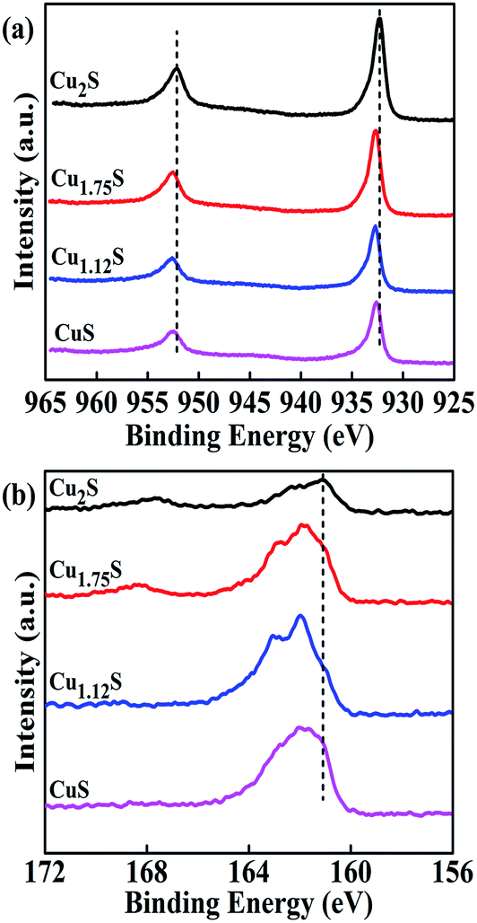 | ||
| Fig. 3 XPS spectra of copper sulfides: (a) Cu 2p and (b) S 2p regions. | ||
Three-dimensional (3D) micrographs of the films were recorded using AFM, as shown in Fig. S2.† The CuS electrode exhibited the lowest root mean square (RMS) surface roughness (43.79 nm) and the films of the Cu2S nanoplatelet and Cu1.75S nanosheet electrodes had intermediate surface roughnesses (RMS = 52.91 nm and 64.27 nm). The roughest film, which had an RMS value of 86.80 nm, was obtained from the Cu1.12S electrode. High superficial roughness improves the catalytic activity of a CE for a polysulfide/sulfide (Sn2−/S2−) or triiodide/iodide (I3−/I−) electrolyte system. Also, the charge transfer resistance at the CE/electrolyte interface would be lower in the cases of CEs based on Cu1.12S nanosphere films, because they offer more electrocatalytically active sites for the reaction of the polysulfide redox couple in the electrolyte.32,35
The growth and variation of nanostructures of CuxS at different concentrations of L-cysteine on the surface of FTO are shown in Fig. 4(a). The presence of carboxyl (–COOH) and amino (–NH2) functional groups in cysteine molecules leads to the formation of hydrogen bonds.39 Hydrogen bonds in the present system act as an “adhesive” to facilitate the self-assembly of small crystallites of cysteine capped with CuI–cysteine complexes, which results in the formation of the nanostructures. When the concentration of L-cysteine added to the solution was 0.005 mM or 0.01 mM, fewer cysteine crystallites capped with CuI–cysteine complexes formed in the solution. Thus, the nanoplatelets and nanosheet-like structures were organized by these organic components via hydrogen bonding interactions. When the concentration of L-cysteine was further increased to 0.02 mM and 0.03 mM, large numbers of cysteine crystallites capped with more CuI–cysteine complexes were assembled into nanospheres and nanoflowers owing to their high concentration and the strong hydrogen bonding interactions among them. Therefore, we concluded that the changes in surface morphology from nanoplatelets to nanosheets to nanospheres to nanoflowers and the thickness of CuxS varied with the concentration of L-cysteine. An improvement in the surface morphology of the CuxS material on the FTO surface would increase the catalytic activity, because the reduction process is responsible for the injection of electrons from the CE into the electrolyte.
 | ||
| Fig. 4 (a) Schematic representation of the L-cysteine-assisted formation of CuxS nanostructures on the surface of FTO substrates. (b) Schematic structure of a TiO2/CdS/CdSe/Mn–ZnSe QDSSC based on a nanosphere-structured Cu1.12S CE. | ||
Fig. 4(b) shows the basic components of a QDSSC. The heart of a QDSSC comprises mesoporous anatase TiO2 nanoparticles deposited on an FTO substrate with a thickness of 7.5 μm. This TiO2 layer is coated with a CdS/CdSe QD photosensitizer. The photoexcitation of electrons occurs from the valence band (VB) to the conduction band (CB) of the QDs, followed by the injection of electrons into the CB of the semiconductor, leaving the QDs in an oxidized state. The oxidized QDs are restored to their original state by the transfer of electrons from the electrolyte, which is known as the QD regeneration process. Regeneration of the QDs by S2− intercepts the recapture of the injected electrons in the CB by the oxidized QDs. Sn2− regenerates S2− by accepting electrons from an external load. In general, electrons flowing from the photoanode to the external load are collected at the CE. The open-circuit voltage (VOC) across the cell is thermodynamically measured by the difference between the quasi-Fermi level (QFL) of the semiconductor and the redox potential of the polysulfide electrolyte.
The absorption spectra of the CuxS thin films are shown in Fig. 5(A) in the wavelength range of 300–900 nm. As shown in Fig. 5(A), the absorbance increased with an increase in the Cu ion concentration of the films, which led to an increase in the thickness of the deposited films. Cu2S and Cu1.75S had absorption minima at 607 and 619 nm, respectively, whereas Cu1.12S and CuS absorbed strongly owing to their high thickness. All the deposited CuxS samples exhibited increased absorption in the longer-wavelength region owing to the intraband absorption of free carriers.47,48
 | ||
| Fig. 5 (A) UV-vis absorption spectra of CuxS thin films. (B) Cyclic voltammograms (CV) of a three-electrode system with a saturated calomel electrode (SCE) as the reference electrode of the CuS electrodes at a scan rate of 100 mV s−1. (C) Nyquist plots of symmetrical dummy cells with (a) Cu2S, (b) Cu1.75S, (c) Cu1.12S, (d) CuS, and (e) Pt electrodes in a polysulfide electrolyte with a composition of 2 M S, 1 M Na2S, and 0.1 M KCl in a solution of methanol and water, which were present in a ratio of 7:3. The frequency range was set from 500 kHz to 100 mHz. Insets in (C) show the high-impedance range for the Pt CE and the equivalent circuit to simulate the EIS curves. (D) Tafel plots of the symmetrical dummy cells fabricated with CuxS and Pt CEs. | ||
To investigate the electrocatalytic activities of the as-prepared CuxS and Pt CEs, CV was performed in a three-electrode system (with Pt mesh and SCE as the counter and reference electrodes, respectively) with an aqueous solution of 0.01 M S, 0.01 M Na2S, and 0.01 M KCl at a scan rate of 100 mV s−1 and the corresponding CV are shown in Fig. 5(B). The current density for the Pt CE was lower than that for the CuxS CEs, because Pt has very low electrocatalytic activity with the polysulfide redox couple. This is caused by its irreversibility and overpotential owing to physisorption.49 The Cu1.12S nanosphere electrode exhibited the highest current density, which accounts for its superior electrocatalytic activity and better photovoltaic performance in a QDSSC. Both Cu2S nanoplatelets and Cu1.75S nanosheets displayed reasonable current densities owing to their moderate electrocatalytic activity, but CuS nanoflowers exhibited much lower electrocatalytic activity. This resulted from its surface morphology, optimized thickness, surface roughness, increased surface-to-volume ratio due to surface uniformity, and also a higher carrier concentration enriched by the presence of optimized sulfur ions.
EIS analysis was conducted with symmetrical dummy cells to determine the electron transport behavior at the CE/electrolyte interface. Fig. 5(C) illustrates the Nyquist plots of CuS and Pt CEs measured in the frequency range of 500 kHz to 100 mHz. The inset shows the equivalent circuit that was used to fit the impedance data curves. In a Nyquist plot, the series resistance (Rs) corresponds to the region of the high-frequency nonzero intercept on the real axis, the left-hand semicircle in the high-frequency region denotes the parallel combination of the charge transfer resistance (Rct) and the constant phase element (CPE) at the CE/electrolyte interface, and the small semicircle in the low-frequency region at the end is assigned to the Warburg impedance (ZW) of the redox couple in the electrolyte.50,51
The values of Rs of the CuS CEs were similar and lower than that of the Pt CE. The Cu1.12S nanosphere CE had the smallest value of Rs, which reflects the strong adhesion of the film to FTO; this allowed fast electrons to be transferred from CuS via FTO into the external circuit. The value of Rs of the Cu1.12S nanosphere CE (8.48 Ω) was lower than those of the Cu2S nanoplatelet (8.50 Ω), Cu1.75S nanosheet (8.51 Ω), CuS nanoflower (8.54 Ω) and Pt (9.33 Ω) CEs, which yielded a slightly higher FF. The value of Rct of Cu1.12S was only 4.02 Ω, which was far lower than the value for Pt (122.25 Ω), which demonstrates that Cu1.12S displayed high catalytic activity for reducing the polysulfide electrolyte. In contrast, the value of Rct of CuS was 5.42 Ω, which demonstrates poor catalytic activity that led to low values of JSC and η. The values of Rct of Cu2S and Cu1.75S were 4.96 and 4.18 Ω, respectively, which indicates that both CEs exhibited reasonable catalytic activity. Furthermore, the values of CPE of the Cu2S, Cu1.75S, Cu1.12S, CuS, and Pt CEs were 522, 578, 744, 388 and 116 μF, respectively. A higher value of CPE corresponds to a larger surface area, which is advantageous for improving the photovoltaic performance of QDSSCs. The values of ZW of the Cu2S, Cu1.75S, Cu1.12S, CuS, and Pt CEs were measured to be 3.41, 3.34, 2.29, 5.7, and 38.94 Ω, respectively. The lower value of ZW of the Cu1.12S CE indicates greater electrolyte diffusion, fast mass transfer of electrons, and improved performance of the QDSSC. The very low values of Rs, Rct, and ZW of the Cu1.12S nanosphere CE in comparison with those of the other CEs also contributed to the high performance of devices fabricated with Cu1.12S as the CE.
To examine the electrochemical catalytic activity of the CuS electrodes, Tafel polarization analysis was performed on the symmetric cells used in EIS. Fig. 5(D) shows the Tafel polarization curves of the CuS and Pt symmetric cells as a function of the voltage (V) vs. the logarithmic current density (logJ). In the plot, the cathodic slope (βc) indicates the reduction of Sn2− to S2− ions, whereas the anodic slope (βa) denotes the oxidation of S2− to Sn2− ions. The Tafel equations for both the cathodic and anodic reactions in a corroding system can be combined to produce the Butler–Volmer equation:52
 | (1) |
The electrochemical catalytic activity of the CEs can be determined from the exchange current density (Jo), which can be calculated from the intercept obtained by extrapolating the cathodic branch of each curve when the overpotential is zero. The slopes of the cathodic and anodic branches for the Cu1.12S electrode are higher than those for the Cu2S, Cu1.75S, CuS, and Pt electrodes, which demonstrates a higher exchange current density (Jo) on the surface of the Cu1.12S electrode. This indicates that the Cu1.12S nanosphere electrode could catalyze the reduction of the polysulfide redox couple as effectively as the Cu2S nanoplatelet, Cu1.75S nanosheet, CuS nanoflower, and Pt electrodes. This observation indicates that the catalytic activity for the reduction of polysulfide was based on the stoichiometry of copper sulfides. The Tafel zone gives information about Jo, which is dependent on the charge transfer resistance (Rct) and is given by the following equation:53
 | (2) |
The limiting current density (Jlim) in the horizontal part can be derived from the high-potential region of the curve. The value of Jlim of the Cu1.12S electrode was higher than those of the Cu2S, Cu1.75S, CuS, and Pt electrodes. A high value of Jlim leads to a higher diffusion coefficient in the reduction of the polysulfide electrolyte, as shown in eqn (3).54 This indicates that the limiting current density depends on the diffusion coefficient of the redox couple electrolyte. The results of this Tafel polarization experiment were consistent with the EIS results.
 | (3) |
To analyze the catalytic activity of the L-cysteine-based CuS CEs in the polysulfide redox electrolyte as hole transporters, we used the CEs in a standard cell device with a CdS/CdSe/Mn–ZnSe photoanode configuration. For a comparative investigation, a QDSSC with a Pt CE was also assembled as a standard cell. Fig. 6(a) shows the resulting J–V curves of the CdS/CdSe/Mn–ZnSe photoanodes with the CuxS CEs recorded under a simulated light source with a power density of 100 mW cm−2. The corresponding open-circuit voltage (VOC), short-circuit current density (JSC), fill factor (FF), and efficiency (η) determined from the J–V curves are summarized in Table 1. Note that QDSSCs based on multiple CuxS cells were fabricated and tested to ensure the reproducibility of our results (see the results of multiple measurements in Table S2 in ESI†).
 | ||
| Fig. 6 (a) J–V characterization of CdS/CdSe/Mn–ZnSe QDSSC devices assembled with L-cysteine-based CuxS CEs and a Pt CE in the presence of a polysulfide electrolyte solution (1 M Na2S, 2 M S, and 0.1 M KCl). (b) Plots of the incident photon-to-current conversion efficiency (IPCE) of fabricated QDSSCs employing CEs based on CuxS and Pt with a S2−/Sn2− redox electrolyte under AM 1.5G simulated solar illumination with an intensity of 100 mW cm−2. | ||
| CE | VOC (V) | JSC (mA cm−2) | FF | η% | Rs (Ω) | RCE (Ω) | Rw (Ω) | Rk (Ω) | Rk/Rw | Ln (μm) | τn (ms) | keff (s−1) | Dn (cm2 s−1) |
|---|---|---|---|---|---|---|---|---|---|---|---|---|---|
| Cu2S | 0.642 | 15.26 | 0.520 | 5.10 | 7.30 | 1.48 | 0.73 | 6.46 | 8.84 | 22.29 | 29.19 | 34.25 | 1.70 × 10⁻⁴ |
| Cu1.75S | 0.642 | 15.85 | 0.522 | 5.32 | 6.35 | 1.33 | 0.65 | 5.81 | 8.98 | 22.47 | 29.45 | 33.95 | 1.71 × 10⁻⁴ |
| Cu1.12S | 0.644 | 17.31 | 0.527 | 5.88 | 5.77 | 1.22 | 0.51 | 5.01 | 9.82 | 23.50 | 30.83 | 32.46 | 1.79 × 10⁻⁴ |
| CuS | 0.641 | 14.44 | 0.527 | 4.88 | 7.94 | 1.52 | 0.84 | 7.25 | 8.63 | 22.03 | 28.96 | 34.53 | 1.67 × 10⁻⁴ |
| Pt | 0.516 | 10.33 | 0.264 | 1.36 | 9.35 | 10.73 | 1.19 | 7.46 | 6.26 | 18.76 | 23.73 | 42.14 | 1.47 × 10⁻⁴ |
The highest photovoltaic performance and power conversion efficiency (η = 5.88%), which were achieved with the Cu1.12S nanosphere CE, were superior to those of the solar cell devices with bare CuS (3.18%),30 Cu2S nanoplatelet (5.10%), Cu1.75S nanosheet (5.32%), and CuS nanoflower (4.88%) CEs. The efficiency of the CuS CEs was much higher than that of the Pt CE (1.36%), as shown in Table 1. For the Pt CE, the values of VOC, JSC, FF, and η were 0.516 V, 10.33 mA cm−2, 0.264, and 1.36%, respectively. The low electrochemical catalytic activity of the Pt CE was due to conjunction with the polysulfide electrolyte, which led to a poisoning effect. In a comparison of the photovoltaic performance of CuxS CEs, an increase in electrocatalytic activity (indicating good agreement with the CV analysis) occurred until the concentration of L-cysteine was 0.02 mM and a drastic reduction occurred above 0.02 mM, which was due to the change in the surface morphology of the CuxS thin films (nanospheres to nanoflowers). As well as the surface morphology, the higher atomic percentage of S in CuS also played a crucial role in improving the electrocatalytic activity of the CE. Even though the CuS CE had a higher atomic percentage of S than the Cu2S, Cu1.75S and Cu1.12S CEs, the photovoltaic performance of the CuS CE was lower than those of the other phases of copper sulfides. This was because the surface morphology of CuS nanoflowers was not efficient for reducing the polysulfide electrolyte at the CE/electrolyte interface (which is consistent with the results of CV, EIS and Tafel analysis).
In general, VOC can be determined from the difference in potential between the Fermi energy level and the potential of the redox couple of the electrolyte. The higher value of VOC (Cu1.12S ≫ Pt) is ascribed to improved electron transfer in the conduction band of TiO2 and the moderately higher resistance when the electrons excited from the Fermi energy level crossed the TiO2 thin film. The values of VOC for all the CuS CEs were similar with negligible variations. Therefore, we conclude from the J–V results that a CuS CE can replace an expensive Pt CE as an efficient CE catalyst in a QDSSC using a polysulfide redox couple electrolyte.
To explain the high photocurrent of QDSSCs based on CuxS CEs, photocurrent action spectra of the QDSSCs were recorded. Fig. 6(b) shows the incident photon-to-electron conversion efficiency (IPCE) spectra of typical QDSSCs based on different CEs, including Cu2S, Cu1.75S, Cu1.12S, CuS and Pt. Apparently, the high photocurrent of cells based on CuxS in comparison with those based on Pt arose from the increase in the IPCE peak in the wavelength range of 380 nm to 650 nm. As IPCE is generally determined from the yields of light harvesting, charge separation and collection, the increase in the IPCE peak here must be due to the improved performance of the photoanode. The peak in the IPCE spectrum of the QDSSC based on Cu1.12S was slightly higher than those of the QDSSCs based on Cu2S, Cu1.75S, and CuS. The IPCE results agreed well with the aforementioned values of JSC of the QDSSCs (Table 1). The increase in the value of JSC for the QDSSC based on Cu1.12S was mainly attributed to its greater electrocatalytic ability for the reduction of polysulfide, which was further confirmed by the following electrochemical analysis, namely, measurements by electrochemical impedance spectroscopy (EIS) and Tafel polarization.
To further investigate electron transport in the QDSSCs based on CuxS CEs, their charge transport properties were determined using EIS measurements under one-sun illumination (AM 1.5G, 100 mW cm−2) by applying a 10 mV AC signal over the frequency range of 0.1 Hz to 500 kHz at a bias of VOC. The measured Nyquist plots of the QDSSCs are shown in Fig. 7(a) and the plots were fitted using the diffusion–recombination model (Fig. 7(b)) proposed by Bisquert.55 The Nyquist plots mainly contain three regions: the high-frequency region represents the parallel combination of the charge transfer resistance (RCE) of the counter electrode and Helmholtz capacitance (CCE) at the CE/electrolyte interface; the middle-frequency region represents the combination of electron transport resistance (Rw = rwL), recombination resistance (Rk = rk/L) and chemical capacitance (Cμ = CμL) at the TiO2/QDs/electrolyte interface; and the low-frequency region corresponds to the Warburg impedance or diffusion resistance (ZN) of the polysulfide redox couple.55,56
 | ||
| Fig. 7 (a) Nyquist plots of fabricated QDSSCs based on CuxS recorded under AM 1.5G solar illumination of 100 mW cm−2. (b) General transmission line model for simulation of the impedance spectra of QDSSCs. (c) Open-circuit voltage decay of QDSSCs with different CuxS nanostructured CEs. (d) Electron lifetime (in log-linear representation) as a function of VOC. | ||
The nonzero intercept on the real axis of the impedance plot represents the series resistance (Rs), which represents the sheet resistance of the FTO substrate. The results of fitting are summarized in Table 1. The following equations can be used to estimate the important parameters of the electron lifetime (τn), diffusion length of electrons (Ln), effective diffusion coefficient (Dn) and reaction rate constant (keff) for electron recombination:
| τn = RkCμ | (4) |
 | (5) |
 | (6) |
 | (7) |
In order to determine the electron lifetime and charge recombination properties of the resulting cell devices, measurements of open-circuit voltage decay (OCVD) were performed. Fig. 7(c) shows plots of the VOC decay rate as a function of time under open-circuit conditions. The plots show that the Cu1.12S cell exhibited a lower voltage decay rate than those of the other CE-based QDSSCs (Cu2S, Cu1.75S, CuS and Pt), which indicated that the recombination rate of photogenerated electrons with oxidized species in the electrolyte was significantly reduced in the Cu1.12S cell. This result is in agreement with those of the EIS study shown above. The decay of voltage with time gives the electron lifetime according to the following equation:57
 | (8) |
In order to examine the stability of the Cu1.12S CE in a polysulfide electrolyte, the assembled QDSSC that incorporated the Cu1.12S CE was disassembled after storage for 5 days and then examined by X-ray diffraction. Fig. S3† shows the XRD patterns of the freshly prepared Cu1.12S CE and disassembled Cu1.12S CE. In comparison with the fresh Cu1.12S CE, it can be seen that the diffraction peaks of Cu1.12S still remained after immersion for 5 days in a polysulfide electrolyte, which indicates that the Cu1.12S catalyst was stable in the polysulfide electrolyte.
The device stability of the best performing QDSSC, which was based on the Cu1.12S nanosphere CE, was examined by exposing it to continuous light illumination for 20 h. Fig. 8 shows stability plots for the Cu1.12S and Pt CEs, which were used to compare the photovoltaic parameters (VOC, JSC, FF, and η). The Cu1.12S CE exhibited little decline in VOC, whereas η, FF, and JSC did not decrease significantly and were nearly constant during irradiation for 20 h. For the first 20 h, not much decline was observed in the photovoltaic parameters because of a capillary effect, which involved slow penetration of the polysulfide redox solution into the pores of TiO2 and an increase in ion transport caused by heating the electrolyte. The value of η of the Cu1.12S CE indicated almost constant efficiency during illumination for 20 h. In contrast, the conversion efficiency of the Pt CE was stable until 12 h and then decreased over the next 8 h from 1.38% to 1.04%. During the measurements, the VOC of the Cu1.12S CE maintained a constant value of 0.642 V. There was a slight increase in JSC from 17.31 to 17.44 mA cm−2 and an increase in FF from 0.529 to 0.538 over the course of illumination for 20 h. During illumination, gains in FF and JSC compensated for losses in VOC, which resulted in almost constant efficiency (5.88 to 6.04%). The increases in power conversion efficiency and performance were due to the positive influence of the Cu1.12S protective layer in comparison with the Pt CE, which improved the electrocatalytic activity of the electrode with the polysulfide electrolyte. All these results indicate the superior photovoltaic performance of the QDSSC based on the Cu1.12S CE and better stability than that of the Pt CE. The good photovoltaic performance of the Cu1.12S CE is ascribed to its enhanced catalytic activity owing to its improved surface morphology, optimized thickness, and optimized atomic percentage of S, which enabled better reduction of the polysulfide electrolyte, and low charge transfer resistance at the CE/electrolyte interface, which resulted in a high FF.
 | ||
| Fig. 8 Temporal evolution of values of the photovoltaic parameters VOC (a), JSC (b), FF (c), and η (d) for Cu1.12S and Pt CEs in CdS/CdSe/Mn–ZnSe QDSSCs under continuous illumination of 100 mW cm−2 over the course of 20 h. | ||
4. Conclusions
In summary, highly electrocatalytically active and phase-controlled CuxS CEs were fabricated on FTO in the presence of L-cysteine using a low-cost and environmentally friendly CBD technique. These were directly used in QDSSCs without any post-treatment. The ratio of Cu/S, stoichiometries (Cu2S, Cu1.75S, Cu1.12S and CuS), thickness, and surface morphology (nanoplatelets, nanosheets, nanospheres and nanoflowers) were dependent on the concentration of L-cysteine. L-Cysteine-based Cu1.12S nanospheres could display high roughness, which favored electrocatalytic performance. In comparison with a Pt CE, the L-cysteine-based CuxS CEs exhibited superior electrocatalytic activity for catalyzing the reduction of a polysulfide electrolyte, contributing to obvious increases in JSC and FF. The optimized L-cysteine concentration in the preparation of CuxS was 0.02 mM. A cell device with a Cu1.12S nanosphere CE achieved a value of η of up to 5.88%, which was superior to those of Cu2S nanoplatelet (5.10%), Cu1.75S nanosheet (5.32%), CuS nanoflower (4.88%) and Pt (1.36%) CEs. Furthermore, the Cu1.12S CE exhibited excellent stability in comparison with that of a Pt CE in a sulfide/polysulfide electrolyte redox system. This strategy paves the way for fabricating stable, cheap, and highly efficient CEs for QDSSCs, which can further improve the performance of QDSSCs via the optimization of the CuxS CE.Conflict of interest
The authors declare no competing financial interest. The samples were analyzed by a transmission electron microscope (TEM, JEM-2011) and a scanning electron microscope (SEM, Hitachi S-4200) with a 4k × 4k CCD camera (Ultra Scan 400SP, Gatan Corp.) at the Busan KBSI, South Korea.Acknowledgements
This research was supported by the Basic Research Laboratory through the National Research Foundations of Korea funded by the Ministry of Science, ICT and Future Planning (NRF-2015R1A4A1041584).References
- W. J. Yue, S. K. Han, R. X. Peng, W. Shen, H. W. Geng, F. Wu, S. W. Tao and M. T. Wang, J. Mater. Chem., 2010, 20, 7570–7578 RSC.
- B. O'Regan and M. Grätzel, Nature, 1991, 353, 737–740 CrossRef.
- J. Wu, Z. Lan, J. Lin, M. Huang, Y. Huang, L. Fan and G. Luo, Chem. Rev., 2015, 115, 2136–2173 CrossRef CAS PubMed.
- M. A. Green and K. Emery, Progress in Photovoltaics: Research and Applications, 1993, 1, 25–29 CrossRef CAS.
- P. V. Kamat, J. Phys. Chem. C, 2008, 112, 18737–18753 CAS.
- P. V. Kamat, Acc. Chem. Res., 2012, 45, 1906–1915 CrossRef CAS PubMed.
- A. J. Nozik, Phys. E, 2002, 14, 115–120 CrossRef CAS.
- Z. X. Pan, K. Zhao, J. Wang, H. Zhang, Y. Y. Feng and X. H. Zhong, ACS Nano, 2013, 7, 5215–5222 CrossRef CAS PubMed.
- W. W. Yu and X. Peng, Angew. Chem., Int. Ed., 2002, 41, 2368–2371 CrossRef CAS.
- R. T. Ross and A. J. Nozik, J. Appl. Phys., 1982, 53, 3813–3818 CrossRef CAS.
- W. W. Yu, L. H. Qu, W. Z. Guo and X. G. Peng, Chem. Mater., 2003, 15, 2854–2860 CrossRef CAS.
- R. Vogel, P. Hoyer and H. Weller, J. Phys. Chem., 1994, 98, 3183–3188 CrossRef CAS.
- M. C. Hanna and A. J. Nozik, J. Appl. Phys., 2006, 100, 074510 CrossRef.
- Y. Luo, D. Li and Q. Meng, Adv. Mater., 2009, 21, 4647–4651 CrossRef CAS.
- C. V. V. M. Gopi, M. V. Haritha, S. K. Kim and H. J. Kim, Dalton Trans., 2015, 44, 630–638 RSC.
- S. Giménez, I. M. Seró, L. Macor, N. Guijarro, T. L. Villarreal, R. Gómez, L. J. Diguna, Q. Shen, T. Toyoda and J. Bisquert, Nanotechnology, 2009, 20, 295204–295209 CrossRef PubMed.
- J. J. Tian, T. Shen, X. G. Liu, C. B. Fei, L. L. Lv and G. Cao, Sci. Rep., 2016, 6, 23094 CrossRef CAS PubMed.
- Z. Pan, I. M. Sero, Q. Shen, H. Zhang, Y. Li, K. Zhao, J. Wang, X. Zhong and J. Bisquert, J. Am. Chem. Soc., 2014, 136, 9203–9210 CrossRef CAS PubMed.
- J. Yang, J. Y. Kim, J. H. Yu, T. Y. Ahn, H. Lee, T. S. Choi, Y. W. Kim, J. Joo, M. J. Ko and T. Hyeon, Phys. Chem. Chem. Phys., 2013, 15, 20517 RSC.
- G. Wang, H. Wei, Y. Luo, H. Wu, D. Li, X. Zhong and Q. Meng, J. Power Sources, 2016, 302, 266–273 CrossRef CAS.
- V. G. Pedro, X. Xu, I. M. Mora-Seró and J. Bisquert, ACS Nano, 2010, 4, 5783–5790 CrossRef PubMed.
- A. Kay and M. Gratzel, Sol. Energy Mater. Sol. Cells, 1996, 44, 99–117 CrossRef CAS.
- J. Yang, J. Wang, K. Zhao, T. Izuishi, Y. Li, Q. Shen and X. Zhong, J. Phys. Chem. C, 2015, 119, 28800–28808 CAS.
- J. Du, Z. Du, J. S. Hu, Z. Pan, Q. Shen, J. Sun, D. Long, H. Dong, L. Sun, X. Zhong and L. J. Wan, J. Am. Chem. Soc., 2016, 138, 4201–4209 CrossRef CAS PubMed.
- K. Kakiage, Y. Aoyama, T. Yano, K. Oya, J. I. Fujisawa and M. Hanaya, Chem. Commun., 2015, 51, 15894–15897 RSC.
- M. Samadpour, A. Irajizad, N. Taghavinia and M. A. Molaei, J. Phys. D: Appl. Phys., 2011, 44, 045103 CrossRef.
- P. Santra and P. V. Kamat, J. Am. Chem. Soc., 2012, 134, 2508–2511 CrossRef CAS PubMed.
- Y. C. Yang, S. L. Yau and Y. L. Lee, J. Am. Chem. Soc., 2006, 128, 3677–3682 CrossRef CAS PubMed.
- M. Ye, C. Chang, N. Zhang, X. Wen, W. Guo and C. Lin, Adv. Energy Mater., 2014, 4, 1301564 CrossRef.
- H. J. Kim, M. S. Lee, C. V. V. M. Gopi, M. V. Haritha, S. S. Rao and S. K. Kim, Dalton Trans., 2015, 44, 11340–11351 RSC.
- A. D. Savariraj, K. K. Viswanathan and K. Prabakar, ACS Appl. Mater. Interfaces, 2014, 6, 19702–19709 CAS.
- M. L. Que, W. X. Guo, X. J. Zhang, X. Y. Li, Q. L. Hua, L. Dong and C. F. Pan, J. Mater. Chem. A, 2014, 2, 13661–13666 CAS.
- C. V. V. M. Gopi, S. S. Rao, S. K. Kim, D. Punnoose and H. J. Kim, J. Power Sources, 2015, 275, 547–556 CrossRef CAS.
- Y. Chen, X. Zhang, Q. Tao, W. Fu, H. Yang, S. Su, Y. Mu, L. Zhou and M. Li, RSC Adv., 2015, 5, 1835–1840 RSC.
- Y. Zhang, C. Shi, X. Dai, F. Liu, X. Fang and J. Zhu, Electrochim. Acta, 2014, 118, 41–44 CrossRef CAS.
- Q. Zhang, Y. Zhang, S. Huang, X. Huang, Y. Luo, Q. Meng and D. Li, Electrochem. Commun., 2010, 12, 327–330 CrossRef CAS.
- V. D. Dao, Y. Choi, K. Yong, L. L. Larina and H. S. Choi, Carbon, 2015, 84, 383–389 CrossRef CAS.
- V. D. Dao, P. Kim, S. Baek, L. L. Larina, K. Yong, R. Ryoo, S. H. Ko and H. S. Choi, Carbon, 2016, 96, 139–144 CrossRef CAS.
- B. Li, Y. Xie and Y. Xue, J. Phys. Chem. C, 2007, 111, 12181–12187 CAS.
- L. Jiang and Y. J. Zhu, Mater. Lett., 2009, 63, 1935–1938 CrossRef CAS.
- H. J. Kim, D. J. Kim, S. S. Rao, A. D. Savariraj, S. K. Kim, M. K. Son, C. V. V. M. Gopi and K. Prabakar, Electrochim. Acta, 2014, 127, 427–432 CrossRef CAS.
- S. J. Bao, Y. Li, C. M. Li, Q. Bao, Q. Lu and J. Guo, Cryst. Growth Des., 2008, 8, 3745–3749 CAS.
- L. Y. Chen, Z. D. Zhang and W. Z. Wang, J. Phys. Chem. C, 2008, 112, 4117–4123 CAS.
- A. Hauch and A. Georg, Electrochim. Acta, 2001, 46, 3457–3466 CrossRef CAS.
- G. Hodes, J. Manassen and D. Cahen, J. Electrochem. Soc., 1980, 127, 544–549 CrossRef CAS.
- V. Krylova and M. Andrulevicius, Int. J. Photoenergy, 2009, 304308 Search PubMed.
- Y. Zhao, H. Pan, Y. Lou, X. Qiu, J. Zhu and C. Burda, J. Am. Chem. Soc., 2009, 131, 4253–4261 CrossRef CAS PubMed.
- S. Eda, K. Moriyasu, M. Fujishima, S. Nomura and H. Tada, RSC Adv., 2013, 3, 10414–10419 RSC.
- J. G. Radich, R. Dwyer and P. V. Kamat, J. Phys. Chem. Lett., 2011, 2, 2453–2460 CrossRef CAS.
- F. Liu, J. Zhu, Y. Xu, L. Zhou, Y. Li, L. Hu, J. Yao and S. Dai, Chem. Commun., 2015, 51, 8108–8111 RSC.
- H. Chae, D. Song, Y. G. Lee, T. Son, W. Cho, Y. B. Pyun, T. Y. Kim, J. H. Lee, F. F. Santiago, J. Bisquert and Y. S. Kang, J. Phys. Chem. C, 2014, 118, 16510–16517 CAS.
- D. K. Yadav, D. S. Chauhan, I. Ahamad and M. A. Quraishi, RSC Adv., 2013, 3, 632–646 RSC.
- M. Wu, X. Lin, A. Hagfeldt and T. Ma, Angew. Chem., Int. Ed., 2011, 50, 3520–3524 CrossRef CAS PubMed.
- M. K. Wang, A. M. Anghel, B. Marsan, N. C. Ha, N. Pootrakulchote, S. M. Zakeeruddin and M. Gratzel, J. Am. Chem. Soc., 2009, 131, 15976–15977 CrossRef CAS PubMed.
- Q. Wang, S. Ito, M. Gratzel, F. Fabregat-Santiago, I. Mora-Sero, J. Bisquert, T. Bessho and H. Imai, J. Phys. Chem. B, 2006, 110, 25210–25221 CrossRef CAS PubMed.
- C. V. V. M. Gopi, M. V. Haritha, Y. S. Lee and H. J. Kim, J. Mater. Chem. A, 2016, 4, 8161–8171 CAS.
- J. Bisquert, A. Zaban, M. Greenshtein and I. Mora-Seró, J. Am. Chem. Soc., 2004, 126, 13550–13559 CrossRef CAS PubMed.
Footnotes |
| † Electronic supplementary information (ESI) available. See DOI: 10.1039/c6ra23763k |
| ‡ These authors have made an equal contribution to this work. |
| This journal is © The Royal Society of Chemistry 2016 |