DOI:
10.1039/C6RA23757F
(Paper)
RSC Adv., 2016,
6, 106150-106159
Synthesis and anti-proliferative evaluation of novel 3,4-dihydro-2H-1,3-oxazine derivatives of bakuchiol†‡
Received
24th September 2016
, Accepted 14th October 2016
First published on 14th October 2016
Abstract
A new series of 3,4-dihydro-2H-1,3-oxazine derivatives of bakuchiol 1 was synthesized through the Mannich-type condensation–cyclization reaction of 1 with formaldehyde and appropriate primary amines. On cytotoxicity evaluation against a panel of four human cancer cell lines, most of the derivatives showed a higher cytotoxic profile than the parent molecule. The best results were observed for compound 15 with IC50 values of 2, 2, 2.4 and 3 μM against MIA-Pa-Ca-2, HCT-116, MCF-7 and HL-60 cells, respectively. A mechanistic study of compound 15 revealed that it caused a loss in the mitochondrial membrane potential in a concentration-dependent manner, accompanied by the activation of caspase-9 and -3, which cleave PARP-1. It also activated caspase-8, which is involved in the extrinsic apoptotic pathway. Therefore, we demonstrated that it induces apoptosis via both intrinsic and extrinsic pathways in human pancreatic cancer MIA-Pa-Ca-2 cells.
Introduction
Natural products (NPs) are an indispensable source of lead structures for drug discovery and development for the treatment of a wide spectrum of diseases.1 Within the sphere of cancer, out of 175 U.S. FDA approved drug entities in the time frame from the 1940s to the end of 2014, 75% were other than synthetic and 49% were either NPs or derived from NPs.2 Furthermore, NPs derived from plants as such or their derivatives have played a vital role in the therapeutic area of cancer, and hence a large number of new chemical entities (NCEs) are in different stages of clinical development.3
Bakuchiol 1, a meroterpene isolated from Psoralea corylifolia (Leguminosae), is known for its various pharmacological activities.4–6 However, the cytotoxicity of bakuchiol has attracted considerable interest as it inhibits the proliferation of various human cancer cells, such as breast (MCF-7), prostate (PC-3), cervical (HeLa), gastric (AGS), lung (A-549), CNS (IMR-32), ovarian (OVCAR-5) and leukaemia (THP-1).7,8,10 The anticancer mechanism of its action involves a loss in the mitochondrial membrane potential, S-phase arrest, caspase 9/3 activation and DNA fragmentation as well as the inhibition of hypoxia-inducible factor-1 (HIF-1) and nuclear factor kappa B (NFκB) in a number of human cancer cell lines.8–10 The natural product 1 has also been found to facilitate tumour necrosis factor related apoptosis inducing ligand (TRAIL) induced apoptosis in human colon cancer cells (HCT-116 and HT-29).11 The key functionalities, such as phenolic-OH, aryl, vinyl and isopropylidene groups, embodied in bakuchiol make it amenable for a variety of chemical transformations. To date, several reports are available wherein considerable structural modification has been done on bakuchiol for the improvement of its anticancer activity.7,10,12–14 A literature survey revealed that 3,4-dihydro-2H-benzo[e][1,3]oxazine moieties exhibit a broad range of pharmacological activities, including anticancer, anti-inflammatory and anti-infective,15–18 which shows their large development value and wide potential as therapeutic agents. Therefore, in a continuation of our interest in the structural modification of natural products for the development of anticancer leads,7,19,20 herein, we report the synthesis of new 3,4-dihydro-2H-1,3-oxazine derivatives of bakuchiol by a Mannich-type condensation–cyclization reaction and their in vitro cytotoxicity evaluation against four human cancer cell lines (MIA-Pa-Ca-2, HCT-116, MCF-7 and HL-60). A mechanistic study was also carried out for the most potent analogue.
Results and discussion
Chemistry
Bakuchiol 1 was isolated in a preparative scale from the seeds of Psoralea corylifolia and then taken for structural modification. The 3,4-dihydro-2H-benzo[e][1,3]oxazines analogues (2–26) were prepared by the treatment of 1 with 37% formaldehyde (w/v) and the respective primary amines in tetrahydrofuran (THF) via a Mannich-type condensation–cyclization reaction (Scheme 1) in good to excellent yields. The structures of all the derivatives were confirmed by spectroscopic techniques (1H NMR, 13C NMR, IR and HRMS). In the 1H NMR, the characteristic singlet of two methylene protons of the 1,3-oxazine ring appeared in the ranges of δ 3.8–4.6 and δ 4.6–5.3, corresponding to Ar–CH2–N and O–CH2–N groups, respectively. In the 13C NMR, Ar–CH2–N carbon appeared at δ 51, while O–CH2–N carbon appeared at δ 81. The IR spectrum showed the characteristic absorption peaks of the benzoxazine ring structure at 1230 cm−1, corresponding to the asymmetric stretching of C–O–C, and at 1018 cm−1, corresponding to the symmetric stretching of C–O–C. Also, C–N stretching absorptions were observed in the range of 1200–1400 cm−1, with the aromatic C–N absorptions at a higher frequency than the aliphatics. Further confirmation for the formation of derivatives 2–26 was also done by HRMS data. The typical synthetic procedure and spectral data of all the derivatives are presented in the Experimental section.
 |
| | Scheme 1 Preparation of 3,4-dihydro-2H-1,3-oxazine derivatives (2–26) of bakuchiol. | |
Biology
Cell growth inhibition. Compound 1 and its synthesized derivatives were screened against a panel of four human cancer cell lines, namely HL-60 (leukaemia), MCF-7 (Breast), HCT-116 (colon) and MIA-Pa-Ca-2 (pancreatic), to evaluate their cytotoxic potential using an MTT assay and the results are summarized in Table 1. In the primary screening, bakuchiol (1) showed only 16–36% growth inhibition at 10 μM concentration, while many of its derivatives displayed ≥90% inhibition at the same concentration against all the experimental cancer cell lines. All the compounds (2–26) showed better growth inhibition compared to the parent molecules against the MIA-Pa-Ca-2 cell lines, while compounds 7, 10, 12–19 and 22–26 showed better inhibition effects against the HL-60 cell lines. Compounds 3, 6–8, 11–19 and 22–26 showed better inhibition effects against MCF-7, while compounds 3, 7, 12–19 and 22–26 showed better inhibition effects against the HCT-116 cell lines compared to the parent molecule. Compounds 7, 12–19 and 22–26 displayed potent cytotoxic effects against all the experimental cancer cell lines. For the HL-60 cell line, maximum effect was observed for two compounds, namely 23 and 24 (99% inhibition); while for the MCF-7 cell line, compounds 7 and 23 displayed the maximum inhibition of 94%. In the case of the colon cancer cell line (HCT-116), compound 15 exhibited the maximum inhibition effects (90%); while in the pancreatic cancer cell line, five compounds, namely 7, 12, 13, 14 and 15, displayed the maximum inhibitions of 96%. Overall, the maximum growth inhibition effects (>90%) against all the cell lines were displayed by compound 15, as can be seen from Table 1.
Table 1 Cytotoxic activity of bakuchiol (1) and its derivatives (2–26) at 10 μM concentrationa
| Cell lines% growth inhibition |
| Compound |
HL-60 |
MCF-7 |
HCT-116 |
MIA-Pa-Ca-2 |
| Bold value indicates ≥50% growth inhibition against the cancer cell lines. |
| 1 |
36 |
16 |
34 |
33 |
| 2 |
1 |
9 |
33 |
47 |
| 3 |
14 |
28 |
39 |
73 |
| 4 |
7 |
14 |
27 |
46 |
| 5 |
13 |
4 |
19 |
57 |
| 6 |
8 |
25 |
17 |
47 |
| 7 |
91 |
94 |
89 |
96 |
| 8 |
6 |
24 |
33 |
48 |
| 9 |
27 |
14 |
16 |
63 |
| 10 |
48 |
4 |
25 |
64 |
| 11 |
14 |
20 |
28 |
65 |
| 12 |
91 |
73 |
89 |
96 |
| 13 |
90 |
93 |
83 |
96 |
| 14 |
92 |
78 |
88 |
96 |
| 15 |
91 |
93 |
90 |
96 |
| 16 |
89 |
88 |
88 |
94 |
| 17 |
87 |
93 |
88 |
93 |
| 18 |
86 |
92 |
88 |
94 |
| 19 |
89 |
93 |
88 |
93 |
| 20 |
1 |
14 |
20 |
46 |
| 21 |
1 |
8 |
30 |
44 |
| 22 |
75 |
55 |
73 |
92 |
| 23 |
99 |
94 |
89 |
93 |
| 24 |
99 |
93 |
88 |
93 |
| 25 |
88 |
93 |
86 |
94 |
| 26 |
87 |
93 |
87 |
94 |
The structure activity relationship of the bakuchiol analogues based on growth inhibition can be summarized as follows: aliphatic primary amine analogues (7, 12–16, 23–26) showed better anticancer effects compared to the parent molecule (1) and aromatic primary amine analogues (2–6, 8–11, 20, 21). Among the aliphatic straight chain primary amine analogues, the cytotoxic activity was found to be enhanced with the increase in the number of carbon atoms along the chain length as a better inhibitory effect was observed for the octyl amine analogue (15) compared to the methyl amine analogue (14). Branched chain aliphatic primary amines analogues (25 and 26) also provided significant cytotoxicity results against all the cell lines examined. Alicyclic (17) and aralkyl (18 and 19) primary amine analogues were found to be more potent against MIA-Pa-Ca-2 and MCF-7 than against the other two cell lines tested. The hydroxyalkyl group-containing analogue (23) also sensitized all the experimental cell lines, with HL-60 the most affected.
Furthermore, the compounds that showed growth inhibition of >50% at 10 μM concentration were screened at four different concentrations (0.1, 1, 5 and 10 μM) along with compound 1 for calculation of their IC50 values. The calculated IC50 of the selected compounds are provided in Table 2. Promising IC50 values (2.0 to 7.0 μM) were observed for compounds 7, 12–19 and 23–26 against all the cell lines examined (HL-60, MIA-Pa-Ca-2, MCF-7 and HCT-116). Compound 15 showed a better cytotoxic profile (2.0 to 3.0 μM) against all the selected cell lines. The pancreatic cell line (MIA-Pa-Ca-2) proved the most sensitive (2.0 to 3.0 μM) towards semi-synthetic analogues, where, in particular, compound 15 exhibited the maximum cytotoxic effect, with an IC50 value of 2.0 μM and hence it was chosen for the further cell death mechanistic study.
Table 2 Ic50 values in μM of bakuchiol and its selected analogues on selected human cancer cell lines
| Compound |
Breast MCF-7 |
Pancreatic MIA-Pa-Ca-2 |
Colon HCT-116 |
Leukaemia HL-60 |
| 1 |
>10 |
>10 |
>10 |
>10 |
| 7 |
3 |
3 |
3.6 |
6 |
| 12 |
3.6 |
3 |
5.5 |
5 |
| 13 |
3 |
2.5 |
2.5 |
6 |
| 14 |
6 |
3 |
6.5 |
7 |
| 15 |
2.4 |
2 |
2 |
3 |
| 16 |
5.2 |
3 |
4 |
6 |
| 17 |
3.6 |
3 |
3 |
3 |
| 18 |
5 |
3 |
3.7 |
3 |
| 19 |
3.2 |
3 |
2.5 |
3 |
| 23 |
6 |
3 |
6 |
3 |
| 24 |
6 |
3 |
3.7 |
4 |
| 25 |
6 |
3 |
3.8 |
5 |
| 26 |
3 |
3 |
4 |
6 |
Compound 15 increases sub-G1 (G0) apoptotic population in MIA-Pa-Ca-2 cells. The extent of apoptotic cell death in MIA-Pa-Ca-2 cells was assessed using flow cytometry through the determination of a sub-G1 cell population by propidium iodide (PI) staining. As depicted in Fig. 1, the percentage of apoptotic cells exposed to compound 15 increased in a concentration-dependent manner after 24 h of incubation. The sub-G1 (G0) apoptotic population was found to be 3, 4 and 25% following 1, 3 and 10 μM concentration of compound 15 treatment compared to the control (untreated cells, 2%).
 |
| | Fig. 1 MIA-Pa-Ca-2 cells (2 × 10⁶) were seeded in 6-well plates and treated with different concentrations of compound 15 (1 μM, 3 μM, and 10 μM) for 24 h to determine the DNA fluorescence and cell cycle phase distribution, as described in the Experimental section. Data were analyzed by Modfit software (Verity Software House Inc., Topsham, ME) for the proportions of the different cell cycle phases. The fraction of cells from the apoptosis, G1, S and G2 phases analyzed from FL2-A vs. cell counts are shown in %. Data are representative of one of three similar experiments. | |
Compound 15 induces apoptotic bodies. The pancreatic cancer cells (MIA-Pa-Ca-2) were treated with compound 15 at 1, 3 and 10 μM concentrations for 24 h and observed under a microscope for any morphological changes that occur during apoptosis. Simultaneously, the nuclear morphology was analyzed through Hoechst staining. Characteristic changes of apoptosis, such as nuclear condensation, membrane blebbing and the formation of apoptotic bodies, were observed in the morphology of the treated cells in a concentration-dependent manner, whereas the nuclei of untreated cells were found to be of normal intact morphology (Fig. 2). These results suggested that compound 15 was able to induce apoptotic cell morphology in MIA-Pa-Ca-2 cells.
 |
| | Fig. 2 Effect of compound 15 on the cellular and nuclear morphology of MIA-Pa-Ca-2 cells. Cells were visualized for cellular and nuclear morphology as described in the Experimental section. The condensed nuclei and the apoptotic bodies are indicated by white arrows. Data are representative of one of three similar experiments, and magnification of the pictures was 30× on an Olympus 1× 70 inverted microscope. | |
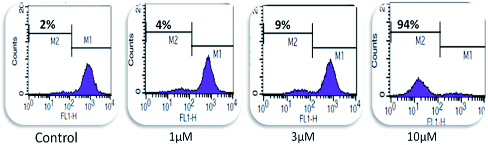
Compound 15 triggers mitochondrial membrane potential loss. Mitochondrial membrane potential (MMP) loss is a key step in the induction of apoptosis in cancer cells. The loss of MMP leads to the depolarization of mitochondrial membrane, resulting in mitochondrial dysfunctioning and ultimately in cell death. The disruption of mitochondrial membrane releases a variety of proteins, which activates procaspase cascade inside the cells and triggers apoptosis.21 The MMP loss in the treated and untreated cells was measured by rhodamine-123 dye (Rh-123), which is reduced by healthy mitochondria into a fluorescent probe whose fluorescence can be measured by a flow cytometer in the FL-1 channel. Compound 15 caused MMP loss in a concentration-dependent manner (Fig. 3). At 1 and 3 μM concentrations, it caused a 4% and 9% loss in the MMP, respectively, while at 10 μM concentration, it caused a 94% loss in the MMP.
 |
| | Fig. 3 Compound 15 induced mitochondrial potential loss in pancreatic cancer MIA-Pa-Ca-2 cells. Cells were treated with compound 15 at 1, 3 and 10 μM concentrations for a 24 h time period. Thereafter, the cells were stained with rhodamine-123 (200 nM), added 40 min before the experiment termination, and analyzed in the FL-1 channel of a flow cytometer. Data are representative of one of three similar experiments. | |
Compound 15 triggers extrinsic and intrinsic apoptosis in MIA-Pa-Ca-2 cells. Compound 15 caused a loss in the MMP. Damaged mitochondria relay signals to downstream elements that further initiate intrinsic apoptotic signals. During apoptosis, cytochrome c is released from the mitochondria, which form an essential part of the apoptosome. This results in the activation of caspase-9, which then processes and activates other caspases.22 The role of caspases (a family of intracellular cysteine proteases) in apoptosis is well known and active caspase-8 or -9 activate the various effector caspases -3, -6 and -7, which cleave the several key proteins required for cellular functioning and survival, where PARP-1 is one of the several known substrates of caspases. The cleavage of PARP-1 by caspases is considered a hallmark of apoptosis.23 Compound 15 activated these caspases and PARP-1 cleavage. It inhibited both procaspase-9 and procaspase-8, which indicates that compound 15 triggers apoptosis via both extrinsic and intrinsic pathways in MIA-Pa-Ca-2 cells (Fig. 4).
 |
| | Fig. 4 Influence of compound 15 on the expression of important proteins involved in the initiation of apoptosis. Cells were treated with 1 and 10 μM concentrations of compound 15 for 24 h. Protein lysates were prepared and electrophoresis was carried out as described in the Experimental section. β-Actin was used as an internal control to represent the same amount of proteins applied for SDS-PAGE. Western blot analyses of the indicated proteins were performed in the whole cell lysate. Data are representative of one of three similar experiments. | |
Experimental
Chemistry
All the reagents and solvents for the synthesis were purchased from Sigma-Aldrich. All the chemical reactions were monitored by TLC on silica gel 60 F254 plates (E. Merck) using 2% ceric ammonium sulphate solution as the spraying reagent for detection of the spots. Purification of all the derivatives was carried out by column chromatography using silica gel 60–120 mesh as the stationary phase. All the NMR spectra were recorded on Bruker DPX 400 and DPX 500 instruments using CDCl3 as the solvent and taking TMS as the internal standard. The chemical shifts are expressed herein in δ and the coupling constant in Hertz. High Resolution Mass Spectra (HRMS) were recorded on an Agilent Technologies 6540 instrument, while the IR spectra were recorded on an FT-IR Bruker (270-30) spectrophotometer.
Isolation of bakuchiol (1)
Bakuchiol was isolated in bulk quantity from a DCM![[thin space (1/6-em)]](https://www.rsc.org/images/entities/char_2009.gif) :MeOH (1:1) extract of seeds of Psoralea corylifolia and characterized by spectroscopic techniques, as previously reported by us.7
:MeOH (1:1) extract of seeds of Psoralea corylifolia and characterized by spectroscopic techniques, as previously reported by us.7
General experimental procedure for the preparation of 3,4-dihydro-2H-1,3-oxazinederivatives (2–26) of bakuchiol
Compounds 2–26 were synthesized by a portion-wise addition of the appropriate primary amine (0.39 mmol) in 3 mL of THF containing 37% aqueous formaldehyde (0.78 mmol), followed by the addition of methanolic solution of bakuchiol (100 mg, 0.39 mmol). The reaction mixture was refluxed for 2–8 h till completion (as monitored by TLC analysis).24 Work-up of the reaction was carried out by diluting the reaction mixture with ice-cold water and extracting it with ethyl acetate (3 times). The combined organic layers were dried over sodium sulphate and concentrated on a rotavapour. The crude product obtained was purified by column chromatography on silica gel 60–120 mesh with EtoAc:hexane (1:49) as the eluent to afford the desired pure products 2–26 in 80–85% yields. The spectral data of all the derivatives (2–26) are given below.
Synthesis of 3,4-dihydro-6-(3,7-dimethyl-3-vinylocta-1,6-dienyl)-3-phenyl-2H-benzo[e][1,3]oxazine (2). Yield: 82%. 1H NMR (400 MHz, CDCl3): δ 1.18 (3H, s, –C(CH![[double bond, length as m-dash]](https://www.rsc.org/images/entities/char_e001.gif) CH2)CH3), 1.48–1.50 (2H, m, CHCH2CH2), 1.58 and 1.67 (3H each, s, C(CH3)2), 1.92–1.97 (2H, m, CHCH2CH2), 4.59 (2H, s, Ar–CH2–N–), 4.97–5.12 (3H, m, –CH2–CH and –C(CHCH2)), 5.33 (2H, s, –O–CH2–N–), 5.87 (1H, dd, J = 12.0, 8.0 Hz, –CHCH2), 6.04 (1H, d, J = 16.0 Hz, Ar–CHCH–), 6.21 (1H, d, J = 16.0 Hz, Ar–CHCH–), 6.73 (1H, d, J = 8.0 Hz, Ar-H), 6.91 (1H, t, J = 8.0 Hz, Ar-H), 7.01 (1H, s, Ar-H), 7.08–7.13 (3H, m, 3 × Ar1-H), 7.24 (2H, dd, J = 8.0, 4.0 Hz, 2 × Ar1-H). 13C NMR (125 MHz, CDCl3): δ 153.57, 148.42, 145.94, 136.12, 131.30, 130.86, 129.29 × 2, 126.55, 125.71, 124.84, 124.30, 121.57, 120.75, 118.46 × 2, 117.00, 111.95, 79.75, 50.49, 42.57, 41.32, 25.73, 23.43, 23.27, 17.69. IR γmax (neat): 3415, 2963, 2922, 2853, 1600, 1496, 1419, 1202, 1117, 1018, 755, 694, 601, 529 cm−1. HRMS m/z calcd for C26H31NO [M + H]+ 374.2478, found 374.2478.
CH2)CH3), 1.48–1.50 (2H, m, CHCH2CH2), 1.58 and 1.67 (3H each, s, C(CH3)2), 1.92–1.97 (2H, m, CHCH2CH2), 4.59 (2H, s, Ar–CH2–N–), 4.97–5.12 (3H, m, –CH2–CH and –C(CHCH2)), 5.33 (2H, s, –O–CH2–N–), 5.87 (1H, dd, J = 12.0, 8.0 Hz, –CHCH2), 6.04 (1H, d, J = 16.0 Hz, Ar–CHCH–), 6.21 (1H, d, J = 16.0 Hz, Ar–CHCH–), 6.73 (1H, d, J = 8.0 Hz, Ar-H), 6.91 (1H, t, J = 8.0 Hz, Ar-H), 7.01 (1H, s, Ar-H), 7.08–7.13 (3H, m, 3 × Ar1-H), 7.24 (2H, dd, J = 8.0, 4.0 Hz, 2 × Ar1-H). 13C NMR (125 MHz, CDCl3): δ 153.57, 148.42, 145.94, 136.12, 131.30, 130.86, 129.29 × 2, 126.55, 125.71, 124.84, 124.30, 121.57, 120.75, 118.46 × 2, 117.00, 111.95, 79.75, 50.49, 42.57, 41.32, 25.73, 23.43, 23.27, 17.69. IR γmax (neat): 3415, 2963, 2922, 2853, 1600, 1496, 1419, 1202, 1117, 1018, 755, 694, 601, 529 cm−1. HRMS m/z calcd for C26H31NO [M + H]+ 374.2478, found 374.2478.
Synthesis of (3-(6-(3,7-dimethyl-3-vinylocta-1,6-dienyl)-2H-benzo[e][1,3]oxazin-3(4H)-yl) phenyl)methanol (3). Yield: 80%. 1H NMR (400 MHz, CDCl3): δ 1.18 (3H, s, –C(CHCH2)CH3), 1.48–1.50 (2H, m, =CHCH2CH2), 1.58 and 1.67 (3H each, s, C(CH3)2), 1.92–1.97 (2H, m, CHCH2CH2), 4.6 (4H, s, Ar–CH2–N– and Ar1–CH2OH), 4.97–5.01 (3H, m, CH2–CH and –CHCH2), 5.33 (2H, s, –O–CH2–N–), 5.87 (1H, dd, J = 12.0, 8.0 Hz, –CHCH2), 6.04 (1H, d, J = 16.0 Hz, Ar–CHCH–), 6.21 (1H, d, J = 16.0 Hz, Ar–CHCH–), 6.73 (1H, d, J = 8.0 Hz, Ar-H), 6.90 (1H, d, J = 8.0 Hz, Ar-H), 6.99–7.01 (2H, m, Ar-H and Ar1-H), 7.10–7.13 (2H, m, 2 × Ar1-H), 7.22 (1H, t, J = 8.0 Hz, Ar1-H). 13C NMR (125 MHz, CDCl3): δ 154.89, 150.02, 147.32, 143.60, 137.59, 132.77, 132.31, 130.93, 127.89, 127.15, 126.22, 125.74, 122.10, 121.42, 118.83, 118.44, 118.12, 113.40, 80.89, 66.79, 51.89, 43.99, 42.70, 27.16, 24.79, 24.67, 19.12. IR γmax (neat): 3381, 2961, 2924, 2855, 1728, 1606, 1498, 1453, 1375, 1236, 1116, 1000, 972, 949, 832, 697, 598 cm−1 HRMS m/z calcd for C27H33NO2 [M + H]+ 404.2584, found 404.2582.
Synthesis of 3-(4-chlorophenyl)-3,4-dihydro-6-(3,7-dimethyl-3-vinylocta-1,6-dienyl)-2H-benzo[e][1,3]oxazine (4). Yield: 83%. 1H NMR (400 MHz, CDCl3): δ 1.18 (3H, s, –C(CHCH2)CH3), 1.48–1.50 (2H, m, CHCH2CH2), 1.58 and 1.67 (3H each, s, C(CH3)2), 1.91–1.98 (2H, m, CHCH2CH2), 4.54 (2H, s, Ar–CH2–N–), 4.98–5.12 (3H, m, CH2–CH and –CHCH2), 5.28 (2H, s, –O–CH2–N), 5.87 (1H, dd, J = 12.0, 8.0 Hz, –CHCH2), 6.04 (1H, d, J = 16.0 Hz, Ar–CHCH–), 6.21 (1H, d, J = 16.0 Hz, Ar–CHCH–), 6.73 (1H, d, J = 8.0 Hz, Ar-H), 6.99–7.01 (3H, m, 1 × Ar1-H and 2 × Ar-H), 7.11–7.19 (3H, m, 3 × Ar1-H). 13C NMR (125 MHz, CDCl3): δ 153.35, 147.08, 145.89, 136.31, 131.33, 131.04, 129.21 × 2, 126.62, 126.45, 125.88, 124.82, 124.29, 120.36, 119.82 × 2, 117.04, 112.00, 79.61, 50.70, 42.59, 41.30, 25.74, 23.41, 23.27, 17.70. IR γmax (neat): 3419, 2963, 2922, 2853, 1594, 1494, 1450, 1374, 1231, 1097, 1018, 971, 952, 812, 530 cm−1. HRMS m/z calcd for C26H30ClNO [M + H]+ 408.2089, found 408.2083.
Synthesis of 3,4-dihydro-3-(4-methoxyphenyl)-6-(3,7-dimethyl-3-vinylocta-1,6-dienyl)-2H-benzo[e][1,3]oxazine (5). Yield: 83%. 1H NMR (400 MHz, CDCl3): δ 1.18 (3H, s, –C(CHCH2)CH3), 1.48–1.51 (2H, m, CHCH2CH2), 1.58 and 1.67 (3H each, s, C(CH3)2), 1.91–1.98 (2H, m, CHCH2CH2), 3.73 (3H, s, Ar1-OCH3), 4.54 (2H, s, Ar–CH2–N–), 4.97–5.01 (3H, m, CH2–CH and –CHCH2), 5.28 (2H, s, –O–CH2–N–), 5.87 (1H, dd, J = 12.0, 8.0 Hz, –CHCH2), 6.04 (1H, d, J = 16.0 Hz, Ar–CHCH–), 6.21 (1H, d, J = 16.0 Hz, Ar–CHCH–), 6.73 (1H, d, J = 8.0 Hz, Ar-H), 6.77–6.81 (2H, m, 2 × Ar1-H), 7.0 (1H, d, J = 1.9 Hz, Ar-H), 7.03–7.07 (2H, m, 2 × Ar1-H), 7.12 (1H, dd, J = 8.5, 2.0 Hz, Ar-H). 13C NMR (125 MHz, CDCl3): δ 155.04, 153.53, 145.92, 142.32, 136.00, 131.32, 130.75, 126.54, 125.67, 124.81, 124.29, 120.90 × 2, 120.72, 116.88, 114.51 × 2, 111.93, 80.98, 55.51, 51.16, 42.55, 41.29, 25.73, 23.38, 23.25, 17.68. IR γmax (neat): 3441, 2962, 2922, 2853, 1612, 1596, 1583, 1503, 1454, 1420, 1374, 1128, 1053, 1029, 945, 746 cm−1. HRMS m/z calcd for C27H33NO2 [M + H]+ 404.2584, found 404.2585.
Synthesis of 3-(4-fluorophenyl)-3,4-dihydro-6-(3,7-dimethyl-3-vinylocta-1,6-dienyl)-2H-benzo[e][1,3]oxazine (6). Yield: 82%. 1H NMR (400 MHz, CDCl3): δ 1.10 (3H, s, –C(CHCH2)CH3), 1.38–1.42 (2H, m, CHCH2CH2), 1.49 and 1.58 (3H each, s, C(CH3)2), 1.83–1.89 (2H, m, CHCH2CH2), 4.44 (2H, s, Ar–CH2–N–), 4.97–5.01 (3H, m, CH2–CH and –CHCH2), 5.17 (2H, s, –O–CH2–N–), 5.78 (1H, dd, J = 12.0, 8.0 Hz, –CHCH2), 5.98 (1H, d, J = 16.0 Hz, Ar–CHCH–), 6.13 (1H, d, J = 16.0 Hz, Ar–CHCH–), 6.65 (1H, d, J = 8.0 Hz, Ar-H), 6.80–6.86 (2H, m, 2 × Ar1-H), 6.91 (1H, d, J = 1.8 Hz, Ar-H), 6.94–6.98 (2H, m, 2 × Ar1-H), 7.05 (1H, dd, J = 8.4, 2.0 Hz, Ar-H). 13C NMR (125 MHz, CDCl3) δ 152.41, 144.84, 143.88, 143.86, 135.85, 130.28, 129.88, 125.44, 124.78, 123.78, 123.26, 119.63, 119.57, 119.40, 115.94, 114.84, 114.66, 110.96, 79.30, 50.05, 41.48, 40.18, 24.80, 22.35, 22.23, 16.67. IR γmax (neat): 3408, 2922, 2852, 1737, 1634, 1509, 1463, 1374, 1229, 1158, 1116, 1018, 972, 948, 918, 827, 669, 537 cm−1. HRMS m/z calcd for C26H30FNO [M + H]+ 392.2384, found 392.2383.
Synthesis of 3-hexyl-3,4-dihydro-6-(3,7-dimethyl-3-vinylocta-1,6-dienyl)-2H-benzo[e][1,3]oxazine (7). Yield: 83%. 1H NMR (400 MHz, CDCl3): δ 0.78–0.82 (3H, m, –N(CH2)5CH3), 1.10 (3H, s, –C(CHCH2)CH3), 1.15–1.25 (6H, m, –NCH2(CH2)3), 1.39–1.48 (4H, m, CHCH2CH2 and –NCH2CH2), 1.50 and 1.59 (3H each, s, C(CH3)2), 1.84–1.90 (2H, m, CHCH2CH2), 2.60–2.65 (2H, m, –N CH2–), 3.87 (2H, s, Ar–CH2–N–), 4.75 (2H, s, –O–CH2–N–), 4.97–5.01 (3H, m, CH2–CH and –CHCH2), 5.78 (1H, dd, J = 12.0, 8.0 Hz, –CHCH2), 5.96 (1H, d, J = 16.0 Hz, Ar–CHCH–), 6.11 (1H, d, J = 16.0 Hz, Ar–CHCH–), 6.62 (1H, d, J = 8.0 Hz, Ar-H), 6.86 (1H, d, J = 4.0 Hz, Ar-H), 7.04 (1H, dd, J = 8.4, 2.0 Hz, Ar-H). 13C NMR (125 MHz, CDCl3): δ 153.47, 145.99, 135.69, 131.19, 130.44, 126.66, 125.47, 125.10, 124.87, 120.09, 116.40, 111.90, 82.42, 51.25, 50.28, 42.35, 41.20, 31.77, 28.13, 26.94, 25.72, 23.46, 23.30, 22.68, 17.67, 14.08. IR γmax (neat): 3345, 2925, 2854, 1610, 1498, 1454, 1376, 1115, 1019, 914, 811, 522 cm−1. HRMS m/z calcd for C26H39NO [M + H]+ 382.3104, found 382.3082.
Synthesis of 3-(3,5-bis(trifluoromethyl)phenyl)-3,4-dihydro-6-(3,7-dimethyl-3-vinylocta-1,6-dienyl)-2H-benzo[e][1,3]oxazine (8). Yield: 82%. 1H NMR (400 MHz, CDCl3): δ 1.19 (3H, s, –C(CHCH2)CH3), 1.47–1.51 (2H, m, CHCH2CH2), 1.58 and 1.67 (3H each, s, C(CH3)2), 1.92–1.98 (2H, m, CHCH2CH2), 4.68 (2H, s, Ar–CH2–N–), 4.97–5.01 (3H, m, CH2–CH and –CHCH2), 5.35 (2H, s, –O–CH2–N–), 5.86 (1H, dd, J = 12.0, 8.0 Hz, –CHCH2), 6.09 (1H, d, J = 16.0 Hz, Ar–CHCH–), 6.21 (1H, d, J = 16.0 Hz, Ar–CHCH–), 6.80 (1H, d, J = 8.0 Hz, Ar-H), 7.04 (1H, d, J = 1.6 Hz, Ar-H), 7.16 (1H, dd, J = 8.5, 1.9 Hz, Ar-H), 7.38 (1H, s, Ar1-H), 7.46 (2H, s, 2 × Ar1-H). 13C NMR (125 MHz, CDCl3): δ 153.08, 149.32, 145.78, 136.89, 132.81, 132.62, 131.61, 131.42, 126.24, 126.17, 124.77, 124.43, 124.22, 122.25, 119.75, 117.36, 117.03, 117.01, 114.27, 112.01, 77.91, 50.79, 42.53, 41.31, 25.69, 23.33, 23.25, 17.62. IR γmax (neat): 3584, 2965, 2922, 2856, 1620, 1500, 1477, 1403, 1385, 1277, 1182, 1135, 1001, 965, 880, 682 cm−1. HRMS m/z calcd for C28H29F6NO [M + H]+ 510.2226, found 510.2211.
Synthesis of 3,4-dihydro-6-(3,7-dimethyl-3-vinylocta-1,6-dienyl)-3-(3-nitrophenyl)-2H-benzo[e][1,3]oxazine (9). Yield: 83%. 1H NMR (400 MHz, CDCl3): δ 1.19 (3H, s, –C(CHCH2)CH3), 1.46–1.51 (2H, m, CHCH2CH2), 1.58 and 1.67 (3H each, s, C(CH3)2), 1.91–1.97 (2H, m, CHCH2CH2), 4.67 (2H, s, Ar–CH2–N–), 4.98–5.11 (3H, m, CH2–CH and –CHCH2), 5.37 (2H, s, –O–CH2–N–), 5.86 (1H, dd, J = 12.0, 8.0 Hz, –CHCH2), 6.06 (1H, d, J = 16.0 Hz, Ar–CHCH–), 6.22 (1H, d, J = 16.0 Hz, Ar–CHCH–), 6.77 (1H, d, J = 8.0 Hz, Ar-H), 7.04 (1H, s, Ar-H), 7.15 (1H, dd, J = 8.5, 1.9 Hz, Ar-H), 7.37 (2H, m, 2 × Ar1-H), 7.70–7.76 (1H, m, Ar1-H), 7.91 (1H, s, Ar1-H). 13C NMR (125 MHz, CDCl3) δ 153.09, 149.25, 149.23, 145.87, 136.61, 131.38, 131.36, 130.06, 126.34, 126.08, 124.77, 124.30, 123.31, 119.97, 117.24, 115.78, 112.37, 112.05, 78.40, 50.51, 42.51, 41.28, 25.70, 23.37, 23.22, 17.67. IR γmax (neat): 3584, 3353, 2922, 2851, 1737, 1649, 1508, 1459, 1245, 1020, 829, 668, 536, 527 cm−1. HRMS m/z calcd for C26H30N2O3 [M + H]+ 419.2329, found 419.2297.
Synthesis of 3,4-dihydro-6-(3,7-dimethyl-3-vinylocta-1,6-dienyl)-3-p-tolyl-2H-benzo[e][1,3]oxazine (10). Yield: 83%. 1H NMR (400 MHz, CDCl3): δ 1.18 (3H, s, –C(CHCH2)CH3), 1.46–1.50 (2H, m, CHCH2CH2), 1.58 and 1.67 (3H each, s, C(CH3)2), 1.92–1.97 (2H, m, CHCH2CH2), 2.23 (3H, s, Ar1–CH3), 4.52 (2H, s, Ar–CH2–N–), 4.98–5.12 (3H, m, CH2–CH and –CHCH2), 5.27 (2H, s, –O–CH2–N–), 5.86 (1H, dd, J = 12.0, 8.0 Hz, –CHCH2), 6.02 (1H, d, J = 16.0 Hz, Ar–CHCH–), 6.21 (1H, d, J = 16.0 Hz, Ar–CHCH–), 6.71 (1H, d, J = 8.0 Hz, Ar-H), 6.97–7.04 (5H, m, 1 × Ar-H, 4 × Ar1-H), 7.10 (1H, dd, J = 8.4, 1.6 Hz, Ar-H). 13C NMR (125 MHz, CDCl3): δ 153.63, 146.18, 146.00, 136.03, 131.34, 131.19, 130.79, 2 × 129.88, 126.67, 125.72, 124.92, 124.41, 120.81, 2 × 118.88, 116.99, 112.03, 80.31, 50.76, 42.65, 41.22, 25.81, 23.47, 23.33, 20.63, 17.78. IR γmax (neat): 3345, 2963, 2921, 2853, 1612, 1514, 1498, 1460, 1374, 1232, 1113, 1017, 971, 915 cm−1. HRMS m/z calcd for C27H33NO [M + H]+ 388.2635, found 388.2629.
Synthesis of 3-(3,4-difluorophenyl)-3,4-dihydro-6-(3,7-dimethyl-3-vinylocta-1,6-dienyl)-2H-benzo[e][1,3]oxazine (11). Yield: 82%. 1H NMR (400 MHz, CDCl3): δ 1.18 (3H, s, –C(CHCH2)CH3), 1.46–1.51 (2H, m, CHCH2CH2), 1.58 and 1.67 (3H each, s, C(CH3)2), 1.92–1.98 (2H, m, CHCH2CH2), 2.2 (3H, s, Ar1–CH3), 4.53 (2H, s, Ar–CH2–N–), 4.98–5.11 (3H, m, CH2–CH and –CHCH2), 5.25 (2H, s, –O–CH2–N–), 5.86 (1H, dd, J = 12.0, 8.0 Hz, –CHCH2), 6.04 (1H, d, J = 16.0 Hz, Ar–CHCH–), 6.21 (1H, d, J = 16.0 Hz, Ar–CHCH–), 6.72–6.80 (2H, m, Ar-H and Ar1-H), 6.87–6.93 (1H, m, 1 × Ar1-H), 6.97–7.04 (2H, m, Ar-H and Ar1-H), 7.14 (1H, dd, J = 8.5, 2.0 Hz, Ar-H). 13C NMR (125 MHz, CDCl3): δ 153.17, 145.86, 136.39, 131.37, 131.14, 126.37, 125.91, 124.80, 124.29, 120.10, 117.51, 117.37, 117.07, 114.38, 112.03, 108.29, 108.13, 79.73, 51.05, 42.70, 41.24, 25.75, 23.36, 23.26, 17.67. IR γmax (neat): 3418, 2966, 2920, 2855, 1732, 1631, 1606, 1583, 1519, 1500, 1453, 1422, 1274, 1228, 954, 915, 812 cm−1. HRMS m/z calcd for C26H29F2NO [M + H]+ 410.2290, found 410.2288.
Synthesis of 3-butyl-3,4-dihydro-6-(3,7-dimethyl-3-vinylocta-1,6-dienyl)-2H-benzo[e][1,3]oxazine (12). Yield: 81%. 1H NMR (400 MHz, CDCl3): δ 0.94 (3H, t, J = 8.0, –N(CH2)3CH3), 1.20 (3H, s, –C(CHCH2)CH3), 1.32–1.41 (2H, m, –NCH2CH2CH2CH3), 1.49–1.58 (4H, m, CHCH2CH2 and –NCH2CH2CH2), 1.60 and 1.69 (3H each, s, C(CH3)2), 1.94–2.00 (2H, m, CHCH2CH2), 2.73–2.76 (2H, m, –NCH2), 3.99 (2H, s, Ar–CH2–N–), 4.87 (2H, s, –O–CH2–N–), 5.0–5.15 (3H, m, CH2–CH and –CHCH2), 5.87 (1H, dd, J = 12.0, 8.0 Hz, –CHCH2), 6.06 (1H, d, J = 16.0 Hz, Ar–CHCH–), 6.24 (1H, d, J = 16.0 Hz, Ar–CHCH–), 6.73 (1H, d, J = 8.0 Hz, Ar-H), 6.99 (1H, d, J = 4.0 Hz, Ar-H), 7.15 (1H, dd, J = 8.4, 2.0 Hz, Ar-H). 13C NMR (125 MHz, CDCl3): δ 153.50, 146.01, 135.74, 131.30, 130.45, 126.60, 125.47, 125.15, 124.88, 120.11, 116.41, 111.89, 82.61, 51.08, 50.24, 42.55, 41.31, 30.28, 25.75, 23.42, 23.27, 20.41, 17.69, 14.02. IR γmax (neat): 3443, 2960, 2928, 2859, 1633, 1613, 1498, 1454, 1375, 1231, 1140, 1118, 916 cm−1. HRMS m/z calcd for C24H35NO [M + H]+ 354.2791, found 354.2776.
Synthesis of 3-heptyl-3,4-dihydro-6-(3,7-dimethyl-3-vinylocta-1,6-dienyl)-2H-benzo[e][1,3]oxazine (13). Yield: 82%. 1H NMR (400 MHz, CDCl3): δ 0.71 (3H, m, –N(CH2)6CH3), 1.01 (3H, s, –C(CHCH2)CH3), 1.11 (8H, m, –NCH2CH2(CH2)4CH3), 1.30–1.38 (4H, m, CHCH2CH2 and –NCH2CH2(CH2)4CH3), 1.41 and 1.50 (3H each, s, C(CH3)2), 1.76–1.81 (2H, m, CHCH2CH2), 2.54 (2H, m, –NCH2), 3.78 (2H, s, Ar–CH2–N–), 4.65 (2H, s, –O–CH2–N–), 4.80–4.95 (3H, m, CH2–CH and –CHCH2), 5.70 (1H, dd, J = 12.0, 8.0 Hz, –CHCH2), 5.86 (1H, d, J = 16.0 Hz, Ar–CHCH–), 6.05 (1H, d, J = 16.0 Hz, Ar–CHCH–), 6.53 (1H, d, J = 8.0 Hz, Ar-H), 6.78 (1H, s, Ar-H), 6.95 (1H, m, Ar-H). 13C NMR (125 MHz, CDCl3): δ 152.46, 144.92, 134.61, 130.15, 129.37, 125.67, 124.47, 124.09, 123.85, 119.05, 115.39, 110.90, 81.55, 50.33, 49.20, 41.51, 40.30, 30.86, 28.23, 27.16, 26.22, 24.72, 22.39, 22.25, 21.65, 16.66, 13.13. IR γmax (neat): 3310, 2957, 2926, 2855, 1633, 1498, 1376, 1230, 1098, 914 cm−1. HRMS m/z calcd for C27H41NO [M + H]+ 396.3261, found 396.3233.
Synthesis of 3,4-dihydro-3-methyl-6-(3,7-dimethyl-3-vinylocta-16-dienyl)-2H-benzo[e][1,3]oxazine (14). Yield: 83%. 1H NMR (400 MHz, CDCl3): δ 1.26 (3H, s, –C(CHCH2)CH3), 1.55–1.59 (2H, m, CHCH2CH2), 1.66 and 1.75 (3H each, s, C(CH3)2), 2.00–2.06 (2H, m, CHCH2CH2), 2.64 (3H, s, –NCH3), 3.98 (2H, s, Ar–CH2–N–), 4.82 (2H, s, –O–CH2–N–), 5.06–5.21 (3H, m, CH2–CH and –C(CHCH2)), 5.95 (1H, dd, J = 17.0, 10.80 Hz, –CHCH2), 6.16 (1H, d, J = 16.0 Hz, Ar–CHCH–), 6.30 (1H, d, J = 16.0 Hz, Ar–CHCH–), 6.79 (1H, d, J = 8.0 Hz, Ar-H), 7.02 (1H, t, J = 8.0 Hz, Ar-H), 7.20 (1H, dd, J = 8.4, 2.0 Hz, Ar-H). 13C NMR (125 MHz, CDCl3): δ 152.99, 145.97, 135.76, 131.20, 130.60, 126.72, 125.57, 125.25, 124.93, 119.68, 116.49, 112.00, 83.89, 52.21, 42.58, 41.36, 39.78, 25.80, 23.46, 23.32, 17.74. IR γmax (neat): 3418, 2962, 2924, 2853, 1633, 1614, 1581, 1499, 1232, 1050, 884, 859, 746 cm−1. HRMS m/z calcd for C21H29NO [M + H]+ 312.2322, found 312.2317.
Synthesis of 3,4-dihydro-6-(3,7-dimethyl-3-vinylocta-1,6-dienyl)-3-octyl-2H-benzo[e][1,3]oxazine (15). Yield: 82%. 1H NMR (400 MHz, CDCl3): δ 0.96 (3H, m, –N(CH2)7CH3), 1.26 (3H, s, –C(CHCH2)CH3), 1.34–1.37 (10H, m, –NCH2 CH2(CH2)5CH3), 1.55–1.63 (4H, m, CHCH2CH2 and –NCH2CH2(CH2)5), 1.66 and 1.75 (3H each, s, C(CH3)2), 2.00–2.04 (2H, m, CHCH2CH2), 2.78 (2H, m, –NCH2), 4.02 (2H, s, Ar–CH2–N–), 4.90 (2H, s, –O–CH2–N–), 5.06–5.20 (3H, m, CH2–CH and –CHCH2), 5.95 (1H, dd, J = 12.0, 8.0 Hz, –CHCH2), 6.13 (1H, d, J = 16.0 Hz, Ar–CHCH–), 6.28 (1H, d, J = 16.0 Hz, Ar–CHCH–), 6.78 (1H, d, J = 8.0 Hz, Ar-H), 7.02 (1H, s, Ar-H), 7.19 (1H, m, Ar-H). 13C NMR (125 MHz, CDCl3): δ 153.52, 145.96, 135.67, 131.16, 130.41, 126.75, 125.54, 125.16, 124.95, 120.11, 116.45, 112.02, 82.56, 51.45, 50.32, 42.64, 41.41, 31.93, 29.58, 29.37, 28.25, 27.33, 25.78, 23.47, 23.31, 22.76, 17.70, 14.21. IR γmax (neat): 3297, 2956, 2924, 2854, 1462, 1376, 1189, 1138, 1116, 1018, 771 cm−1. HRMS m/z calcd for C28H43NO [M + H]+ 410.3417, found 410.3377.
Synthesis of 3,4-dihydro-6-(3,7-dimethyl-3-vinylocta-1,6-dienyl)-3propyl-2H-benzo[e][1,3]oxazine (16). Yield: 82%. 1H NMR (400 MHz, CDCl3): δ 1.00 (3H, t, J = 8.0 Hz, –N(CH2)2CH3), 1.27 (3H, s, –C(CHCH2)CH3), 1.55–1.65 (4H, m, CHCH2CH2 and–NCH2CH2CH3), 1.66 and 1.75 (3H each, s, C(CH3)2), 2.01–2.07 (2H, m, CHCH2CH2), 2.76 (2H, m, –NCH2), 4.03 (2H, s, Ar–CH2–N–), 4.91 (2H, s, –O–CH2–N–), 5.06–5.20 (3H, m, CH2–CH and –CHCH2), 5.95 (1H, dd, J = 12.0, 8.0 Hz, –CHCH2), 6.13 (1H, d, J = 16.0 Hz, Ar–CHCH–), 6.28 (1H, d, J = 16.0 Hz, Ar–CHCH–), 6.78 (1H, d, J = 8.0 Hz, Ar-H), 7.02 (1H, s, Ar-H), 7.20 (1H, m, Ar-H). 13C NMR (125 MHz, CDCl3): δ 153.56, 145.99, 135.66, 131.17, 130.44, 126.78, 125.54, 125.16, 124.95, 120.14, 116.48, 111.98, 82.64, 53.30, 50.25, 42.59, 41.38, 25.80, 23.48, 23.34, 21.37, 17.73, 11.76. IR γmax (neat): 3299, 3081, 2962, 2926, 2873, 2854, 2750, 1730, 1633, 1612, 1581, 1498, 1454, 1376, 1343, 1230, 1141, 1117, 969, 939, 912, 812, 746 cm−1. HRMS m/z calcd for C23H33NO [M + H]+ 340.2635, found 340.2640.
Synthesis of 3-cyclohexyl-3,4-dihydro-6-(3,7-dimethyl-3-vinylocta-1,6-dienyl)-2H-benzo[e][1,3]oxazine (17). Yield: 82%. 1H NMR (400 MHz, CDCl3): δ 1.00–1.26 (9H, m, –C(CHCH2)CH3) and cyclohexyl (–CH2–)3, 1.38–1.42 (2H, m, CHCH2CH2), 1.49 and 1.58 (3H each, s, C(CH3)2), 1.62–1.66 (2H, m, cyclohexyl –CH(–CH2–)2), 1.86 (4H, m, CHCH2CH2 and cyclohexyl –CH(–CH2–)2), 2.58 (1H, m, –NCH), 4.03 (2H, s, Ar–CH2–N–), 4.91 (2H, s, –O–CH2–N), 5.06–5.20 (3H, m, CH2–CH and –CHCH2), 5.95 (1H, dd, J = 12.0, 8.0 Hz, –CHCH2), 6.13 (1H, d, J = 16.0 Hz, Ar–CHCH–), 6.28 (1H, d, J = 16.0 Hz, Ar–CHCH–), 6.78 (1H, d, J = 8.0 Hz, Ar-H), 7.02 (1H, s, Ar-H), 7.20 (1H, m, Ar-H). 13C NMR (125 MHz, CDCl3): δ 153.30, 144.85, 134.44, 130.08, 129.05, 125.69, 124.21, 123.85, 123.46, 120.50, 115.48, 110.87, 79.21, 57.59, 46.31, 41.49, 40.25, 2 × 30.60, 24.85, 24.68, 2 × 24.42, 22.37, 22.22, 16.62. IR γmax (neat): 3437, 3081, 2927, 2854, 1733, 1581, 1498, 1450, 1418, 1376, 1330, 1230, 1125, 1109, 915, 746 cm−1. HRMS m/z calcd for C26H37NO [M + H]+ 380.2948, found 380.2947.
Synthesis of 3,4-dihydro-6-(3,7-dimethyl-3-vinylocta-1,6-dienyl)-3-phenethyl-2H-benzo[e][1,3]oxazine (18). Yield: 82%. 1H NMR (400 MHz, CDCl3): δ 1.35 (3H, s, –C(CHCH2)CH3), 1.69–1.73 (2H, m, CHCH2CH2), 1.79 and 1.88 (3H each, s, C(CH3)2), 2.12 (2H, m, CHCH2CH2), 2.97–3.01 (2H, m, NCH2CH2Ar1), 3.12–3.16 (2H, m, NCH2CH2Ar1) 4.12 (2H, s, Ar–CH2–N–), 4.98 (2H, s, –O–CH2–N–), 5.15–5.29 (3H, m, CH2–CH and –C(CHCH2)), 6.02 (1H, dd, J = 17.0, 10.80 Hz, –CHCH2), 6.20 (1H, d, J = 16.0 Hz, Ar–CHCH–), 6.38 (1H, d, J = 16.0 Hz, Ar–CHCH–), 6.87 (1H, d, J = 8.0 Hz, Ar-H), 7.09 (1H, d, Ar-H), 7.26–7.41 (6H, m, 1 × Ar-H and 5 × Ar1-H). 13C NMR (125 MHz, CDCl3): δ 153.64, 146.08, 139.97, 135.87, 131.33, 130.63, 2 × 128.88, 2 × 128.50, 126.85, 126.29, 125.73, 125.28, 125.07, 120.16, 116.68, 112.15, 82.71, 53.23, 50.55, 42.58, 41.40, 34.93, 25.99, 23.60, 23.47, 17.85. IR γmax (neat): 3440, 3027, 2963, 2923, 2854, 1729, 1633, 1612, 1581, 1497, 1454, 1374, 1329, 1231, 1125, 1020, 971, 913, 812, 746, 698 cm−1. HRMS m/z calcd for C28H35NO [M + H]+ 402.2791, found 402.2788.
Synthesis of 3-(3,4-dimethoxyphenethyl)-3,4-dihydro-6-(3,7-dimethyl-3-vinylocta-1,6-dienyl)-2H-benzo[e][1,3]oxazine (19). Yield: 82%. 1H NMR (400 MHz, CDCl3): δ 1.09 (3H, s, –C(CHCH2)CH3), 1.35–1.41 (2H, m, CHCH2CH2), 1.48 and 1.57 (3H each, s, C(CH3)2), 1.83–1.88 (2H, m, CHCH2CH2), 2.68–2.72 (2H, m, –NCH2CH2Ar1), 2.86–2.90 (2H, m, –NCH2CH2Ar1), 3.71 and 3.72 (3H each, s, Ar1-OCH3), 3.88 (2H, s, Ar–CH2–N–), 4.75 (2H, s, –O–CH2–N–), 4.88–5.03 (3H, m, CH2–CH and –C(CHCH2)), 5.77 (1H, dd, J = 17.0, 10.80 Hz, –CHCH2), 5.96 (1H, d, J = 16.0 Hz, Ar–CHCH–), 6.13 (1H, d, J = 16.0 Hz, Ar–CHCH–), 6.60–6.66 (4H, m, 1 × Ar-H and 3 × Ar1-H), 6.85 (1H, d, J = 4.0 Hz, Ar-H), 7.02 (1H, m, Ar-H). 13C NMR (125 MHz, CDCl3): δ 153.51, 148.88, 147.55, 145.98, 135.78, 132.41, 131.15, 130.53, 126.69, 125.59, 125.12, 124.88, 120.64, 120.10, 116.56, 112.02, 111.97, 111.27, 82.65, 55.88, 55.84, 53.20, 50.46, 42.54, 41.39, 34.62, 25.84, 23.45, 23.32, 17.73. IR γmax (neat): 3079, 2961, 2927, 2854, 1633, 1610, 1590, 1515, 1504, 1463, 1417, 1330, 1234, 1156, 1141, 1030, 936, 915, 810, 746 cm−1. HRMS m/z calcd for C30H39NO3 [M + H]+ 462.3003, found 462.2992.
Synthesis of 3,4-dihydro-3-(2-methoxyphenyl)-6-(3,7-dimethyl-3-vinylocta-1,6-dienyl)-2H-benzo[e][1,3]oxazine (20). Yield: 84%. 1H NMR (400 MHz, CDCl3): δ 1.18 (3H, s, –C(CHCH2)CH3), 1.46–1.50 (2H, m, CHCH2CH2), 1.57 and 1.67 (3H each, s, C(CH3)2), 1.91–1.98 (2H, m, CHCH2CH2), 3.90 (3H, s, Ar1-OCH3), 4.53 (2H, s, Ar–CH2–N–), 4.97–5.12 (3H, m, CH2–CH and –CHCH2), 5.30 (2H, s, –O–CH2–N–), 5.86 (1H, dd, J = 12.0, 8.0 Hz, –CHCH2), 6.04 (1H, d, J = 16.0 Hz, Ar–CHCH–), 6.21 (1H, d, J = 16.0 Hz, Ar–CHCH–), 6.73–6.88 (3H, m, 1 × Ar-H and 2 × Ar1-H), 6.99–7.03 (2H, m, 2 × Ar1-H), 7.11–7.18 (2H, m, 2 × Ar-H). 13C NMR (125 MHz, CDCl3): δ 153.35, 152.14, 145.94, 137.63, 135.90, 131.29, 130.76, 126.69, 125.75, 124.84, 124.37, 124.26, 121.11, 2 × 121.05, 116.87, 111.99, 111.20, 80.60, 55.48, 50.41, 42.54, 41.30, 25.80, 23.43, 23.30, 17.74. IR γmax (neat): 3441, 2962, 2922, 2853, 1612, 1596, 1583, 1503, 1454, 1420, 1374, 1128, 1053, 1029, 945, 746 cm−1. HRMS m/z calcd for C27H33NO2 [M + H]+ 404.2584, found 404.2582.
Synthesis of 3,4-dihydro-3-(2,3-dihydrobenzo[1,4]dioxin-6-yl)-6-(3,7-dimethyl-3-vinylocta-1,6-dienyl)-2H-benzo[e][1,3]oxazine (21). Yield: 82%. 1H NMR (400 MHz, CDCl3): δ 1.18 (3H, s, –C(CHCH2)CH3), 1.45–1.50 (2H, m, CHCH2CH2), 1.58 and 1.67 (3H each, s, C(CH3)2), 1.92–1.98 (2H, m, CHCH2CH2), 4.17–4.19 (4H, m, –O(CH2)2O–), 4.50 (2H, s, Ar–CH2–N–), 4.97–5.12 (3H, m, CH2–CH and –CHCH2), 5.24 (2H, s, –O–CH2–N–), 5.86 (1H, dd, J = 12.0, 8.0 Hz, –CHCH2), 6.03 (1H, d, J = 16.0 Hz, Ar–CHCH–), 6.20 (1H, d, J = 16.0 Hz, Ar–CHCH–), 6.58–6.65 (2H, m, Ar-H and Ar1-H), 6.71–6.77 (2H, m, 2 × Ar1-H), 6.99 (1H, d, J = 4.0 Hz, Ar-H), 7.10–7.13 (1H, m, Ar-H). 13C NMR (125 MHz, CDCl3): δ 153.42, 145.96, 143.70, 143.07, 138.81, 135.99, 131.32, 130.76, 126.50, 125.71, 124.90, 124.34, 120.64, 117.59, 116.96, 112.77, 111.96, 108.54, 80.70, 64.54, 64.23, 51.0, 41.60, 41.22, 25.75, 23.44, 23.23, 17.75. IR γmax (neat): 3079, 2967, 2924, 2873, 2797, 1727, 1625, 1612, 1588, 1513, 1504, 1245, 1205, 1113, 1069, 945, 905, 754, 709 cm−1. HRMS m/z calcd for C28 H33NO3 [M + H]+ 432.2533, found 432.2549.
Synthesis of 3-((furan-2-yl)methyl-3,4-dihydro)-6-(3,7-dimethyl-3-vinylocta-1,6-dienyl)-2H-benzo[e][1,3]oxazine (22). Yield: 81%. 1H NMR (400 MHz, CDCl3): δ 1.25 (3H, s, –C(CHCH2)CH3), 1.56–1.58 (2H, m, CHCH2CH2), 1.65 and 1.74 (3H each, s, C(CH3)2), 1.99–2.05 (2H, m, CHCH2CH2), 3.96 (2H, s, –N–CH2–C–), 4.04 (2H, s, Ar–CH2–N–), 4.91 (2H, s, –O–CH2–N–), 5.05–5.20 (3H, m, CH2–CH and –CHCH2), 5.93 (1H, dd, J = 12.0, 8.0 Hz, –CHCH2), 6.11 (1H, d, J = 16.0 Hz, Ar–CHCH–), 6.26–6.30 (2H, m, Ar–CHCH– and Ar1–CH), 6.37 (1H, m, Ar1–CH), 6.81 (1H, d, J = 8.0 Hz, Ar-H), 7.01 (1H, m, Ar-H), 7.21 (1H, m, Ar-H), 7.46 (1H, m, Ar1–CH). 13C NMR (125 MHz, CDCl3): δ 153.22, 151.64, 145.93, 142.68, 135.96, 131.25, 130.82, 126.68, 125.67, 125.23, 124.91, 119.46, 116.63, 112.06, 110.23, 109.05, 81.93, 49.35, 48.09, 42.55, 41.52, 25.99, 23.42, 23.30, 17.75. IR γmax (neat): 3418, 2965, 2917, 2854, 1728, 1633, 1612, 1581, 1499, 1420, 1363, 1330, 1230, 1124, 1013, 969, 938, 812, 736, 599 cm−1. HRMS m/z calcd for C25H31NO2 [M + H]+ 378.2428, found 378.2414.
Synthesis of 4-(6-(3,7-dimethyl-3-vinylocta-1,6-dienyl)-2H-benzo[e][1,3]oxazin-3(4H)-yl) butan-1-ol (23). Yield: 83%. 1H NMR (400 MHz, CDCl3): δ 1.18 (3H, s, –C(CHCH2)CH3), 1.46–1.51 (2H, m, CHCH2CH2), 1.58 (3H, s, C–CH3), 1.67 (7H, s, C–CH3 and –NCH2(CH2)2 CH2OH), 1.95 (2H, m, CHCH2CH2), 2.78 (2H, m, –NCH2), 3.63 (2H, m, –CH2OH), 3.98 (2H, s, Ar–CH2–N–), 4.85 (2H, s, –O–CH2–N–), 4.98–5.12 (3H, m, CH2–CH and –CHCH2), 5.86 (1H, dd, J = 12.0, 8.0 Hz, –CHCH2), 6.04 (1H, d, J = 16.0 Hz, Ar–CHCH–), 6.21 (1H, d, J = 16.0 Hz, Ar–CHCH–), 6.72 (1H, d, J = 8.0 Hz, Ar-H), 6.97 (1H, s, Ar-H), 7.14 (1H, m, Ar-H). 13C NMR (125 MHz, CDCl3): δ 153.04, 145.91, 136.00, 131.31, 130.75, 126.51, 125.16, 124.87, 124.81, 119.50, 116.48, 111.96, 82.13, 62.64, 51.15, 49.91, 42.55, 41.28, 31.17, 25.74, 25.15, 23.39, 23.25, 17.69. IR γmax (neat): 3417, 2091, 1657, 1651, 1644, 1633, 1451, 1132, 1115, 1071, 748, 666 cm−1. HRMS m/z calcd for C24H35NO2 [M + H]+ 370.2741, found 370.2722.
Synthesis of 2-(6-(3,7-dimethyl-3-vinylocta-1,6-dienyl)-2H-benzo[e][1,3]oxazin-3(4H)-yl) ethanamine (24). Yield: 81%. 1H NMR (400 MHz, CDCl3): δ 1.19 (3H, s, –C(CHCH2)CH3), 1.46–1.51 (2H, m, CHCH2CH2), 1.58 and 1.67 (3H each, s, C(CH3)2), 1.92–1.98 (2H, m, CHCH2CH2), 2.58 (4H, s, –N(CH2)2NH2), 3.93 (2H, s, Ar–CH2–N–), 4.77 (2H, s, –O–CH2–N–), 4.98–5.12 (3H, m, CH2–CH and –CHCH2), 5.87 (1H, dd, J = 12.0 Hz, 8.0 Hz, –CHCH2), 6.03 (1H, d, J = 16.0 Hz, Ar–CHCH–), 6.22 (1H, d, J = 16.0 Hz, Ar–CHCH–), 6.72 (1H, d, J = 8.0 Hz, Ar-H), 6.97 (1H, s, Ar-H), 7.13 (1H, m, Ar-H). 13C NMR (125 MHz, CDCl3): δ 152.94, 145.96, 135.90, 131.35, 130.69, 126.64, 125.58, 125.23, 124.86, 119.69, 116.48, 111.97, 83.92, 2 × 52.22, 42.54, 41.31, 39.78, 25.75, 23.44, 23.30, 17.73. IR γmax (neat): 3418, 2924, 2852, 2080, 1633, 1613, 1499, 1446, 1231, 916, 747 cm−1. HRMS m/z calcd for C22H32N2O [M + H]+ 341.2587, found 341.2626.
Synthesis of 3,4-dihydro-3-isobutyl-6-(3,7-dimethyl-3-vinylocta-1,6-dienyl)-2H-benzo[e][1,3]oxazine (25). Yield: 83%. 1H NMR (400 MHz, CDCl3): δ 0.93 (6H, d, J = 8.0 Hz, –NCH2CH(CH3)2), 1.20 (3H, s, –C(CHCH2)CH3), 1.48–1.51 (2H, m, CHCH2CH2), 1.59 and 1.68 (3H each, s, C(CH3)2), 1.84 (1H, m, –NCH2CH(CH3)2), 1.96 (2H, m, CHCH2CH2), 2.51 (2H, d, J = 8.0 Hz, –NCH2–), 3.93 (2H, s, Ar–CH2–N–), 4.81 (2H, s, –O–CH2–N–), 5.00–5.14 (3H, m, CH2–CH and –CHCH2), 5.87 (1H, dd, J = 12.0, 8.0 Hz, –CHCH2), 6.03 (1H, d, J = 16.0 Hz, Ar–CHCH–), 6.22 (1H, d, J = 16.0 Hz, Ar–CHCH–), 6.72 (1H, d, J = 8.0 Hz, Ar-H), 6.97 (1H, s, Ar-H), 7.13 (1H, m, Ar-H). 13C NMR (125 MHz, CDCl3): δ 153.71, 145.98, 135.72, 131.17, 130.38, 126.87, 125.51, 125.24, 125.00, 120.23, 116.54, 112.00, 83.58, 59.54, 50.69, 42.69, 41.43, 26.85, 25.84, 23.51, 23.71, 2 × 20.71, 17.79. IR γmax (neat): 3437, 2967, 2925, 2098, 1650, 1644, 1634, 1499, 1462, 1384, 1331, 1170, 970, 811 cm−1. HRMS m/z calcd for C24H35NO [M + H]+ 354.2791, found 354.2788.
Synthesis of 3,4-dihydro-3-isopropyl-6-(3,7-dimethyl-3-vinylocta-1,6-dienyl)-2H-benzo[e][1,3]oxazine (26). Yield: 83%. 1H NMR (400 MHz, CDCl3): δ 1.05 (6H, d, J = 8.0 Hz, –NCH(CH3)2), 1.10 (3H, s, –C–CH3), 1.40–1.42 (2H, m, CHCH2CH2), 1.49 and 1.58 (3H each, s, C(CH3)2), 1.84–1.90 (2H, m, CHCH2CH2), 2.97–3.03 (1H, m, CH(CH3)2), 3.93 (2H, s, Ar–CH2–N–), 4.84 (2H, s, –O–CH2–N–), 4.99–5.03 (3H, m, CH2–CH and –CHCH2), 5.78 (1H, dd, J = 12.0, 8.0 Hz, –CHCH2), 5.94 (1H, d, J = 16.0 Hz, Ar–CHCH–), 6.13 (1H, d, J = 16.0 Hz, Ar–CHCH–), 6.60 (1H, d, J = 8.0 Hz, Ar-H), 6.88 (1H, s, Ar-H), 7.01 (1H, m, Ar-H). 13C NMR (125 MHz, CDCl3): δ 154.43, 145.46, 135.07, 131.16, 130.32, 127.31, 126.28, 125.36, 124.72, 121.32, 116.11, 111.94, 80.67, 50.27, 47.52, 42.58, 41.28, 25.64, 23.50, 23.29, 2 × 21.37, 17.60. IR γmax (neat): 3437, 2967, 2925, 2098, 1650, 1644, 1634, 1499, 1462, 1384, 1331, 1170, 970, 811 cm−1. HRMS m/z calcd for C23H33NO [M + H]+ 340.2635, found 340.2615.
Biology
Chemicals and antibodies. Propidium iodide (PI), rhodamine 123 (Rh-123), DNase-freeRNase, 3-(4,5-dimethylthiazole-2-yl)-2,5-diphenyltetrazolium bromide (MTT), Eukaryotic proteinase inhibitor cocktail, Streptomycin and Kanamycin were purchased from M/s Sigma chemicals Co., St. Louis, USA. Mouse anti-human antibodies to PARP-1 (#SC8007), procaspase-9 (#SC56077), procaspase-3 (#SC7272), β-actin (#SC-8432) and goat anti-mouse IgGHRP (#SC2031) were from M/s Santa Cruz, USA. Mouse anti-caspase-8 (#AM46) was from M/s BD, Pharmingen. The electrophoresis reagents and protein markers were from M/s Bio-Rad, USA, while the Hyper film and ECL reagents were from M/s Amersham Biosciences (GE Healthcare), UK. All these chemicals used in the present study were of molecular biology grade.
Cell culture and treatment. The human leukemia cancer cell line HL-60, breast cancer cell line MCF-7, colon cancer cell line HCT-116 and pancreatic cancer MIA-Pa-Ca-2 cell line were purchased from ECACC, UK. Cells were grown in RPMI/DMEM/MEM growth medium containing 10% FCS, 100 mg mL−1 kanamycin and streptomycin. Cells were grown in a CO2 incubator (Thermocon Electron Corporation, Houston, TX, USA) having 95% humidity and 37 °C temperature. Cells were treated with compounds dissolved in DMSO, while the untreated control cultures received only DMSO with a concentration of <0.2%.
Cytotoxicity (MTT) assay. Human pancreatic MIA-Pa-Ca-2 cells were seeded (16000 cells per well/100 μL of media) into 96-well plates. After 24 h, the cells were treated with the given compounds and placed in an incubator for 48 h. MTT dye was added to the plates 4 h prior to the experiment termination at a concentration of 2.5 mg mL−1, dissolved in media. MTT formazon crystals formed were dissolved in DMSO and the absorbance at 570 nm was recorded.
Cell cycle analysis. MIA-Pa-Ca-2 cells (2 × 10⁶/2 mL medium) were seeded in 6-well culture plates. After 24 h, the cells were treated at 1, 3 and 10 μM concentrations of compound 15 for 24 h. The cells were collected at 400 × g, washed with PBS twice and then fixed overnight in 70% ethyl alcohol at 4 °C. The next day, the cells were collected at 400 × g, washed with PBS twice, suspended in PBS and subjected to RNase digestion (200 mg mL−1) at 37 °C for 60 min. Cells were then stained with PI and acquired in a flow cytometer. The resulting DNA distributions were analyzed by Modfit (Verity Software House Inc., Topsham, ME) for the proportions of cells in the G0-G1, S-phase, and G2-M phases of the cell cycle. The fluorescence intensity of the sub-G1 cell fraction (DNA < 2n) represent the apoptotic cell population.
Mitochondrial membrane potential. Cells (2 × 10⁶/2 mL medium/6 well plate) were treated with compound 15 at 1, 3 and 10 μM concentrations for 24 h. Rhodamine-123 (Rh-123) was added 40 min prior to the experiment termination. Cells were collected at 400 × g, washed with PBS twice and the changes in MMP due to mitochondrial damage were analyzed in the FL-1 channel on a flow cytometer.
Cell lysates preparation for western blots analysis. The human pancreatic MIA-Pa-Ca-2 cell line was treated with compound 15 at 1 and 10 μM concentrations for a 24 h time period. The cells were collected by centrifugation at 400 × g at 4 °C, washed with PBS twice and the cell pellets collected were lysed with a cold cell lysis buffer (RIPA, Sigma) having a protease inhibitors NaF (50 mM), NaVO4 (0.5 mM), PMSF (2 mM) and protease inhibitor cocktail (1% v/v) for 40 min and then centrifuged at 12000 × g for 15 min at 4 °C. After that, the supernatant was collected and then subjected to western blot analysis.
Western blot analysis. Protein measurement was done by using the Bio-Rad protein assay kit, and an equal amount of protein lysates (50–70 mg) were subjected to SDS-PAGE analysis. Proteins were electro-transferred to a PVDF membrane for 1.40 h at 4 °C at 100 V using a Bio-Rad electrode assembly. After the transfer of proteins, the membrane was blocked by incubation with 5% non-fat milk or 3% bovine serum albumin in Tris-buffered saline containing 0.1% Tween-20 (TBST) for 50 min at room temperature to prevent any non-specific binding of the desired antibody. Blots were incubated with respective primary antibodies for 4 h at room temperature. They were washed twice with TBST and were incubated with horseradish peroxidase (HRP) conjugated secondary antibodies for another 1 h. Membranes were washed three times with a 15 min interval with TBST buffer and the signals were detected using an ECL plus chemiluminescence kit on X-ray film.
Acknowledgements
This research was supported by a grant from the CSIR 12th five-year project BSC0108. The authors are also thankful to the staff of instrumentation division of our institute for recording the spectroscopic (NMR and HRMS) data.
Notes and references
- D. J. Newman and G. M. Cragg, J. Nat. Prod., 2012, 75, 311–335 CrossRef CAS PubMed.
- D. J. Newman and G. M. Cragg, J. Nat. Prod., 2016, 79, 629–661 CrossRef CAS PubMed.
- M. S. Butler, A. A. B. Robertson and M. A. Cooper, Nat. Prod. Rep., 2014, 31, 1612–1661 RSC.
- M. V. Reddy, N. Thota, P. L. Sangwan, P. Malhotra, F. Ali, I. A. Khan, S. S. Chimni and S. Koul, Eur. J. Med. Chem., 2010, 45, 3125–3134 CrossRef CAS PubMed.
- G. Jiangning, W. Xinchu, W. Hou, L. Qinghua and B. Kaishun, Food Chem., 2005, 91, 287–292 CrossRef.
- Y. H. Choi, G. H. Yon, K. S. Hong, D. S. Yoo, C. W. Choi, W. K. Park, J. Y. Kong, Y. S. Kim and S. Y. Ryu, Planta Med., 2008, 74, 1405–1408 CrossRef CAS PubMed.
- R. Majeed, M. V. Reddy, P. K. Chinthakindi, P. L. Sangwan, A. Hamid, G. Chashoo, A. K. Saxena and S. Koul, Eur. J. Med. Chem., 2012, 49, 55–67 CrossRef CAS PubMed.
- Z. Chen, K. Jin, L. Gao, G. Lou, Y. Jin, Y. Yu and Y. Lou, Eur. J. Pharmacol., 2010, 643, 170–179 CrossRef CAS PubMed.
- E. J. Park, Y. Z. Zhao, Y. C. Kim and D. H. Sohn, Eur. J. Pharmacol., 2007, 559, 115–123 CrossRef CAS PubMed.
- C. Z. Wu, S. S. Hong, X. F. Cai, N. T. Dat, J. X. Nan, B. Y. Hwang, J. J. Lee and D. Lee, Bioorg. Med. Chem. Lett., 2008, 18, 2619–2623 CrossRef CAS PubMed.
- M. H. Park, J. H. Kim, Y. H. Chung and S. H. Lee, Biochem. Biophys. Res. Commun., 2016, 473, 586–591 CrossRef CAS PubMed.
- H. Chen, X. Du, W. Tang, Y. Zhou, J. Zuo, H. Feng and Y. Li, Bioorg. Med. Chem., 2008, 16, 2403–2411 CrossRef CAS PubMed.
- M. R. Cha, C. W. Choi, J. Y. Lee, Y. S. Kim, G. H. Yon, S. U. Choi and S. Y. Ryu, Bull. Korean Chem. Soc., 2012, 33, 2378–2380 CrossRef CAS.
- L. N. Gautam, T. Ling, W. Lang and F. Rivas, Eur. J. Med. Chem., 2016, 113, 75–80 CrossRef CAS PubMed.
- J. B. Chylinska, T. Urbanski and M. Mordarski, J. Med. Chem., 1963, 6, 484–487 CrossRef CAS PubMed.
- M. Akhter, S. Habibullah, S. M. Hasan, M. M. Alam, N. Akhter and M. Shaquiquazzaman, Med. Chem. Res., 2011, 20, 1147–1153 CrossRef CAS.
- N. A. Shakil, A. Pandey, M. Singh, J. Kumar, S. K. Awasthi, P. C. Srivastava, M. K. Singh and R. P. Pandey, J. Environ. Sci. Health, Part B, 2010, 45, 108–115 CrossRef CAS PubMed.
- B. P. Mathew, A. Kumar, S. Sharma, P. K. Shukla and M. Nath, Eur. J. Med. Chem., 2010, 45, 1502–1507 CrossRef CAS PubMed.
- I. Khan, S. K. Guru, S. K. Rath, P. K. Chinthakindi, B. Singh, S. Koul, S. Bhushan and P. L. Sangwan, Eur. J. Med. Chem., 2016, 108, 104–116 CrossRef CAS PubMed.
- R. Majeed, A. Hamid, P. L. Sangwan, P. K. Chinthakindi, S. Koul, S. Rayees, G. Singh, D. M. Mondhe, M. J. Mintoo, S. K. Singh, S. K. Rath and A. K. Saxena, Cell Death Dis., 2014, 5, e1459 CrossRef CAS PubMed.
- J. D. Ly, D. R. Grubb and A. Lawen, Apoptosis, 2003, 8, 115–128 CrossRef CAS PubMed.
- S. P. Cullen and S. J. Martin, Cell Death Differ., 2009, 16, 935–938 CrossRef CAS PubMed.
- G. V. Chaitanya, A. J. Steven and P. P. Babu, Cell Commun. Signaling, 2010, 8, 31 CrossRef CAS PubMed.
- W. J. Burke, J. Am. Chem. Soc., 1949, 71, 609–612 CrossRef CAS.
Footnotes |
| † IIIM communication no. 1907/2016. |
| ‡ Electronic supplementary information (ESI) available: Spectroscopic data of the compounds (NMR, HRMS) associated with this article can be found, in the online version. See DOI: 10.1039/c6ra23757f |
| § Current address: Indian Pharmacopoeia Commission,Ghaziabad-201002, India |
|
| This journal is © The Royal Society of Chemistry 2016 |
Click here to see how this site uses Cookies. View our privacy policy here.