DOI:
10.1039/C6RA23507G
(Paper)
RSC Adv., 2016,
6, 110525-110534
In vivo target bio-imaging of cerebral ischemic stroke by real-time labeling of zinc†
Received
21st September 2016
, Accepted 12th November 2016
First published on 15th November 2016
Abstract
Stroke is a sudden onset disease of the human brain, which has become the second-leading global cause of death after ischemic heart disease. Nowadays, it is especially important to find a rapid, simple, non-toxic targeted imaging reagent. In this contribution we have exploited the potential of zinc gluconate as a novel diagnostic reagent for cerebral ischemic stroke. Our observations demonstrate that zinc ions can readily cross the blood brain barrier in ionic form and assemble under the ischemic and hypoxic condition of stroke supported by inflammation, and in situ biosynthesize fluorescent zinc nanoclusters. This may provide a new strategy for the diagnosis of cerebral ischemic stroke through in vivo fluorescence bioimaging after further investigation.
Introduction
Stroke is a sudden onset disease of the human brain that caused 6.5 million deaths worldwide in 2013 and has become the second-leading global cause of death after ischemic heart disease.1 On an average, every fourth minute one person dies of a stroke with a 30% higher incidence in female than male individuals.2 Stroke can be divided into cerebral ischemic (CIS), intracerebral hemorrhage and subarachnoid hemorrhage, in which the later accounts for more than 80% among the reported stroke cases annually.2 Morbidity of stroke is influenced by multiple risk factors including high blood pressure,3 diabetes mellitus,4 cardiac arrhythmia,5,6 high blood cholesterol and lipid level,7 smoking,8 family history and genetics,9 etc. At the onset of CIS, the regional blood circulation is compromised and brain is injured due to ischemia and hypoxia.10 CIS is attributed to atherosclerosis or embolism of main arteries responsible for supply to brain tissue,11,12 that may also lead to stenosis or even occlusion. In some cases, the exogenous matter or antigen (solid, gas or liquid) may hamper the cerebro-cervical arterial flow resulting in a partial or complete blocking of blood supply and induce the onset of CIS.10 Various mechanisms are considered responsible for the pathogenesis of stroke, however, inflammation and ischemic injury are believed as major etiological factors for stroke progression.13 It has been reported that the patients with arterial inflammatory or thrombosis associate disease and weak blood vessels are more likely to promote CIS.14,15 Stroke is also one of the most expensive diseases to ameliorate in developed countries,16,17 which affects body function, communication ability, sensory impact and brain functions (i.e. thinking, emotion and memory).18 So far, there is still no satisfactory treatment for CIS. Fortunately, a larger part of brain tissue is surrounding the ischemic core, known as the penumbra, and its injury would be maximum reduced if blood supply is restored promptly. Till date, the common treatment given is using thrombolysis and drugs to dissolve thrombus, i.e., recombinant tissue plasminogen activator (rt-PA) is a drug of choice for acute ischemic stroke.19,20 It is well known that the time window to dissolve thrombus is very short,21 whereas, the medication for thrombus dissolving is counterproductive to hemorrhagic stroke treatment, as it will decrease the tensile strength and blood flow of vessels. Hence, it is highly demanded to distinguish between the ischemic and hemorrhagic type of stroke. The commonly used method to differentiate between the stroke types are cerebral computed tomography (CT) or magnetic resonance imaging (MRI), which are exorbitant and time costly. Above all, in the future ischemic stroke treatment, it becomes especially important to find a rapid, simple, non-toxic targeted imaging reagent.
In a human body zinc has a close relationship with synthesis of more than 300 types of enzymes.22 Zinc has a catalytic effect and can block the free radicals production, which can also stabilize the structure and function of cell membrane.23 The decrease in zinc concentration will bring about change in the arterial morphology or biochemical reactions, which may lead to atherosclerosis and cardiovascular diseases. Our recent research has demonstrated that metal ions, including zinc, gold, silver or platinum ions can in situ biosynthesize nanoclusters in tumors due to relatively higher levels of free radicals and special region microenvironment in tumor, which could be used for early diagnosis and bioimaging of neoplasms.24–27 These findings encourage us to exploit the potential of biocompatible zinc gluconate as a novel diagnostic reagent for other than neoplastic maladies. In this contribution we have explored the potential of zinc gluconate as a novel diagnostic reagent for cerebral ischemic stroke. And our observations demonstrate that zinc gluconate can readily cross the blood brain barrier in ionic form, which could be further biosynthesized in situ to the fluorescent zinc nanoclusters under the ischemic and hypoxic condition of stroke supported by inflammation. This strategy of the in situ biosynthesis of fluorescence zinc nanoclusters in target diseased sites are facile and significant, which could be utilized for early diagnosis and rapid bioimaging of cerebral ischemic stroke.
Experimental
Laser confocal fluorescence imaging
Normal synovial cell lines and fibroblast synoviocytes in logarithmic phase were cultured in 6-well plate, then 0.5 mL zinc gluconate (2 mmol, 5 mmol or 10 mmol) was added to the cell culture medium, whereas 0.5 mL PBS was introduced as control. After 24 hours, all the cells were washed three times with PBS and fixed with paraformaldehyde (4%) solution prior to confocal fluorescence imaging (Leica TCS SP2).
Animal modelling and selection
All chemicals were purchased from Sigma Aldrich for experiments. To prepare the CIS model, C57/black mice were treated with middle cerebral artery occlusion (MCAO) by intraluminal thread technique to cause focal cerebral infarction.28 All animals were provided with water and food through a semi-barrier system and maintained in clean facilities with a 12 hours light/dark cycle. All animal experiments were conducted following the guidelines of the Animal Research Ethics Board of Southeast University and were approved by the National Institute of Biological Science and Animal Care Research Advisory Committee of Southeast University.
Mice were randomly assigned to groups for experimental purposes including normal group (NOR, n = 3), control group (mice of which were CIS models without injection of zinc gluconate) (CON, n = 3) and experimental group (mice of which were CIS models with treatment) (CIS, n = 3).
In vivo bio-imaging
At first mice were anesthetized and then fur from their whole bodies were removed to avoid noise. Perkin Elmer in vivo imaging system (IVIS Lumina XRMS Series III, with excitation wavelength of 420 nm and emission wavelength of 670 nm) was used for bio-imaging of the brain, the mice were fully anesthetized by inhalation of a mixture of oxygen with 5% isoflurane. Fluorescence imaging of all the models were observed at time intervals of post-injection of 0.1 mL 10 mmol L−1 zinc gluconate solution for 1 hour (H), 4 hours, 18 hours, 24 hours, 30 hours, etc. The regions of interest (ROI) analysis was measured under the assistance of Perkin Elmer Image software.
Histopathology analyses of tissues
The vital tissues samples were harvested from mice after zinc gluconate solution injection via tail-vein post 48 hours. Liver, spleen and kidney were obtained from the mice of NOR, CON and CIS groups, then fixed in 4% paraformaldehyde solution. The organs were embedded in paraffin, sectioned and stained with hematoxylin and eosin (H&E). The histological sections were observed by using a BX53 microscopy system (Olympus, Tokyo, Japan) which was equipped with a color CCD (DMK 41BU02, Sony Company, Tokyo, Japan).
ELISA
Blood was collected by 2 mL intracardiac injection under standard operative protocols and serum was removed from blood by centrifugation.29 ELISA was performed on the serum samples of normal mice and CIS mice to find the concentration change after the MCAO surgery. ELISA ready set go kit (R&D Systems, Inc.) was used for both interleukin (IL) 17 and tumor necrosis factor (TNF-α) according to instructions provided by the manufacturer.
Extraction of zinc nano-clusters
To extract the biosynthesized zinc nano-clusters (Zn NCs), we followed previously reported procedure.26 Briefly, brain tissue was obtained from the CIS group mice and was desiccative by keeping in a freeze drying equipment (Xianou-12N, ATPIO, Inc.) for 24 hours. The dry brain was then grinded in a mortar and was dissolved in ultrapure water. A repetitive freeze thaw method was used, straight after being kept in the liquid nitrogen for 10 min the brain tissue solution was removed to the 37 °C water bath for 15 min, the procedure was repeated 5 times, then the lysed tissue solution was centrifuged by a high speed centrifuge (CT15RT) for 10 min@8000 RPM and the supernatant containing biosynthesized Zn NCs was retained for further characterization.
Zn NCs characterization
To characterize the Zn NCs extracted from brains, UV-Vis absorption spectra was measured by the ultraviolet visible spectrophotometer (Thermo scientific biomate-3S), and fluorescence spectra was measured by LS-55 fluorescence spectrophotometer (PerkinElmer Company, USA). Energy dispersive spectroscopy (EDS) were taken on a field-emission scanning electron microscope (Zeiss, Ultra Plus). A JEM-2100 transmission electron microscope (TEM) was used to characterize the size and its in situ distribution. A 7.0 T Micro-MR (Pharma Scan 70/17 Brucker) was used for Magnetic Resonance Imaging (MRI) imaging. Zeta potential was measured by a zeta potential measurement analyzer (Malvern, ZS90). Fluorescence lifetime was measured by a fluorescence spectrophotometer (FL3-TCSPC). The Inductively Coupled Plasma (ICP)-atomic emission spectrometry was determined on Thermo Electron Model IRIS Intrepid II XDL.
Cell culture
Normal synovial cell lines and fibroblast synoviocytes were cultured in DMEM standard medium (high glucose, Hyclone, USA) containing 10% fetal bovine serum (FBS) (Hyclone) and 1% penicillin–streptomycin solution (Hyclone) at 37 °C temperature with 5% CO2 and 95% humidity.
Data analysis
Data were expressed as the means ± SD (standard deviation) from at least three independent experiments. For significance, one-tailed unpaired Student's t-test and p < 0.05 was considered as significant.
Results and discussion
Fluorescence imaging
In our recent studies, the potential of fluorescent zinc nanoclusters biosynthesized from zinc gluconate have been successfully exploited for the bioimaging of cancer cells.27,30 The concentration of reactive oxygen species (ROS), such as H2O2, is relatively higher in cancer cells than that of normal cells.31,32 It is well-known that the ischemia of central nervous system and anoxic injury are often associated with inflammation. In the damage tissue of central nervous system, like ischemia, TNF-α level as well as IL-17 significantly increased, which usually indicates the severity of inflammation.33 Same results were observed for the MCAO model serum IL-17 and TNF-α level, shown in Fig. S8.† Similarly, the tissue with inflammatory lesions in human body is usually accompanied by high expression of free radicals and specific microenvironment similar to that of tumors. In view of these observations, we have further simulated the inflammation condition in ischemic tissue (Fig. 1). As shown in Fig. S1,† laser confocal imaging of normal synovial cells and fibroblast synoviocytes that cultured with PBS or zinc gluconate demonstrated that zinc gluconate could be utilized as a special agent to target the inflammatory region, where the fluorescent zinc nanoclusters can be readily biosynthesized by reacting with the highly concentrated hydroxyl radicals in inflamed tissues. The TNF-α and IL-17 level of fibroblast synoviocytes, which represent inflammatory cells, are higher than that of normal synovial cells. This is similar to the in vivo bioimaging results on CIS mice model, as shown in Fig. 2, 3 and S2.† During an acute CIS, higher number of ROS is generated and relatively higher oxidative stress has been proved an important mediator of tissue injury in CIS.34
 |
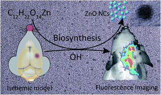
| Fig. 1 Scheme illustration of the fluorescence imaging of ischemic model mice brain. Through intravenous injection of zinc gluconate, we could readily realize in vivo fluorescence imaging by real-time labeling the relevant brain regions of CIS model mice based on the in situ biosynthesis of fluorescence zinc nanoclusters in target diseased sites. | |
 |
| Fig. 2 In vivo fluorescence imaging. (a–e) In vivo fluorescence imaging of mice from control group (pre injection of zinc gluconate) and CIS group (post injection of 0.1 mL 10 mmol L−1 zinc gluconate solution) at different time intervals (λex: 420 nm, λem: 670 nm). (f) Relative fluorescence intensity of normal group and CIS group at different time intervals (0–24 h). | |
 |
| Fig. 3 Fluorescence imaging of major organs. Fluorescence imaging of major organs (i.e., l for liver, h for heart, s for spleen, k for kidney, lu for lung and br for brain) from (a) normal group, (b) control group and (c) CIS group at 48 h after intravenous injection of 0.1 mL 10 mmol L−1 zinc gluconate (λex: 420 nm, λem: 670 nm). (d) Relative fluorescence intensity of different organs from three groups. | |
As shown in Fig. 2, in vivo fluorescence imaging of CIS mice model brains were specially explored in this study. Under excitation wavelength of 420 nm and emission wavelength 670 nm, zinc gluconate which were injected via tail-vein could spontaneously produce strong fluorescence in brain, especially on the right side where the embolism occurred. The fluorescence could be obviously seen 4 hours post-injection, while the relative fluorescence intensity increased considerably along with longer circulation and more accumulation of zinc, until the strongest fluorescence detected when 16 hours post-injection. In contrast, the fluorescence intensity of normal mice group (NOR) was much lower and had no apparent changes (Fig. 2(f)).
The fluorescence imaging of major organs such as liver, spleen, lungs and kidney of three groups were obtained together with brain to explore the precise targeting ability and bio distribution of zinc. As shown in Fig. 3, the results suggest that there is a weak fluorescence in all of the major organs except brain of CIS group, where the fluorescence intensity in diseased sites is significantly strong. This indicates that the circulation and accumulation of zinc in brain could be biosynthesized in situ to fluorescent zinc nanoclusters under the ischemic and hypoxic conditions of stroke supported by inflammation.
Histopathology
On the basis of above observations, histopathology study was further explored to evaluate the relevant biocompatibility of zinc gluconate treatment. Fig. 4 shows histopathological results of tissues obtained from harvested organs. H&E stained tissue sections of CIS model mice after injected with zinc gluconate show no histopathological lesions or abnormalities in liver, spleen or kidney. Meanwhile, no changes were found in drinking, eating, weight, activity and neurological status in NOR, CON and CIS groups. All these observations revealed that zinc gluconate has no side effects to major organs, which is consistent with the conclusions that properly dose of zinc can significantly repair the damaged neuron cell.23
 |
| Fig. 4 Histopathologic analysis of H&E-stained tissue sections. Histopathologic analysis of H&E-stained tissue sections from organs of CIS group after injection of 0.1 mL 10 mmol L−1 zinc gluconate. No appreciable pathological changes were found in all these organs (a for liver, b for spleen and c for kidney). | |
Characterization of the biosynthesized zinc nanoclusters
In view of the studies described above, we have further characterized the biosynthesized zinc nanoclusters. In Fig. 5(a), energy dispersive spectroscopy results suggested that no elemental impurities were presented in the biosynthesized zinc nanoclusters (Zn NCs). As shown in Fig. 5(b), the biosynthesized Zn NCs displayed remarkable fluorescence with emission peak wavelength at around 665 nm and excitation wavelength at 420 nm. Meanwhile, TEM analysis suggested that Zn NCs were almost spherical and had no noticeable trend to agglomerate (Fig. 5(c)), and more than 80% ranged between 2.05 to 2.65 nm in diameter with a distribution peak at 2.35 nm (Fig. 5(d)). Moreover, HR-TEM study of the biosynthesized Zn NCs indicated the interplanar spacing of ca. 0.17 nm, which is in agreement with that of the (110) lattice plane of nano ZnO (Fig. 5(e) and (f)). The element concentration of Zn in the extract of CIS part determined by ICP-AES is 0.056 mM. Besides, our observations illustrate that the fluorescence lifetime and mean zeta potential of the biosynthesized zinc nanoclusters were 1.1102 ns (Fig. S5†) and −63.6 ± 7.96 mV (Fig. S6†), respectively.
 |
| Fig. 5 Characterization of the biosynthesized zinc nanoclusters extracted from CIS mouse brain tissue. (a) EDS spectroscopy, (b) fluorescence emission spectrum (ex: 420 nm) (c) TEM of the biosynthesized zinc nanoclusters extracted from the brain of CIS group. (d) Diameter measurement (mean value 2.35 nm). (e) High resolution TEM and (f) intensity distribution show 0.17 nm interplanar spacing of the zinc nanoclusters. | |
As stated above, in this contribution we have described a new strategy of the biosynthesized zinc nanoclusters from zinc gluconate for rapid in vivo fluorescence imaging of cerebral ischemic stroke (CIS). Our findings demonstrate that self-biosynthesized zinc nanoclusters are powerful fluorescence imaging probes promising for the rapid CIS diagnosis, which is biocompatible and also convenient to realize accurate bioimaging.
It is desirable that all materials must be safe and nontoxic to body systems when used for biomedical applications. Our fluorescence imaging (Fig. 3) and histopathology (Fig. 4) studies of vital organs including kidney, lungs, spleen and liver proved the safety of zinc gluconate by presenting no histopathological abnormalities.35 The space between Bowman's capsule and glomerulus was normal. Besides, lungs had normal alveoli, epithelial lining and pneumocytes in bronchioles while the splenic nodules of spleen were also normal. Similarly, the hepatocytes and vasculature (i.e., portal, artery and bile duct) in liver were normal with no inflammatory infiltrate. During the whole experimental process mice exhibited no abnormal behaviour in eating, drinking, body weight, hyper activeness or nervous signs. These results demonstrate the biocompatibility and safety of zinc gluconate with no side effects, when compared with control group (Fig. S7†).
Oxidative stress may lead to ischemic cell death, which involves the formation of ROS/RNS (reactive nitrogen species) via various injury mechanisms, such as inflammation, reperfusion injury, Ca2+ overload and mitochondrial inhibition.36 Our previous study demonstrated that zinc gluconate or AuCl4− can biosynthesize the fluorescent ZnO NCs or Au NCs for the rapidly targeting fluorescence bioimaging of Alzheimer's disease.37–39 Similarly, when brain is short of blood, it also generates superoxide (O2−), which is the basic radical from which H2O2 is formed. Furthermore, H2O2 is the source of OH−, and OH− with zinc ions can form ZnO nanoclusters (NCs) through biosynthesis, which may result in a pathway as below.
On the basis of above study, we have further collected the extracts from the brain of CIS model mice and characterized the biosynthesized zinc nanoclusters. The fluorescence spectra of the as-prepared ZnO NCs biosynthesized in situ from zinc gluconate show emission peak at ca. 665 nm, which is in accordance with that previously reported in the literature40 as well as those results of both in vitro and in vivo fluorescence bioimaging study. In addition, the characterization of the extracts by EDS, TEM and HR-TEM also proved that zinc gluconate molecules can readily pass through blood–brain barrier and accumulate in the brain, especially the embolism region, for the biosynthesis of the fluorescent ZnO NCs. This suggests that zinc gluconate could be conveniently used as a biocompatible agent for the in vivo biosynthesis of fluorescent nanoprobes for the in situ high-spatiotemporal labelling of ischemic sites and achieving early rapid-targeting imaging of CIS. The relevant fluorescence halflife and apparent zeta potential of extracted zinc nanoclusters indicated good fluorescent property and negatively charged NCs, which could be another reason of the related targeting efficiency of the NCs biosynthesis process to CIS.
Conclusions
In summary, through all these observations of the biosynthesized zinc nanoclusters, which could be readily extracted from the brain tissues, we confirmed that the zinc gluconate could accurately assemble in the region of ischemia, hypoxia and inflammatory lesion of stroke, and then in situ biosynthesize the fluorescent ZnO NCs for the rapidly targeting fluorescence bioimaging in vivo of the CIS.
Acknowledgements
This work is supported by National High Technology Research & Development Program of China (2015AA020502) and the National Natural Science Foundation of China (81325011, 21327902 and 21175020). H. J. acknowledges support from the Fundamental Research Funds for the Central Universities (2242016K41023), China.
Notes and references
- M. Naghavi, H. Wang, R. Lozano, A. Davis, X. Liang, M. Zhou, S. E. Vollset, A. A. Ozgoren, S. Abdalla, F. Abd-Allah, M. I. A. Aziz, S. F. Abera, V. Aboyans, B. Abraham, J. P. Abraham, K. E. Abuabara, I. Abubakar, L. J. Abu-Raddad, N. M. E. Abu-Rmeileh, T. Achoki, A. Adelekan, Z. N. Ademi, K. Adofo, A. K. Adou, J. C. Adsuar, J. Aernlov, E. E. Agardh, D. Akena, M. J. Al Khabouri, D. Alasfoor, M. Albittar, M. A. Alegretti, A. V. Aleman, Z. A. Alemu, R. Alfonso-Cristancho, S. Alhabib, M. K. Ali, R. Ali, F. Alla, F. Al Lami, P. Allebeck, M. A. AlMazroa, R. A.-S. Salman, U. Alsharif, E. Alvarez, N. Alviz-Guzman, A. A. Amankwaa, A. T. Amare, O. Ameli, H. Amini, W. Ammar, H. R. Anderson, B. O. Anderson, C. A. T. Antonio, P. Anwari, H. Apfel, S. A. Cunningham, V. S. A. Arsenijevic, A. Al, M. M. Asad, R. J. Asghar, R. Assadi, L. S. Atkins, C. Atkinson, A. Badawi, M. C. Bahit, T. Bakfalouni, K. Balakrishnan, S. Balalla, A. Banerjee, R. M. Barber, S. L. Barker-Collo, S. Barquera, L. Barregard, L. H. Barrero, T. Barrientos-Gutierrez, A. Basu, S. Basu, M. O. Basulaiman, J. Beardsley, N. Bedi, E. Beghi, T. Bekele, M. L. Bell, C. Benjet, D. A. Bennett, I. M. Bensenor, H. Benzian, A. Bertozzi-Villa, T. J. Beyene, N. Bhala, A. Bhalla, Z. A. Bhutta, B. Bikbov, A. Bin Abdulhak, S. Biryukov, J. D. Blore, F. M. Blyth, M. A. Bohensky, G. Borges, D. Bose, S. Boufous, R. R. Bourne, L. N. Boyers, M. Brainin, M. Brauer, C. E. G. Brayne, A. Brazinova, N. Breitborde, H. Brenner, A. D. M. Briggs, J. C. Brown, T. S. Brugha, G. C. Buckle, L. N. Bui, G. Bukhman, M. Burch, I. R. C. Nonato, H. Carabin, R. Cardenas, J. Carapetis, D. O. Carpenter, V. Caso, C. A. Castaneda-Orjuela, R. E. Castro, F. Catala-Lopez, F. Cavalleri, J.-C. Chang, F. C. Charlson, X. Che, H. Chen, Y. Chen, J. S. Chen, Z. Chen, P. P.-C. Chiang, O. Chimed-Ochir, R. Chowdhury, H. Christensen, C. A. Christophi, T.-W. Chuang, S. S. Chugh, M. Cirillo, M. M. Coates, L. E. Coffeng, M. S. Coggeshall, A. Cohen, V. Colistro, S. M. Colquhoun, M. Colomar, L. T. Cooper, C. Cooper, L. M. Coppola, M. Cortinovis, K. Courville, B. C. Cowie, M. H. Criqui, J. A. Crump, L. Cuevas-Nasu, I. d. C. Leite, K. C. Dabhadkar, L. Dandona, R. Dandona, E. Dansereau, P. I. Dargan, A. Dayama, V. De la Cruz-Gongora, S. F. de la Vega, D. De Leo, L. Degenhardt, B. del Pozo-Cruz, R. P. Dellavalle, K. Deribe, D. C. D. Jarlais, M. Dessalegn, G. A. deVeber, S. D. Dharmaratne, M. Dherani, J.-L. Diaz-Ortega, C. Diaz-Torne, D. Dicker, E. L. Ding, K. Dokova, E. R. Dorsey, T. R. Driscoll, L. Duan, H. C. Duber, A. M. Durrani, B. E. Ebel, K. M. Edmond, R. G. Ellenbogen, Y. Elshrek, S. P. Ermakov, H. E. Erskine, B. Eshrati, A. Esteghamati, K. Estep, T. Fuerst, S. Fahimi, A. S. Fahrion, E. J. A. Faraon, F. Farzadfar, D. F. J. Fay, A. B. Feigl, V. L. Feigin, M. M. Felicio, S.-M. Fereshtehnejad, J. G. Fernandes, A. J. Ferrari, T. D. Fleming, N. Foigt, K. Foreman, M. H. Forouzanfar, F. G. R. Fowkes, U. Fra Paleo, R. C. Franklin, N. D. Futran, L. Gaffikin, K. Gambashidze, F. G. Gankpe, F. A. Garcia-Guerra, A. C. Garcia, J. M. Geleijnse, B. D. Gessner, K. B. Gibney, R. F. Gillum, S. Gilmour, I. Abdelmageem, M. Ginawi, M. Giroud, E. L. Glaser, S. Goenka, H. G. Dantes, P. Gona, D. Gonzalez-Medina, C. Guinovart, R. Gupta, R. Gupta, R. A. Gosselin, C. C. Gotay, A. Goto, H. N. Gowda, N. Graetz, K. F. Greenwell, H. C. Gugnani, D. Gunnell, R. A. Gutierrez, J. Haagsma, N. Hafezi-Nejad, H. Hagan, M. Hagstromer, Y. A. Halasa, R. R. Hamadeh, H. Hamavid, M. Hammami, J. Hancock, G. J. Hankey, G. M. Hansen, H. L. Harb, H. Harewood, J. M. Haro, R. Havmoeller, R. J. Hay, S. I. Hay, M. T. Hedayati, I. B. H. Pi, K. R. Heuton, P. Heydarpour, H. Higashi, M. Hijar, H. W. Hoek, H. J. Hoffman, J. C. Hornberger, H. D. Hosgood, M. Hossain, P. J. Hotez, D. G. Hoy, M. Hsairi, G. Hu, J. J. Huang, M. D. Huffman, A. J. Hughes, A. Husseini, C. Huynh, M. Iannarone, K.M. Iburg, B. T. Idrisov, N. Ikeda, K. Innos, M. Inoue, F. Islami, S. Ismayilova, K. H. Jacobsen, S. Jassal, S. P. Jayaraman, P. N. Jensen, V. Jha, G. Jiang, Y. Jiang, J. B. Jonas, J. Joseph, K. Juel, E. K. Kabagambe, H. Kan, A. Karch, C. Karimkhani, G. Karthikeyan, N. Kassebaum, A. Kaul, N. Kawakami, K. Kazanjan, D. S. Kazi, A. H. Kemp, A. P. Kengne, A. Keren, M. Kereselidze, Y. S. Khader, S. E. A. H. Khalifa, E. A. Khan, G. Khan, Y.-H. Khang, C. Kieling, Y. Kinfu, J. M. Kinge, D. Kim, S. Kim, M. Kivipelto, L. Knibbs, A. K. Knudsen, Y. Kokubo, S. Kosen, M. Kotagal, M. A. Kravchenko, S. Krishnaswami, H. Krueger, B. K. Defo, E. J. Kuipers, B. K. Bicer, C. Kulkarni, V. S. Kulkarni, K. Kumar, R. B. Kumar, G. F. Kwan, H. Kyu, T. Lai, A. L. Balaji, R. Lalloo, T. Lallukka, H. Lam, Q. Lan, V. C. Lansingh, H. J. Larson, A. Larsson, P. M. Lavados, A. E. B. Lawrynowicz, J. L. Leasher, J.-T. Lee, J. Leigh, M. Leinsalu, R. Leung, C. Levitz, B. Li, Y. Li, Y. Li, C. Liddell, S. S. Lim, G. M. F. de Lima, M. L. Lind, S. E. Lipshultz, S. Liu, Y. Liu, B. K. Lloyd, K. T. Lofgren, G. Logroscino, S. J. London, J. Lortet-Tieulent, P. A. Lotufo, R. M. Lucas, R. Lunevicius, R. A. Lyons, S. Ma, V. M. P. Machado, M. F. MacIntyre, M. T. Mackay, J. H. MacLachlan, C. Magis-Rodriguez, A. A. Mahdi, M. Majdan, R. Malekzadeh, S. Mangalam, C. C. Mapoma, M. Marape, W. Marcenes, C. Margono, G. B. Marks, M. B. Marzan, J. R. Masci, M. T. Q. Mashal, F. Masiye, A. J. Mason-Jones, R. Matzopolous, B. M. Mayosi, T. T. Mazorodze, J. J. McGrath, A. C. McKay, M. McKee, A. McLain, P. A. Meaney, M. M. Mehndiratta, F. Mejia-Rodriguez, Y. A. Melaku, M. Meltzer, Z. A. Memish, W. Mendoza, G. A. Mensah, A. Meretoja, F. A. Mhimbira, T. R. Miller, E. J. Mills, A. Misganaw, S. K. Mishra, C. N. Mock, T. E. Moffitt, N. M. Ibrahim, K. A. Mohammad, A. H. Mokdad, G. L. Mola, L. Monasta, J. d. l. C. Monis, J. C. M. Hernandez, M. Montico, T. J. Montine, M. D. Mooney, A. R. Moore, M. Moradi-Lakeh, A. E. Moran, R. Mori, J. Moschandreas, W. N. Moturi, M. L. Moyer, D. Mozaffarian, U. O. Mueller, M. Mukaigawara, E. C. Mullany, J. Murray, A. Mustapha, P. Naghavi, A. Naheed, K. S. Naidoo, L. Naldi, D. Nand, V. Nangia, K. M. V. Narayan, D. Nash, J. Nasher, C. Nejjari, R. G. Nelson, M. Neuhouser, S. P. Neupane, P. A. Newcomb, L. Newman, C. R. Newton, M. Ng, F. N. Ngalesoni, G. Nguyen, N. Nhung Thi Trang, M. I. Nisar, S. Nolte, O. F. Norheim, R. E. Norman, B. Norrving, L. Nyakarahuka, S. Odell, M. O'Donnell, T. Ohkubo, S. L. Ohno, B. O. Olusanya, S. B. Omer, J. N. Opio, O. E. Orisakwe, K. F. Ortblad, A. Ortiz, M. L. K. Otayza, A. W. Pain, J. D. Pandian, C. I. Panelo, J. Panniyammakal, C. Papachristou, A. J. Paternina Caicedo, S. B. Patten, G. C. Patton, V. K. Paul, B. Pavlin, N. Pearce, C. A. Pellegrini, D. M. Pereira, S. C. Peresson, R. Perez-Padilla, F. P. Perez-Ruiz, N. Perico, A. Pervaiz, K. Pesudovs, C. B. Peterson, M. Petzold, B. K. Phillips, D. E. Phillips, M. R. Phillips, D. Plass, F. B. Piel, D. Poenaru, S. Polinder, S. Popova, R. G. Poulton, F. Pourmalek, D. Prabhakaran, D. Qato, A. D. Quezada, D. A. Quistberg, F. Rabito, A. Rafay, K. Rahimi, V. Rahimi-Movaghar, S. U. R. Rahman, M. Raju, I. Rakovac, S. M. Rana, A. Refaat, G. Remuzzi, A. L. Ribeiro, S. Ricci, P. M. Riccio, L. Richardson, J. H. Richardus, B. Roberts, D. A. Roberts, M. Robinson, A. Roca, A. Rodriguez, D. Rojas-Rueda, L. Ronfani, R. Room, G. A. Roth, D. Rothenbacher, D. H. Rothstein, J. T. Rowley, N. Roy, G. M. Ruhago, L. Rushton, S. Sambandam, K. Soreide, M. Y. Saeedi, S. Saha, R. Sahathevan, M. A. Sahraian, B. W. Sahle, J. A. Salomon, D. Salvo, G. M. J. Samonte, U. Sampson, J. R. Sanabria, L. Sandar, I. S. Santos, M. Satpathy, M. Sawhney, M. Saylan, P. Scarborough, B. Schoettker, J. C. Schmidt, I. J. C. Schneider, A. E. Schumacher, D. C. Schwebel, J. G. Scott, S. G. Sepanlou, E. E. Servan-Mori, K. Shackelford, A. Shaheen, S. Shahraz, M. Shakh-Nazarova, S. Shangguan, J. She, S. Sheikhbahaei, D. S. Shepard, K. Shibuya, Y. Shinohara, K. Shishani, I. Shiue, R. Shivakoti, M. G. Shrime, I. D. Sigfusdottir, D. H. Silberberg, A. P. Silva, E. P. Simard, S. Sindi, J. A. Singh, L. Singh, E. Sioson, V. Skirbekk, K. Sliwa, S. So, M. Soljak, S. Soneji, S. S. Soshnikov, L. A. Sposato, C. T. Sreeramareddy, J. R. D. Stanaway, V. K. Stathopoulou, K. Steenland, C. Stein, C. Steiner, A. Stevens, H. Stoeckl, K. Straif, K. Stroumpoulis, L. Sturua, B. F. Sunguya, S. Swaminathan, M. Swaroop, B. L. Sykes, K. M. Tabb, K. Takahashi, R. T. Talongwa, F. Tan, D. Tanne, M. Tanner, M. Tavakkoli, B. T. Ao, C. M. Teixeira, T. Templin, E. Y. Tenkorang, A. S. Terkawi, B. A. Thomas, A. L. Thorne-Lyman, A. G. Thrift, G. D. Thurston, T. Tillmann, D. L. Tirschwell, I. M. Tleyjeh, M. Tonelli, F. Topouzis, J. A. Towbin, H. Toyoshima, J. Traebert, B. X. Tran, T. Truelsen, U. Trujillo, M. Trillini, Z. T. Dimbuene, M. Tsilimbaris, E. M. Tuzcu, C. Ubeda, U. S. Uchendu, K. N. Ukwaja, E. A. Undurraga, A. J. Vallely, S. van de Vijver, C. H. van Gool, Y. Y. Varakin, T. J. Vasankari, A. M. N. Vasconcelos, M. S. Vavilala, N. Venketasubramanian, L. Vijayakumar, S. Villalpando, F. S. Violante, V. V. Vlassov, G. R. Wagner, S. G. Waller, J. Wang, L. Wang, X. Wang, Y. Wang, T. S. Warouw, S. Weichenthal, E. Weiderpass, R. G. Weintraub, W. Wenzhi, A. Werdecker, K. R. R. Wessells, R. Westerman, H. A. Whiteford, J. D. Wilkinson, T. N. Williams, S. M. Woldeyohannes, C. D. A. Wolfe, T. M. Wolock, A. D. Woolf, J. Q. Wong, J. L. Wright, S. Wulf, B. Wurtz, G. Xu, Y. C. Yang, Y. Yano, H. Yatsuya, P. Yip, N. Yonemoto, S.-J. Yoon, M. Younis, C. Yu, K. Y. Jin, M. E. S. Zaki, M. F. Zamakhshary, H. Zeeb, Y. Zhang, Y. Zhao, Y. Zheng, J. Zhu, S. Zhu, D. Zonies, X. N. Zou, J. R. Zunt, T. Vos, A. D. Lopez, C. J. L. Murray and G. B. D. M. C. D. Colla, Lancet, 2015, 385, 117–171 CrossRef.
- D. Mozaffarian, E. J. Benjamin, A. S. Go, D. K. Arnett, M. J. Blaha, M. Cushman, S. R. Das, S. de Ferranti, J. P. Despres, H. J. Fullerton, V. J. Howard, M. D. Huffman, C. R. Isasi, M. C. Jimenez, S. E. Judd, B. M. Kissela, J. H. Lichtman, L. D. Lisabeth, S. Liu, R. H. Mackey, D. J. Magid, D. K. McGuire, E. R. Mohler III, C. S. Moy, P. Muntner, M. E. Mussolino, K. Nasir, R. W. Neumar, G. Nichol, L. Palaniappan, D. K. Pandey, M. J. Reeves, C. J. Rodriguez, W. Rosamond, P. D. Sorlie, J. Stein, A. Towfighi, T. N. Turan, S. S. Virani, D. Woo, R. W. Yeh, M. B. Turner, C. American Heart Association Statistics and S. Stroke Statistics, Circulation, 2016, 133, e38–60 CrossRef PubMed.
- L. Castilla-Guerra and C. Fernandez-Moreno Mdel, J. Stroke, 2016, 18, 31–37 CrossRef PubMed.
- M. Lee, Y. L. Wu and B. Ovbiagele, J. Stroke, 2016, 18, 60–65 CrossRef PubMed.
- P. A. Wolf, R. D. Abbott and W. B. Kannel, Stroke, 1991, 22, 983–988 CrossRef CAS PubMed.
- T. J. Wang, J. M. Massaro, D. Levy, R. S. Vasan, P. A. Wolf, R. B. D'Agostino, M. G. Larson, W. B. Kannel and E. J. Benjamin, J. Am. Med. Assoc., 2003, 290, 1049–1056 CrossRef PubMed.
- X. Wang, Y. Dong, X. Qi, C. Huang and L. Hou, Stroke, 2013, 44, 1833–1839 CrossRef CAS PubMed.
- T. H. Park, Y. Ko, S. J. Lee, K. B. Lee, J. Lee, M. K. Han, J. M. Park, Y. J. Cho, K. S. Hong, D. H. Kim, J. K. Cha, M. S. Oh, K. H. Yu, B. C. Lee, B. W. Yoon, J. S. Lee, J. Lee and H. J. Bae, J. Stroke, 2015, 17, 302–311 CrossRef PubMed.
- S. Seshadri, A. Beiser, A. Pikula, J. J. Himali, M. Kelly-Hayes, S. Debette, A. L. DeStefano, J. R. Romero, C. S. Kase and P. A. Wolf, Circulation, 2010, 121, 1304–1312 CrossRef PubMed.
- G. A. Donnan, M. Fisher, M. Macleod and S. M. Davis, Lancet, 2008, 371, 1612–1623 CrossRef CAS.
- P. Libby, Nature, 2002, 32, 868–874 CrossRef PubMed.
- A. J. Lusis, Nature, 2000, 407, 233–241 CrossRef CAS PubMed.
- K. W. Muir, P. Tyrrell, N. Sattar and E. Warburton, Curr. Opin. Neurol., 2007, 20, 334–342 CrossRef CAS PubMed.
- P. Amarenco, J. Labreuche and P. J. Touboul, Atherosclerosis, 2008, 196, 489–496 CrossRef CAS PubMed.
- Y. Zhang, J. Tuomilehto, P. Jousilahti, Y. Wang, R. Antikainen and G. Hu, Stroke, 2012, 43, 1768–1774 CrossRef CAS PubMed.
- C. Ellis, A. N. Simpson, H. Bonilha, P. D. Mauldin and K. N. Simpson, Stroke, 2012, 43, 1429–1431 CrossRef PubMed.
- M. A. Gardner, N. K. Hills, S. Sidney, S. C. Johnston and H. J. Fullerton, Neurology, 2010, 74, 372–378 CrossRef CAS PubMed.
- M. W. Brault, J. Hootman, C. G. Helmick, K. A. Theis and B. S. Armour, Centers for Disease and Prevention, Morb. Mortal. Wkly. Rep., 2009, 58, 421–426 Search PubMed.
- J. M. Wardlaw, V. Murray, E. Berge, G. del Zoppo, P. Sandercock, R. L. Lindley and G. Cohen, Lancet, 2012, 379, 2364–2372 CrossRef CAS.
- J. M. Wardlaw, G. J. D. Zoppo, T. Yamaguchi and E. Berge, Cochrane Database Syst. Rev., 2001, 7, CD000213 Search PubMed.
- J. R. Marler, T. Brott, J. Broderick, R. Kothari, M. Odonoghue, W. Barsan, T. Tomsick, J. Spilker, R. Miller, L. Sauerbeck, J. Jarrell, J. Kelly, T. Perkins, T. McDonald, M. Rorick, C. Hickey, J. Armitage, C. Perry, K. Thalinger, R. Rhude, J. Schill, P. S. Becker, R. S. Heath, D. Adams, R. Reed, M. Klei, S. Hughes, J. Anthony, D. Baudendistel, C. Zadicoff, M. Rymer, I. Bettinger, P. Laubinger, M. Schmerler, G. Meirose, P. Lyden, K. Rapp, T. Babcock, P. Daum, D. Persona, M. Brody, C. Jackson, S. Lewis, J. Liss, Z. Mahdavi, J. Rothrock, T. Tom, R. Zweifler, J. Dunford, J. Zivin, R. Kobayashi, J. Kunin, J. Licht, R. Rowen, D. Stein, J. Grisolia, F. Martin, E. Chaplin, N. Kaplitz, J. Nelson, A. Neuren, D. Silver, T. Chippendale, E. Diamond, M. Lobatz, D. Murphy, D. Rosenberg, T. Ruel, M. Sadoff, J. Schim, J. Schleimer, R. Atkinson, D. Wentworth, R. Cummings, R. Frink, P. Heublein, J. C. Grotta, T. Degraba, M. Fisher, A. Ramirez, S. Hanson, L. Morgenstern, C. Sills, W. Pasteur, F. Yatsu, K. Andrews, C. Villarcordova, P. Pepe, P. Bratina, L. Greenberg, S. Rozek, K. Simmons, T. G. Kwiatkowski, S. H. Horowitz, R. Libman, R. Kanner, R. Silverman, J. Lamantia, C. Mealie, R. Duarte, R. Donnarumma, M. Okola, V. Cullin, E. Mitchell, S. R. Levine, C. A. Lewandowski, G. Tokarski, N. M. Ramadan, P. Mitsias, M. Gorman, B. Zarowitz, J. Kokkinos, J. Dayno, P. Verro, C. Gymnopoulos, R. Dafer, L. Dolhaberriague, K. Sawaya, S. Daley, M. Mitchell, M. Frankel, B. Mackay, C. Barch, J. Braimah, B. Faherty, J. Macdonald, S. Sailor, A. Cook, H. Karp, B. Nguyen, J. Washington, J. Weissman, M. Williams, T. Williamson, M. Kozinn, L. Hellwick, E. C. Haley, T. P. Bleck, W. S. Cail, G. H. Lindbeck, M. A. Granner, S. S. Wolf, M. W. Gwynn, R. W. Mettetal, C. W. J. Chang, N. J. Solenski, D. G. Brock, G. F. Ford, G. L. Kongable, K. N. Parks, S. S. Wilkinson, M. K. Davis, G. L. Sheppard, D. W. Zontine, K. H. Gustin, N. M. Crowe, S. L. Massey, M. Meyer, K. Gaines, A. Payne, C. Bales, J. Malcolm, R. Barlow, M. Wilson, C. Cape, T. Bertorini, K. Misulis, W. Paulsen, D. Shepard, B. C. Tilley, K. M. A. Welch, S. C. Fagan, M. Lu, S. Patel, E. Masha, J. Verter, J. Boura, J. Main, L. Gordon, N. Maddy, T. Chociemski, J. Windham, H. S. Zadeh, W. Alves, M. F. Keller, J. R. Wenzel, N. Raman, L. Cantwell, A. Warren, K. Smith, E. Bailey, K. M. A. Welch, B. C. Tilley, J. Froehlich, J. Breed, J. D. Easton, J. F. Hallenbeck, G. Lan, J. D. Marsh and M. D. Walker, N. Engl. J. Med., 1995, 333, 1581–1587 CrossRef CAS PubMed.
- B. L. Vallee and D. S. Auld, Biochemistry, 1990, 29, 5647–5659 CrossRef CAS PubMed.
- J. E. Cummings and J. P. Kovacic, J. Vet. Emerg. Crit. Care, 2009, 19, 215–240 CrossRef PubMed.
- J. Wang, G. Zhang, Q. Li, H. Jiang, C. Liu, C. Amatore and X. Wang, Sci. Rep., 2013, 3, 1157 Search PubMed.
- S. Gao, D. Chen, Q. Li, J. Ye, H. Jiang, C. Amatore and X. Wang, Sci. Rep., 2014, 4, 4384 Search PubMed.
- D. Chen, C. Zhao, J. Ye, Q. Li, X. Liu, M. Su, H. Jiang, C. Amatore, M. Selke and X. Wang, ACS Appl. Mater. Interfaces, 2015, 7, 18163–18169 CAS.
- M. Su, J. Ye, Q. Li, W. Ge, Y. Zhang, H. Jiang, C. Amatore and X. Wang, RSC Adv., 2015, 5, 74844–74849 RSC.
- T. Hayashi, N. Noshita, T. Sugawara and P. H. Chan, J. Cereb. Blood Flow Metab., 2003, 23, 166–180 CrossRef CAS PubMed.
- S. Parasuraman, R. Raveendran and R. Kesavan, J. Pharmacol. Pharmacother., 2010, 1, 87–93 CrossRef CAS PubMed.
- M. N. Su, J. Ye, Q. W. Li, S. H. Li, W. Ge, X. Chen, H. Jiang and X. M. Wang, Chin. Chem. Lett., 2015, 26, 1400–1402 CrossRef CAS.
- H. Chang, X. Wang, K. K. Shiu, Y. Zhu, J. Wang, Q. Li, B. Chen and H. Jiang, Biosens. Bioelectron., 2013, 41, 789–794 CrossRef CAS PubMed.
- D. Trachootham, J. Alexandre and P. Huang, Nat. Rev. Drug Discovery, 2009, 8, 579–591 CrossRef CAS PubMed.
- T. Shichita, Y. Sugiyama, H. Ooboshi, H. Sugimori, R. Nakagawa, I. Takada, T. Iwaki, Y. Okada, M. Iida, D. J. Cua, Y. Iwakura and A. Yoshimura, Nat. Med., 2009, 15, 946–950 CrossRef CAS PubMed.
- S. Cuzzocrea, D. P. Riley, A. P. Caputi and D. Salvemini, Pharmacol. Rev., 2001, 53, 135–159 CAS.
- A. Nyska, Y. S. Schiffenbauer, C. T. Brami, R. R. Maronpot and Y. Ramot, Polym. Adv. Technol., 2014, 25, 461–467 CrossRef CAS.
- J. T. Coyle and P. Puttfarcken, Science, 1993, 262, 689–695 CAS.
- L. Lai, C. Zhao, X. Li, X. Liu, H. Jiang, M. Selke and X. Wang, RSC Adv., 2016, 6, 30081–30088 RSC.
- L. Lanmei, Z. Chunqiu, S. Meina, Y. Jing, J. Hui and W. Xuemei, Sci. Bull., 2015, 60, 1465–1467 CrossRef.
- L. Lai, C. Zhao, M. Su, X. Li, X. Liu, H. Jiang, C. Amatore and X. Wang, Biomater. Sci., 2016, 4, 1085–1091 RSC.
- H. P. Wang, H. Jiang and X. M. Wang, Chem. Commun., 2010, 46, 6900–6902 RSC.
Footnote |
† Electronic supplementary information (ESI) available. See DOI: 10.1039/c6ra23507g |
|
This journal is © The Royal Society of Chemistry 2016 |
Click here to see how this site uses Cookies. View our privacy policy here.