Preventive effects of zingerone on cardiac mitochondrial oxidative stress, calcium ion overload and adenosine triphosphate depletion in isoproterenol induced myocardial infarcted rats

Kunchpillai Lakshmanan Hemalatha and Ponnian Stanely Mainzen Prince*
Department of Biochemistry and Biotechnology, Annamalai University, Annamalai Nagar-608002, Tamil Nadu, India. E-mail: ps_mainzenprince@yahoo.co.in; Fax: +91 4144 239141; Tel: +91 4144 239141

Received 19th September 2016 , Accepted 18th November 2016

First published on 21st November 2016


Abstract

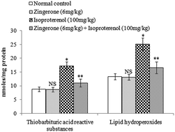
Cardiac mitochondrial oxidative stress, calcium ion (Ca2+) overload and adenosine triphosphate (ATP) depletion play an important role in the pathogenesis of myocardial infarction. The preventive effects of zingerone on oxidative stress, Ca2+ overload and ATP depletion in isoproterenol induced myocardial infarction were evaluated in rats. Rats were pretreated with zingerone (6 mg kg−1 body weight) daily for a period of 14 days. Isoproterenol (100 mg kg−1 body weight) was injected subcutaneously into rats twice at an interval of 24 h (on 15th and 16th day) to induce myocardial infarction. Isoproterenol induced myocardial infarcted rats showed a significant increase in the levels/concentrations of cardiac diagnostic marker, heart mitochondrial lipid peroxidation, Ca2+, and a significant decrease in the activities/concentrations of heart mitochondrial superoxide dismutase, glutathione peroxidase, reduced glutathione, tricarboxylic acid cycle and respiratory chain enzymes and ATP. Zingerone pretreatment showed significant preventive effects on all the biochemical parameters evaluated. Furthermore, the biochemical findings were correlated with transmission electron microscopic study on the structure of heart mitochondria. The in vitro study revealed the reactive oxygen species (ROS) scavenging effects of zingerone. Thus zingerone prevented mitochondrial damage by preventing cardiac mitochondrial oxidative stress, Ca2+ overload and ATP depletion in isoproterenol induced myocardial infarcted rats. The observed effects could be due to zingerone's anti-inotropic and ROS scavenging properties.


1. Introduction

Myocardial infarction is the acute condition of necrosis of the myocardium that occurs as a result of imbalance between coronary blood supply and myocardial demand. Pharmacological induction of myocardial infarction by subcutaneous administration of isoproterenol in rats is convenient because of the relatively small size of coronary arteries.1 Isoproterenol causes oxidative stress in the myocardium, resulting in gross and microscopic infarct in rat's heart muscle.2 An excessive β-adrenergic stimulation by isoproterenol produces more cyclic adenosine monophosphate which leads to enhanced release of calcium ions (Ca2+) by the sarcoplasmic reticulum in the heart. Subsequent increase in inotropy and generation of lipid peroxides during ischemia could damage mitochondrial membrane and affect mitochondrial function.3 This is accompanied by damage of mitochondria with oxidative stress, and inactivation of tricarboxylic acid cycle and respiratory chain enzymes and depletion of adenosine triphosphate (ATP).4 By studying the biochemical alterations occurring in an animal model, it is possible to gain more insight into the mechanisms leading to the altered metabolic processes occurring in the human myocardial infarction.5

Mitochondria are vital subcellular organelles for cellular oxidative process and are also the main source of reactive oxygen species (ROS) in the cell and they carry out oxidative phosphorylation. Mitochondria are the main source of energy and a decrease in oxygen supply during myocardial infarction impairs energy production by mitochondria. Both ATP synthesis and electron transport chain are located in mitochondria, which is required for cardiac contraction and relaxation. Prolonged oxidative stress in failing myocardium results in damage to mitochondrial DNA, ROS generation and consequent cellular injury leading to functional decline. Thus, mitochondria serve both as a source and target of ROS mediated injury in failing heart. Murray et al.6 suggested that benefit may be derived from the development of therapies aimed at preserving cardiac mitochondrial function.

Recently, there has been an upsurge of interest to explore the cardioprotective potential of natural products. The rhizome of Zingiber officinale (ginger) is consumed worldwide as a spice and flavoring agent. Zingerone, 4-(4-hydroxy-3-methoxyphenyl)-2-butanone, isolated from ginger is a phenolic alkanone.7 In our previous phases of experiment, we reported the preventive effects of zingerone (6 mg kg−1 body weight) on altered serum LPO and nonenzymatic antioxidants, adenosine triphosphatases and electrolyte imbalance, left ventricular dysfunction, myocardial infarct size and antihyperlipidaemic and antihypertrophic effects in isoproterenol induced myocardial infarcted rats.7–9 Recently, we published the anti-inflammatory and anti-thrombotic effects of zingerone in isoproterenol induced myocardial infarcted rats.10 In this study, we reported the effects of zingerone on inflammatory markers and histopathology of thrombosis and heart in myocardial infarcted rats. Based on the above proven preventive effects of zingerone on isoproterenol induced myocardial infarction model, we hypothesized that zingerone may have a protective role on cardiac mitochondrial damage. Hence, in the next phase of our experiment, we evaluated the preventive effects of zingerone on heart mitochondrial oxidative stress, Ca2+ overload and ATP depletion in isoproterenol induced myocardial infarcted rats. The in vitro ROS scavenging effects of zingerone was evaluated to understand the mechanism of action.

2. Materials and methods

2.1. Chemicals

Zingerone, isoproterenol hydrochloride, nitroblue tetrazolium, phenazine methosulphate, butylated hydroxy toluene, glutathione and cytochrome-c were purchased from Sigma Chemical Co., St. Louis, MO, USA. All other chemicals and solvents used were of analytical grade.

2.2. Experimental animals and diet

The whole experiment was performed according to the guidelines of the Committee for the Purpose of Control and Supervision of Experiments on Animals, New Delhi, India and approved by the Animal Ethical Committee of Annamalai University (Proposal No: 878; Approval Date: 10/01/2012). All animals received humane care and the study protocols comply with the institution's guidelines. This study was conducted in healthy male albino Wistar rats (Rattus norvegicus) weighing 180–200 g, (8–9 weeks old) obtained from the Central Animal House, Department of Experimental Medicine, Rajah Muthiah Institute of Health Sciences, Annamalai University, Tamil Nadu, India. They were housed in polypropylene cages (47 × 34 × 20 cm) (3 rats per cage) lined with husk, renewed every 24 hours under a 12[thin space (1/6-em)]:[thin space (1/6-em)]12 hours light dark cycle at around 22 °C and had free access to tap water and food. The rats were fed on a standard pellet diet (Pranav Agro Industries Limited, Maharashtra, India).

2.3. Induction of experimental myocardial infarction and experimental design

Isoproterenol hydrochloride (100 mg kg−1 body weight) dissolved in saline was injected subcutaneously into rats twice at an interval of 24 h to induce myocardial infarction.2,8 The experimental design consists of four groups of rats. Group I: normal untreated rats (n = 6). Group II: rats were treated with zingerone (6 mg kg−1 body weight) orally daily for a period of 14 days (n = 6). Group III: rats were induced myocardial infarction by subcutaneous injection of isoproterenol (100 mg kg−1 body weight) on 15th and 16th day (n = 6). Group IV: rats were pretreated with zingerone (6 mg kg−1 body weight) orally daily for a period of 14 days and were then induced myocardial infarction by subcutaneous injection of isoproterenol (100 mg kg−1 body weight) on 15th and 16th day (n = 6). Zingerone was dissolved in saline and administered 2 ml to rats orally using an intragastric tube daily for a period of 14 days. Normal control (Group I) and isoproterenol control (Group III) rats were received 2 ml saline alone orally using an intragastric tube daily for 14 days. The dosage (6 mg kg−1 body weight) and duration of pretreatment (14 days) of zingerone was based on our earlier studies.7–10

At the end of experimental period, after 12 h of second dose of isoproterenol injection (i.e. on 17th day), all the rats were anesthetized with pentobarbital sodium (60 mg kg−1 body weight) and then sacrificed by cervical decapitation. Blood was collected in dry test tubes without anticoagulant for serum. The heart was dissected out immediately and stored for mitochondrial isolation. All the enzyme assays were performed on 17th day.

2.4. The in vivo studies

2.4.1. Estimation of serum cardiac troponin-T. The level of cardiac troponin-T in serum was estimated by electro chemiluminescence immunoassay (Elecsys troponin-T Stat reagent kit, Roche Diagnostics, Mannheim, Germany).
2.4.2. Isolation of heart mitochondrial fraction. The mitochondrial fraction of the heart tissue was isolated by the standard procedure of Takasawa et al.11 The heart tissue was put into ice-cold 50 mM Tris–HCl (pH 7.4) containing 0.25 M sucrose and homogenized. The homogenates were centrifuged at 700 × g for 20 min, and then the supernatants obtained were centrifuged at 9000 × g for 15 min. Then, the pellets were washed with 10 mM Tris–HCl (pH 7.8) containing 0.25 M sucrose and finally resuspended in the same buffer.
2.4.3. Estimation/assay of lipid peroxidation products and antioxidants in the heart mitochondrial fraction. Thiobarbituric acid reactive substances and lipid hydroperoxides were estimated in the heart mitochondrial fraction by the methods of Fraga et al.12 and Jiang et al.13 respectively. Superoxide dismutase and glutathione peroxidase activities in the heart mitochondria were assayed by the methods of Kakkar et al.14 and Rotruck et al.15 respectively. The concentration of reduced glutathione in the heart mitochondria was estimated by the method of Ellman.16
2.4.4. Assay/estimation of heart mitochondrial enzymes, Ca2+, ATP and protein. The activities of isocitrate dehydrogenase and malate dehydrogenase in the heart mitochondrial fraction were assayed by the method of King17 and Mehler et al.18 respectively. Further, the activities of α-ketoglutarate dehydrogenase and nicotinamide adenine dinucleotide dehydrogenase in the heart mitochondrial fraction were assayed by the method of Reed and Mukherjee19 and Minakami et al.20 respectively. Cytochrome-c-oxidase activity in the heart mitochondrial fraction was assayed by the method of Pearl et al.21 The levels of heart mitochondrial Ca2+ were measured by the O-cresolphthalein complexone method using a reagent kit (Span Diagnostics Limited, India). Heart mitochondrial ATP concentration was estimated by Williams and Coorkey.22 The content of protein in the heart tissue homogenate was determined by the method of Lowry et al.23
2.4.5. Transmission electron microscopic study on heart mitochondria. Small pieces of heart were taken from normal and experimental rats on 17th day and rinsed in 0.1 M phosphate buffer (pH 7.2). Approximately, 1 mm heart pieces were trimmed and immediately fixed into 3% ice-cold glutaraldehyde in 0.1 M-phosphate buffer (pH 7.2) and kept at 4 °C for 12 h. Then, tissue processing for transmission electron microscopic study was carried out. The grids containing sections were stained with 2% uranyl acetate and 0.2% lead acetate. Then, the sections were examined under a transmission electron microscope (4000×).

2.5. The in vitro studies

2.5.1. The O2˙ scavenging effect of zingerone in vitro. O2˙ scavenging activity of zingerone in vitro was determined by the method of Nishikimi et al.24 Varying volumes of zingerone (5, 10, 15, 20, 25 and 30 μM) were taken in different test tubes and then added 1.0 ml of reduced nicotinamide adenine dinucleotide solution (14.68 μM of reduced nicotinamide adenine dinucleotide in 100 mM phosphate buffer, pH 7.4) and 1.0 ml of nitroblue tetrazolium (100 μM of nitroblue tetrazolium in 100 mM phosphate buffer, pH 7.4) and mixed well. The reaction was initiated by the addition of 100 μM of phenazine methosulfate (60 μM in 100 mM of phosphate buffer, pH 7.4). The reaction mixture was incubated at 30 °C for 15 min and the absorbance was read at 560 nm in a spectrophotometer with a reagent blank containing double distilled water instead of zingerone. Decrease in the absorbance of reaction mixture indicates increased O2˙ scavenging of zingerone.
% scavenging of O2˙ = control-OD − test-OD/control-OD × 100
2.5.2. H2O2 scavenging of zingerone in vitro. The ability of zingerone to scavenge H2O2 was determined according to the method of Ruch et al.25 A solution of H2O2 (40 mM) was prepared in phosphate buffer (pH 7.4). Zingerone (5–30 μM) in distilled water was added to a H2O2 solution (0.6 ml, 40 mM H2O2). Absorbance of H2O2 at 230 nm was determined after 10 min against a blank solution containing the phosphate buffer without H2O2. The percentage of H2O2 scavenging activity of zingerone was calculated using the following formula.
% scavenging activity of H2O2 = control-OD − test-OD/control-OD × 100
2.5.3. The OH˙ scavenging effect of zingerone in vitro. The zingerone's OH˙ scavenging activity in vitro was determined by the method of Halliwell and Gutteridge.26 Varying volumes of zingerone (5, 10, 15, 20, 25 and 30 μM), were taken in different test tubes and then mixed with 0.1 ml of 100 mM potassium dihydrogen phosphate–dipotassium hydrogen phosphate buffer, 0.2 ml of 500 mM ferric chloride, 0.1 ml of 1 mM ascorbic acid, 0.1 ml of 1 mM EDTA, 0.1 ml of 10 mM H2O2 and 0.2 ml of 2-deoxyribose in a total volume of 1.0 ml. They were mixed well and incubated at room temperature for 60 min. Then, 1.0 ml of 1% thiobarbituric acid (1 g in 100 ml of 0.05 N sodium chloride) and 1.0 ml of 28% trichloro acetic acid were added and all the tubes were kept in a boiling water bath for 30 min. The absorbance was read in a spectrophotometer at 532 nm with a reagent blank containing double distilled H2O instead of zingerone. Decrease in the absorbance of reaction mixture reveals increased OH˙ scavenging activity of zingerone.
% scavenging of OH˙ = control-OD − test-OD/control-OD × 100

2.6. Statistical analysis

Statistical analysis was performed by one-way analysis of variance followed by Duncan's Multiple Range Test (DMRT) using Statistical Package for the Social Science software package version 12.00. Results were expressed as mean ± standard deviation (S. D.) for 6 rats in each group. P values < 0.05 were considered significant.

3. Results

Isoproterenol induced myocardial infarcted rats showed considerable (P < 0.05) increased level of serum cardiac troponin-T compared to normal control rats. Pretreatment with zingerone (6 mg kg−1 body weight) daily for a period of 14 days considerably (P < 0.05) lowered the level of serum cardiac troponin-T in isoproterenol induced myocardial infarcted rats compared to isoproterenol alone induced myocardial infarcted rats (Fig. 1).
image file: c6ra23330a-f1.tif
Fig. 1 Level of serum cardiac troponin-T. Each column is mean ± S.D.; n = 6; NS as compared to normal control (Group I); *P < 0.05 as compared to normal control; **P < 0.05 as compared to isoproterenol control, DMRT.

Isoproterenol induced myocardial infarcted rats showed a significant (P < 0.05) increase in the concentration of thiobarbituric acid reactive substances and lipid hydroperoxides in the heart mitochondria compared to normal control rats. Oral pretreatment with zingerone significantly (P < 0.05) decreased the concentration of thiobarbituric acid reactive substances and lipid hydroperoxides in the heart mitochondria of isoproterenol induced myocardial infarcted rats compared to isoproterenol alone induced myocardial infarcted rats (Fig. 2).


image file: c6ra23330a-f2.tif
Fig. 2 Concentration of heart mitochondrial thiobarbituric acid reactive substances and lipid hydroperoxides. Each column is the mean ± S.D.; n = 6; NS as compared to normal control (Group I); *P < 0.05 as compared to normal control (Group I); **P < 0.05 as compared to isoproterenol control (Group III), DMRT.

Isoproterenol induced myocardial infarcted rats revealed a significant (P < 0.05) decrease in the activities of superoxide dismutase, glutathione peroxidase and the concentration of reduced glutathione in the heart mitochondria compared to normal control rats. Pretreatment with zingerone significantly (P < 0.05) increased the above mentioned antioxidants in the heart mitochondria of isoproterenol induced myocardial infarcted rats compared to isoproterenol alone induced myocardial infarcted rats (Fig. 3).


image file: c6ra23330a-f3.tif
Fig. 3 Activities/concentration of heart mitochondrial superoxide dismutase, glutathione peroxidase and reduced glutathione. Each column is the mean ± S.D.; n = 6; NS as compared to normal control (Group I); *P < 0.05 as compared to normal control (Group I); **P < 0.05 as compared to isoproterenol control (Group III), DMRT, units; superoxide dismutase: units per mg protein; one unit of superoxide dismutase is defined as the enzyme concentration required inhibiting the optical density at 560 nm of chromogen production by 50% in one min; glutathione peroxidase: nmoles of reduced glutathione oxidized per min per 100 mg protein; *units for reduced glutathione: nmoles of reduced glutathione reduced per 100 mg protein.

The activities of tricarboxylic acid cycle enzymes such as isocitrate dehydrogenase, malate dehydrogenase, α-ketoglutarate dehydrogenase and respiratory chain enzymes such as NADH–dehydrogenase (Fig. 4A) and cytochrome-c-oxidase (Fig. 4B) were significantly (P < 0.05) decreased in the heart mitochondria of isoproterenol induced myocardial infarcted rats compared to normal control rats. Zingerone pretreatment significantly (P < 0.05) increased the activities of tricarboxylic cycle and respiratory chain enzymes in isoproterenol induced myocardial infarcted rats compared to isoproterenol alone induced myocardial infarcted rats.


image file: c6ra23330a-f4.tif
Fig. 4 (A and B) Activities of tricarboxylic acid cycle and respiratory chain enzymes. Each column is the mean ± S.D.; n = 6; NS as compared to normal control (Group I); *P < 0.05 as compared to normal control (Group I); **P < 0.05 as compared to isoproterenol control (Group III), DMRT. *Units: activity is expressed as nmoles of α-ketoglutarate formed per h per mg protein for isocitrate dehydrogenase; nmoles of nicotinamide adenine dinucleotide oxidized per min per mg protein for malate dehydrogenase; nmoles of ferrocyanide formed per h per mg protein for α-ketoglutarate dehydrogenase; nmoles of nicotinamide adenine dinucleotide oxidized per min per mg protein for NADH–dehydrogenase.

The concentration of Ca2+ was significantly (P < 0.05) increased and the concentration of ATP was significantly (P < 0.05) decreased in the heart mitochondrial fraction of isoproterenol induced myocardial infarcted rats compared to normal control rats. Zingerone pretreatment significantly (P < 0.05) decreased the concentration of Ca2+ and significantly (P < 0.05) increased the concentration of ATP in the heart mitochondrial fraction of isoproterenol induced myocardial infarcted rats compared to isoproterenol alone induced myocardial infarcted rats (Fig. 5).


image file: c6ra23330a-f5.tif
Fig. 5 Concentration of Ca2+ and ATP in heart mitochondria. Each column is the mean ± S.D.; n = 6; NS as compared to normal control (Group I); *P < 0.05 as compared to normal control (Group I); **P < 0.05 as compared to isoproterenol control (Group III), DMRT.

We examined the preventive effects of zingerone on the structure of heart mitochondria by transmission electron microscopic study to confirm the biochemical findings. The normal control rat's heart mitochondria (Group-I) revealed normal architecture (Fig. 6A). Also, zingerone treated rat's heart mitochondria (Group-II) showed normal architecture without any damage (Fig. 6B). But, isoproterenol induced myocardial infarcted rat's heart mitochondria (Group-III) showed loss of cristae (←) with vacuolation (↑), swollen mitochondria (↓) with irregular size and shape (→) (Fig. 6C). Zingerone pretreated isoproterenol induced myocardial infarcted rat's heart mitochondria (Group-IV) revealed near normal architecture (Fig. 6D).


image file: c6ra23330a-f6.tif
Fig. 6 (A–D) Transmission electron microscopic study on the structure of heart mitochondria (A) normal control rat's heart mitochondria (Group-I) showing normal architecture (4000×); (B) zingerone treated rat's heart mitochondria (Group-II) revealing normal architecture without any damage (4000×); (C) isoproterenol induced myocardial infarcted rat's heart mitochondria (Group-III) showing loss of cristae (←), vacuolation (↑), swollen mitochondria (↓) with irregular size and shape (→) (4000×); (D) zingerone pretreated isoproterenol induced myocardial infarcted rat's heart mitochondria (Group-IV) showing near normal architecture without swelling, vacuolation, and disruption of cristae (4000×).

Fig. 7A–C depicts the percentage in vitro scavenging effects of zingerone on ROS such as O2˙, H2O2 and OH˙. Five different concentrations of zingerone (5, 10, 15, 20, 25 and 30 μM) were tested for its O2˙, H2O2 and OH˙ scavenging effects. Zingerone scavenged O2˙, H2 O2, OH˙ in vitro in a concentration dependent manner. The percentage scavenging effect of zingerone increased with increasing concentration. The percentage scavenging of O2˙ and H2O2 at five concentrations of zingerone (5, 10, 15, 20, 25 and 30 μM) were found to be 17.2%, 30.13%, 42.4%, 56.9%, 70.26%, 82.53% (Fig. 7A) and 29.12%, 37.87%, 46.12%, 55.62%, 63.75% and (Fig. 7B) respectively. Further, the scavenging of OH˙ at different concentrations of zingerone 5, 10, 15, 20, 25 and 30 μM were observed to be 72.8% 14.62%, 27.97%, 39.67%, 52.2%, 65.3% and 74.7% (Fig. 7C) respectively. Thus, zingerone in vitro scavenged ROS such as O2˙, H2O2, and OH˙.


image file: c6ra23330a-f7.tif
Fig. 7 (A–C) The in vitro scavenging effects of zingerone on ROS such as O2˙, H2O2 and OH˙. Each column is the average of triplicate experiments.

For all the biochemical parameters and the structure of cardiac mitochondria evaluated, zingerone (6 mg kg−1 body weight) administration to normal rats (Group-II) did not reveal any effect indicating this dose appears safe.

4. Discussion

Isoproterenol induced myocardial infarction in animals is a well established, standardized and common model to evaluate the protective/preventive effect of various cardioprotective agents/phytoconstituents.3,27 The isoproterenol induced myocardial infarction occurs as a result of intense inotropic and chronotropic actions of isoproterenol with lower morbidity and mortality of animals. Further, there is no need of administering general anesthesia and no chance of getting foreign body in the heart of animals in isoproterenol induced myocardial infarction model as compared to the physical occlusion of the coronary artery model in animals. Reperfusion is possible after isoproterenol induction and the occlusion and survival rates are consistent and reproducible after vessel occlusion.28

It is well known that isoproterenol causes similar electrophysiological, biochemical, pathophysiological, histological and molecular changes in rat's heart as that of human.3–6 In this experiment, we report the preventive effects of zingerone on oxidative stress, Ca2+ overload, ATP depletion and altered structure of heart mitochondria in the mitochondrial fraction of heart in isoproterenol induced myocardial infarcted rats. Further, the present study is performed in mitochondrial fraction of the heart and is entirely different from the previous reports published on zingerone in isoproterenol induced myocardial infarcted rats in our laboratory.

Cardiac troponin-T is one of the sensitive cardiac markers in experimentally induced myocardial infarction and human myocardial infarction. An increase in serum cardiac troponin-T level observed in myocardial infarcted rats might be due to isoproterenol induced cardiac damage. Zingerone (6 mg kg−1 body weight) pretreatment daily for a period of 14 days restricted the leakage of cardiac troponin-T from the myocardium into the circulation, by virtue of its cardioprotective effects.

We further studied the underlying mechanism of action of zingerone in vivo. Oxidative stress has been implicated in the pathogenesis of cardiovascular diseases. Antioxidants constitute the foremost defense system that limits the toxicity associated free radicals. The equilibrium between antioxidants and free radicals is an important for the effective removal of oxidative stress in intracellular organelles. However, in myocardial infarction, the generation of ROS can dramatically upset this balance with an increased demand on the antioxidant defense system. Therapeutic intervention that could improve impaired antioxidant defense mechanisms or diminish ROS production in the ischemic myocardium has been of great interest. Activated LPO is an important pathogenic event in myocardial infarction. Halliwell and Gutteridge,26 reported that mitochondrial membrane contains relatively large amount of polyunsaturated fatty acids in its phospholipids, which are highly susceptible to LPO, an important deterioration in biological membrane. The elevated levels of LPO products such as thiobarbituric acid reactive substances and lipid hydroperoxides observed in the isoproterenol induced cardiac mitochondria reveal oxidative stress. Thus, accelerated lipid peroxidation damages both the mitochondrial structure and function of isoproterenol induced myocardial infarcted rats. Zingerone pretreatment prevented lipid peroxide mediated heart mitochondrial membrane damage by scavenging excessive ROS produced by isoproterenol metabolism, thereby preventing heart mitochondrial damage, by its antilipid peroxiodation effect.

In this study, we examined the effects of zingerone on the activities of superoxide dismutase, glutathione peroxidase, and the concentration of reduced glutathione in myocardial infarcted rat's heart. The metabolism of isoproterenol produces quinones, which react with oxygen to produce O2˙ and H2O2 leading to oxidative stress and depletion of endogenous antioxidant system. The elevated level of O2˙ generated at the site of damage is the reason for the declined activity of superoxide dismutase observed in the isoproterenol induced cardiac mitochondria. Reduced mitochondrial reduced glutathione levels are a major mechanism for inducing mitochondrial dysfunction. Furthermore, isoproterenol reduces glutathione levels, leading to the loss of membrane integrity and inducing heart contractile dysfunction and myocyte toxicity, finally causing myocardial necrosis.3 The decreased cardiac mitochondrial glutathione peroxidase activity observed in the isoproterenol induced myocardial infarcted rats is due to the reduced availability of reduced glutathione, the substrate of glutathione peroxidase and this decrease causes more isoproterenol induced damage, which leads to change in mitochondrial composition and function.27 Pretreatment with zingerone improved the above mentioned antioxidants by scavenging excessive ROS and prevented oxidative stress in cardiac mitochondria of isoproterenol induced myocardial infarcted rats, which could be attributed to the antioxidant property of zingerone.

Targeting the mitochondrial tricarboxylic acid cycle enzymes and respiratory chain whose activity is decreased in ischemic conditions is a new approach with huge therapeutic prospective. Further, tricarboxylic acid cycle enzymes such as isocitrate dehydrogenase, malate dehydrogenase, α-ketoglutarate dehydrogenase and the respiratory chain enzymes such as cytochrome-c-oxidase and nicotinamide adenine dinucleotide dehydrogenase present the mitochondrial membrane received much attention because of their involvement in the synthesis of ATP. Hence, we evaluated the effect of zingerone on these enzymes in the cardiac mitochondria of myocardial infarcted rats. The tissue hypoxia and the excessive ROS produced by isoproterenol lowered the activities of isocitrate dehydrogenase, malate dehydrogenase and α-ketoglutarate dehydrogenase in the cardiac mitochondria of myocardial infarcted rats. Further, the unavailability of cardiolipin due to enhanced phospholipid degradation is the reason for the lowered activities of nicotinamide adenine dinucleotide dehydrogenase and cytochrome-c-oxidase in isoproterenol induced myocardial infarcted rats.29 Pretreatment with zingerone scavenged excessive ROS and improved the activities of these tricarboxylic acid cycle and respiratory chain enzymes, thereby preventing ATP depletion and impaired mitochondrial function in isoproterenol induced myocardial infarcted rats.

The positive inotropism is one of the main mechanisms of isoproterenol induced cardiac mitochondrial damage. Ca2+ is a key second messenger for the regulation of cardiac mitochondrial functions. The observed increased levels of cardiac mitochondrial Ca2+ could be due to enhanced Ca2+ uptake by isoproterenol induced myocardial infarcted rats.27 The increased mitochondrial Ca2+ uptake stimulates mitochondrial nitric oxide production via the activation of mitochondrial nitric oxide synthase. Nitric oxide modulates mitochondrial oxygen uptake, ROS generation and ATP production, and thus represents a crucial link for excitation-metabolism as well as excitation–contraction coupling in cardiac cells.30 Further, the accumulation of heart mitochondrial Ca2+ may inhibit the electron transport and oxidative phosphorylation or activate the key enzymes responsible for ROS generation leading to overproduction of ROS. Zingerone pretreated isoproterenol induced myocardial infarcted rats decreased the Ca2+ overload by virtue of its negative inotropic effects and prevented the cardiac mitochondria from oxidative stress.

Mitochondria produce 95% of the total ATP used by typical vertebrate cells. The cardiomyocytes are the most energy demanding cells in the body and is totally dependent on oxidative phosphorylation to supply large amount of ATP required for beat-by-beat contraction and relaxation.31 Further, there is a close association between ATP depletion and the metabolic changes on the onset of swelling, loss of ionic gradients and alterations in mitochondrial membrane structure and function. Sparing of ATP during myocardial infarction would preserve cellular integrity and functions in myocardial infarction, thereby favoring full recovery. Hence, we evaluated the effect of zingerone on cardiac mitochondrial ATP depletion in myocardial infarcted rats. The activated LPO and overload of Ca2+ in the cardiac mitochondria are the reasons for the decreased ATP levels noted in isoproterenol induced myocardial infarcted rats. Pretreatment with zingerone enhanced the concentration of ATP by decreasing Ca2+ and LPO in the mitochondrial fraction of heart in isoproterenol induced myocardial infarcted rats. Thus, improved ATP concentration prevents swelling, loss of ionic gradients and maintains the structure and function of heart mitochondria in zingerone pretreated isoproterenol induced myocardial infarcted rats.

A change in the mitochondrial morphology is a key indicator of cellular pathology. The isoproterenol induced rat's heart mitochondria revealed loss of cristae, vacuolation, change in shape and size by transmission electron microscopic study. The swollen morphology is typical for mitochondria that have been subjected to ischemic and hypoxic conditions which could be due to the accumulation of LPO products as a result of reduced glutathione depletion. Further, ATP depletion observed in the heart mitochondria of myocardial infarcted rats causes swelling of mitochondria. Zingerone prevents glutathione and ATP depletion and restores near normal mitochondrial architecture in isoproterenol induced myocardial infarcted rats, by its ROS scavenging property.

ROS are implicated as mediators of tissue injury in myocardial infarction. Isoproterenol metabolism yields quinones that react with oxygen to produce ROS such as O2˙ and H2O2, thus injuring myocardial cells. That is why the ROS scavenging effects of zingerone on O2˙, OH˙ and H2O2 was evaluated. The results of the current study revealed that zingerone is a potent scavenger of ROS such as O2˙, OH˙ and H2 O2. Thus, the excessive ROS produced by isoproterenol metabolism are scavenged by zingerone and prevented oxidative stress in isoproterenol induced myocardial infarcted rats, by virtue of its ROS scavenging effects.

5. Conclusions

In conclusion, zingerone prevents cardiac mitochondrial oxidative stress, Ca2+ overload and ATP depletion in isoproterenol induced myocardial infarcted rats and restores normal cardiac mitochondrial structure and function by virtue of its negative inotropic and ROS scavenging effects. This can also be evidenced from transmission electron microscopic study. Also, daily intake of zinger may reduce the risk of myocardial infarction in humans. These results are rational to understand the preventive effects of zingerone on cardiac mitochondrial protection against myocardial infarction, in which oxidative stress, Ca2+ overload and ATP depletion is known to contribute to the pathogenesis.

Acknowledgements

The authors are grateful to Dr Puspha Viswanathan, The Head, Department of Electron Microscopy, Cancer Institute, Adyar, Chennai, Tamil Nadu, India, for carrying out the transmission electron microscopic study on the structure of heart mitochondria.

References

  1. G. Rona, C. I. Chappel, T. Balazs and R. Gaudry, AMA Arch. Pathol., 1959, 67, 443–455 CAS.
  2. P. Stanely Mainzen Prince, Eur. J. Pharmacol., 2011, 671, 95–101 CrossRef PubMed.
  3. M. Marikannan and S. Darlin Quine, Food Res. Int., 2012, 45, 1–8 CrossRef.
  4. S. Prabhu, M. Jainu, K. E. Sabitha and C. S. Shyamala Devi, Vasc. Pharmacol., 2006, 44, 519–525 CrossRef CAS PubMed.
  5. M. Rajadurai and P. Stanely Mainzen Prince, Toxicology, 2007, 232, 216–225 CrossRef CAS PubMed.
  6. A. J. Murray, L. M. Edwards and K. Clarke, Curr. Opin. Clin. Nutr. Metab. Care, 2007, 10, 704–711 CrossRef CAS PubMed.
  7. K. L. Hemalatha and P. Stanely Mainzen Prince, J. Biochem. Mol. Toxicol., 2015, 29, 63–69 CrossRef CAS PubMed.
  8. K. L. Hemalatha and P. Stanely Mainzen Prince, Eur. J. Pharmacol., 2015, 746, 198–205 CrossRef CAS PubMed.
  9. K. L. Hemalatha and P. Stanely Mainzen Prince, J. Biochem. Mol. Toxicol., 2015, 29, 182–188 CrossRef CAS PubMed.
  10. K. L. Hemalatha and P. Stanely Mainzen Prince, Eur. J. Pharmacol., 2016, 791, 595–602 CrossRef CAS PubMed.
  11. M. Takasawa, M. Hayakawa, S. Sugiyama, K. Hattori, T. Ito and T. Ozawa, Exp. Gerontol., 1993, 28, 269–280 CrossRef CAS PubMed.
  12. C. G. Fraga, B. E. Leibovitz and A. L. Tappel, Free Radical Biol. Med., 1988, 4, 155–161 CrossRef CAS PubMed.
  13. Z. Y. Jiang, J. V. Hunt and S. P. Wolff, Anal. Biochem., 1992, 202, 384–389 CrossRef CAS PubMed.
  14. P. Kakkar, B. Das and P. N. Viswanathan, Indian J. Biochem. Biophys., 1984, 21, 130–132 CAS.
  15. J. T. Rotruck, A. L. Pope, H. E. Ganther, A. B. Swanson, D. G. Hafeman and W. G. Hoekstra, Science, 1973, 179, 588–590 CAS.
  16. G. L. Ellman, Arch. Biochem. Biophys., 1959, 82, 70–77 CrossRef CAS PubMed.
  17. J. King, Isocitrate dehydrogenase, Practical Clinical Enzymology, ed. J. C. King and D. Van, Nostrand Co, London, 1965, p. 363 Search PubMed.
  18. A. H. Mehler, A. Kornberg, S. Grisolia and S. Ochoa, J. Biol. Chem., 1948, 174, 961–977 CAS.
  19. L. J. Reed and R. B. Mukherjee, Alpha ketoglutarate dehydrogenase complex from Escherichia coli, Methods in enzymology, ed. J. M. Lowenstein, Academic Press, London, 1969, pp. 53–61 Search PubMed.
  20. S. Minakami, R. L. Ringler and T. P. Singer, J. Biol. Chem., 1962, 237, 569–576 CAS.
  21. W. Pearl, J. Cascarano and B. W. Zweifach, J. Histochem. Cytochem., 1963, 11, 102–104 CrossRef CAS.
  22. J. R. Williams and B. E. Coorkey, Assay of intermediates of the citric acid cycle and related compounds by flourimetric enzymatic methods, Methods in Enzymology, ed. J. M. Lowenstein, Academic Press, New York, 1967, pp. 488–492 Search PubMed.
  23. O. H. Lowry, N. J. Rosebrough, A. L. Farr and R. J. Randall, J. Biol. Chem., 1951, 193, 265–275 CAS.
  24. M. Nishikimi, N. Appaji and K. Yagi, Biochem. Biophys. Res. Commun., 1972, 46, 849–853 CrossRef CAS PubMed.
  25. R. J. Ruch, S. J. Cheng and J. E. Klaunig, Carcinogenesis, 1989, 10, 1003–1008 CrossRef CAS PubMed.
  26. B. Halliwell and J. M. Gutteridge, Methods Enzymol., 1990, 186, 1–85 CAS.
  27. T. Sangeetha and S. Darlin Quine, Mol. Cell. Biochem., 2009, 328, 1–8 CrossRef CAS PubMed.
  28. S. A. Saeed and S. Ahmed, J Coll Phys Surg Pakistan, 2006, 16, 324–328 Search PubMed.
  29. S. Suchalatha, P. Srinivasan and C. S. Shyamala Devi, Chem.-Biol. Interact., 2007, 169, 145–153 CrossRef CAS PubMed.
  30. E. N. Dedkova and L. A. Blatter, Cell Calcium, 2008, 44, 77–91 CrossRef CAS PubMed.
  31. A. P. Halestrap, S. J. Clarke and I. Khaliulin, Biochim. Biophys. Acta, 2007, 1767, 1007–1031 CrossRef CAS PubMed.

This journal is © The Royal Society of Chemistry 2016
Click here to see how this site uses Cookies. View our privacy policy here.