Analysis of burn-in photo degradation in low bandgap polymer PTB7 using photothermal deflection spectroscopy

Mushfika Baishakhi Upama*, Matthew Wright, Binesh Puthen-Veettil, Naveen Kumar Elumalai, Md Arafat Mahmud, Dian Wang, Kah Howe Chan, Cheng Xu, Faiazul Haque and Ashraf Uddin*
School of Photovoltaic and Renewable Energy Engineering, University of New South Wales, 2052, Sydney, Australia. E-mail: m.upama@student.unsw.edu.au; a.uddin@unsw.edu.au

Received 19th September 2016 , Accepted 27th October 2016

First published on 27th October 2016


Abstract

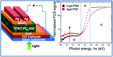
The efficiency of organic photovoltaic devices continues to increase; however, their limited stability is currently a barrier to the commercial prospects of the technology. Burn-in photo degradation, caused by continuous illumination under a light source, can cause a significant reduction in device performance. Our aim was to investigate this degradation pathway for the high-efficiency polymer PTB7, which was compared to the well-studied P3HT:PC71BM material system. In this study, we compared the burn-in aging profile for organic solar cells containing either P3HT or PTB7 as the donor polymer. This showed that PTB7:PC71BM solar cells exhibit a severe initial reduction in performance, due mainly to reduced short circuit current density (Jsc), during the 5 hour test period. P3HT:PC71BM cells were relatively stable during this test. Photothermal deflection spectroscopy (PDS), which provides sensitive measurement of sub bandgap absorption, was employed to discover the underlying mechanism causing this discrepancy. In PTB7-based devices, a significant increase in sub bandgap absorption was observed after illumination, which was attributed to the formation of sub bandgap trap states. This mechanism was identified as a contributing factor to the severe burn-in for PTB7-based organic solar cells. No such increase was observed for P3HT:PC71BM films.


1. Introduction

Organic photovoltaics (OPVs) promise low-cost fabrication of lightweight, flexible solar modules, due to the use of solution processing. Improvements in device processing, including the development of novel solar polymers,1 interfacial engineering2 and morphology control3 have led to increases in the power conversion efficiency (PCE) of OPV devices, with a recent report of 11.7% for a single junction OPV cell.4 As such, one critical remaining barrier to commercialisation is their poor environmental stability.5 Past research on the stability of OPV devices has shown a characteristic rapid degradation at the beginning of the aging curve.6 This significant, initial reduction in overall photovoltaic performance, referred to as ‘burn-in’, requires further study to unravel the surrounding mechanisms. This is particularly important considering that the aging profile of an OPV cell depends on the solar polymer used in the active layer; different polymers are prone to different failure mechanisms,7,8 such that novel polymers which have recently yielded promising results may be subject to a variety of degradation mechanisms. The main factors causing OPV devices to degrade are light, heat, oxygen and water.9 To provide a thorough analysis of changes in the photoactive layer due to light-induced degradation, sensitive characterisation tools, such as photo thermal deflection spectroscopy (PDS) are required. PDS, first developed by Jackson et al.,10,11 is often the best way to measure the absorption coefficient of thin films due to its high sensitivity. Traditional reflection and transmission spectroscopy has limited sensitivity, especially for small absorption near the band-edge. PDS, on the other hand, allows direct measurement of absorption by measuring the subtle temperature change in a surrounding liquid medium brought on by absorbed energy from the sample. PTB7 is a particularly promising solar polymer, due to lower band gap and higher hole mobility, when compared to P3HT.12 P3HT is the most commonly employed polymer for OPV devices, the degradation pathways for P3HT:PCBM cells have been heavily studied.

Herein, we use PDS to investigate the light-induced burn-in degradation in both P3HT:PC71BM and PTB7:PC71BM OPV devices. To the best of our knowledge, this is the first report of a degradation study of PTB7 using PDS to investigate sub bandgap absorption. No change in the PDS spectra for P3HT:PC71BM cells was observed after exposure to light over the 5 hour test period, however, a significant increase in sub bandgap absorption was observed in PTB7. This absorption is indicative of increased trap states. We propose this formation of trap states as a key degradation mechanism in PTB7 based solar cells.

2. Experimental detail

2.1 Device fabrication

Plain and ITO-coated glass substrates (12 mm × 12 mm) were used for all films and devices. They were first cleaned by ultrasonication in soapy deionized (DI) water, DI water, acetone, and isopropanol. PDS samples were prepared by spin coating active layer solution (PTB7:PC71BM and P3HT:PC71BM) directly on plain glass substrates. PTB7 and PC71BM (1-Material, Inc.; polymer: fullerene ratio of 1[thin space (1/6-em)]:[thin space (1/6-em)]1.5, concentration: 25.0 mg mL−1) were mixed in chlorobenzene with 3.0 vol% 1,8-diiodooctane in a N2-filled glovebox and stirred overnight at 60 °C. P3HT:PC71BM active layer solution was made by stirring the mixture of P3HT, and PC71BM (1[thin space (1/6-em)]:[thin space (1/6-em)]1 ratio) in chlorobenzene solution, purchased from 1-Material inc. To fabricate the complete device, ZnO sol–gel solution (0.48 M) was prepared by dissolving zinc acetate dihydrate (Zn(CH3COO)2·2H2O, Sigma-Aldrich, >99.0%, 0.109 g) and ethanolamine (NH2CH2CH2OH, Sigma-Aldrich, >99.5%, 32 μL) in 2-methoxyethanol (CH3OCH2CH2OH, Sigma-Aldrich, 99.8%, anhydrous, 1 mL). It was then spin-cast on top of the pre-cleaned ITO glass substrates at 4000 rpm for 60 s. These samples were then annealed at 170 °C for 30 min. PTB7:PC71BM and P3HT:PC71BM solutions were then spin cast on top of the ZnO coated films at 900 rpm for 120 s and 1200 rpm for 30 s respectively. P3HT:PC71BM devices and films were annealed at 110 °C for 10 min. The active layer coated substrates were loaded in a vacuum chamber (10−6 torr) where a 10 nm film of MoO3 and 100 nm film of Ag were deposited through a shadow mask by thermal evaporation. The device area used was 0.12 cm2. At least six devices were made for each polymer.

2.2 Device characterization

The current density–voltage (JV) measurements were performed using an IV5 solar cell IV testing system from PV measurements, Inc. (using a Keithley 2400 source meter) under illumination power of 100 mW cm−2 by an AM 1.5G solar simulator (Oriel model 94023A; 100 mW cm−2). To record burn-in losses, the samples were illuminated by the same simulated sunlight source at 1 sun intensity in encapsulation and under open-circuit conditions. The test was conducted over a 5 hour period, during which time the photo-induced degradation is most severe. The device temperatures were measured by GM1350 50:1 LCD Infrared Thermometer Digital Gun. The temperatures were maintained near room temperature (25–30 °C) by forced air cooling during the aging process in order to minimize thermally induced degradation effects. For optical characterization of fresh and aged films, a UV-VIS-NIR spectrometer (Perkin Elmer – Lambda 950) was used. The film morphology was observed by atomic force microscopy (AFM) with a Bruker Dimension ICON SPM. X-ray diffraction (XRD) with CuKα radiation was performed at an angle ranging from 2° to 30° by step-scanning with a step size of 0.020°. Photoluminescence (PL) spectra were measured using a 1/4 meter monochromator (Cornerstone™ 260) equipped with a silicon charge-coupled device (CCD) camera. The continuous wave (CW) laser (405 nm, 50 mW) was used in conjunction with an OD1 filter as the excitation source and the luminescence was detected by the CCD. The laser lines were filtered out from the detected signal by a 568 nm low pass filter. The spectra were obtained using a 2 second integration time. Regarding PDS, the excitation light source or the pump beam was the output from a 250 W QTH lamp spectrally filtered by a Cornerstone™ 130 monochromator. The output of the monochromator was further filtered with order sorting filters. A probe laser beam of 635 nm was used in conjunction with a four quadrant position detector to detect the beam deflection caused by the heat gradient. The sample was immersed in Fluorinert FC72™ in a quartz cuvette. An ANFATEC™ lock-in amplifier connected to a computer was used to record the deflection data at each wavelength.

3. Results and discussion

3.1 Burn-in degradation vs. device performance

A degradation of the PCE over the 5 hour test period is observed for both P3HT and PTB7 based devices, this is displayed in Fig. 1(a). The PCE of the P3HT device is relatively stable under illumination during the 5 hour test period; it reduces slightly to 88% of its initial value. In contrast, a rapid reduction in PCE is seen for the PTB7-based device. It reduces to 39% of its initial PCE value after 5 hours of illumination. Fig. 1(b) shows the normalized photovoltaic parameters for the P3HT device. In this case, the reduction in PCE is mainly caused by a reduction in Voc, which degrades to 87% of its initial value. The Jsc and FF remain almost unchanged. Fig. 1(c) shows the normalized photovoltaic parameters for the PTB7 device. All parameters degrade for this case. The largest degradation is observed in the Jsc, which reduces to only 64% of its initial value. FF and Voc also degrade to 85% and 71% of their initial values, respectively. The reduction in Voc observed in both material systems may be related to the ZnO buffer layer. Kam et al.13 showed that under UV illumination, a significant reduction in Voc was observed. This was attributed to shunting channels in ZnO at the ZnO/active layer interface, induced by the illumination. A similar phenomenon was observed under concentrated sunlight by Manor et al.14 Comparing the two material systems, we can see that one of the main differences is the reduction in Jsc for PTB7, whilst that parameter remains almost unchanged for P3HT. Evidently, the aging profile is different for P3HT and PTB7, indicating that a different mechanism underpins the aging process. As all other layers in the devices are identical, this varied mechanism must arise from the photoactive material.
image file: c6ra23288d-f1.tif
Fig. 1 (a) Normalized values of PCE tracked over 5 hours of photo-degradation for P3HT:PC71BM and PTB7:PC71BM devices under illumination power of 100 mW cm−2. The rate of efficiency loss is higher in PTB7:PC71BM devices compared to P3HT:PC71BM devices. (b and c) Normalized values of photovoltaic parameters (PCE, Voc, Jsc and FF) tracked over 5 hours of photo-degradation for P3HT:PC71BM and PTB7:PC71BM devices respectively.

3.2 PDS analysis of fresh and degraded devices

To uncover the mechanism causing this difference in Jsc reduction after aging for PTB7 and P3HT, photothermal deflection spectroscopy (PDS) measurements were employed. PDS was used to measure the absorption coefficient of the active layer blend in the sub bandgap region. The PDS spectra of PTB7:PC71BM and P3HT:PC71BM blend films are displayed in Fig. 2. Photo-absorption in a semiconductor occurs in three regions, these regions are marked in Fig. 2. Absorption in region 1, for energies larger than the bandgap, is caused by a band-to-band (HOMO–LUMO) transition. This can be easily measured using a typical UV-VIS spectrometer. Absorption in region 2 occurs at energies slightly lower than the optical bandgap. The absorption in this region reduces exponentially as the photon energy reduces. The absorption in this region is caused by tail states at the band edge.15 The slope of the curve in this region is indicative of the structural order of the semiconductor. From this region, the Urbach energy, Eu, can be calculated, which gives a quantitative description of the disorder in semiconductor.15 The Urbach energy for the samples used in this work (both fresh and aged) has been calculated according to eqn (1), which describes the absorption coefficient:
 
image file: c6ra23288d-t1.tif(1)
where α0 is a constant, h is Planck's constant, ν is the frequency and Eu is the Urbach energy. A summary of the extracted Urbach energy values can be found in Table 1. The details of this calculation are shown in Fig. S3.

image file: c6ra23288d-f2.tif
Fig. 2 Photothermal deflection spectroscopy (PDS) absorption spectra of P3HT:PC71BM and PTB7:PC71BM films under both fresh and aged conditions. Region 1 is related to absorption due to band-to-band (HOMO–LUMO) transition. In region 2, absorption is caused by tail states at the band edge. The slope of this region can lead to the calculation of Urbach energy, which is indicative of structural disorder in semiconductor. Region 3 is related to absorption at low photon energies due to trap energy level within the bandgap caused by sub bandgap defects. Unlike P3HT, in aged PTB7 based devices, absorption in region 3 is higher (shown by green arrow) which indicates formation of sub bandgap trap states due to burn-in photo-degradation. The inset displays the fitted absorption coefficient, α (cm−1) of all the films solely in region-3, showing the distribution of sub-bandgap absorption along photon energy.
Table 1 The list of Urbach energies of fresh and aged P3HT:PC71BM and PTB7:PC71BM films extracted from region-2 of PDS
Film description Urbach energy (meV)
Fresh P3HT:PC71BM 109.9
Aged P3HT:PC71BM 109.8
Fresh PTB7:PC71BM 48.9
Aged PTB7:PC71BM 71.1


Absorption in region 3 occurs at low photon energies. The magnitude of absorption in this region is orders of magnitude lower, which is why sensitive techniques such as PDS are required. From Fig. 2, the absorption of the P3HT:PC71BM films in both region 2 and 3 is unchanged after exposure to light over the 5 hour test period. The Urbach energy of P3HT:PC71BM film is unchanged, even after photo-ageing, (for fresh film, Eu = 109.9 meV and for aged film, Eu = 109.8 meV). By contrast, the absorption of the PTB7:PC71BM sample is largely affected by exposure to light. The slope in region 2 is clearly reduced after aging. The calculated Urbach energy increases from 48.9 meV in the fresh film, to 71.1 meV in the aged film. An increase in Urbach energy is indicative of increased structural disorder.15,16 This may be related to a broadening of the available density of states (DOS), due to the formation of shallow trap states. A similar observation was seen by Heumueller et al. in another amorphous material (PCDTBT) using charge extraction (CE) technique.17 After exposure to light, the absorption in region 3 is significantly increased for the PTB7:PC71BM film. This is further displayed in Fig. 3, which compares fitted curves of the absorption in this region. The area under the curve is directly related to the number of defect states.18 After aging in the PTB7:PC71BM film, this area increased by a factor of 2.6. However, the area under the curve for P3HT:PC71BM increased only by a factor of 1.14. Details of this calculation are presented in the ESI material (Fig. S4). Absorption in this region is related to sub bandgap defects which cause a trap energy level within the bandgap.7 This observation implies that the photodegradation increases the density of sub bandgap trap states. Due to the controlled environment in this investigation, it is likely that these sub bandgap trap states are caused by an intrinsic effect, such as a photoinduced morphological disorder, rather than an extrinsic defect, like the incorporation of O2.19 Peters et al. observed an increase in sub bandgap absorption due to the formation of trap states in PCDTBT:PC70BM films.7 As with PTB7, PCDTBT is a relatively amorphous polymer. This increase in the density of trap states can increase the rate of recombination, which will limit Jsc. This matches the PCE curves for PTB7:PC71BM cells displayed in Fig. 1, for which the Jsc for PTB7 reduces significantly. The link between PDS and Jsc is also consistent for P3HT:PC71BM material system; no observable increase in sub bandgap absorption was seen in PDS, whilst the Jsc remained almost unchanged. The structure of the two polymers is quite different; P3HT is semi-crystalline20 whilst PTB7 is largely amorphous in nature.21–23 For these material systems, it appears the amorphous polymer is more prone to photodegradation.


image file: c6ra23288d-f3.tif
Fig. 3 Fitted absorption coefficient, α (cm−1) of all the films solely in region-3, showing the distribution of sub-bandgap absorption along photon energy.

3.3 Absorbance and PL analysis of fresh and degraded devices

Fig. 4 displays the absorption and PL spectra for fresh and aged PTB7:PC71BM and P3HT:PC71BM blend films. The spectra measured at intermediate aging times between 0 and 5 hours are displayed in Fig. S5 and S6. As shown in Fig. 4(a), the PL yield for fresh P3HT:PC71​BM (1[thin space (1/6-em)]:[thin space (1/6-em)]1) film is extremely low, which matches with previous literature.24,25 This low PL yield is attributed to exciton quenching and effective charge separation at the polymer: fullerene interface. When compared to the fresh film, the PL yield of the aged film is increased by a factor of 27, this increase in PL signal is due to the photogenerated excitons in P3HT that do not take part in charge separation. From the absorption spectra, also shown in Fig. 4(a), it is evident that there is no significant difference in the photo absorption between fresh and aged films. As such, the increased PL peak is indicative of inefficient charge separation and charge transfer between the electron donating P3HT polymer and electron accepting PCBM molecules, rather than exciton generation. The characteristic PL emission peak of fresh films, at around 720 nm,25 does not undergo a peak-shift in the aged films. The consistency of the PL peak position in fresh and aged films strongly suggests that the phase separation and the aggregated P3HT and PCBM domains in the P3HT:PC71​BM films are not affected by photo-aging of the film.26 Negligible change in film morphology is also evident from the AFM and XRD of fresh and aged P3HT:PC71BM films (Fig. S7 and S8).
image file: c6ra23288d-f4.tif
Fig. 4 (a) PL emission spectra (right) and absorption spectra (left) of fresh and aged P3HT:PC71​BM (1[thin space (1/6-em)]:[thin space (1/6-em)]1) film. (b) PL emission spectra (right) and absorption spectra (left) of fresh and aged PTB7:PC71​BM (1[thin space (1/6-em)]:[thin space (1/6-em)]1.5) film. The black dashed vertical lines identify the absorption and PL emission peak positions. The absorption displayed in this figure is measured using reflection/transmission measurements from a UV/VIS spectrometer.

Fig. 4(b) displays the absorption for fresh and aged PTB7:PC71BM films. In the fresh film, characteristic peaks of PTB7 can be seen at 615 nm and 685 nm, which is in accordance with previous literature.27,28 The peaks (denoted as ABS0-0 and ABS0-1) are related to 0-0 and 0-1 transition between the ground state energy and the first two vibrational levels of the excited state.27 Characteristic peaks of PC71BM can be seen at 375 nm and 475 nm (Fig. 4(b)).29 In the aged films, the PC71BM characteristics peaks remain unaltered, indicating that changes in absorption are related to PTB7. After aging, the absorption intensities (ABS0-1 and ABS0-0) of PTB7 diminish and a spectral blue shift is observed. This phenomenon may be caused by a decreased interchain interaction of the conjugated polymer.27 As the temperature of the experiment was controlled, this spectral blue shift is caused by light induced degradation. Alem et al. observed a similar blue shift in the absorption of different polymers containing benzo dithiophene (BDT) units after light exposure.30 PTB7 is comprised of a BDT unit and an electron-withdrawing thienothiophene (TT) unit. This is shown schematically in Fig. S9. The blue shift was attributed to a reduction of the main chain conjugation, caused by a reaction with oxygen in the BDT unit, which is also present in PTB7. The PL spectra for fresh and aged PTB7:PC71BM films are also shown in Fig. 4(b). For both films, two dominant peaks corresponding to 0-0 and 0-1 transitions at 720 nm and 780 nm, respectively, are present in the PL spectra. The magnitude of the PL signal increases in the aged film, indicating increased radiative recombination of excitons and less effective charge separation between donor polymer and acceptor fullerene.31 In the fresh film, the magnitude of the 0-0 and 0-1 peaks are similar. The ratio of the peaks, RPL = PL0-0/PL0-1, is close to 1. After aging, the 0-1 transition peak (PL′′0-1) undergoes a slight red shift. Additionally, the ratio of the PL peaks changes, the intensity of the low energy peak, PL0-1, increases more than PL0-0, such that RPL = 0.75. RPL < 1 implies the formation of an aggregated phase in the conjugated polymer.32 As shown in Fig. 1(c), the Jsc in PTB7:PC71​BM device drastically reduces with photo-aging. The aggregates formed during photo-aging may serve as electron traps and recombination centers in aged PTB7.33 This supports the PDS data, which shows that the formation of trap states, due to photoinduced aging, occurs in PTB7, but not in P3HT. Evidence of trap-assisted recombination, in both the PDS and PL signals, implies that recombination may occur through both radiative and non-radiative pathways.

4. Conclusion

In conclusion, we compared the burn-in degradation for P3HT:PC71BM and PTB7:PC71BM organic solar cells after illumination over a 5 hour period. Devices with P3HT were relatively stable during the test period, however, the PTB7:PC71BM devices exhibited a significant reduction in efficiency, largely due to reduced Jsc. Photothermal deflection spectroscopy measurements showed an increase in sub bandgap absorption in PTB7:PC71BM films after illumination. This indicates the formation of sub bandgap trap states. No change in sub bandgap absorption was observed in P3HT:PC71BM films. We believe that the formation of these trap states is the mechanism underpinning the severe initial degradation in PTB7-based devices.

Acknowledgements

The authors would like to thank the Australian Centre for Advanced Photovoltaics, UNSW staff and technicians for their support. We are grateful to all of our OPV group members for useful discussions and support during this work. We also acknowledge Future Solar Technologies for providing funding.

References

  1. M. Helgesen, R. Sondergaard and F. C. Krebs, J. Mater. Chem., 2010, 20, 36–60 RSC.
  2. H.-L. Yip and A. K. Y. Jen, Energy Environ. Sci., 2012, 5, 5994–6011 CAS.
  3. C. R. McNeill, Energy Environ. Sci., 2012, 5, 5653–5667 CAS.
  4. J. Zhao, Y. Li, G. Yang, K. Jiang, H. Lin, H. Ade, W. Ma and H. Yan, Nat. Energy, 2016, 1, 15027 CrossRef.
  5. M. Jørgensen, K. Norrman and F. C. Krebs, Sol. Energy Mater. Sol. Cells, 2008, 92, 686–714 CrossRef.
  6. M. O. Reese, S. A. Gevorgyan, M. Jørgensen, E. Bundgaard, S. R. Kurtz, D. S. Ginley, D. C. Olson, M. T. Lloyd, P. Morvillo, E. A. Katz, A. Elschner, O. Haillant, T. R. Currier, V. Shrotriya, M. Hermenau, M. Riede, K. R. Kirov, G. Trimmel, T. Rath, O. Inganäs, F. Zhang, M. Andersson, K. Tvingstedt, M. Lira-Cantu, D. Laird, C. McGuiness, S. Gowrisanker, M. Pannone, M. Xiao, J. Hauch, R. Steim, D. M. DeLongchamp, R. Rösch, H. Hoppe, N. Espinosa, A. Urbina, G. Yaman-Uzunoglu, J.-B. Bonekamp, A. J. J. M. van Breemen, C. Girotto, E. Voroshazi and F. C. Krebs, Sol. Energy Mater. Sol. Cells, 2011, 95, 1253–1267 CrossRef CAS.
  7. C. H. Peters, I. T. Sachs-Quintana, W. R. Mateker, T. Heumueller, J. Rivnay, R. Noriega, Z. M. Beiley, E. T. Hoke, A. Salleo and M. D. McGehee, Adv. Mater., 2011, 24, 663–668 CrossRef PubMed.
  8. E. Voroshazi, I. Cardinaletti, T. Conard and B. P. Rand, Adv. Energy Mater., 2014, 4, 1400848 CrossRef.
  9. W. Greenbank, L. Hirsch, G. Wantz and S. Chambon, Appl. Phys. Lett., 2015, 107, 263301 CrossRef.
  10. W. B. Jackson, N. M. Amer, A. C. Boccara and D. Fournier, Appl. Opt., 1981, 20, 1333–1344 CrossRef CAS PubMed.
  11. A. C. Boccara, W. Jackson, N. M. Amer and D. Fournier, Opt. Lett., 1980, 5, 377–379 CrossRef CAS PubMed.
  12. Y. Liang, Z. Xu, J. Xia, S.-T. Tsai, Y. Wu, G. Li, C. Ray and L. Yu, Adv. Mater., 2010, 22, E135–E138 CrossRef CAS PubMed.
  13. Z. Kam, X. Wang, J. Zhang and J. Wu, ACS Appl. Mater. Interfaces, 2015, 7, 1608–1615 CAS.
  14. A. Manor, E. A. Katz, T. Tromholt and F. C. Krebs, Adv. Energy Mater., 2011, 1, 836–843 CrossRef CAS.
  15. T. Gotoh, S. Nonomura, S. Hirata and S. Nitta, Appl. Surf. Sci., 1997, 113, 278–281 CrossRef.
  16. S. De Wolf, J. Holovsky, S.-J. Moon, P. Löper, B. Niesen, M. Ledinsky, F.-J. Haug, J.-H. Yum and C. Ballif, J. Phys. Chem. Lett., 2014, 5, 1035–1039 CrossRef CAS PubMed.
  17. T. Heumueller, T. M. Burke, W. R. Mateker, I. T. Sachs-Quintana, K. Vandewal, C. J. Brabec and M. D. McGehee, Adv. Energy Mater., 2015, 5, 1500111 CrossRef.
  18. W. B. Jackson and N. M. Amer, Phys. Rev. B: Condens. Matter Mater. Phys., 1982, 25, 5559–5562 CrossRef CAS.
  19. J. A. Carr and S. Chaudhary, Energy Environ. Sci., 2013, 6, 3414–3438 CAS.
  20. S. S. van Bavel, M. Bärenklau, G. de With, H. Hoppe and J. Loos, Adv. Funct. Mater., 2010, 20, 1458–1463 CrossRef CAS.
  21. B. A. Collins, Z. Li, J. R. Tumbleston, E. Gann, C. R. McNeill and H. Ade, Adv. Energy Mater., 2013, 3, 65–74 CrossRef CAS.
  22. L. Lu and L. Yu, Adv. Mater., 2014, 26, 4413–4430 CrossRef CAS PubMed.
  23. M. R. Hammond, R. J. Kline, A. A. Herzing, L. J. Richter, D. S. Germack, H.-W. Ro, C. L. Soles, D. A. Fischer, T. Xu, L. Yu, M. F. Toney and D. M. DeLongchamp, ACS Nano, 2011, 5, 8248–8257 CrossRef CAS PubMed.
  24. J. Piris, T. E. Dykstra, A. A. Bakulin, P. H. M. v. Loosdrecht, W. Knulst, M. T. Trinh, J. M. Schins and L. D. A. Siebbeles, J. Phys. Chem. C, 2009, 113, 14500–14506 CAS.
  25. Y. Kim, S. A. Choulis, J. Nelson, D. D. C. Bradley, S. Cook and J. R. Durrant, J. Mater. Sci., 2005, 40, 1371–1376 CrossRef CAS.
  26. P.-T. Huang, P.-F. Huang, Y.-J. Horng and C.-P. Yang, J. Chin. Chem. Soc., 2013, 60, 467–472 CrossRef CAS.
  27. F. Bencheikh, D. Duché, C. M. Ruiz, J.-J. Simon and L. Escoubas, J. Phys. Chem. C, 2015, 119, 24643–24648 CAS.
  28. S. Cho, B. S. Rolczynski, T. Xu, L. Yu and L. X. Chen, J. Phys. Chem. B, 2015, 119, 7447–7456 CrossRef CAS PubMed.
  29. G. J. Hedley, A. J. Ward, A. Alekseev, C. T. Howells, E. R. Martins, L. A. Serrano, G. Cooke, A. Ruseckas and I. D. W. Samuel, Nat. Commun., 2013, 4, 2867 Search PubMed.
  30. S. Alem, S. Wakim, J. Lu, G. Robertson, J. Ding and Y. Tao, ACS Appl. Mater. Interfaces, 2012, 4, 2993–2998 CAS.
  31. K. Tvingstedt, K. Vandewal, F. Zhang and O. Inganäs, J. Phys. Chem. C, 2010, 114, 21824–21832 CAS.
  32. H. Yamagata and F. C. Spano, J. Chem. Phys., 2012, 136, 184901 CrossRef CAS PubMed.
  33. F. C. Spano, Acc. Chem. Res., 2010, 43, 429–439 CrossRef CAS PubMed.

Footnote

Electronic supplementary information (ESI) available. See DOI: 10.1039/c6ra23288d

This journal is © The Royal Society of Chemistry 2016
Click here to see how this site uses Cookies. View our privacy policy here.