
Enhanced lithium storage capabilities of NiO@Si core–shell nanowall arrays by voltage-control technique and their use as anode materials for lithium-ion batteries
Shali Wu,
Ning Du*,
Hao Wu,
Chengmao Xiao,
Wenjia Zhao and
Deren Yang
State Key Laboratory of Silicon Materials, School of Materials Science and Engineering, Cyrus Tang Center for Sensor Materials and Applications, Zhejiang University, Hangzhou 310027, People's Republic of China. E-mail: dna1122@zju.edu.cn; Fax: +86-571-87952322; Tel: +86-571-87953190
First published on 2nd November 2016
Abstract
In this work, we demonstrate the synthesis of Ni–Li2O@Si core–shell nanowall arrays through the lithiation of pre-synthesized NiO@Si core–shell nanowall arrays during the first lithium insertion process. When we control the potential window between 0.01–1.0 V, the electrochemically active NiO irreversibly reacts with Li to form Ni–Li2O nanowall cores. These Ni–Li2O cores function as both a stable mechanical support and an efficient electron conducting pathway in the later cycles while the coated Si shell functions as the active material during the charge and discharge process. The elaborately designed electrode shows better cycling and rate performance when acting as a lithium-ion battery anode compared to a planar Ni@Si electrode.
1. Introduction
Rechargeable lithium-ion batteries are commonly recognized as the most promising power sources for modern portable equipment and vehicles. However, as the most widely used anode material for commercial lithium-ion batteries, graphite has a relative low theoretical capacity (∼370 mA h g−1), which greatly limits the application of lithium-ion batteries in high power density devices. Silicon (Si), which has the highest theoretical gravimetric capacity (∼4200 mA h g−1),1–3 is considered as one of the most promising candidates. In addition, Si has the advantages of low discharge potential (0.37 V vs. Li/Li+), being non-toxic and having abundant reserves. However, pure silicon anodes undergo large volume change (up to 300%) during a lithium insertion and extraction process, resulting in structural damage, capacity fading and poor cycling life.4,5To circumvent these issues, tremendous efforts have been made. Designing the nano/microstructures of Si materials such as nanoparticles,6,7 nanospheres,8 nanowires,9,10 nanotubes,11,12 thin films,13,14 nanoporous Si15,16 and three-dimensional network current collector supported Si17,18 is one of the effective approaches. Among these structures, nanowall arrays in situ grown on a current collector represent good performance. The open, porous and connective electrode configuration provides ideal electrolyte and lithium ion paths, shortens lithium diffusion distances, and enhances interface contact and structure stability, which is beneficial for the high-rate performance of the batteries.19,20 For instance, Wang et al. reported hierarchical nanoarchitectured Ni3S2/Ni arrays grown on the Ni foam fabricated by a hydrothermal method, which exhibited a sustained reversible capacity at 2C rate with the loss of 18% after 20 cycles.21 Besides, the inactive/active core–shell structures can also enhance the conductivity and accommodate the volume change.22,23
Herein, we demonstrate the construction of Ni–Li2O@Si nanowall arrays as high performance anode materials for lithium ion batteries. NiO nanowall arrays24,25 are firstly prepared in situ onto a Ni foam substrate by a hydrothermal route and followed by the deposition of Si through Rf-sputtering. Subsequently, we control the working voltage to the range of 0.01–1 V to prevent the reaction between Ni and Li2O during the delithiation process. Thus we obtain a novel material of Ni–Li2O@Si nanowall arrays after the first cycle. To prepare Ni@Si nanowall arrays, the synthesis method usually involves complicated electrochemical deposition or thermal reduction of pre-prepared NiO precursor. Therefore, compared to previous work, the synthesis route of the voltage-control method is simplified and facile since the phase-transition process of NiO has been incorporated into the electrochemical testing procedure, which could be extended to other Si-based materials. In the composite core, the robust Li2O matrix functions as mechanical support and the Ni nanoparticles in contact with each other function as a built-in current collector, which allows effective electron conduction. The as-synthesized nanowall arrays show outstanding capacity retention and long cycle life when applied as an anode material for lithium ion batteries.
2. Experimental section
2.1 Synthesis of NiO nanowall arrays on Ni foam
Firstly, a piece of Ni foam with the size of 2 × 4 cm2 was put into the diluted hydrochloric acid for 8 min to remove the NiO layer on the surface. Then we rinsed it with deionized water and absolute ethanol for several times. One side of the Ni foam was coated with a polytetrafluoroethylene tape to protect it from solution contamination.0.3 g NiNO3·6H2O and 1.0 g CO(NH2)2 were dissolved in 70 mL deionized water followed by stirring for 20 min. The homogeneous solution was then transferred into a Teflon-lined stainless steel autoclave. The washed Ni foam was then placed into the homogeneous solution vertically. The autoclave was sealed and placed in an oven, reacting at 100 °C for 90 min. After the reaction, the product was cooled down to room temperature and then taken out and rinsed with deionized water and ethanol for several times. Afterwards it was dried under vacuum at 40 °C for 1 h. Finally, the product was calcined in a furnace at 350 °C in a nitrogen or argon atmosphere with the ramping rate of 5 °C min−1 for 2 h.
2.2 Synthesis of NiO@Si nanowall arrays
Si films were deposited onto the substrate by Rf-sputtering with a 99.999% Si target for 30 min. The working pressure was 2 Pa and the power was 80 W.2.3 Materials characterization and electrochemical measurement
The product was characterized by X-ray diffraction (XRD), scanning electron microscopy (SEM HITACH S4800) and transmission electron microscopy (TEM FEI F20) accompanied with an energy-dispersive X-ray spectrometer (EDX). The Raman spectrum was recorded on a HR800 Raman spectrometer. The excitation was carried out with a 532 nm laser at the power of 10 mW.2.4 Electrochemical measurement
2025 coin type half cells were assembled in an argon filled glove box (Mbraun, Labstar, Germany) to determine the electrochemical properties of the products with a lithium foil (15 mm diameter) as the counter electrode. The electrolyte solution was 1 M solution of LiPF6 in ethylene carbonate (EC) and dimethylcarbonate (DMC) (1![[thin space (1/6-em)]](https://www.rsc.org/images/entities/char_2009.gif) :1 by volume). The oxygen and water contents were less than 5 ppm. Galvanostatic cycling test of the assembled cells was carried out on a Land CT2001A system with voltage cut-off of 0.01–1 V vs. Li/Li+ at a current density of 2.1 A g−1 (1C = 4.2 A g−1). Cyclic voltammetry in the potential range of 0.01–1 V was carried out on MSTAT4 (Arbin Instruments) system at a scan rate of 0.1 mV s−1. In the electrochemical impedance spectroscopy (EIS) measurement, the amplitude applied to the cells was 5 mV and the frequency range was from 0.01 Hz to 100 kHz.
:1 by volume). The oxygen and water contents were less than 5 ppm. Galvanostatic cycling test of the assembled cells was carried out on a Land CT2001A system with voltage cut-off of 0.01–1 V vs. Li/Li+ at a current density of 2.1 A g−1 (1C = 4.2 A g−1). Cyclic voltammetry in the potential range of 0.01–1 V was carried out on MSTAT4 (Arbin Instruments) system at a scan rate of 0.1 mV s−1. In the electrochemical impedance spectroscopy (EIS) measurement, the amplitude applied to the cells was 5 mV and the frequency range was from 0.01 Hz to 100 kHz.
3. Results and discussion
Fig. 1a shows the fabrication process of Ni–Li2O@Si nanowall arrays. NiO nanowall arrays were firstly grown on a Ni foam through a hydrothermal method followed by the thermal treatment. Si films were then deposited by a Rf-sputtering method. With the control of potential window, the lithiation process of NiO became irreversible and hence the formation of Ni–Li2O@Si core–shell nanowall arrays. During the first lithiation process, NiO nanowalls reacted with Li according to the following reaction: NiO + 2Li+ + 2e− → Ni + Li2O, and irreversibly transformed into nickel nanoparticles. The schematic illustration of the working mechanism is shown in Fig. 1b.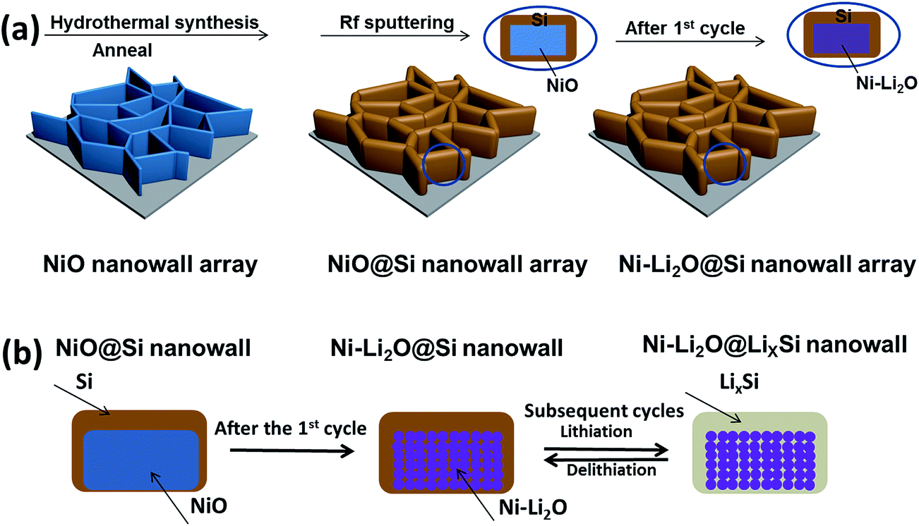 | ||
| Fig. 1 Schematic illustration for (a) the synthetic process of Ni–Li2O@Si core–shell nanowall arrays and (b) the working mechanism. | ||
As the XRD patterns for NiO@Si nanowall arrays on a Ni foam substrate shown (Fig. 2a), all the Bragg peaks are consistent with those of the NiO phase except for the peaks originating from the Ni substrate. There are no peaks of Si phase detected likely due to the amorphous nature of the deposited Si. To eliminate the influence of the original oxidation of Ni foam, we also show the XRD patterns for a Ni foam substrate (Fig. 2b). In Fig. 2b, only the diffraction peaks of Ni can be observed, confirming that the NiO is obtained afterwards. To further demonstrate the existence of NiO and Si, we have the Raman spectrum of the as-synthesized material in Fig. 2c and the peak near 490 cm−1 represents the amorphous Si layer in allowable error while the two peaks in 1400 cm−1 and 1600 cm−1 demonstrate the existence of NiO.
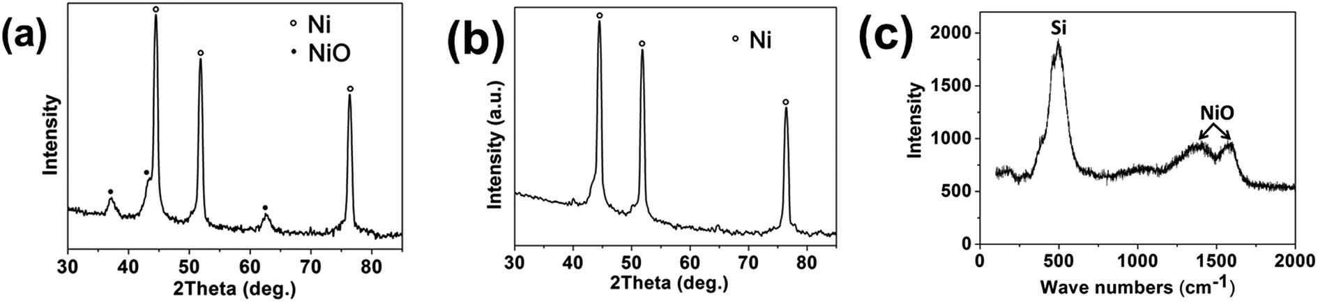 | ||
| Fig. 2 XRD patterns of (a) NiO@Si nanowall arrays on Ni foam substrate and (b) pure Ni foam substrate. (c) Raman spectrum of NiO@Si nanowall arrays. | ||
Fig. 3a is the SEM image of the as-synthesized NiO nanowall arrays. The NiO nanowall arrays exhibit a porous structure with some interconnected nanoflakes. The thickness of the NiO nanowalls is about 20–30 nm. After the deposition of Si layer for 30 min, the thickness of NiO@Si nanowall arrays is greatly increased to about 80–100 nm as shown in Fig. 3b and c. The inset in Fig. 3c shows the cross sectional view of the NiO@Si nanowall arrays, where Si was deposited from top to bottom onto the surface of NiO nanowall arrays. Fig. 3d is the EDX spectrum of NiO@Si nanowall arrays with the peaks of Si, O and Ni, which comes from the substrate and NiO nanowalls, respectively.
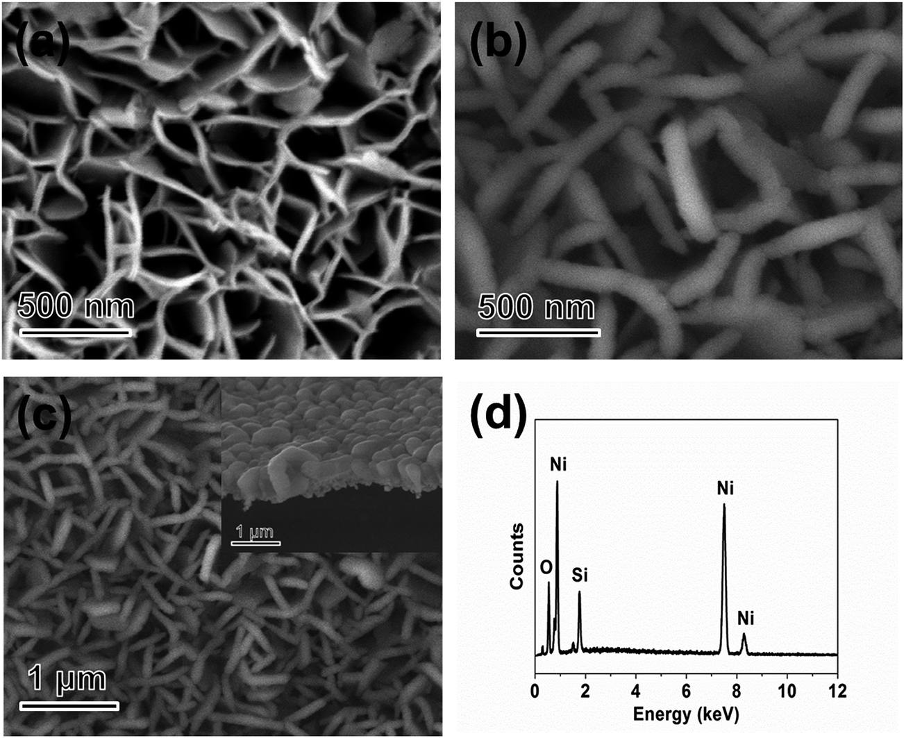 | ||
| Fig. 3 SEM images of (a) NiO nanowall arrays, (b) and (c) NiO@Si core–shell nanowall arrays and (d) corresponding EDX spectrum of NiO@Si core–shell nanowall arrays. | ||
TEM, HRTEM, STEM and EDX analyses are used to demonstrate the core–shell structure and composition of NiO@Si nanowalls. The core–shell structure can be observed distinctly in the TEM and STEM images of an individual NiO@Si nanowall (Fig. 4a and c). As the HRTEM image of the NiO@Si nanowall (Fig. 4b) shown, the nanowall is comprised of small nanocrystals. The lattice fringes with the lattice spacing of 0.21 nm correspond to the {200} planes of NiO.22 The line scanning of the NiO@Si core–shell nanowall (Fig. 4d) shows the element distribution of O, Si and Ni, which further confirms the core–shell structure (Fig. 4c).
 | ||
| Fig. 4 (a) TEM image and (b) HRTEM image of an individual NiO@Si core–shell nanowall; (c) HAADF-STEM image and (d) compositional line profiles of O, Si and Ni for the NiO@Si core–shell nanowall. | ||
The electrochemical performance of the composite material has been systematically measured by assembling half cells with the nanowall arrays directly as the working electrode and lithium foil as the counter electrode. Fig. 5a and b present the first three cyclic voltammogram (CV) curves of the Ni–Li2O nanowall arrays material between 0.01–1 V versus Li/Li+ at a scan rate of 0.1 mV s−1 at room temperature. In the first cycle (Fig. 5a), we have a cathodic peak at ∼0.2 V, which is ascribed to the insertion of the lithium ion to form LixSi.26,27 The peaks at ∼0.55 V and ∼0.8 V is observed corresponding to the initial reduction of NiO to metallic Ni, with Li2O coming into being and the formation of solid electrolyte interphase (SEI) layer.28,29 In the anodic process, two peaks at ∼0.4 V and 0.5 V represent the extraction of lithium from LixSi alloy.26,30 It should be pointed out that the anodic peaks of NiO located at ∼1.25 V and ∼2.2 V.29,31 An enlarged view of this segment is provided as an inset for a better look. Herein the Ni and Li2O cannot get back to NiO and Li by controlling the potential window between 0.01–1 V. Consequently, we obtain Ni–Li2O@Si nanowall arrays material after the first cycle. As a result, we can only find typical redox peaks of Si in the second and third cycles (Fig. 5b) with Ni–Li2O nanowall arrays serving as mechanical support and current collector.
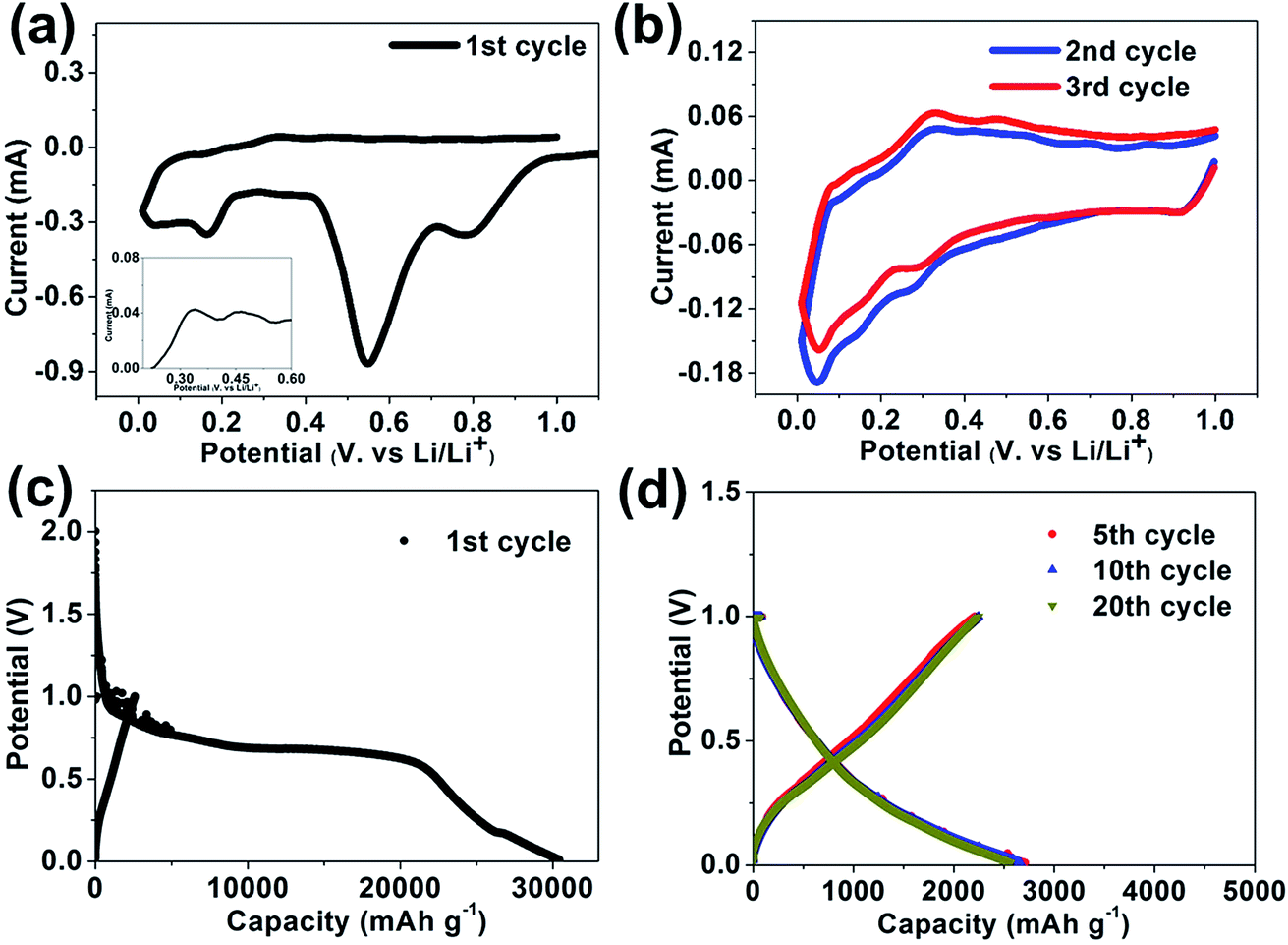 | ||
| Fig. 5 (a) The 1st, (b) 2nd and 3rd cyclic voltammograms curves of NiO@Si core–shell nanowall arrays electrodes between 0–1.0 V versus Li/Li+ at a scan rate of 0.1 mV s−1. (c) The 1st, (d) 5th, 10th and 20th galvanostatic charge/discharge profiles plotted of NiO@Si core–shell nanowall arrays electrodes at the current density of 2.1 A g−1. | ||
Fig. 5c and d show the charge–discharge curves for the as-synthesized materials on the 1st (Fig. 5c) and 5th, 10th and 20th (Fig. 5d) cycles at a current density of 0.5C (2.1 A g−1) with the potential window between 0.01–1 V. In the first discharge curve, sloping profiles are between 0.25 and 0.01 V, corresponding to lithiation behavior of amorphous Si. We can also see plateaus at 0.55 V and 0.8 V, which is consistent with the CV curves, referring to the insertion of lithium ions into the NiO and the formation of the SEI layer. These two plateaus disappear at the cycles afterwards for the reason that the potential window (0.01–1 V) has excluded the delithiation potential of NiO and the lithium ions can no longer come out while the plateaus for the SEI layer disappear as the SEI layer becomes stable.
Fig. 6a exhibits the charge and discharge capacity of Ni–Li2O@Si nanowall arrays versus cycle number compared with planar Ni@Si and NiO@Si nanowall arrays (the time of Rf-sputtering of Si is the same). The potential window of Ni–Li2O@Si nanowall arrays and planar Si was controlled to 0.01–1 V while NiO@Si nanowall arrays was controlled to 0.01–3 V at the same current density of 0.5C. As we can see, the first charge and discharge capacities of the composite are 2687 and 30425 mA h g−1 (only with the respect to the mass of Si (0.05 mg cm−2), the mass of NiO is not counted). The irreversible capacity loss in the first cycle is mainly due to the irreversible reaction of Li with NiO. Besides, the formation of the SEI and the irreversible reaction of Li with the dangling bonds and surface oxides that possibly introduced in Si during sample transportation and battery assembly also contribute to the loss of capacity. As we can see, the specific capacity of Ni–Li2O@Si electrode still remains 1690 mA h g−1 after 500 cycles while the planar Ni@Si electrode shows the capacity of only 151 mA h g−1. It is believed that the enhanced performance can be attributed to the ideal electrolyte and lithium ion paths provide by the optimized electrode configuration of Ni–Li2O@Si nanowall arrays anode. In the composite core, the Li2O matrix functions as mechanical support and the Ni nanoparticles in contact with each other function as a built-in current collector, which allows effective electron conduction. Furthermore, this kind of structure can also shorten the diffusion distance for lithium and enhance the interface contact. On the other side, when the potential window was controlled to 0.01–3 V, the active anode material refers to NiO@Si composite material and the specific capacity decreases from 925 mA h g−1 to 400 mA h g−1 after 60 cycles. To further explain the reason of low coulombic efficiency during the first few cycles, we have the profile of the irreversible capacity of both Ni–Li2O@Si core–shell nanowall arrays and NiO nanowall arrays at the potential window of 0.01–1.0 V. As is shown in Fig. 6b, these two profiles mostly overlaps, indicating the irreversible capacity of Ni–Li2O@Si nanowall arrays during the initial stage is mainly contributed by the irreversible reaction between NiO and Li. Nevertheless, after the first dozens of cycles, the reaction tends to slow down, and the coulombic efficiency of the Ni–Li2O@Si nanowall arrays stays above 97% from the 80th cycle as shown in Fig. 6a.
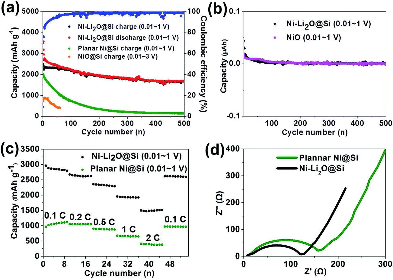 | ||
| Fig. 6 (a) Cycling performance of Ni–Li2O@Si core–shell nanowall arrays (0.01–1 V), planar Ni@Si (0.01–1 V) NiO@Si core–shell nanowall arrays (0.01–3 V). (b) Irreversible capacity of Ni–Li2O@Si core–shell nanowall arrays and NiO nanowall arrays (0.01–1 V). (c) Rate performance of Ni–Li2O@Si core–shell nanowall arrays and planar Ni@Si electrode at varies current densities. (d) Nyquist plots of Ni–Li2O@Si core–shell nanowall arrays and planar Ni@Si after 10 cycles by applying a sine wave with amplitude of 5 mV over the frequency range 100 kHz to 0.01 Hz. | ||
As shown in Fig. 6c, the rate capacity of the Ni–Li2O@Si nanowall arrays and planar Ni@Si nanowall arrays is investigated. When the current rate increased from 0.1C, 0.2C, 0.5C, 1C to 2C, the specific capacities of Ni–Li2O@Si nanowall arrays changed from 2849.6, 2618.8, 2326.0, 1928.6 to 1486.5 mA h g−1, respectively, exhibiting decent capacity retention while the planar Ni@Si nanowall arrays display a quite low capacity during any current rate compared to the other one. When the current went back to 0.1C, 92% of the capacity can be recovered approximately, indicating the preeminent reversibility of the electrode material. The core–shell nanowall arrays structure can accommodate the volume change as well as enhance the conductivity, which results in the improved capacity retention.
Fig. 6d presents the Nyquist plots of Ni–Li2O@Si nanowall arrays and planar Ni@Si electrode after 10 cycles at room temperature in the frequency range of 100 kHz to 0.01 Hz with a sine wave. Both of the two curves show the similar shape of Nyquist plots, composed of a depressed semicircle where a high-frequency semicircle and a medium semicircle overlap each other and followed by an inclines line in low-frequency region. In impedance spectroscopy, it is well accepted that the high frequency semicircle is dominated by the SEI film and/or contact resistance, the semicircle of medium frequency region is attributed to the interfacial charge transfer impedance. In the low frequency range, the straight line corresponds to the lithium-diffusion process within electrodes.32 The diameter of the Ni–Li2O@Si nanowall arrays electrode is obviously smaller than that of the planar Ni@Si electrode, which indicates the core–shell nanowall arrays electrode has a lower transfer resistance as well as a faster transfer reaction during the working process. These results further verify that the core–shell nanowall arrays contribute to the enhanced conductivity of the electrode.
Table 133–39 shows the electrochemical performance of other Si-based materials in previous works. In comparison with other materials, the Ni–Li2O@Si nanowall arrays herein presented a better cycling performance (with a reversible capacity of 2231 mA h g−1 after 100 cycles).
| Si-based materials | Main body structure | 1st discharge capacity (mA h g−1) | 1st charge capacity (mA h g−1) | Coulombic efficiency (%) | Reversible capacity after x cycles (mA h g−1) | Ref. |
|---|---|---|---|---|---|---|
| Si/SiOx/graphite | Si crystals (∼5 nm) dispersed in solid SiOx | 1516 | 1002 | 66.1 | 710 (x = 100) | 33 |
| Si@SiOx/C | C-coated solid Si microparticles (∼1 μm) | 1805 | 913 | 50.6 | 885 (x = 60) | 34 |
| 3D c-Si | (1) Si nanocrystals (∼100 nm); (2) interconnected pores (∼200 nm) | 3138 | 2820 | 88 | 2780 (x = 100) | 35 |
| Core–shell bulk@nanowire Si | (1) SiO2 coating (∼7 nm); (2) nanowire shell (∼1.4 μm) | 2279 | 2117 | 92 | 1757 (x = 50) | 36 |
| Porous Si@C | Carbon layer (40 nm thick) | 2530 | 2390 | 94.4 | 2050 (x = 50) | 37 |
| c-mSi | (1) Si nanowires (5–8 μm in length); (2) full of nanopores (∼10 nm) | 2440 | 2150 | 88 | 1850 (x = 20) | 38 |
| SiOx–PANI–AG | (1) Full of nanopores (∼3 nm); (2) well-embedded Si nanocrystals (∼5 nm); (3) PANI–Ag nanolayer coating (∼10 nm) | 2573 | 1533 | 59.6 | 1149 (x = 100) | 39 |
| Ni–Li2O@SI nanowall | Core–shell Ni–Li2O@Si nanowall | 30425 |
2687 | 8.8 | 2231 (x = 100) | This work |
To demonstrate the Ni–Li2O@Si nanowall structure directly, we have the HRTEM image of Ni–Li2O@Si (Fig. 7a) arrays after 5 cycles. The lattice fringes spaced by 2.17 Å correspond to Ni {002} distance, while fringes spaced by 2.6 Å correspond to Li2O {111} distance. The SAED pattern (Fig. 7b), which shows rings made up of discrete spots related to polycrystalline nature of the material, further evidences the composition of the nanowall. These rings are unambiguously indexed indicating the presence of amorphous Li2O and metallic nickle.
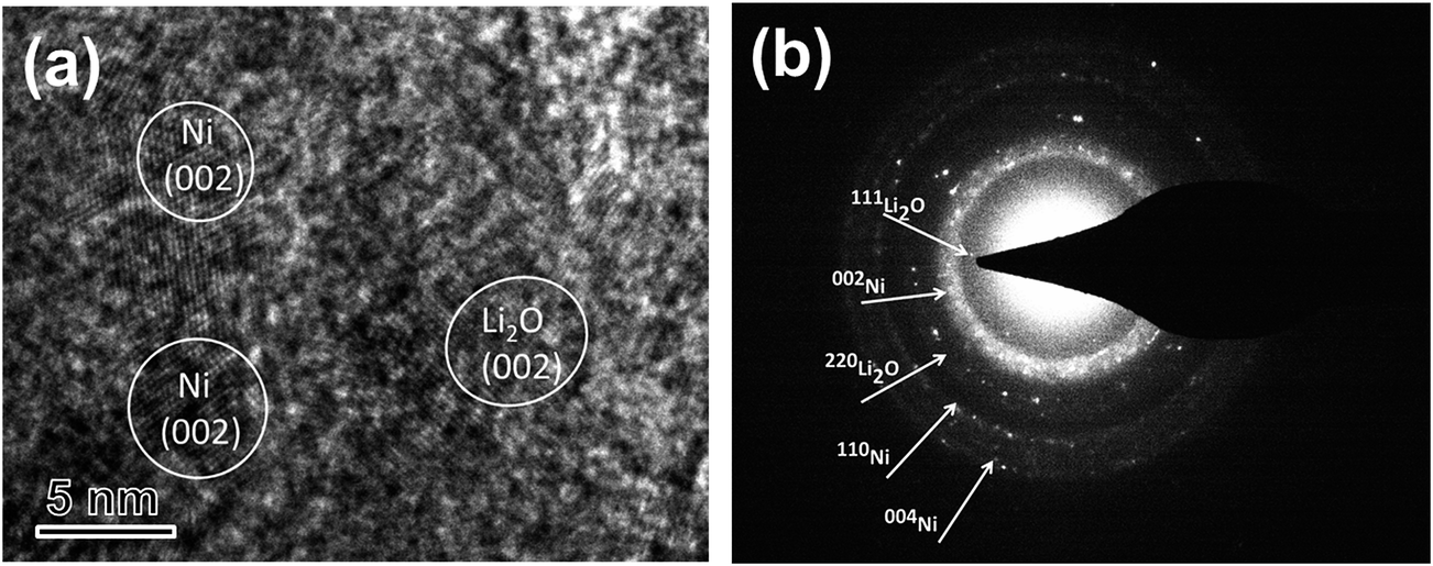 | ||
| Fig. 7 (a) HRTEM image and (b) SAED pattern of a Ni–Li2O@Si nanowall recovered from fully charged battery cells. | ||
To further testify the stability of Ni–Li2O@Si nanowall array electrode, the morphologies of Ni–Li2O@Si nanowall arrays and planar Ni@Si after 20 cycles at a current density of 2.1 A g−1 were characterized (Fig. 8). The morphology of the core–shell nanowall arrays (Fig. 8a) is mostly retained while the planar Ni@Si film (Fig. 8b) displays distinct cracking and pulverization. This difference manifests that the core–shell nanowall arrays structure can remit the volume change during the lithium alloying/dealloying process to a great extent which may also be ascribed to the improved electrochemical performance of the electrode.
 | ||
| Fig. 8 SEM images of (a) the Ni–Li2O core–shell nanowall arrays and (b) planar Ni@Si after 20 cycles at the current density of 0.5C. | ||
4. Conclusions
In conclusion, NiO@Si nanowall arrays were fabricated and subsequently formed to Ni–Li2O@Si nanowall arrays via voltage-control technique. The as-synthesized material showed excellent cycle performance (1690 mA h g−1 after 500 cycles at the current rate of 0.5C) and high rate performance. The core–shell nanowall arrays structure can remit the volume change and enhance the conductivity during the lithium alloying/dealloying process, which may be ascribed to the improved electrochemical performance of the electrode. This voltage-control method can be applied to other composite materials in the area of rechargeable lithium ion batteries.Acknowledgements
The authors would like to appreciate the financial supports from the Fundamental Research Funds for the Central Universities and the Program for Innovative Research Team in University of Ministry of Education of China (IRT13R54).References
- J. M. Tarascon and M. Armand, Nature, 2001, 414, 359–367 CrossRef CAS PubMed.
- M. Winter, J. O. Besenhard, M. E. Spahr and P. Novak, Adv. Mater., 1998, 10, 725–763 CrossRef CAS.
- J. B. Goodenough and Y. Kim, Chem. Mater., 2009, 22, 587–603 CrossRef.
- C. K. Chan, H. L. Peng, G. Liu, K. Mcllwrath, X. F. Zhang, R. A. Huggins and Y. Cui, Nat. Nanotechnol., 2010, 4, 1443–1450 CAS.
- B. A. Boukamp, G. C. Lesh and R. A. Huggins, J. Electrochem. Soc., 1981, 128, 725–729 CrossRef CAS.
- A. Magasinski, P. Dixon, B. Hertzberg, A. Kvit, J. Alaya and G. Yushin, Nat. Mater., 2010, 9, 353–358 CrossRef CAS PubMed.
- J. R. Szczech and S. Jin, Energy Environ. Sci., 2011, 4, 56–72 CAS.
- Y. Yao, M. T. McDowell, I. Ryu, H. Wu, N. A. Liu, L. B. Hu, W. D. Nix and Y. Cui, Nano Lett., 2011, 11, 2949–2954 CrossRef CAS PubMed.
- H. Wu and Y. Cui, Nano Today, 2012, 7, 414–429 CrossRef CAS.
- X. H. Wu, L. Q. Zhang, L. Zhong, Y. Liu, H. Zheng, J. W. Wang, J. H. Cho, S. A. Dayeh, S. T. Picraux and J. P. Sullivan, Nano Lett., 2011, 11, 2251–2258 CrossRef PubMed.
- M. H. Park, M. G. Kim, J. Joo, K. Kim, J. Kim, S. Ahn, Y. Cui and J. Cho, Nano Lett., 2009, 9, 3844–3947 CrossRef CAS PubMed.
- M. R. Zamfir, H. T. Nguyen, E. Moyen, Y. H. Lee and D. Pribat, J. Mater. Chem. A, 2013, 1, 9566–9586 CAS.
- C. M. Hwang, C. H. Lim, J. H. Yang and J. W. Park, J. Power Sources, 2010, 3, 124–129 Search PubMed.
- T. Takamura, S. Ohara, M. Uehara, J. Suzuki and K. Sekine, J. Power Sources, 2003, 129, 96–100 CrossRef.
- H. Kim, B. Han, J. Uehara, J. Choo and J. Cho, Angew. Chem., Int. Ed., 2008, 47, 10151–10154 CrossRef CAS PubMed.
- Z. Y. Jiang, C. L. Li, S. J. Hao, K. Zhu and P. Zhang, Electrochim. Acta, 2014, 115, 393–398 CrossRef CAS.
- Y. P. Liu, K. Huang, Y. Fan, Q. Zhang, F. Sun, T. Gao, L. W. Yang and J. X. Zhong, Electrochim. Acta, 2012, 88, 766–771 CrossRef.
- W. Wang, M. Tian, Y. J. Wei, S. H. Lee, Y. C. Lee and R. G. Yang, Nano Energy, 2013, 2, 942–950 Search PubMed.
- J. P. Liu, Y. Y. Li, X. T. Huang, R. M. Ding, Y. Y. Hu, J. Jiang and L. Liao, J. Mater. Chem., 2009, 19, 1859–1864 RSC.
- Y. G. Li, B. Tan and Y. Y. Wu, Nano Lett., 2008, 8, 265–270 CrossRef CAS PubMed.
- Q. Wang, R. Gao and J. H. Li, Appl. Phys. Lett., 2007, 90, 143107 CrossRef.
- Z. Wei, R. S. Li, T. Huang and A. S. Yu, J. Power Sources, 2013, 128, 165–172 CrossRef.
- H. Liu, L. B. Hu, Y. S. Meng and Q. Li, Nanoscale, 2013, 5, 10376–10383 RSC.
- Q. Wang, C. Y. Zhang, W. F. Shan, L. L. Xing and X. Y. Xue, Mater. Lett., 2014, 118, 66–68 CrossRef CAS.
- L. L. Xing, C. X. Cui, B. He, Y. X. Nie, P. Deng and X. Y. Xue, Mater. Lett., 2013, 96, 158–161 CrossRef CAS.
- T. Jiang, S. C. Zhang, X. P. Qiu, W. T. Zhu and L. Q. Chen, Electrochem. Commun., 2006, 11, 930–934 Search PubMed.
- M. Green, E. Fielder, B. Scrosati, M. Wachtler and J. Serra Moreno, Electrochem. Solid-State Lett., 2003, 6, A75–A79 CrossRef CAS.
- Y. J. Mai, X. H. Xia, R. Chen, C. D. Gu, X. L. Wang and J. P. Tu, Electrochim. Acta, 2012, 67, 73–78 CrossRef CAS.
- Y. Q. Zhou and Y. Wang, Nanoscale, 2011, 3, 2615–2620 RSC.
- W. R. Liu, Z. Z. Guo, W. S. Young, D. T. Shieh, H. C. Wu, M. H. Yang and N. L. Wu, J. Power Sources, 2005, 140, 139–144 CrossRef CAS.
- B. Varghese, M. V. Reddy, Z. Yanwu, C. S. Lit, T. C. Hoong, G. V. S. Rao, B. V. R. Chowdari, A. T. S. Wee, C. T. Lim and C. H. Sow, Chem. Mater., 2008, 20, 3360–3367 CrossRef CAS.
- J. X. Yu, N. Du, H. Zhang and D. R. Yang, RSC Adv., 2013, 3, 7713–7717 RSC.
- C. M. Park, W. Choi, Y. Hwa, J. H. Kim, G. Jeong and H. J. Sohn, Chem. Commun., 2010, 20, 4854–4860 CAS.
- H. C. Tao, M. Huang, L. Z. Fan and X. Qu, Solid State Ionics, 2012, 220, 1–6 CrossRef CAS.
- H. Kim, B. Han, J. Choo and J. Cho, Angew. Chem., Int. Ed., 2008, 47, 10151–10154 CrossRef CAS PubMed.
- S. Sim, P. Oh, S. Park and J. Cho, Adv. Mater., 2013, 25, 4498–4503 CrossRef CAS PubMed.
- B. M. Bang, J. I. Lee, H. Kim, J. Cho and S. Park, Adv. Energy Mater., 2012, 2, 878–883 CrossRef CAS.
- B. M. Bang, H. Kim, H. K. Song, J. Cho and S. Park, Energy Environ. Sci., 2011, 4, 5013–5019 CAS.
- P. J. Zhang, L. B. Wang, J. Xie, L. W. Su and C. A. Ma, J. Mater. Chem A, 2014, 2, 3776–3782 CAS.
| This journal is © The Royal Society of Chemistry 2016 |