The effect of doped heteroatoms (nitrogen, boron, phosphorus) on inhibition thermal oxidation of reduced graphene oxide

Xiaowei Mua, Bihe Yuanb, Xiaming Fenga, Shuilai Qiua, Lei Song*a and Yuan Hu*a
aState Key Laboratory of Fire Science, University of Science and Technology of China, Hefei 230026, China. E-mail: leisong@ustc.edu.cn; yuanhu@ustc.edu.cn; Fax: +86-551-63601664; Tel: +86-551-63601664
bSchool of Resources and Environmental Engineering, Wuhan University of Technology, Wuhan 430070, China

Received 25th August 2016 , Accepted 28th October 2016

First published on 28th October 2016


Abstract

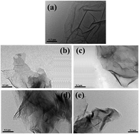
The doping of nitrogen into reduced graphene oxide (NRGO) is achieved by reduction of graphene oxide with hydrazine hydrate and ammonium hydroxide. Boron (BRGO) or phosphorus (PRGO) doped reduced graphene oxide (RGO) is obtained by annealing of RGO (prepared by reduction with sodium borohydride) with boric acid or phosphoric acid, respectively. The successful preparation of the doped RGO is confirmed by Fourier transform infrared spectroscopy, X-ray diffraction, Raman spectroscopy, X-ray photoelectron spectroscopy and transmission electron microscopy. A remarkable enhancement in thermal oxidative stability of RGO is achieved by doping of these heteroatoms. The enthalpy values of BRGO and PRGO during the thermal oxidation decrease remarkably compared with that of RGO, indicating the reduced heat release by the doped heteroatoms. The mechanism for improvement in thermal oxidative resistance by doping of heteroatoms is demonstrated clearly. Doping of boron atom not only lowers the electrons density on the reactive carbon atoms and the Fermi level of carbon, but also contribute to graphitization of RGO, leading to inhibition of thermal oxidation of RGO. Phosphorus containing complexes, in the forms of metaphosphates, C–O–PO3 and C–PO3 groups, can poison the active sites on RGO and function as physical barrier for the access of oxygen. Retarding oxidation of RGO against air may be strengthened by forming more thermostable structure in NRGO, for example: pyrrolic-N (NC2), pyridinic-N (NC2) and graphitic-N (NC3). The doping of heteroatoms will provide an important strategy for broadening RGO application at the elevated temperature.


1. Introduction

Carbon materials, which are classified as carbon fibers, carbon–carbon composites and graphite according to intrinsic structure, show wide applications as supercapacitors, electrodes, fuel cells, hydrogen storage materials, etc.1–3 However, there is still an obstacle for more extensive use of carbon materials because of their relatively poor thermal oxidative resistance.4 There are various methods to overcome this drawback of carbon materials, including increasing the graphitization degree by heat treatment,5 covering with an inhibition layer on the surface,2,6 and doping heteroatoms into carbon materials.7–10 Generally, doping of heteroatoms, combined with heat treatment, is applied to improve thermal oxidative resistance of carbon materials.

Graphene, one-atom-thick sp2 carbon network, is widely investigated around the world. Generally, the thermal oxidative resistance of graphite or perfect graphene is better than that of reduced graphene oxide (RGO) prepared by chemical methods, due to the defects, vacancies and functional groups in the RGO.11 Inspired by the fact that oxidation inhibition of carbon materials is enhanced significantly by doping of heteroatoms, it is logical to expect that thermal oxidative stability of RGO can be strengthened in the same way. There have been a large amount of articles concerning doped graphene published so far.3,12–17 However, there is few report about inhibiting thermal oxidation of graphene by doping heteroatoms. To verify the assumption, we have prepared boron/nitrogen and phosphorus/nitrogen co-doped RGO. In comparison with neat RGO, the temperature at maximum weight loss rate of boron/nitrogen and phosphorus/nitrogen co-doped RGO increase by as much as 83 °C and 161 °C, respectively.17 The RGO in the previous work was prepared by hydrazine reduction and nitrogen atoms were inevitably introduced. Thus, it is hard to evaluate the contribution of the single doped heteroatom to thermal oxidative stability of RGO.

In this study, RGO was synthesized by reduction of GO with sodium borohydride (NaBH4), thus, the nitrogen atoms are absent in the RGO obtained. Then, boron and phosphorus were introduced by thermal annealing. Their thermal oxidative stability was compared with that of RGO prepared by hydrazine. The influence of the single kind of doped heteroatom on the thermal oxidative stability of the doped RGO was investigated. Furthermore, the mechanism for enhanced thermal oxidative resistance of RGO by doping heteroatoms was demonstrated further.

2. Experimental

2.1. Materials

Graphite powder, hydrochloric acid (HCl, 37%), phosphoric acid (H3PO4), boric acid (H3BO3), hydrazine hydrate (N2H4·H2O, 85%), ammonium hydroxide (NH3·H2O, 25–28%), NaBH4 and sodium carbonate (Na2CO3) were supplied by Sinopharm Chemical Reagent Co., Ltd. (China). All reagents were used as received without further purification.

2.2. Preparation of GO and RGO

Graphene oxide (GO) was prepared from graphite powder by modified Hummers method.18 In this work, two kinds of RGO were synthesized.19–21 Briefly, 0.5 g of GO was dispersed in 400 mL deionized water in a three-neck flask with 30 min ultrasonication. Then, 2.5 mL of NH3·H2O and 1 mL of N2H4·H2O were added in the GO dispersion and the mixture was heated at 100 °C for 1 h. For the reduction of GO with NaBH4, 2 g of GO was dispersed in 800 mL of water and the PH value was adjusted to 9–10 by 5 wt% Na2CO3 aqueous solution. 20 g of NaBH4 was added and the system was heated at 80 °C for 1 h. The RGO obtained was filtered and washed with deionized water, finally dried at 80 °C oven for 12 h.

2.3. Preparation of RGO, NRGO, BRGO and PRGO

0.3 g of RGO reduced by NaBH4 and 1.5 g of H3PO4 or H3BO3 were dispersed in 100 mL of deionized water under ultrasonication for 30 min. Then this dispersion was dried at 120 °C for 24 h. Finally, the RGO mixture containing H3PO4 or H3BO3, and RGO prepared by N2H4·H2O or NaBH4 were annealed at 600 °C for 30 min under the protection of argon atmosphere. The samples obtained were washed with dilute HCl aqueous solution and deionized water, and then dried at 80 °C for 12 h. The RGO prepared from N2H4·H2O and thermal annealing was designated as NRGO. The boron or phosphorus doped RGO were named as BRGO or PRGO, respectively.

2.4. Characterization

Fourier transform infrared (FTIR) spectrophotometer (Nicolet Instrument Co., USA) was used to analyze chemical bonds of the samples and the wavenumber range was 4000–400 cm−1. Thermogravimetric analysis/differential scanning calorimetry (TGA/DSC) was recorded on a SDT Q600 thermoanalyzer (TA Instruments Inc., USA) at a liner heating rate of 10 °C min−1 under air atmosphere. Raman spectra of samples were obtained from 500 to 2000 cm−1 on a LABRAM-HR laser confocal Raman spectroscope (Jobin Yvon Co., Ltd., France) with a 514.5 nm argon laser line. X-ray diffraction (XRD) patterns were obtained on a Rigaku TTR-III X-ray diffractometer (Rigaku Co., Japan) with Cu Kα radiation (λ = 0.1542 nm). X-ray photoelectron spectroscopy (XPS) was recorded on a VG ESCALAB 250 electron spectrometer (Thermo VG. Scientific Ltd., UK) with an Al Kα line (1486.6 eV) as the X-ray source. Transmission electron microscopy (TEM) images were performed on a JEM-2100F microscope (JEOL Co., Ltd., Japan) with an acceleration voltage of 200 kV. The sample dispersions were dripped on copper grids for testing.

3. Results and discussion

FTIR is employed to characterize the chemical structure of samples. Fig. 1 depicts the FTIR spectra of GO, RGO, NRGO, BRGO and PRGO. The characteristic absorption peaks of GO correspond to its oxygen functional groups. The peak at 3422 cm−1 is attributed to the stretching vibration of O–H or the trapped water.22 The absorption peak at 1715, 1572, 1213 and 1038 cm−1 belong to C[double bond, length as m-dash]O stretching vibration, C[double bond, length as m-dash]C stretching vibration of the unoxidized graphitic domains, C–O–C stretching vibration and C–OH stretching vibration, respectively.23 It is obvious that the stretching vibration of C[double bond, length as m-dash]O bond in RGO disappears compared with that of GO, which can be illuminated by the fact that the GO was reduced successfully. There is unmarked difference among the FTIR spectra of RGO, NRGO, BRGO and PRGO. The peak at 1088 cm−1 in the RGO spectrum is assigned to the stretching vibration of C–O.24,25 The characteristic peaks for the oxygen functional groups almost vanish in the spectrum of NRGO, due to strong reduction effect of N2H4·H2O and NH3. The wide peak at 1103 cm−1 may be ascribed to C–O or C–N bond.26,27 The broad peak at 1106 cm−1 in the spectrum of BRGO is assigned to C–O or C–B stretching vibration.28 The peaks appearing at 1178 cm−1 and 1019 cm−1 in the spectrum of PRGO are due to the stretching vibration of C–P and P–O bonds, respectively.29
image file: c6ra21329d-f1.tif
Fig. 1 FTIR spectra of GO, RGO, NRGO, BRGO and PRGO.

Crystal structure of the samples is characterized with XRD and the results are shown in Fig. 2. The interlayer spacing of GO calculated from the characteristic (002) diffraction peak at 11.58° is 0.746 nm. The diffraction peak centered at approximately 26.1° is showed in the pattern of RGO, confirming that GO was reduced successfully.30,31 The XRD patterns of the doped RGO are similar to that of RGO, indicating that the doping has negligible influence on the crystal structure of RGO.


image file: c6ra21329d-f2.tif
Fig. 2 XRD patterns of GO, RGO, NRGO, BRGO and PRGO.

The graphitization degree and defect density of the samples are investigated by Raman spectra. The peaks at 1355 cm−1 and 1398 cm−1 for GO are corresponding to D and G band, respectively. The intensity ratio of D to G band (ID/IG) is usually employed to measure of specific value of sp3-hybridized carbon atoms in the disordered graphite layer compared to sp2-hybridized carbon atoms in graphite layer.32 As shown in Fig. 3, the ID/IG value of BRGO is lower than those of NRGO and PRGO, which may be explained by the fact that boron can catalyze the graphitization of RGO.33 The ID/IG values of NRGO and PRGO are higher than that of RGO. This may be explained that the defect degree of RGO is changed along with doping heteroatoms.34 Besides, red-shift or blue-shift of D and G bands are observed in the Raman spectra of the doped RGO compared with that of RGO, because of redistribution electron density of RGO by the heteroatoms.30,35


image file: c6ra21329d-f3.tif
Fig. 3 Raman spectra of GO, RGO, NRGO, BRGO and PRGO.

Chemical composition and bond state of the samples are investigated by XPS spectra, as shown in Fig. 4. The peaks of nitrogen, boron and phosphorus are observed, confirming the successful preparation of doped RGO. The detailed XPS data are presented in Table 1. The atomic percentage of the doped heteroatoms in NRGO, BRGO and PRGO is 2.29 at%, 1.98 at% and 8.27 at%, respectively. The high-resolution XPS spectra of GO, RGO, NRGO, BRGO and PRGO are depicted in Fig. 5–9. The characteristic peaks of C1s in Fig. 5 are assigned to C–C (284.8 eV), C–OH (285.5), C–O–C (287.4 eV), C[double bond, length as m-dash]O (288.5 eV) and π–π* shake-up satellite peak (290.0 eV).36,37 The high-resolution XPS spectra of RGO can be divided into four peaks in Fig. 6, for example, C–C (284.6 eV), C–OH (286.0), C–O–C (287.3 eV) and π–π* shake-up satellite peak (290.8 eV).36,38,39 In Fig. 7a, the C–N peak at 285.2 eV is observed in C1s XPS spectrum of NRGO.40 There are pyridinic-N (398.6 eV), pyrrolic-N (400.2 eV) and graphitic-N (401.9 eV) in N1s XPS spectrum of NRGO in Fig. 6b.41–43 As shown in Fig. 8a, a new peak at 288.2 eV in C1s XPS spectrum of BRGO is corresponding to C–O–B bond.44 In Fig. 8b, three characteristic peaks are shown in B1s XPS spectrum, including BC3 (189.8 eV), BC2O (191.4 eV) and BCO2 (192.9 eV).43 It can be confirmed that C–P (285.2 eV) and C–O–P (286.7 eV) bonds are in C1s XPS spectrum of PRGO in Fig. 9a.35,45,46 The characteristic peaks of P2p are shown in Fig. 9b. The peaks at 132.7, 134.3 and 135.1 eV belong to PO3–C, PO3–O–C and metaphosphates, respectively.47 These results confirmed that the heteroatoms were doped successfully into graphene.


image file: c6ra21329d-f4.tif
Fig. 4 XPS wide scanning spectra of GO, RGO, NRGO, BRGO and PRGO.
Table 1 XPS data of GO, RGO, NRGO, BRGO and PRGO
Sample C (at%) O (at%) N (at%) B (at%) P (at%)
GO 67.44 32.56      
RGO 92.24 7.76
NRGO 92.45 5.26 2.29
BRGO 88.49 9.53 1.98
PRGO 50.56 41.17 8.27


image file: c6ra21329d-f5.tif
Fig. 5 High-resolution C1s XPS spectrum of GO.

image file: c6ra21329d-f6.tif
Fig. 6 High-resolution C1s XPS spectrum of RGO.

image file: c6ra21329d-f7.tif
Fig. 7 High-resolution (a) C1s and (b) N1s XPS spectra of NRGO.

image file: c6ra21329d-f8.tif
Fig. 8 High-resolution (a) C1s and (b) B1s XPS spectra of BRGO.

image file: c6ra21329d-f9.tif
Fig. 9 High-resolution (a) C1s and (b) P2p XPS spectra of PRGO.

TEM images of samples are shown in Fig. 10. There is slight distinction between the doped RGO and RGO. The doped RGO nanosheets are more crumpled than that of RGO and GO, which is ascribed to structure distortion caused by the doped heteroatoms. The bond length and bond angle of C–C are changed when the heteroatoms are incorporated into RGO nanosheets by substituting carbon atoms.35,48


image file: c6ra21329d-f10.tif
Fig. 10 TEM micrographs of (a) GO, (b) RGO, (c) NRGO, (d) BRGO and (e) PRGO.

Thermal stability of the samples under air atmosphere is shown by TGA/DSC in Fig. 11. The detailed data are listed in Table 2. The temperature at 5 wt% weight loss (T5 wt%) is named as initial degradation temperature. The main weight loss stage for RGO and doped RGO is assigned to the thermal oxidation. The T5 wt% of NRGO, BRGO and PRGO are increased by 100 °C, 139 °C and 220 °C, respectively, compared with that of RGO. The temperature at maximum weight loss rate (Tmax) of NRGO, BRGO and PRGO is 142 °C, 182 °C and 318 °C higher than that of RGO, respectively. The degradation stage below 200 °C in the TGA curve of PRGO is corresponding to the loss of water in the sample. In addition, the values of maximum mass loss rate of the doped RGO decrease significantly compared with that of RGO. Enthalpy values of samples during thermal oxidation under air atmosphere are characterized by TGA/DSC in Fig. 12 and detailed enthalpy values of the samples are listed in Table 3. The enthalpy values of BRGO and PRGO decrease significantly compared with that of RGO. It is shown that the thermal oxidation of BRGO and PRGO is inhibited. In conclusion, the thermal oxidative stability of graphene is enhanced impressively by the doping of heteroatoms (nitrogen, boron and phosphorus). PRGO shows the best thermal oxidative resistance. Thermal oxidative stability of heteroatom doped RGO is in the order of PRGO > BRGO > NRGO.


image file: c6ra21329d-f11.tif
Fig. 11 (a) TGA and (b) DTG curves of RGO, NRGO, BRGO and PRGO under air.
Table 2 TGA data of RGO, NRGO, BRGO and PRGO under air atmosphere
Sample T5 wt% (°C) Tmax (°C)
RGO 410 470
NRGO 510 612
BRGO 549 652
PRGO 625 788


image file: c6ra21329d-f12.tif
Fig. 12 DSC curves of RGO, NRGO, BRGO and PRGO under air.
Table 3 Thermal oxidation enthalpy values of RGO, NRGO, BRGO and PRGO under air atmosphere
Sample ΔH (J g−1)
RGO 2644.7
NRGO 2834.5
BRGO 1213.6
PRGO 707.6


To study the thermal oxidative resistance of doped RGOs further, the RGO and doped RGOs were calcined at 700 °C in muffle furnace under air atmosphere for 5 min. The gained oxidized products were characterized by FTIR and XPS to figure out the variation in their chemical composition. FTIR spectra of oxidized samples are shown in Fig. 13. It is obvious that no new chemical bond of oxidized products occurs. Thus, the chemical composition of samples after calcination is similar to that of untreated specimens. High-resolution C1s XPS spectra of oxidized RGO and doped RGOs are displayed in Fig. 14. The detailed XPS data of samples are listed in Table 4. In Fig. 14, it can be seen that the content of C–OH and C–O–C band of oxidized samples increases and decreases, respectively. It is referred from Table 4 that heteroatom content of oxidized products decreases except for BRGO. It may be explained that there is a good compatibility between boron and carbon atom due to similar covalent radius between them.49 The ratio (Cox/Ca) between oxidized carbons and unoxidized carbons (aliphatic and aromatic carbons) can function as a standard of thermal oxidative resistance.50 As shown in Table 4, the values of (BA)/A of treated doped RGOs are smaller than that of oxidized RGO, which means the present of heteroatom inhibits the thermal oxidation of samples. Besides, the Cox/Ca values of BRGO and PRGO after calcination are lower than that before annealing, which can be illuminated that the thermal oxidative resistance of BRGO and PRGO is enhanced after calcining. In conclusion, the thermal oxidative resistance of RGO is enhanced by heteroatom doping.


image file: c6ra21329d-f13.tif
Fig. 13 FTIR spectra of RGO, NRGO, BRGO and PRGO after calcination.

image file: c6ra21329d-f14.tif
Fig. 14 High-resolution C1s XPS spectra of (a) RGO, (b) NRGO, (c) BRGO and (d) PRGO after calcination at 700 °C.
Table 4 XPS data of RGO, NRGO, BRGO and PRGO after calcination at 700 °Ca
Sample C (at%) O (at%) N (at%) B (at%) P (at%) A B (BA)/A
a A: Cox/Ca value of samples after calcination, B: Cox/Ca value of samples before calcination.
RGO 88.63 11.37 0.208 0.263 26.4%
NRGO 90.51 7.58 1.91 0.274 0.327 19.3%
BRGO 79.13 14.18 6.69 0.379 0.278 −26.6%
PRGO 73.83 21.42 4.75 0.326 0.307 −5.8%


The oxidation of RGO occurs at the active sites on nanosheets, like defects, vacancies and functional groups.17,51 In this study, thermal oxidative resistance of RGO is enhanced by two ways. The first is heat treatment. The elimination of functional groups and increased graphitization of RGO are achieved after heat treatment, which will contribute to the enhancement in thermal oxidative stability of doped RGO. Incorporating of heteroatoms into RGO is another more efficient way to enhancing thermal stability, according to TGA results. Boron atom is electron-poor and is inclined to attract the electrons in carbon atoms, resulting in the collapsing of C–C band and the rearrangement of the crystal lattice. Thus, the graphitization degree of RGO is increased.49 Besides, substitutional boron atoms in the RGO result in redistribution of π-electrons in the graphene layer and lowering the Fermi level of carbon, leading to poorer electrons donation ability of carbon.5,33 The ability of the atom to donate electrons in oxidation reaction is regarded as oxidation reactivity of the atom.52 Thus, boron doped RGO is less susceptible to oxidation. Meanwhile, the electrons density on the reactive carbon atoms is reduced by substitution of boron, leading to a suppression effect on oxygen chemisorption.53 As confirmed by Raman results, the order and crystallite dimensions of RGO increase due to the boron-catalyzed graphitization.54 Phosphorus possesses the best inhibition effect on the thermal oxidation of RGO. Excess phosphorus containing deposit, in the forms of metaphosphates, C–O–PO3 and C–PO3 groups, may be dispersed as discrete clusters on the basal plane of RGO.55 A part of deposit forms complexes with the carbon on the active sites of RGO, leading to poisoning of active sites.56 In another word, inhibition oxidation of RGO is achieved through forming of C–O–P bond and it can block active sites on graphene.4,57,58 Some phosphorus-containing products may work as a barrier for access of oxygen to RGO surface. In this study, poisoning of active sites by the formed C–O–P bond may be the main factor affecting the enhancement in thermal oxidative stability of RGO due to no obvious deposit found in the TEM image of PRGO. Pyrrolic-N (NC2), pyridinic-N (NC2) and graphitic-N (NC3) are formed after incorporating nitrogen atoms into RGO. It is confirmed that stability of these nitrogen bonds against combustion are higher than graphene (CC3) fragments.42 Therefore, thermal oxidative stability of nitrogen doped RGO is strengthened. The doped boron and phosphorus atoms have better effect on the thermal oxidative stability of RGO than that of nitrogen.

4. Conclusions

NRGO, BRGO and PRGO were prepared successfully by thermal annealing. Thermal oxidative stability of the doped RGO was enhanced obviously. The T5 wt% of NRGO, BRGO and PRGO was increased by 100 °C, 139 °C and 220 °C compared with that of RGO, respectively. The enthalpy values of BRGO and PRGO during thermal oxidation decrease remarkably compared with that of RGO. The mechanism for the enhancement in thermal oxidative stability of the doped RGO was proposed. Electrons density and the Fermi level of reactive carbon atoms were lowered by boron doping, resulting in inhibition oxidation of RGO. The doping process of boron results in the enhanced graphitization of BRGO. Metaphosphates, C–O–PO3 and C–PO3 groups, which were formed during the heat treatment of PRGO, could poison active sites on RGO. The mechanism for retarding oxidation of NRGO against air was due to the formed more thermostable structures, such as pyrrolic-N (NC2), pyridinic-N (NC2) and graphitic-N (NC3). This work may provide guidance to relevant research works and broaden the application of RGO in high temperature equipment.

Acknowledgements

The authors acknowledge the research grants from the National Basic Research Program of China (973 Program) (2014CB931804), the Fundamental Research Funds for the Central Universities (WK2320000032) and the National Natural Science Foundation of China (21374111).

References

  1. D. W. McKee, Carbon, 1972, 10, 491–493 CrossRef CAS.
  2. W. M. Lu and D. D. L. Chung, Carbon, 2002, 40, 1249–1254 CrossRef CAS.
  3. J. Han, L. L. Zhang, S. Lee, J. Oh, K.-S. Lee, J. R. Potts, J. Ji, X. Zhao, R. S. Ruoff and S. Park, ACS Nano, 2013, 7, 19–26 CrossRef CAS PubMed.
  4. Y. J. Lee and L. R. Radovic, Carbon, 2003, 41, 1987–1997 CrossRef CAS.
  5. L. Xia, F. Jin, T. Zhang, X. Hu, S. Wu and G. Wen, Corros. Sci., 2015, 99, 240–248 CrossRef CAS.
  6. A. P. Wieber, J. E. Guzman and E. E. Wolf, Carbon, 2006, 44, 2069–2079 CrossRef CAS.
  7. D. Mang, H. P. Boehm, K. Stanczyk and H. Marsh, Carbon, 1992, 30, 391–398 CrossRef CAS.
  8. S. Ragan and G. T. Emmerson, Carbon, 1992, 30, 339–344 CrossRef CAS.
  9. T. Sogabe, T. Matsuda, K. Kuroda, Y. Hirohata, T. Hino and T. Yamashina, Carbon, 1995, 33, 1783–1788 CrossRef CAS.
  10. D. J. Allardice and P. L. Walker Jr, Carbon, 1970, 8, 375–385 CrossRef CAS.
  11. E. Rodriguez and R. Garcia, Fuel, 2012, 93, 288–297 CrossRef CAS.
  12. T. Shirasaki, A. Derré, M. Ménétrier, A. Tressaud and S. Flandrois, Carbon, 2000, 38, 1461–1467 CrossRef CAS.
  13. E. Beheshti, A. Nojeh and P. Servati, Carbon, 2011, 49, 1561–1567 CrossRef CAS.
  14. L. Yang, S. Jiang, Y. Zhao, L. Zhu, S. Chen, X. Wang, Q. Wu, J. Ma, Y. Ma and Z. Hu, Angew. Chem., Int. Ed., 2011, 50, 7132–7135 CrossRef CAS PubMed.
  15. Y. Wen, B. Wang, C. Huang, L. Wang and D. Hulicova-Jurcakova, Chem.–Eur. J., 2015, 21, 80–85 CrossRef CAS PubMed.
  16. M. Latorre-Sanchez, A. Primo and H. Garcia, Angew. Chem., Int. Ed., 2013, 52, 11813–11816 CrossRef CAS PubMed.
  17. B. Yuan, W. Xing, Y. Hu, X. Mu, J. Wang, Q. Tai, G. Li, L. Liu, K. M. Liew and Y. Hu, Carbon, 2016, 101, 152–158 CrossRef CAS.
  18. W. S. Hummers and R. E. Offeman, J. Am. Chem. Soc., 1958, 80, 1339 CrossRef CAS.
  19. Y. Zhao, Y. Zhou, R. O'Hayre and Z. Shao, J. Phys. Chem. Solids, 2013, 74, 1608–1614 CrossRef CAS.
  20. R. Ramachandran, M. Saranya, V. Velmurugan, B. P. C. Raghupathy, S. K. Jeong and A. N. Grace, Appl. Energy, 2015, 153, 22–31 CrossRef CAS.
  21. S. Hyeon-Jin, K. Ki Kang, A. Benayad, Y. Seon-Mi, P. Hyeon Ki, J. In-Sun, J. Mei Hua, J. Hae-Kyung, K. Jong Min, C. Jae-Young and L. Young Hee, Adv. Funct. Mater., 2009, 19, 1987–1992 CrossRef.
  22. Z.-z. Yang, Q.-b. Zheng, H.-x. Qiu, J. Li and J.-h. Yang, New Carbon Mater., 2015, 30, 41–47 CrossRef.
  23. C. K. Chua and M. Pumera, J. Mater. Chem. A, 2013, 1, 1892–1898 CAS.
  24. A. Ambrosi, C. K. Chua, A. Bonanni and M. Pumera, Chem. Mater., 2012, 24, 2292–2298 CrossRef CAS.
  25. L. Junfeng, L. Hong, Y. Zhilong and L. Jianbao, Carbon, 2011, 49, 3024–3030 CrossRef.
  26. Z. Lin, G. H. Waller, Y. Liu, M. Liu and C.-p. Wong, Carbon, 2013, 53, 130–136 CrossRef CAS.
  27. Q. Shou, J. Cheng, L. Zhang, B. J. Nelson and X. Zhang, J. Solid State Chem., 2012, 185, 191–197 CrossRef CAS.
  28. T. V. Khai, H. G. Na, D. S. Kwak, Y. J. Kwon, H. Ham, K. B. Shim and H. W. Kim, Chem. Eng. J., 2012, 211–212, 369–377 CrossRef CAS.
  29. S. Some, I. Shackery, S. J. Kim and S. C. Jun, Chem.–Eur. J., 2015, 21, 15480–15485 CrossRef CAS PubMed.
  30. F. Niu, L.-M. Tao, Y.-C. Deng, Q.-H. Wang and W.-G. Song, New J. Chem., 2014, 38, 2269–2272 RSC.
  31. K. R. Lee, K. U. Lee, J. W. Lee, B. T. Ahn and S. I. Woo, Electrochem. Commun., 2010, 12, 1052–1055 CrossRef CAS.
  32. Z. R. Tang, Y. Zhang, N. Zhang and Y. J. Xu, Nanoscale, 2015, 7, 7030–7034 RSC.
  33. L. R. Radovic, M. Karra, K. Skokova and P. A. Thrower, Carbon, 1998, 36, 1841–1854 CrossRef CAS.
  34. T. Lin, F. Huang, J. Liang and Y. Wang, Energy Environ. Sci., 2011, 4, 862–865 Search PubMed.
  35. R. Li, Z. Wei, X. Gou and W. Xu, RSC Adv., 2013, 3, 9978–9984 RSC.
  36. H.-L. Ma, H.-B. Zhang, Q.-H. Hu, W.-J. Li, Z.-G. Jiang, Z.-Z. Yu and A. Dasari, ACS Appl. Mater. Interfaces, 2012, 4, 1948–1953 CAS.
  37. X. Xu, T. Yuan, Y. Zhou, Y. Li, J. Lu, X. Tian, D. Wang and J. Wang, Int. J. Hydrogen Energy, 2014, 39, 16043–16052 CrossRef CAS.
  38. B. Yuan, C. Bao, L. Song, N. Hong, K. M. Liew and Y. Hu, Chem. Eng. J., 2014, 237, 411–420 CrossRef CAS.
  39. X. Xu, T. Yuan, Y. Zhou, Y. Li, J. Lu, X. Tian, D. Wang and J. Wang, Int. J. Hydrogen Energy, 2014, 39, 16043–16052 CrossRef CAS.
  40. B. Yuan, C. Bao, X. Qian, L. Song, Q. Tai, K. M. Liew and Y. Hu, Carbon, 2014, 75, 178–189 CrossRef CAS.
  41. Y. Wang, B. Zhang, M. Xu and X. He, RSC Adv., 2015, 5, 86746–86753 RSC.
  42. S. Sandoval, N. Kumar, A. Sundaresan, C. N. R. Rao, A. Fuertes and G. Tobias, Chem.–Eur. J., 2014, 20, 11999–12003 CrossRef CAS PubMed.
  43. C. H. Choi, M. W. Chung, H. C. Kwon, S. H. Park and S. I. Woo, J. Mater. Chem. A, 2013, 1, 3694–3699 CAS.
  44. H. Fang, C. Yu, T. Ma and J. Qiu, Chem. Commun., 2014, 50, 3328–3330 RSC.
  45. A. M. Puziy, O. I. Poddubnaya and A. M. Ziatdinov, Appl. Surf. Sci., 2006, 252, 8036–8038 CrossRef CAS.
  46. H. L. Poh, Z. Sofer, M. Novacek and M. Pumera, Chem.–Eur. J., 2014, 20, 4284–4291 CrossRef CAS PubMed.
  47. J. Maria Rosas, R. Ruiz-Rosas, J. Rodriguez-Mirasol and T. Cordero, Carbon, 2012, 50, 1523–1537 CrossRef.
  48. A. M. Puziy, O. I. Poddubnaya, R. P. Socha, J. Gurgul and M. Wisniewski, Carbon, 2008, 46, 2113–2123 CrossRef CAS.
  49. J. Chen, C. Tian, X.-G. Luan, P. Xiao and X. Xiong, Materials Science and Engineering: A, 2014, 610, 126–131 CrossRef CAS.
  50. X. Wang, L. Song, H. Yang, H. Lu and Y. Hu, Ind. Eng. Chem. Res., 2011, 50, 5376–5383 CrossRef CAS.
  51. R. Narasimman and K. Prabhakaran, Carbon, 2013, 55, 305–312 CrossRef CAS.
  52. X. Ma, Q. Wang, L. Q. Chen, W. Cermignani, H. H. Schobert and C. G. Pantano, Carbon, 1997, 35, 1517–1525 CrossRef CAS.
  53. X. Wu and L. R. Radovic, Carbon, 2005, 43, 1768–1777 CrossRef CAS.
  54. R. H. Bragg, Am. Ceram. Soc. Bull., 1984, 63, 1002 Search PubMed.
  55. X. X. Wu and L. R. Radovic, Carbon, 2006, 44, 141–151 CrossRef CAS.
  56. E. E. Wolf, A. P. Wieber and J. E. Guzman, Carbon, 2006, 44, 2069–2079 CrossRef.
  57. S. G. Oh and N. M. Rodriguez, J. Mater. Res., 1993, 8, 2879–2888 CrossRef CAS.
  58. L. Zhao, X. Cao, W. Zheng and Y. Kan, PLoS One, 2014, 9, 115373 Search PubMed.

This journal is © The Royal Society of Chemistry 2016
Click here to see how this site uses Cookies. View our privacy policy here.