DOI:
10.1039/C6RA21113E
(Paper)
RSC Adv., 2016,
6, 93356-93362
Activation of radical addition to graphene by chemical hydrogenation†
Received
22nd August 2016
, Accepted 24th September 2016
First published on 26th September 2016
Abstract
We report several methods of chemical dehydrogenation of hydrogenated graphene (HG), characterizing the results using Raman, X-ray photoelectron spectroscopy, and electrical conductivity measurements. Notably, the hydrogen–graphene bonds appear to activate the graphene toward subsequent reaction such that, in several cases, the addition of the dehydrogenating agent to the graphene accompanies the removal of hydrogen. We compare the uptake of chemical groups on HG to those on pristine graphene and find that HG reacts more readily than pristine graphene with radical generators such as chlorine and AIBN.
Introduction
Graphene, a two-dimensional hexagonal carbon allotrope, has a variety of interesting physical properties1 with potentially wide-ranging applications such as novel thermal materials,2–4 chemical and biological sensors,5–7 and functional polymer composites.8–10 As the field of graphene science expands, researchers have increasingly focused on tuning graphene's properties via chemical modification. While several families of reactions have already been demonstrated on graphene,11,12 further expanding graphene's chemistry will likely require multi-step functionalization where the graphene is activated with a harsh reagent followed by a milder reagent to reach a final chemical functionalization unobtainable in a single step. Derived materials such as graphene oxide can be quite chemically versatile, owing to a variety of pendant functional groups.10,13 In addition, a few papers have explored multistep functionalization of graphene with fluorination or chlorination followed by nucleophilic substitution to obtain otherwise inaccessible products.14–16 Activation with fluorine is noteworthy since C–F bonds tend to be strong and chemically inert. However, fluorine's sp3 attachment to a tertiary graphene carbon is geometrically strained, weakening the C–F bond and making it susceptible to further chemical attack.17,18 The weakness of this bond also appears in the relative instability of highly fluorinated single-layer graphene, which steadily loses roughly half its fluorine over a few weeks.19 Indeed, removal of fluorine from graphene has been achieved via thermal, chemical, and mechanochemical means.14,15,20 However, functionalization reactions on graphene oxide and graphene halides are often irreversible, in that attempts to restore the superlative properties of pristine graphene typically meet with difficulty.17,21 These considerations can limit the applicability of graphene oxide and halide derivatives where chemical reversibility is desired.
Graphene hydrogenation11,22–24 offers more promise for full recovery of graphene's properties. As an example of the applicability of reversible chemistry on graphene, our group recently demonstrated a clean graphene transfer method which allows transfer of hydrogenated graphene without use of a polymer support.25 Like fluorine, the covalent C–H bond on HG is sufficiently weak to be broken thermally or mechanochemically,20,23,26 and nearly pristine graphene can be recovered by thermally dehydrogenating the functionalized graphene after transfer. However, many substrates of interest, such as biological and fragile polymer species, are not compatible with the elevated temperatures used in dehydrogenation. For transfer of HG to achieve the widest applicability, it is necessary to devise mild room-temperature dehydrogenation methods.
Recent theoretical work has suggested that HG might react with some radical species,27 in spite of theoretical studies predicting fully hydrogenated graphene to be more thermodynamically stable than pristine graphene.28 These results suggest that the C–H bond might be chemically reactive, in analogy with the aforementioned C–F bond of graphene fluoride.15,16
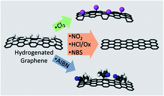
In this paper, we report the results of possible HG dehydrogenation routes. We find several mild reactions from hydrogenated to dehydrogenated graphene, as shown in Fig. 1. Interestingly, some of the reactions which remove hydrogen also add chemical groups to the graphene more effectively than addition of these groups to pristine graphene. Thus, hydrogenation can be used both as a convenient graphene transfer technology and a starting point for further chemistry on graphene.
 |
| Fig. 1 Reaction scheme outlining the main results of this paper. Perfluorobutyl iodide (PFBI) and N-bromosuccinimide (NBS) under UV light dehydrogenate HG without concomitant uptake of heteroatom groups. Dehydrogenation without activation is also observed upon exposure of HG to nitrogen oxides and nonaqueous solutions of HCl with a mild oxidant. Upon exposure to chlorine or azoisobutyronitrile (AIBN), a large increase in surface heteroatoms is observed. | |
Experimental
Materials and methods
All reagents were purchased from Sigma-Aldrich and, unless otherwise indicated, were used without further purification. Of particular importance to the results in this paper, 1H,1H,2H-perfluorohexene was obtained with a listed purity of ≥98.5%. The dichloromethane referred to as oDCM was from a lot whose chemical quality test dates from November 1998. Single-layer graphene was grown on copper from methane decomposition via a CVD method, transferred onto Si/SiOx wafers, and hydrogenated via Birch reduction, as previously described.23,29,30 Raman spectra were taken on a Renishaw spectrometer exciting with an Ar-ion laser at 514.5 nm. X-ray photoelectron spectra (XPS) were taken with a Thermo Kalpha spectrometer with the Al Kα line as the X-ray source and the data was analyzed using Avantage software. This software includes built-in sensitivity ratios for all elemental spectral lines, allowing us to directly report the atomic ratios of various elements. Collinear 4-point probe conductivity measurements were taken on a Keithley 4200-SCS. UV photodehydrogenation experiments (with PFBI or NBS) were performed using a 4 W UV wand at a wavelength of 254 nm. These experiments were carried out by either (1) immersing the samples in the reagent in a small open dish that was covered completely by the UV wand to prevent evaporation or (2) inserting the samples into a custom-built liquid-tight PTFE reaction cell with a quartz window for UV exposure (Fig. S1 in ESI†). All other experiments were carried out in scintillation vials. After reaction, all samples were rinsed with hexanes, acetone, and ethanol. Chlorine gas was generated by dropwise addition concentrated HCl onto solid potassium permanganate. The HCl/H2O2 experiments used 0.1 mL each of 37% aqueous HCl and 30% aqueous H2O2 in 10 mL dichloromethane, shaken briefly. The HCl/ammonium persulfate (APS) experiments used 0.1 mL of 37% aqueous HCl and 25 mg APS in 10 mL dichloromethane and sonicated for 10 minutes. Both of these preparations include aqueous and organic layers which separate, so the mixtures are shaken briefly with MgSO4 to remove excess water and homogenize the reagents. Gaseous mixed nitrogen oxides were generated by preparing a saturated solution of FeSO4·7H2O in 1.8 M H2SO4 and adding the solution dropwise onto solid sodium nitrite. This reaction produces colorless NO, which immediately reacts with oxygen in the air to give red-brown NO2. With regard to gas exposure of graphene, we obtained the most consistent results when the system was relatively free of water vapor, as condensation of water on the HG surface tends to delaminate and tear the graphene sheet.25
Analysis of Raman spectra
We rely heavily on Raman spectroscopy to determine the extent of dehydrogenation of HG. The most common approach to Raman analysis of graphene chemistry uses the intensity ratio of D (∼1345 cm−1) and G (∼1585 cm−1) peaks to determine defect density.31 However, the trend in D/G ratio versus defect density is non-monotonic. Because these experiments probe dehydrogenation of graphene on both sides of the maximum of this non-monotonic curve, reporting D/G ratios alone could be misleading depending on which side of the maximum the ratios fall for any given experiment. To circumvent this problem, we instead use the ratio of integrated intensities of the G and 2D (∼2690 cm−1) peaks (G/2D ratio) because it increases monotonically with defect density and changes rapidly in the region of interest.32 Strong dehydrogenation of HG decreases the G/2D ratio from much greater than 1 to less than 1. We also point out that after hydrogenation, the spectra often slope sharply upward above ∼2500 cm−1 due to the strong photoluminescence of highly hydrogenated graphene.33 This photoluminescence disappears from the Raman spectra of HG under mild conditions, most notably including heating from laser illumination, making it an unreliable measure of graphene functionalization. The disappearance of photoluminescence is not necessarily accompanied by other changes such as the D peak reduction or 2D peak restoration, which are better indicators of large-scale removal of defects from graphene. The baselines of the Raman spectra were fit with a spline curve to deconvolute the 2D peak intensity from the photoluminescence intensity.
Results and discussion
Dehydrogenation with minimal activation
1. Perfluorobutyl iodide. When highly insulating HG was immersed in commercially obtained 1H,1H,2H-perfluorohexene (PFH) and exposed to UV light, the Raman spectral features and the conductivity of pristine graphene recovered over a period of several hours (Fig. 2a and b). At the same time XPS showed a small amount of fluorine on the graphene surface (Fig. S2 in ESI†). We also noted that the reaction could be photomasked, as shown in the Raman map in Fig. 2c, where half of the HG was exposed to UV light and PFH, and the other half remained in the dark. During this reaction, the PFH changed from colorless to purple. Apparently, the commercial synthesis of PFH proceeds from addition of perfluorobutyl iodide (PFBI) to ethylene,34 and so the small (∼1%) PFBI impurity in the PFH might be photolyzed to form iodine. We conjectured that dehydrogenation could be caused either by free iodine or by perfluorobutyl radicals and that the latter would attach to the graphene, explaining the XPS fluorine peak.
 |
| Fig. 2 (a) Raman spectra of HG exposed to 254 nm UV light and PFH for 1, 2, 4, and 8 h. Note the decreasing intensity of the D peak and the increasing intensity of the 2D peak. (b) Resistance measurements for graphene, HG, and HG exposed to 254 nm UV light and PFH for 1, 4, and 8 h, showing recovery of conductivity. (c) Raman map of D/G integrated peak intensity ratio for HG exposed to 254 nm UV light and PFH for 4 h. Chromium as a photomask was deposited over the quartz window of the reaction cell. The darker right side of the image is the area under this photomask, while the left side was unmasked. (d) Raman spectra of HG before (top) and after (bottom) exposure to PFBI under 254 nm UV light. Note the recovery of the 2D peak intensity relative to the G peak intensity after PFBI exposure. The sharply upward sloping spectrum of HG before exposure is due to strong photoluminescence, as mentioned in the Materials and methods section of the main text. | |
To test these hypotheses, we prepared a 0.1% by volume solution of PFBI in hexanes and reacted it with HG. Raman spectra of the resultant material showed significant removal of the functional groups from the graphene basal plane (Fig. 2d), with the G/2D ratio, which scales with the extent of chemical functionalization, dropping from 3.13 ± 0.83 before PFBI treatment to 0.41 ± 0.03 after PFBI treatment. Sheet resistivity is another sensitive measure of the presence of covalently bonded functional groups on graphene, increasing dramatically when functional groups are attached. The sheet resistance of graphene before the Birch reduction was 0.52 ± 0.08 kΩ □−1, while after the Birch reduction, the resistance was too high to be measured with our equipment (>10 GΩ). Treating the HG with PFBI restored the sheet resistance to 4.88 ± 2.18 kΩ □−1, within an order of magnitude of the starting material, but at least 6 orders of magnitude less than HG. Moreover, after PFBI treatment a small fluorine peak was observed in the XPS; however, its position at 686.6 eV was not entirely consistent with a perfluoro species.35 A shoulder also appeared on the silicon XPS peak. Together, these results—especially the Raman data—strongly suggest that impurities such as PFBI in PFH are at least partially responsible for removing hydrogen from HG upon exposure to UV light.
2. N-Bromosuccinimide. N-Bromosuccinimide (NBS) is a common source of bromine radicals. We exposed HG to a saturated diethyl ether solution of N-bromosuccinimide (NBS) and irradiated with 254 nm UV light. The HG showed significant dehydrogenation within 10 minutes, with a G/2D ratio of 3.75 ± 0.67 before NBS treatment versus 0.41 ± 0.06 after treatment (Fig. 3a). Also, the sheet resistance recovered from >10 GΩ □−1 before NBS treatment (0.44 ± 0.05 kΩ □−1 before Birch reduction) to 4.19 ± 2.85 kΩ □−1 afterwards. The large standard deviation in these measurements is likely due to some tearing observed in the graphene sheet after hydrogenation and NBS exposure. In the absence of UV exposure, the recovery was much less pronounced, with a G/2D ratio of 3.16 ± 0.34 beforehand versus 2.06 ± 0.84 afterwards and no recovery of conductivity. In addition, XPS showed that reaction of NBS with HG left behind a small amount of bromine—barely discernible above the noise—but no nitrogenous species (Fig. 3b). A comparative reaction with pristine graphene also produced a small D peak in the Raman spectrum (Fig. 3c) and showed a bromine peak in the XPS similar to what was observed in the case of HG (Fig. 3d).
 |
| Fig. 3 (a) Raman spectra of HG before (top) and after (bottom) treatment with NBS under UV light. (b) N 1s, C 1s, and Br 3d regions of the XPS spectrum for HG reacted with NBS for 10 minutes under UV light. (c) Raman spectra of pristine graphene before (top) and after (bottom) treatment with NBS under UV light. Note the appearance of the small D peak at 1335 cm−1 after the reaction, indicating an increase in defect density. (d) N 1s, C 1s, and Br 3d regions of the XPS spectrum for pristine graphene under identical conditions to the sample in this Fig. 3b. | |
Both fluorinated and chlorinated graphene are stable covalent compounds,17,36 whereas brominated and iodinated graphene readily decompose into the elements.37,38 The instability of graphene bromide explains the relatively small quantity of bromine observed in the XPS of NBS-reacted HG. The bromine cannot stably bind to the graphene basal plane, so any residual bromine is likely bound to edges or vacancies where strong sp2 C–Br bonds can form. Our observation of a similar amount of bromine in the reaction product of NBS with pristine graphene corroborates this hypothesis, since pristine graphene should have edge and vacancy defect densities comparable to HG.
3. Nitrogen oxides. Nitrogen oxides are both strong oxidants and radical species. HG exposed to NOx vapors for 25 minutes showed significant recovery in the Raman spectrum (Fig. 4a) as well as in the electrical characteristics. The G/2D ratio in the Raman spectrum recovered from 2.64 ± 1.27 before the reaction to 0.50 ± 0.05 afterwards. In addition, the conductivity showed substantial recovery upon NOx exposure. Pristine graphene had a sheet resistance of 0.55 ± 0.07 kΩ □−1 and HG before NOx exposure exhibited no measurable resistance below 10 GΩ. After NOx exposure, the sheet resistance recovered to 2.39 ± 1.53 kΩ □−1, consistent with removal of covalently bound hydrogen atoms. NOx did not form covalent bonds with graphene, and any byproducts can be nearly completely eliminated from the surface with a simple wash in ethanol. As shown in the XPS (Fig. 4b), the N/C ratio was measured to be very small, at 0.0157 ± 0.0109.
 |
| Fig. 4 (a) Raman spectra of HG before (top) and after (bottom) treatment with NOx gas. (b) C 1s and N 1s regions of the XPS spectrum of HG treated with NOx gas. | |
4. HCl/oxidizer. Serendipitously, we observed that exposure of HG to dichloromethane from a ∼18 years old bottle (referred to as oDCM—all other uses of “dichloromethane” hereafter refer to new dichloromethane) caused nearly complete dehydrogenation within 30 minutes, whereas new dichloromethane did not react with HG. When exposed to oxygen and light, dichloromethane slowly decomposes into chloroform, HCl, phosgene, and other chlorinated species.39 A series of experiments showed that chloroform, neat oxalyl chloride (a phosgene simulant), and HCl bubbled through dichloromethane all have a negligible effect on HG, indicating that another decomposition product was the dehydrogenation agent (Fig. S3 in ESI†). The dehydrogenation was dramatically reduced if the oDCM was shaken in a separation funnel with either aqueous NH4OH, NaBH4 in methanol, or neat cyclohexene (Fig. S4 in ESI†). While the first two reagents will eliminate HCl and phosgene, cyclohexene should not react with either substance, ruling out these decomposition products.To test if free Cl2 was the oxidant, we bubbled freshly generated Cl2 gas through dichloromethane for 30 seconds until the solution turned vivid yellow. Cyclohexene was mixed with this solution and with oDCM and the reaction products were compared via GC-MS. Reaction with Cl2-saturated dichloromethane yielded only 1,2-dichlorocyclohexane, whereas reaction with oDCM additionally yielded chlorocyclohexane. Ultimately, we obtained a product mixture similar to that of the oDCM reaction by exposing cyclohexene to a suspension in dichloromethane of concentrated hydrochloric acid and ammonium persulfate (APS), an oxidizer. Thus, the most likely active species in oDCM were HCl and an oxidizer that likely generated Cl radicals and Cl2 gas in situ.
We can mimic the oDCM reactivity by exposing HG to a dichloromethane suspension of 37% HCl and either APS or H2O2. These reactions almost completely recover pristine graphene with negligible addition of chlorine as shown in the Raman spectra and XPS (Fig. 5 and S5 in ESI†). Using H2O2 in water as an oxidizer was undesirable because the presence of extra water facilitated delamination of HG from its substrate and left the remaining graphene severely damaged.25 This problem was mitigated by using APS as an oxidizer and drying the suspension with anhydrous magnesium sulfate before carrying out the reaction. The G/2D Raman peak ratio for HG decreased from 2.78 ± 0.72 to 0.37 ± 0.09 upon a 15 minute exposure to the HCl/APS mixture in dichloromethane, and the D peak which measures defect density showed a remarkable decrease in intensity as well. In addition, the HCl/APS reaction with HG restored sheet resistance from >10 GΩ (0.63 ± 0.06 kΩ □−1 before Birch hydrogenation) to 5.27 ± 1.70 kΩ □−1. Pristine graphene did not react under these conditions. Oxidizer alone does not restore HG (Fig. S6 in ESI†). Specifically, HG immersed in a mixture of 0.5 mL 30% aqueous H2O2 in 9.5 mL of ethanol and irradiated with 254 nm UV light showed little change in its Raman spectrum after 1.5 hours. Also, HG exposed to dichloromethane with 25 mg APS in 0.1 mL water showed minimal recovery in the Raman spectrum after 15 minutes.
 |
| Fig. 5 (a) Raman spectra of HG before (top) and after (bottom) treatment with APS and 37% aqueous HCl in dichloromethane. (b) C 1s and Cl 2p regions of the XPS spectrum of HG treated with APS and 37% aqueous HCl in dichloromethane. | |
Dehydrogenation with significant activation
1. Chlorine. To further explore the notion that Cl2 was generated in situ in the reaction of HG with HCl and an oxidizer, we exposed HG to Cl2-saturated dichloromethane for 5 minutes. This reaction removed much of the hydrogen as shown in the Raman spectrum (Fig. 6a), where the initial G/2D ratio of 2.75 ± 0.54 decreased to 0.41 ± 0.03 after Cl2 treatment. HG reacted with either oDCM (Fig. S7 in ESI†) or Cl2-bubbled dichloromethane (Fig. 6b) left substantial chlorine content on the graphene, with XPS spectra showing a Cl/C atomic ratio of 0.0198 ± 0.0025 for oDCM and 0.0313 ± 0.0047 for Cl2-bubbled dichloromethane. In contrast, pristine graphene exposed to Cl2-bubbled dichloromethane showed no chlorine by XPS (Fig. 6c) and no D peak in the Raman spectrum (Fig. S8 in ESI†), indicating that it was essentially inert. This is consistent with earlier work on chlorographene, which asserted that gaseous chlorine does not react with pristine graphene in the absence of UV radiation.36 Beyond adding chlorine to the graphene, this process heavily restores the graphene. Before the reaction, the graphene gave a sheet resistance of 1.93 ± 0.35 kΩ □−1 which increased to open-circuit (Rsheet > 10 GΩ □−1) after hydrogenation before dramatically restoring to 13.67 ± 3.60 kΩ □−1 after reaction with Cl2. These results suggest that both molecular Cl2 and the Cl radicals produced by reaction of HCl with oxidizer remove hydrogen from HG, but the HCl/oxidizer combination adds Cl less efficiently to the graphene than Cl2, which is advantageous if chlorination is not desired.
 |
| Fig. 6 (a) Raman spectra of HG before (top) and after (bottom) exposure to Cl2 dissolved in dichloromethane. (b) C 1s and Cl 2p regions of the XPS spectrum for HG reacted with Cl2 dissolved in dichloromethane. (c) C 1s and Cl 2p regions of the XPS spectrum for pristine graphene, showing the absence of a chlorine peak. | |
2. AIBN. Azoisobutyronitrile (AIBN) is a common non-oxidizing radical initiator. We reacted HG with a 0.15 M solution of AIBN in acetonitrile under 254 nm UV light for 1 hour. As Fig. 7a shows, the Raman spectra of HG before and after reaction are essentially unchanged, with a G/2D ratio of 2.93 ± 0.67 before and 2.98 ± 0.41 after reaction with AIBN. Furthermore, reaction with AIBN did not recover the electrical conductivity of graphene. However, the characteristic optical contrast of graphene, which disappears upon extensive hydrogenation, returned after treatment with AIBN. AIBN has been reported to react with pristine graphene under high-temperature conditions,40 and we observed a small D peak, along with CN stretching modes, in the Raman spectrum (Fig. 7c) of pristine graphene exposed to AIBN under our experimental conditions. We hypothesized that AIBN was removing hydrogen from HG to restore optical contrast and simultaneously reacting with the graphene basal plane to maintain the defect density observed in the Raman spectra. XPS (Fig. 7b) on the material showed a N/C atomic ratio of 0.0902 ± 0.0117. Assuming that each nitrogen represents one isobutyronitrile radical (IBN·) attached, and taking into account the 4 carbons per IBN·, this gives a density of roughly 1 IBN· group per 7 graphene carbons, an extremely dense packing. Compare this to the ratio found on pristine graphene after the same reaction (Fig. 7d): an N/C ratio of 0.0389 ± 0.0038, or roughly 1 IBN· group per 22 graphene carbons. It should be noted that XPS probably overestimates the density, as the AIBN could undergo radical oligomerization which would inflate the density. Yet, oligomerization would also occur with pristine graphene, so this observation still supports the assertion that HG reacts more readily with radical species than does pristine graphene, and further, that the reactivity is not limited to halogenated radicals.
 |
| Fig. 7 (a) Raman spectra of HG before (top) and after (bottom) treatment with AIBN under UV light. (b) N 1s and C 1s regions of the XPS spectrum of HG treated with AIBN under UV light. (c) Raman spectra of pristine graphene before (top) and after (bottom) treatment with AIBN under UV light. The small peaks that appear after AIBN treatment are likely from CN stretches on AIBN. (d) N 1s and C 1s regions of the XPS spectrum of pristine graphene treated with AIBN under UV light. | |
Conclusions
In conclusion, we have shown that several radical generators—halogens and fluoroalkyl halides as well as NOx and the non-oxidizing radical initiator AIBN—react with HG to dehydrogenate the material. Some of these species, in particular Cl2 and AIBN, undergo reactive chemisorption to HG in addition to dehydrogenation. Compared with pristine graphene, both Cl2 and AIBN reacted more readily with HG. These results suggest that radical species generated during the course of the reaction remove hydrogen from HG and can replace it in cases where a stable bond can form between graphene and the radical moiety. Therefore, hydrogenation of graphene can serve as an initial step that chemically activates graphene to reactions which might otherwise be difficult or impossible on pristine graphene.
Acknowledgements
This research was developed with funding from the Defense Advanced Research Projects Agency (MIPR HR0011512805). The views, opinions and/or findings expressed are those of the author and should not be interpreted as representing the official views or policies of the Department of Defense or the U. S. Government. This work was also supported by a Naval Research Laboratory Karle Fellowship and base programs.
References
- A. K. Geim and K. S. Novoselov, Nat. Mater., 2007, 6, 183–191 CrossRef CAS PubMed.
- J. Gu, J. Du, J. Dang, W. Geng, S. Hu and Q. Zhang, RSC Adv., 2014, 4, 22101–22105 RSC.
- J. Gu, N. Li, L. Tian, Z. Lv and Q. Zhang, RSC Adv., 2015, 5, 36334–36339 RSC.
- J. Gu, X. Yang, Z. Lv, N. Li, C. Liang and Q. Zhang, Int. J. Heat Mass Transfer, 2016, 92, 15–22 CrossRef CAS.
- T. Wang, D. Huang, Z. Yang, S. Xu, G. He, X. Li, N. Hu, G. Yin, D. He and L. Zhang, Nano-Micro Lett., 2016, 8, 95–119 CrossRef.
- S. P. Mulvaney, R. Stine, N. C. Long, C. R. Tamanaha and P. E. Sheehan, BioTechniques, 2014, 57, 21–30 CrossRef CAS PubMed.
- S. S. Varghese, S. Lonkar, K. K. Singh, S. Swaminathan and A. Abdala, Sens. Actuators, B, 2015, 218, 160–183 CrossRef CAS.
- Y. Wang, Q. He, H. Qu, X. Zhang, J. Guo, J. Zhu, G. Zhao, H. A. Colorado, J. Yu, L. Sun, S. Bhana, M. A. Khan, X. Huang, D. P. Young, H. Wang, X. Wang, S. Wei and Z. Guo, J. Mater. Chem. C, 2014, 2, 9478–9488 RSC.
- J. Zhu, Z. Luo, S. Wu, N. Haldolaarachchige, D. P. Young, S. Wei and Z. Guo, J. Mater. Chem., 2012, 22, 835–844 RSC.
- J. Zhu, M. Chen, Q. He, L. Shao, S. Wei and Z. Guo, RSC Adv., 2013, 3, 22790–22824 RSC.
- A. Hirsch, J. M. Englert and F. Hauke, Acc. Chem. Res., 2013, 46, 87–96 CrossRef CAS PubMed.
- A. Criado, M. Melchionna, S. Marchesan and M. Prato, Angew. Chem., Int. Ed., 2015, 54, 10734–10750 CrossRef CAS PubMed.
- D. R. Dreyer, S. Park, C. W. Bielawski and R. S. Ruoff, Chem. Soc. Rev., 2010, 39, 228–240 RSC.
- R. Stine, J. W. Ciszek, D. E. Barlow, W.-K. Lee, J. T. Robinson and P. E. Sheehan, Langmuir, 2012, 28, 7957–7961 CrossRef CAS PubMed.
- K. E. Whitener, R. Stine, J. T. Robinson and P. E. Sheehan, J. Phys. Chem. C, 2015, 119, 10507–10512 CAS.
- M. Dubecký, E. Otyepková, P. Lazar, F. Karlický, M. Petr, K. Čépe, P. Banáš, R. Zbořil and M. Otyepka, J. Phys. Chem. Lett., 2015, 6, 1430–1434 CrossRef PubMed.
- J. T. Robinson, J. S. Burgess, C. E. Junkermeier, S. C. Badescu, T. L. Reinecke, F. K. Perkins, M. K. Zalalutdniov, J. W. Baldwin, J. C. Culbertson, P. E. Sheehan and E. S. Snow, Nano Lett., 2010, 10, 3001–3005 CrossRef CAS PubMed.
- Z. M. Ao, Q. G. Jiang, S. Li, H. Liu, F. M. Peeters, S. Li and G. X. Wang, ACS Appl. Mater. Interfaces, 2015, 7, 19659–19665 CAS.
- R. Stine, W.-K. Lee, K. E. Whitener, J. T. Robinson and P. E. Sheehan, Nano Lett., 2013, 13, 4311–4316 CrossRef CAS PubMed.
- J. R. Felts, A. J. Oyer, S. C. Hernández, K. E. Whitener Jr, J. T. Robinson, S. G. Walton and P. E. Sheehan, Nat. Commun., 2015, 6, 6467 CrossRef CAS PubMed.
- V. B. Mohan, R. Brown, K. Jayaraman and D. Bhattacharyya, Mater. Sci. Eng., B, 2015, 193, 49–60 CrossRef CAS.
- D. C. Elias, R. R. Nair, T. M. G. Mohiuddin, S. V. Morozov, P. Blake, M. P. Halsall, A. C. Ferrari, D. W. Boukhvalov, M. I. Katsnelson, A. K. Geim and K. S. Novoselov, Science, 2009, 323, 610–613 CrossRef CAS PubMed.
- K. E. Whitener Jr, W. K. Lee, P. M. Campbell, J. T. Robinson and P. E. Sheehan, Carbon, 2014, 72, 348–353 CrossRef.
- Z. Q. Yang, Y. Q. Sun, L. B. Alemany, T. N. Narayanan and W. E. Billups, J. Am. Chem. Soc., 2012, 134, 18689–18694 CrossRef CAS PubMed.
- K. E. Whitener, W. K. Lee, N. D. Bassim, R. M. Stroud, J. T. Robinson and P. E. Sheehan, Nano Lett., 2016, 16, 1455–1461 CrossRef CAS PubMed.
- W.-K. Lee, K. E. Whitener, J. T. Robinson and P. E. Sheehan, Adv. Mater., 2015, 27, 1774–1778 CrossRef CAS PubMed.
- P. Rubio-Pereda and N. Takeuchi, J. Phys. Chem. C, 2013, 117, 18738–18745 CAS.
- J. O. Sofo, A. S. Chaudhari and G. D. Barber, Phys. Rev. B: Condens. Matter Mater. Phys., 2007, 75, 4 CrossRef.
- X. S. Li, C. W. Magnuson, A. Venugopal, R. M. Tromp, J. B. Hannon, E. M. Vogel, L. Colombo and R. S. Ruoff, J. Am. Chem. Soc., 2011, 133, 2816–2819 CrossRef CAS PubMed.
- J. W. Suk, A. Kitt, C. W. Magnuson, Y. Hao, S. Ahmed, J. An, A. K. Swan, B. B. Goldberg and R. S. Ruoff, ACS Nano, 2011, 5, 6916–6924 CrossRef CAS PubMed.
- M. M. Lucchese, F. Stavale, E. H. M. Ferreira, C. Vilani, M. V. O. Moutinho, R. B. Capaz, C. A. Achete and A. Jorio, Carbon, 2010, 48, 1592–1597 CrossRef CAS.
- E. H. Martins Ferreira, M. V. O. Moutinho, F. Stavale, M. M. Lucchese, R. B. Capaz, C. A. Achete and A. Jorio, Phys. Rev. B: Condens. Matter Mater. Phys., 2010, 82, 125429 CrossRef.
- R. A. Schafer, J. M. Englert, P. Wehrfritz, W. Bauer, F. Hauke, T. Seyller and A. Hirsch, Angew. Chem., Int. Ed., 2013, 52, 754–757 CrossRef PubMed.
- H.-J. Lehmler, Chemosphere, 2005, 58, 1471–1496 CrossRef CAS PubMed.
- G. Nansé, E. Papirer, P. Fioux, F. Moguet and A. Tressaud, Carbon, 1997, 35, 175–194 CrossRef.
- B. Li, L. Zhou, D. Wu, H. Peng, K. Yan, Y. Zhou and Z. Liu, ACS Nano, 2011, 5, 5957–5961 CrossRef CAS PubMed.
- R. Zbořil, F. Karlický, A. B. Bourlinos, T. A. Steriotis, A. K. Stubos, V. Georgakilas, K. Šafářová, D. Jančík, C. Trapalis and M. Otyepka, Small, 2010, 6, 2885–2891 CrossRef PubMed.
- F. Karlický, R. Zbořil and M. Otyepka, J. Chem. Phys., 2012, 137, 034709 CrossRef PubMed.
- Dichloromethane, MSDS No. 270997, Sigma-Aldrich, St. Louis, MO, Version 5.9.
- X. Y. Zhang, M. S. Han, S. Chen, L. Bao, L. Li and W. J. Xu, RSC Adv., 2013, 3, 17689–17692 RSC.
Footnotes |
† Electronic supplementary information (ESI) available: Experimental apparatus, Raman, XPS, and electronic measurements of PFH- and oDCM-treated HG, as well as miscellaneous control experiment measurements. See DOI: 10.1039/c6ra21113e |
‡ Present address: Keysight Technologies, Santa Rosa, CA 95403. |
|
This journal is © The Royal Society of Chemistry 2016 |
Click here to see how this site uses Cookies. View our privacy policy here.