DOI:
10.1039/C6RA20413A
(Paper)
RSC Adv., 2016,
6, 112634-112640
High strength chitosan rod reinforced by non-covalent functionalized multiwalled carbon nanotubes via an in situ precipitation method
Received
12th August 2016
, Accepted 15th November 2016
First published on 24th November 2016
Abstract
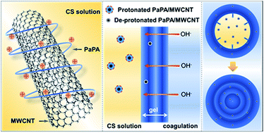
Chitosan (CS) has been widely used as temporary mechanical supporter for the regeneration of bone, owing to its biocompatibility, biodegradability and versatility in orthopedic treatment. CS rod material is a promising candidate for internal fixation devices. However, the mechanical strength of existing CS rod materials is still unsatisfactory. In the present work, multiwalled carbon nanotubes (MWCNTs) were non-covalently functionalized by poly(p-aminophenylacetylene) (PaPA). CS/MWCNTs composite rods were subsequently fabricated via a unique in situ precipitation method. The resultant composite rods were studied in view of their microscopic morphology, crystallinity, mechanical strength, and biocompatibility. Results indicated that the composite rods showed great improvement in mechanical strength and exhibited good biocompatibility, which made the material a promising candidate for bone fracture internal fixation.
Introduction
Chitosan (CS) is a polycationic biopolymer generally obtained by alkaline deacetylation of chitin. CS is currently receiving considerable attention due to its intrinsic properties: biocompatibility, biodegradability and inherent wound healing properties.1–8 These desirable properties enabled the application of CS as temporary mechanical supporter for the regeneration of bone.9–14 In the fabrication of these materials, the processability of CS had been a challenge. Due to the multiple inter-/intramolecular hydrogen-bonds, melt extrusion appeared to be unsuitable for the process of pure CS. Thus an in situ precipitation method had been developed.15,16 This method employed the gelation process of solubilized CS in acidic aqueous medium. CS becomes a polyelectrolyte because of the protonation of –NH2 groups.17 Then the CS solution can be neutralized and transformed into hydrogel by alkali coagulation bath.18 The in situ precipitation method is a simple way to fabricate CS-based devices in various shapes and dimensions. Three-dimensional CS rods had been prepared by in situ precipitation method.19–21 The bending strength of pure CS rods made by in situ precipitation method was approximately 92.4 MPa.15 However, for clinical application such as internal fixation of bone fracture, adequate initial mechanical strength was required. The mechanical strength of pure CS rods was lower than the quota.
In order to improve the mechanical performance of CS rods, various strategies had been employed.22–24 Multiwalled carbon nanotubes (MWCNTs) have found important applications in high-performance composites due to their extraordinary tensile strength.25–27 As in the case of CS-based materials, many works about CS/MWCNTs composites have been reported, with enhanced properties like elasticity and thermal stability.28–30 Moreover, studies had been carried out on the response of bone cells to MWCNTs.31–35 Results suggest that MWCNTs have beneficial effects on bone growth. As a result, it is rational to recognize that MWCNTs may effectively improve the mechanical strength of CS rods as reinforcement.
MWCNTs themselves are known to be insoluble in any solvents. However, to make CS/MWCNTs composite rods, a uniform CS/MWCNTs solution was required by the in situ precipitation method. Thus it is essential to realize the uniform dispersion of MWCNTs in aqueous media. Fortunately, some strategies are available to fulfil such requirement. The effective dispersion of MWCNTs in aqueous medium had been realized by both covalent and non-covalent surface modification.36–38
Among these strategies, non-covalent functionalization of MWCNTs with poly(phenylacetylene) (PPA) is a very promising approach.39–41 PPA and its derivatives containing aromatic pendants showed strong power to disperse MWCNTs in proper solvents, by wrapping PPA chains onto the surface of MWCNTs. Moreover, PPAs modified with amino and carboxylic groups could even realize desirable dispersion of MWCNTs in aqueous medium. In addition to fulfil the primary requirement of in situ precipitation, this strategy also shows other merits. Non-covalent functionalization of MWCNTs with PPAs is harmless to the inherent mechanical properties of MWCNTs, which benefits the fabrication of high performance composite material. Good biocompatibility and cytophilic property of PPAs have been verified, which is favourable in the view of biomedical applications.42
In the present work, one derivative of PPAs, poly(p-aminophenylacetylene) (PaPA) was employed in the in situ precipitation to fabricate CS/MWCNTs composite rods. Compared to pure CS rods, the resultant composite rods exhibit obvious enhancement in bending modulus and strength. By in situ precipitation, MWCNTs could be uniformly incorporated in CS hydrogel matrix with 3D sophisticated structure. Such morphology cannot be achieved by simple blending nor layer-by-layer process. This work also provided new thought for the preparation of CS/MWCNTs composites.
Experimental
1. Materials
Chitosan (biomedical grade, Mη = 5.63 × 10⁵, D.D. = 91%, Qingdao Haihui Bioengineering Co., Ltd), acetic acid (CP, Yixing Niujia Chemical Reagent Plant), sodium hydroxide (AR, Hangzhou Xiaoshan Chemical Reagent Corporation), MWCNTs (20–30 nm in diameter, about 30 μm in length, Shenzhen Nanotech Port Co., Ltd). 4-Aminophenylacetylene is commercially available and purchased from Aldrich. The catalyst of Rh+(nbd)[C6H5B–(C6H5)3] was prepared in our laboratory, where nbd is the abbreviation of 2,5-norbornadiene.
2. Preparation of water soluble polymer–MWCNTs hybrids
The synthesis and characterization of poly(p-aminophenylacetylene) (PaPA) were carried out in the same procedures as reported in literature.39 PaPA is readily soluble in acidic aqueous solution, and protonated PaPA is derived (Scheme 1). Simple mixing and stirring of the protonated PaPA with MWCNTs afforded polymer/MWCNTs with a moderate water solubility.
 |
| Scheme 1 Synthetic route for the protonated PaPA. | |
3. Preparation of CS-based composite rods
Acetic acid aqueous solution (2%, v/v) was added into an aqueous solution containing PaPA/MWCNTs. Then desired amount of CS powder was dissolved in the mixture. The final detailed composition of three different kinds of rod samples was listed in Table 1. A cylindrical semipermeable membrane was used as mold. Subsequently, the cylindrical mold was filled with the solution mentioned above, followed by precipitation in 5% (w/v) NaOH aqueous solution to form gel rods. CS gel rods were washed with deionized water repeatedly to remove OH−. Finally, the rods were air-dried in oven at 60 °C, and the dried rods (Fig. 1c) were used for the required measurements.
Table 1 Detailed composition of rod samples
Sample |
CS (g) |
PaPA (mg) |
MWCNTs (mg) |
Pure CS rods |
10 |
0 |
0 |
CS/MWCNTs-1 |
10 |
20 |
4 |
CS/MWCNTs-2 |
10 |
40 |
8 |
 |
| Fig. 1 (a) Pure CS solution and PaPA/MWCNTs–CS solution; (b) concentric multi-layered structure in the CS gel rod, scale bar: 1 cm; (c) digital photo of CS/MWCNTs composite rod, scale bar: 2 cm. | |
4. In vitro cell test
CS solution containing PaPA/MWCNTs was casted into membrane, soaked in 5% (w/v) sodium hydroxide aqueous solution, then washed with deionized water, at last air-dried in oven at 60 °C. The membranes sterilized by ultraviolet irradiation were cut into square pieces (1 cm × 1 cm), and placed in a culture plate. The in vitro cell tests of the hybrid membranes were performed using MG63 cells. The human osteosarcoma cell line MG-63 (CRL-1427TM, ATCC) was obtained from Cell Bank of Shanghai Institute of Biochemistry and Cell Biology, Chinese Academy of Sciences (Shanghai, China). MG63 cells were plated at density of 2.0 × 10⁴ cells per mL in Dulbecco's modified Eagle's medium (DMEM, Gibco). The medium was supplemented with 10% fetal bovine serum (FBS, Gibco), 100 U mL−1 penicillin and 100 μg mL−1 streptomycin (Thermofisher Scientific). Cultivation was conducted for 6 h, 24 h, 48 h, 72 h at 37 °C in 5% CO2 in a humidified incubator. After being cultured for desired time, disks from each group were removed from the plates and gently washed with PBS. The cells on the disks were fixed with 3% glutaraldehyde in PBS for 24 h at 4 °C. After being thoroughly washed with PBS, the specimens were dehydrated in a graded series of alcohol (30–100%). After the disks were dehydrated twice in each ethanol concentration (15 min each time), they were freeze dried, sputter-coated with gold, and examined under SEM. The cultivation was also conducted on pure CS material by the same procedure, which served as the control group.
5. Mechanical properties
The bending strength and modulus of CS/MWCNTs composite rods were tested by three point bending test on a universal mechanical testing apparatus (Shenzhen Reger Company, China). The CS/MWCNTs composite rods were cylindrical. The dimensions of a rod were determined by a vernier caliper. The span length in three point bending test was 40 mm and loading rate was 2 mm min−1. The ultimate bending strength (σb) and bending modulus (Eb) of the rods were calculated according to eqn (1) and (2), respectively43,44 |
 | (1) |
|
 | (2) |
where Fmax is the maximum bending force recorded (N), L the support span (mm), d the diameter of the rod (mm), and ΔF/Δl the slope of the force–deflection curve of the initial linear section (N mm−1).
6. Instrumentations
The morphology and dispersion of MWCNTs in the CS matrix were investigated with TEM (JEOL, Japan, JEM-1200EX) and SEM (Japan, HITACHI S-4800) techniques. The composite rod was cut into ultrathin sections before TEM test, and coated by gold before SEM observation. All of the samples were air-dried in oven at 60 °C for 2 h to remove the moisture before mechanical testing.
Results and discussion
1. Preparation of uniform CS/MWCNTs acidic solution
The PaPA polymer chains have amino groups in the pendants, and could be protonated and soluble in the acidic aqueous solution. On the other hand, as mentioned above, CS could also be protonated in such medium to form solution. Thus, the PaPA could be a perfect candidate for the uniform dispersion of MWCNTs in CS solution.
By wrapping of PaPA polymer chains onto the surface of MWCNTs, the PaPA/MWCNTs hybrid was formed, which showed solubility in organic solvents. The wrapping had been confirmed experimentally by our previous investigation.41 In the present work, acidic aqueous solution was utilized as solvent. The protonated PaPA bestowed the PaPA/MWCNTs hybrid with the solubility up to 316 mg L−1 in aqueous solution (Scheme 2a). As shown in Fig. 1a, a PaPA/MWCNTs–CS solution was obtained. Compared to pure CS solution, the PaPA/MWCNTs–CS solution presented a color of dark green due to the existence of MWCNTs, but the latter solution is also clear.
 |
| Scheme 2 Scheme of the preparation of CS/MWCNTs composite rod by in situ precipitation method. (a) PaPA/MWCNTs hybrids in CS solution; (b) gelation process of PaPA/MWCNTs–CS solution, the protonated and de-protonated PaPA/MWCNTs hybrids were only schematically illustrated. | |
In addition to macroscopic observation, the microscopic investigation of MWCNTs was also performed. The typical morphology of MWCNTs used for the in situ precipitation was shown in Fig. 2. The MWCNTs had an average diameter of approximately 20–30 nm before wrapped by PaPA (Fig. 2a). This value was in accordance with the product description. Then after hybridization with PaPA, the average diameter of the nano-tubular entities increased to about 60–70 nm (Fig. 2b). A thin coating layer could be observed on the surface of MWCNTs with lower contrast, which corresponded to the PaPA component. The protonation of this PaPA thin coating layer facilitated the exfoliation of MWCNTs from the bundles, and prohibited the re-aggregation of the separated MWCNTs via static electric repulsion as well. These results indicated the realization of uniform dispersion of MWCNTs in the aqueous solution, which laid the foundation of in situ precipitation of composite material.
 |
| Fig. 2 TEM images of (a) MWCNTs in the primary form and (b) PaPA/MWCNTs hybrids. | |
2. Preparation of CS/MWCNTs composite rod by in situ precipitation
As introduced above, the premise of in situ precipitation is the uniform dispersion of MWCNTs in the CS solution. This was due to the unique structure of CS rod and formation process of this method. The predecessor of ultimate rod was a CS hydrogel rod. This CS hydrogel rod had concentric multi-layered structure (Fig. 1b), which could effectively stop the propagation of cracks and greatly increase the bending strength after turned to dry material.21 The concentric multi-layered structure originated from the unique gelation process of the acidic CS solution. It has been reported in our previous study, this gelation process possessed a layer-wise character due to the advance of c(OH−) equipotential surface.45
This character brings spatiotemporal sequence to the system, which means there was always some solution under the gel–sol surface before the entire system turned into gel. As a result, if the reinforcements were simply mixed in the solution, sedimentation and aggregation would last the whole gelation process and became aggravated with time. Thus the uniform dispersion of MWCNTs in the gel rod would be impossible. However, if the MWCNTs were functionalized with PaPA, the situation would be quite different. The preparation mechanism was demonstrated in Scheme 2b. After the uniform and stable PaPA/MWCNTs–CS solution was obtained, its gelation process was the same as that of pure CS solution essentially. When OH− diffused to certain position, CS macromolecules became de-protonated and transformed to gel. In the meantime, protonated PaPA also went through de-protonation. As a consequence, the MWCNTs wrapped by PaPA chains were embedded in situ in the CS matrix.
3. Microscopic morphology of CS/MWCNTs composite rod
The microscopic morphology of CS/MWCNTs composite rod was shown in Fig. 3. The observation demonstrated the morphology of the cross-section of CS/MWCNTs rods after mechanical measurement. Results showed that the typical multi-layered structure was successfully maintained in the composite rod (Fig. 3a). The arrows directed to the rod-like nanostructures projecting out of the fracture surface (Fig. 3b), which can be assigned to the hybrid nanotubes. SEM images revealed the morphology of the nanotubes on the fracture surface, while TEM observation provided more information on reinforcements inside the matrix (Fig. 3c). The nanostructure with higher contrast revealed that nanotubes were uniformly dispersed and embedded in the CS matrix. Microscopic observation indicated that, nanotubes were separately dispersed in the CS matrix and no bundles could be observed in the whole vision. Since the matrix was created by in situ gelation of CS solution, the uniform dispersion of MWCNTs in the CS solution greatly contributed to the uniform distribution of reinforcements in rods.
 |
| Fig. 3 Morphology of CS/MWCNTs-1 composite rod. (a) Multi-layered structure in the composite rod; (b and c) MWCNTs in the CS matrix; (a and b) are SEM images and (c) is TEM image. | |
4. Influence of MWCNTs on the crystallinity of CS rod
After in situ precipitation, the composite gel rod was dried to prepare the ultimate rod material, and the rod materials were studied by X-ray diffraction. The profile of in situ precipitated CS/MWCNTs composite rod showed peaks at 2θ = 10° and 2θ = 20° (Fig. 4). These peaks are characteristic diffraction peaks of CS, corresponding to 020 reflection and 110 reflection of CS crystalline, respectively.46 Whereas the XRD profile of pure CS rod only showed a very broad peak at 2θ = 20°.47 The degree of crystallinity and the size of the crystals can be evaluated by the area and the full width at half maximum (FWHM) of diffraction peak, respectively.48 Results showed that in situ precipitated CS/MWCNTs composite rod had higher diffraction peak value and smaller FWHM. This indicated that in situ precipitated CS/MWCNTs composite rod has advantage over pure CS rod in both the degree of crystallinity and the size of the crystals. The existence of MWCNTs played the role of nuclear agent to improve the crystallinity of CS.49 Thus, by adding small amount of MWCNTs into CS matrix, crystallinity of CS has been increased. The increase of crystallinity will in turn enhanced the mechanical strength of the rod material.
 |
| Fig. 4 XRD profiles of CS rod and CS/MWCNTs-2 composite rod. | |
5. Mechanical properties of CS/MWCNTs composite rod
The utmost goal of the present work is to improve the bending strength of pure CS rods. To quantitatively evaluate the improvement, comparative experiments had been performed on both pure CS rods and CS/MWCNTs composite rods. The data are list in Table 2, together with a reference data of a commercially available sample used for internal fixation of bone fracture (Dikfix Ltd., Co.). The CS/MWCNTs composite rod showed superior mechanical properties to pure CS rod. Higher mechanical performance could be obtained with increased amount of PaPA/MWCNTs. Bending strength and bending modulus of composite rod could reach 135.5 MPa and 4.8 GPa, respectively. It is noteworthy that the bending strength is approximate to the quota of the nails made in Dikfix. It is clear that, by PaPA functionalization and in situ precipitation, the introduction of MWCNTs in CS matrix had successfully achieved the utmost goal. The efficient improvement of mechanical properties is associated with the good dispersion of MWCNTs in the CS matrix.
Table 2 Bending strength and bending modulus of different bone fracture internal fixation devicesa
Sample |
Bending strength (MPa) |
Bending modulus (GPa) |
The data in the table are the average values of 5 parallel measurements. The data are extracted from ref. 15. |
Pure CS rods |
92.4 ± 2.0 |
4.1 ± 0.1 |
CS/MWCNTs-1 |
97.7 ± 1.1 |
4.1 ± 0.1 |
CS/MWCNTs-2 |
135.5 ± 1.3 |
4.8 ± 0.1 |
Dikfixb |
130.0 |
2.0–3.0 |
Additionally, there was another noteworthy point about the in situ prepared composite rod. A small fraction of reinforcements dispersed in the polymer matrix can significantly improve the mechanical strength of the composites. This phenomenon was more evident in the in situ prepared composite rods compared with the composite rods by blending method. It has been reported that, CS/MWCNTs composite rod can be prepared by a blending method, assisted by ultrasonic dispersion. When prepared by blending method, the bending strength increased by 12.5% with 0.25 wt% of MWCNTs.49 While in the present work, with only 0.08 wt% of MWCNTs, the bending strength increased by 46.1% in comparison with the pure CS rods. It is highly possible that this phenomenon is related to the dispersion and the improved interface between reinforcement and matrix. In the fracture surface of composite rod by blending method, nanotubes were pulled out from CS matrix with lots of holes formed.49 By this way, MWCNTs embedded in the CS matrix may absorb energy when the composite went through destruction, so MWCNTs could endure external stress. However, the fracture surface of in situ prepared composite rod indicated that the situation was quite different in this case. The possible reason is that the introduction of PaPA and the good dispersion of MWCNTs in CS solution led to the improved interface of reinforcement and matrix. Thus, during the bending process, the reinforcement tend to absorb energy by breaking rather than being pulled out of the system.
6. Cell proliferation on CS/MWCNTs composite material
Cell adhesion, spreading and migration on substrates are the first sequential reactions upon contact of the cell with a material surface, which are crucial for cell survival.50 The in vitro cell responses to the CS/MWCNTs composites were examined in terms of the initial attachment and proliferation. The cellular behavior on biomaterials is an important factor for evaluation of the biocompatibility of biomaterial. The morphology of the cells adhered to the CS/MWCNTs composite material was observed by SEM, as shown in Fig. 5. After 6 h (Fig. 5a), a small amount of cells with round shape were observed to adhere on the material. When observed after 24 h (Fig. 5b), the amount of cells increased and some cells spread out through cytoskeletal processes. After 48 h (Fig. 5c), cells attached firmly on the material, with fully extended pseudopodia. After 72 h (Fig. 5d), the increase of cell was quite obvious and cells contact with each other in full vision. In addition, compared with pure CS material (Fig. 5e–h), MG63 cells cultured on CS/MWCNTs composite material showed better conditions. The results indicated that cells adhere well and grow actively on the CS/MWCNTs composite material. The composite material showed good biocompatibility and is favored for cell adhesion, spreading and migration.
 |
| Fig. 5 SEM microscopic observation of MG63 cells cultured on the surface of materials, after incubation of 6 h, 24 h, 48 h and 72 h; (a–d) cultured on the surface of CS/MWCNTs composite material, and (e–h) cultured on the surface of pure CS material as control group; scale bar: 100 μm. | |
Conclusion
CS/MWCNTs composite rods were prepared via in situ precipitation method, assisted by PaPA functionalization. The bending strength and bending modulus of the resultant composite rods reached 135.5 MPa and 4.8 GPa, respectively, increased by 46.1% and 17.1% compared with pure CS rods. The experimental data indicated that the improvement of mechanical strength is more evident for CS/MWCNTs composite rods via PaPA functionalization than that via blending method. The key mechanical properties of the resultant composite rods are approximate to the commercially available materials. Additionally, the composite material showed good biocompatibility and is favored for biomedical applications. Overall, the strategy reported in the present work is facile to the preparation of CS/MWCNTs composite rods which could be a promising candidate for bone fracture internal fixation.
Acknowledgements
This work was financially supported by the Key Science Technology Innovation Team of Zhejiang Province (No. 2013TD02), the National Natural Science Foundation of China (No. 21104067, 21274127, 21374099 and 51473144).
Notes and references
- N. Bhattarai, J. Gunn and M. Q. Zhang, Mater. Sci. Semicond. Process., 2010, 62, 83–99 CAS.
- G. D. Wu and H. Xiao, RSC Adv., 2015, 5, 105084–105089 RSC.
- X. F. Huang, J. W. Jia, Z. K. Wang and Q. L. Hu, Chin. J. Polym. Sci., 2015, 33, 284–290 CrossRef CAS.
- Z. K. Wang, Y. L. Liu, J. W. Jia, S. J. Chen, W. Qin, Q. L. Hu and B. Z. Tang, J. Mater. Chem. B, 2016, 4, 5265–5271 RSC.
- J. Fang, K. Zhang, J. Jia, Z. Wang and Q. Hu, RSC Adv., 2015, 5, 99418–99424 RSC.
- K. Zhang, P. Zhuang, Z. Wang, Y. Li, Z. Jiang, Q. Hu, M. Liu and Q. Zhao, Carbohydr. Polym., 2012, 90, 1515–1521 CrossRef CAS PubMed.
- X. F. Huang, Y. C. Pang and Y. L. Liu, RSC Adv., 2016, 6, 64357–64363 RSC.
- X. F. Huang, Y. F. Sun, J. Y. Nie, W. T. Lu, L. Yang, Z. L. Zhang, H. P. Yin, Z. K. Wang and Q. L. Hu, Int. J. Biol. Macromol., 2015, 75, 322–329 CrossRef CAS PubMed.
- J. Y. Nie, Z. K. Wang, J. Z. Zhang, L. Yang, Y. C. Pang and Q. L. Hu, RSC Adv., 2015, 5, 68243–68250 RSC.
- J. Y. Nie, Z. K. Wang, K. Zhang and Q. L. Hu, RSC Adv., 2015, 5, 37346–37352 RSC.
- A. Di Martino, M. Sittinger and M. V. Risbud, Biomaterials, 2005, 26, 5983–5990 CrossRef CAS PubMed.
- J. Z. Zhang, J. Y. Nie, Q. R. Zhang, Y. L. Li, Z. K. Wang and Q. L. Hu, J. Biomater. Sci., Polym. Ed., 2014, 25, 61–74 CrossRef CAS PubMed.
- K. Zhang, M. Zhao, L. Cai, Z. K. Wang, Y. F. Sun and Q. L. Hu, Chin. J. Polym. Sci., 2010, 28, 555–561 CrossRef CAS.
- L. Yang, W. T. Lu, Y. C. Pang, X. F. Huang, Z. K. Wang, A. Qin and Q. L. Hu, RSC Adv., 2016, 6, 71567–71573 RSC.
- Q. L. Hu, X. Z. Qian, B. Q. Li and J. C. Shen, Chem. Res. Chin. Univ., 2003, 24, 528–531 CAS.
- J. H. Ke, Z. K. Wang, Y. Z. Li, Q. L. Hu and J. Feng, Chin. J. Polym. Sci., 2012, 30, 436–442 CrossRef CAS.
- J. Y. Nie, Z. K. Wang and Q. L. Hu, Sci. Rep., 2016, 6, 36005 CrossRef CAS PubMed.
- J. Y. Nie, Z. K. Wang and Q. L. Hu, Sci. Rep., 2016, 6, 36053 CrossRef CAS PubMed.
- Z. K. Wang and Q. L. Hu, Acta Phys.-Chim. Sin., 2010, 26, 2053–2056 CAS.
- Z. K. Wang and Q. L. Hu, Biomed. Mater., 2010, 5, 045007 CrossRef PubMed.
- Z. K. Wang, Q. L. Hu and Y. X. Wang, Sci. China: Chem., 2011, 54, 380–384 CrossRef CAS.
- Z. K. Wang, Q. L. Hu and L. Cai, Int. J. Polym. Sci., 2010, 2010, 369759 Search PubMed.
- K. Shen, Q. L. Hu, Z. K. Wang and J. Qu, Mater. Sci. Eng., C, 2011, 31, 866–872 CrossRef CAS.
- Z. K. Wang, Q. L. Hu, X. G. Dai, H. Wu, Y. X. Wang and J. C. Shen, Polym. Compos., 2009, 30, 1517–1522 CrossRef CAS.
- D. E. H. Jones, Nature, 1996, 381, 384 CrossRef CAS.
- K. Kang, H. Park, J. Kim and C. Lee, Appl. Surf. Sci., 2015, 356, 1039–1051 CrossRef CAS.
- A. M. Visco, A. Pistone, V. Brancato, D. Iannazzo and M. Fazio, Polym. Compos., 2016, 37, 1007–1015 CrossRef CAS.
- A. A. Azeez, K. Y. Rhee, S. J. Park, H. J. Kim and D. H. Jung, Composites, Part B, 2013, 50, 127–134 CrossRef CAS.
- J. Venkatesan, Z. J. Qian, B. Ryu, N. A. Kumar and S. K. Kim, Carbohydr. Polym., 2011, 83, 569–577 CrossRef CAS.
- J. Yan, T. H. Wu, Z. Z. Ding and X. K. Li, Carbohydr. Polym., 2016, 136, 1288–1296 CrossRef CAS PubMed.
- N. Narita, Y. Kobayashi, H. Nakamura, K. Maeda, A. Ishihara, T. Mizoguchi, Y. Usui, K. Aoki, M. Simizu, H. Kato, H. Ozawa, N. Udagawa, M. Endo, N. Takahashi and N. Saito, Nano Lett., 2009, 9, 1406–1413 CrossRef CAS PubMed.
- J. L. Xu, K. A. Khor, J. J. Sui and W. N. Chen, Mater. Sci. Eng., C, 2009, 29, 44–49 CrossRef CAS.
- B. Das, P. Chattopadhyay, S. Maji, A. Upadhyay, M. Das Purkayastha, C. L. Mohanta, T. K. Maity and N. Karak, Biomed. Mater., 2015, 10, 025011 CrossRef PubMed.
- P. Khalid, M. A. Hussain, P. D. Rekha and A. B. Arun, Hum. Exp. Toxicol., 2015, 34, 548–556 CAS.
- G. Lalwani, A. Gopalan, M. D'Agati, J. S. Sankaran, S. Judex, Y. X. Qin and B. Sitharaman, J. Biomed. Mater. Res., Part A, 2015, 103, 3212–3225 CrossRef CAS PubMed.
- Y. K. Kang, O. S. Lee, P. Deria, S. H. Kim, T. H. Park, D. A. Bonnell, J. G. Saven and M. J. Therien, Nano Lett., 2009, 9, 1414–1418 CrossRef CAS PubMed.
- B. I. Kharisov, O. V. Kharissova, H. L. Gutierrez and U. O. Mendez, Ind. Eng. Chem. Res., 2009, 48, 572–590 CrossRef CAS.
- X. H. Peng and S. S. Wong, Adv. Mater., 2009, 21, 625–642 CrossRef CAS.
- H. Zhao, W. Z. Yuan, J. Mei, L. Tang, X. Q. Liu, M. Yan, X. Y. Shen, J. Z. Sun, A. J. Qin and Z. Tang, J. Polym. Sci., Part A: Polym. Chem., 2009, 47, 4995–5005 CrossRef CAS.
- W. Z. Yuan, J. Z. Sun, Y. Q. Dong, M. Haussler, F. Yang, H. P. Xu, A. J. Qin, J. W. Y. Lam, Q. Zheng and B. Z. Tang, Macromolecules, 2006, 39, 8011–8020 CrossRef CAS.
- W. Z. Yuan, L. Tang, H. Zhao, J. K. Jin, J. Z. Sun, A. J. Qin, H. P. Xu, J. H. Liu, F. Yang, Q. Zheng, E. Q. Chen and B. Z. Tang, Macromolecules, 2009, 42, 52–61 CrossRef CAS.
- F. Salhi, K. K. L. Cheuk, Q. H. Sun, J. W. Y. Lam, J. A. K. Cha, G. Li, B. S. Li, J. D. Luo, J. W. Chen and B. Z. Tang, J. Nanosci. Nanotechnol., 2001, 1, 137–141 CrossRef CAS PubMed.
- A. Saikku-Backstrom, R. M. Tulamo, T. Pohjonen, P. Tormala, J. E. Raiha and P. Rokkanen, J. Mater. Sci.: Mater. Med., 1999, 10, 1–8 CrossRef CAS PubMed.
- K. Shen and Q. L. Hu, Mater. Lett., 2011, 65, 1503–1505 CrossRef CAS.
- J. Y. Nie, W. T. Lu, J. J. Ma, L. Yang, Z. K. Wang, A. Qin and Q. L. Hu, Sci. Rep., 2015, 5, 7635 CrossRef CAS PubMed.
- Z. Wang, J. Nie, W. Qin, Q. Hu and B. Tang, Nat. Commun., 2016, 7, 12033 CrossRef CAS PubMed.
- R. J. Samuels, J. Polym. Sci., Part B: Polym. Phys., 1981, 19, 1081–1105 CrossRef CAS.
- G. Bhagavannarayana, S. Parthiban and S. Meenakshisundaram, J. Appl. Crystallogr., 2006, 39, 784–790 CrossRef CAS.
- Z. K. Wang, Q. L. Hu and L. Cai, Chin. J. Polym. Sci., 2010, 28, 801–806 CrossRef CAS.
- J. Jia, Z. Wang, W. Lu, L. Yang, Q. Wu, W. Qin, Q. Hu and B. Z. Tang, J. Mater. Chem. B, 2014, 2, 8406–8411 RSC.
Footnote |
† J. Y. N. and Z. K. W. contributed equally to this work. J. Y. N. and Z. K. W. designed and conducted the experiments, analysed the experimental data and wrote the paper; Y. Z., S. D. W. and H. Y. L completed the cell experiments; H. Z. synthesized poly(p-aminophenylacetylene); Z. K. W., A. J. Q., Q. L. H., J. Z. S. and B. Z. T. supervised the projects. |
|
This journal is © The Royal Society of Chemistry 2016 |
Click here to see how this site uses Cookies. View our privacy policy here.