Hydralazine inhibits amyloid beta (Aβ) aggregation and glycation and ameliorates Aβ1–42 induced neurotoxicity

Kedar B. Batkulwarab, Asis K. Janabc, Rashmi K. Godbolea, Puneet Khandelwalbc, Neelanjana Sengupta bc and Mahesh J. Kulkarni*ab
aMass-spectrometry and Proteomics Facility, Division of Biochemical Sciences, CSIR-National Chemical Laboratory, Pune-411008, India. E-mail: mj.kulkarni@ncl.res.in; Fax: +91 20 25902648; Tel: +91 20 25902541
bAcademy of Scientific and Innovative Research (AcSIR), CSIR-National Chemical Laboratory, Pune 411008, India
cPhysical Chemistry Division, CSIR-National Chemical Laboratory, Pune-411008, India

Received 10th August 2016 , Accepted 7th November 2016

First published on 7th November 2016


Abstract

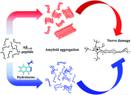
Alzheimer's disease (AD) is a neurodegenerative disorder affecting millions of people worldwide, characterized by senile plaques formed due to deposition of insoluble aggregates of amyloid beta (Aβ) peptide in neuronal synapses of the brain. The synthesis of the Aβ peptide and its aggregation is central to AD related pathogenesis. Post-translational modifications such as glycation, is known to exacerbate Aβ aggregation and neurodegeneration. Thus inhibitors of glycation can potentially inhibit Aβ aggregation and attenuate the progression of neurodegeneration. In the present study, we have evaluated the effect of hydralazine on Aβ aggregation and fibril formation by employing thioflavin-T (ThT), 1-anilino-8-naphthalene sulfonic acid (ANS) fluorescence assays, atomic force microscopy and static light scattering assay. From the results of these experiments, it is evident that hydralazine inhibits Aβ aggregation and fibril formation. Circular dichroism (CD) analysis revealed that hydralazine prevents β-sheet formation of Aβ peptide thereby inhibiting amyloid aggregation. Furthermore, molecular dynamics (MD) simulations studies also revealed that hydralazine binds to Aβ monomers as well as protofibrils and potentially destabilizes Aβ monomer–monomer interaction and protofibrils, thereby possibly alleviating Aβ neurotoxicity. This was evident by 3[4,5-dimethylthiazol-2-yl]2,5-diphenyl-tetrazolium bromide (MTT) assay, which confirmed that hydralazine ameliorates Aβ induced neuronal toxicity. This study suggests that hydralazine is a potential candidate for drug repositioning for the management of AD. However, it has to be re-engineered to reduce vasoactive effects and improve blood brain barrier permeability for future use in AD therapeutics.


Introduction

Alzheimer's disease (AD) is a progressive neurodegenerative disorder that is rapidly emerging as a major cause of dementia in aged people. The disease is characterized by the presence of senile plaques and neurofibrillary tangles, primarily in the cerebral cortex and hippocampus regions of the brain.1–3 Plaques are mainly formed by the aggregation of extracellular deposits of amyloid beta peptides. These peptides are formed, when the amyloid precursor protein (APP) undergoes sequential proteolytic cleavages by the beta (β) and gamma (γ) secretases.4 These monomeric Aβ peptides undergo fibrillation involving a complex process of nucleation and lead to formation of various aggregates such as oligomers and protofibrils that cause cellular dysfunction and neuronal death.5–7 The positive correlation between Aβ aggregation and synaptic dysfunction suggests the crucial role of Aβ aggregation in AD advancement.8

Alzheimer's disease is also referred as type III diabetes, because of its prevalence in diabetic people.9,10 Diabetes accelerates non-enzymatic glycation11 and glycation of Aβ1–42 enhances its aggregation, aggravates neurotoxicity and AD pathogenesis.12 Glycated Aβ1–42 is associated with upregulation of the receptor for advanced glycation end products (RAGE) and activation of glycogen synthase kinase-3 (GSK-3), suggesting a crucial role of glycation in Aβ1–42 toxicity.13,14 Hence, it is conjectured that glycation inhibitors can potentially reduce Aβ1–42 aggregation and neuronal toxicity.

The multifactorial nature of AD has rendered challenges to develop effective drugs. Acetyl cholinesterase (AChE) inhibitors such as donepezil, tacrine, galantamine, rivastigmine and the N-methyl-D-aspartate (NMDA)-receptor antagonist memantine are the only FDA approved drugs currently used to treat AD.15 However, these drugs only improve cognitive function and provide temporary relief in diseased people. The widely used strategies to reduce Aβ production are (i) development of γ-secretase and β-secretase inhibitors,16,17 (ii) reducing the APP expression and (iii) inhibition of beta amyloid aggregation.18 As the secretases (β and γ) and APP are required for normal physiological brain function, investigating amyloid aggregation inhibitors would be a better strategy to treat AD. Hence, there is an urgent need to develop therapeutics that can potentially reduce Aβ oligomerization and fibril formation.

Several small molecules and peptides have been reported to be inhibitors/modulators of Aβ1–42 aggregation;18 however none of them have passed clinical trials. The major concern of these inhibitors was a lack of selectivity, harmful side effects, poor bioavailability, and lack of information about their mechanism of action.19–22 We previously reported “drug repositioning” as a promising strategy to develop anti Alzheimer's drugs, as it has several advantages over classical drug discovery approach, like reduced time and cost required for clinical trials. Using this approach, in our earlier study, we have elucidated protriptyline as a promising candidate for AD treatment.23 Here we report that hydralazine (Fig. 1), an antihypertensive drug and a glycation inhibitor24 acts as a potent inhibitor of Aβ1–42 aggregation. Further, it was interesting to observe reduced cytotoxicity of Aβ1–42 upon hydralazine treatment in neuronal cells. For additional insights, we performed atomistic molecular dynamics (MD) simulation studies of hydralazine binding with a putative Aβ1–42 monomer, as well as its protofibrillar aggregate. Driven primarily by electrostatic interactions, hydralazine is observed to bind preferentially to the turn region of both the monomeric and the protofibrillar forms, and result in sharp destabilization of the inter-monomeric interactions. The current study provides evidence of usefulness of evaluating hydralazine as a potential drug candidate in AD therapeutics.


image file: c6ra20225j-f1.tif
Fig. 1 Structure of hydralazine.

Materials and methods

Materials

1–42 peptide, ammonium hydroxide (NH4OH), Thioflavin T (ThT), ANS, MTT, sodium phosphate monobasic and dibasic, and other chemicals were purchased from Sigma-Aldrich. Hydralazine was generous gift from Dr Gurjar M. K., Director (R and D), Emcure Pharmaceuticals, Pune. All solutions were prepared with deionized water collected from a Millipore water purification system. Neuro2a cells (Murine neuroblastoma) were supplied by National Centre for Cell Science, Pune. Dulbecco's Modified Eagle's Medium (DMEM), Trypsin Phosphate Versene Glucose (TPVG), and Fetal Bovine Serum (FBS) solutions were procured from Life technologies.

Solution preparation

Lyophilized Aβ1–42 powder was dissolved in 10% ammonium hydroxide (0.1 mg mL−1), incubated for 10 min at room temperature followed by sonication for 5 min and then centrifugation at 16[thin space (1/6-em)]000 rpm to remove preformed aggregates.25 The ammonium hydroxide was removed by lyophilisation to yield a salt free monomers of the peptide. Hydralazine stock solution of 4 mM was prepared in phosphate buffer saline (PBS).

ThT assay

1–42 (22 μM) dissolved in 10 mM PBS (150 mM NaCl, pH 7.4) was incubated with or without 10 μM hydralazine at 37 °C. For each assay, 50 μL aliquot was taken from either Aβ1–42 or an Aβ1–42–hydralazine mixture and mixed with 150 μL of 20 μM ThT-containing phosphate buffer saline (10 mM, 150 mM NaCl, pH 7.4).23 ThT fluorescence was recorded at 485 nm, with an excitation wavelength of 440 nm, using a Thermo Scientific Varioskan Flash Multimode Plate Reader (Thermo scientific, Germany).

ANS assay

The binding of nonpolar ANS dye to hydrophobic patches formed due to aggregation was analyzed by measuring the fluorescence on the Thermo Scientific Varioskan Flash Multimode Plate Reader (Thermo scientific, Germany). 1.5 μL of 20 mM ANS was mixed with 200 μL of Aβ1–42 reaction with or without hydralazine which was then excited at 375 nm and the emission was recorded between 400–700 nm.23

Atomic force microscopy

The atomic force microscopy (AFM) measurements were performed on silicon wafers drop casted with Aβ1–42 aggregates formed in presence and absence of hydralazine using a Multimode scanning probe microscope equipped with a Nanoscope IV controller from Veeco Instrument Inc., Santa Barbara, CA. All the AFM measurements were done under ambient conditions using the tapping-mode AFM probes model – Tap190Al purchased from Budget Sensors. The radius of tip used in this study was less than 10 nm, and its height was ∼17 μm. The cantilever used had a resonant frequency of ca. 162 kHz and nominal spring constant of ca. 48 N m−1 with a 30 nm thick aluminium reflex coating on the back side of the cantilever of the length 225 μm. For each sample, three locations with a surface area of 7 × 7 μm2, each were imaged at a rate of 1 Hz and at a resolution of 512 × 512.23

Circular dichroism spectroscopy

The CD spectra of Aβ1–42 in presence and absence of hydralazine were recorded in wavelength range of 190–250 nm on a Jasco-J815 spectropolarimeter at room temperature. Each CD spectrum was accumulated from three scans at scan speed of 50 nm min−1 with cell path length of 0.2 cm. CD signals from buffer were subtracted from the sample spectra and converted to mean residual ellipticity (MRE) in deg cm2 dmol−1 defined as
image file: c6ra20225j-t1.tif
where M is the molecular weight of the protein, θλ is ellipticity in millidegree, d is the path length in cm, c is the protein concentration in mg mL−1 and r is the number of amino acid residues in the protein. Further, secondary structure content of the Aβ1–42 was calculated using the CDPro software package.

Light scattering analysis

Rayleigh light-scattering experiments were carried out with the Perkin-Elmer Luminescence spectrometer LS50B to follow Aβ1–42 aggregation in presence and absence of hydralazine. The excitation and emission wavelength used were 400 nm. The excitation slit width was 8 nm and emission slit width was 2.5 nm. The scattering intensity was monitored for 60 s.

Cell cytotoxicity assay

Neuro2a cells were grown in monolayer cultures and maintained in DMEM containing 10% Fetal Bovine Serum (FBS) in humidified air containing 5% CO2 at 37 °C. The cultured cells were then seeded to a sterile 96 well plate at a density of 10[thin space (1/6-em)]000 cells per well. After serum starvation of 4 hours, cells were treated with different concentrations of Aβ1–42 alone, Aβ1–42 with hydralazine and varying concentrations of hydralazine alone for 12 hours. The viability of the cells was determined by MTT assay.

AGE fluorescence assay

1–42 (22 μM) was incubated in the presence and absence of glucose (250 mM) and hydralazine (10 μM) for 7 days in phosphate buffer. AGE specific fluorescence of glycated Aβ1–42 was measured at excitation of 370 nm and emission was scanned from 400–600 nm.26

Molecular dynamics simulation

Details of full-length Aβ1–42 monomer structure generation were reported in our previous study.27 We have also calculated mean 15N and 13Cα of the Aβ protein using the SHIFTS program28 and are compared with the reported experimental data.29 Significant β-sheet population in central hydrophobic core and C-terminal region of Aβ are consistent with previous experimental and computational studies.29–31 Five hydralazine molecules were placed randomly in the vicinity of the Aβ monomer, and the hydralazine-Aβ protofibrillar complex was obtained by placing five hydralazine molecules in the vicinity of Aβ protofibrillar structure obtained from the PDB structure 2M4J.32 The resulting systems were solvated by placing them in an equilibrated box of TIP3P water33 such that there was a minimum distance of 15 Å between any protein or drug atom and a box edge. The NH3+ and COO groups were added to the N- and C-termini of the peptide units, respectively. Requisite counter ions were added to neutralize the system. All simulations in this study were performed with the NAMD 2.9 simulation package,34 using the CHARMM22 all-atom force field with CMAP correction for the Aβ protein.35,36 Visualizations were made using the VMD package.37 Force field parameters for hydralazine molecule were generated using the SwissPARAM38 webserver and the partial charges were further refined via electronic structure calculations using Gaussian09. Bonds involving hydrogen atoms were held fixed with SHAKE algorithm.39 Energy minimizations, using the conjugate gradient technique, were performed initially on all the systems followed by simulations in the isothermal–isobaric (NPT) ensemble. Constant temperature of 310 K was maintained using Langevin dynamics, using a collision frequency of 1 ps−1 and constant pressure of 1 atmosphere was maintained using the Langevin piston Nosé–Hoover algorithm.40,41 The simulation timestep was 2 fs and three-dimensional orthorhombic periodic boundary conditions were employed. Full electrostatic interactions were calculated with particle-mesh Ewald method42 and the cutoff for non-bonded interactions was set to 12 Å with a smoothing function starting from 10.5 Å. Each system was sampled for a cumulative time of ∼250 ns with three trajectories.

Statistical analysis

All experiments were performed in triplicates and data are expressed as means ± SD. Statistical significance for glycation inhibition and light scattering assay was determined by using one-way ANOVA followed by Bonferroni test and t test respectively. P values <0.05 were considered as significant.

Results and discussion

Hydralazine inhibits Aβ1–42 fibril formation

Thioflavin T (ThT) is a widely used dye to quantify and visualize the amyloid beta aggregates in vitro and in vivo respectively. ThT binds to β-sheet-rich structures usually present in misfolded protein aggregates such as in Aβ aggregates leading to enhanced fluorescence signal.43 In the current study, we used ThT fluorescence assay to monitor Aβ1–42 aggregation in presence and absence of hydralazine in vitro. In absence of hydralazine, Aβ1–42 showed characteristic sigmoidal curve and plateau after 24 h which corresponds to the formation of β-sheet-rich Aβ1–42 fibrils (Fig. 2). Whereas, in presence of hydralazine, fluorescence maxima (44.23) is significantly lower than the ThT plateau (78.16) recorded from Aβ1–42 alone (Fig. 2) suggesting reduction in Aβ aggregation by hydralazine. Further decline in ThT fluorescence upon subsequent incubation for 96 h, signifies the potential of hydralazine to destabilize Aβ1–42 aggregates.
image file: c6ra20225j-f2.tif
Fig. 2 Thioflavin T aggregation assay demonstrating the change in fluorescence during incubation of Aβ1–42 solution at 37 °C in the absence (red solid curve) and presence of 10 μM hydralazine (blue dotted curve). Error bars indicate standard deviations of three replicates.

1–42 aggregates are highly hydrophobic due to presence of β-sheet-rich oligomers and fibrils. Differences in hydrophobicity of aggregates formed in presence and absence of hydralazine were studied by exploiting the interaction of Aβ1–42 aggregates with ANS, which binds to hydrophobic patches of protein leading to fluorescence.441–42 incubated with hydralazine showed decrease in ANS fluorescence (Fig. 3). In presence of hydralazine, ANS fluorescence maxima (12.58) is significantly lower than the fluorescence (19.84) recorded from Aβ1–42 alone suggests decreased ANS binding to Aβ1–42 in presence of hydralazine. The blue shift along with enhanced fluorescence indicates the formation of β-sheets rich Aβ1–42 aggregates in absence of hydralazine.45


image file: c6ra20225j-f3.tif
Fig. 3 Change in fluorescence emission spectra for ANS binding to Aβ1–42 in the absence (red solid curve) and presence of hydralazine (blue dotted curve) after incubation of 48 h.

We further performed atomic force microscopy to visualize Aβ1–42 aggregates. The AFM study indicates a reduction in the fibrillar density and a high degree of size dispersion resulting from hydralazine treatment (Fig. 4). It was evident from the AFM images that the Aβ1–42 peptide undergoes aggregation and leads to fibril formation (Fig. 4A), whereas, hydralazine treatment effectively reduced Aβ1–42 aggregation/fibril formation (Fig. 4B). Additionally, static light scattering experiments were performed to investigate the relative decrease in average molecular mass/aggregate size of amyloid aggregates. Presence of hydralazine led to reduced light scattering due to smaller Aβ1–42 aggregates as compared to only Aβ1–42 solution (Fig. 5).


image file: c6ra20225j-f4.tif
Fig. 4 AFM topography images and height profiles of Aβ1–42 solution after incubation of 48 h. (A) Aβ1–42 solution only (incubated in absence of hydralazine) (B) Aβ1–42 solution coincubated with hydralazine. The scale is 7 × 7 μm2.

image file: c6ra20225j-f5.tif
Fig. 5 Bar graph demonstrating the comparison of Rayleigh light scattering of Aβ1–42 aggregates in absence (red) and presence of hydralazine (blue) after incubation of 48 h (*p < 0.05).

Next, circular dichroism (CD) spectroscopy was used to study conformational changes in Aβ1–42 incubated for 48 h. Aβ1–42 aggregates formed in absence of hydralazine showed characteristic CD spectrum with minima at 217 nm which indicates abundance of β-sheet-rich oligomers and fibrils (Fig. 6A).23 In presence of hydralazine, Aβ1–42 showed unstructured conformation. CD pro analysis indicates an increase in helices from 0.9% to 8.1% and decrease in beta sheets from 46.4% to 35.9% in Aβ1–42 upon hydralazine treatment (Fig. 6B). These results are very well in accordance with the ThT aggregation assay, ANS assay, light scattering assay and AFM results.


image file: c6ra20225j-f6.tif
Fig. 6 (A) CD spectra illustrating the mean residual ellipticity of Aβ1–42 solutions incubated in the absence (red solid) and presence of hydralazine (blue dotted) for 48 h (B) CD pro analysis elucidating the conformational change in the secondary structure of Aβ1–42 aggregates formed in absence (red) and presence of 10 μM hydralazine (blue).

Hydralazine decreases Aβ1–42 induced cytotoxicity

Cytotoxicity of Aβ1–42 aggregates was studied using MTT cell viability assay in neuro2a cells (Fig. 7). Cells were treated with different concentrations of hydralazine (5 μM, 10 μM and 20 μM) for 12 h; all these concentrations were found to be nontoxic to cells. Further, cells were treated with Aβ1–42 aggregates at concentrations of 0.5 μM, 1 μM and 2 μM for 12 h. Aβ1–42 aggregates at concentrations of 1 μM and 2 μM were toxic to cells and reduced viability up to 65 ± 9% and 51 ± 9% respectively. However, Aβ1–42 aggregates formed in presence of hydralazine were found to be non toxic or less toxic and rescued cell viability to 98 ± 2% and 88 ± 4% respectively.
image file: c6ra20225j-f7.tif
Fig. 7 Cell viability of neuro2a cells after treating with different concentrations of hydralazine (5 μM, 10 μM and 20 μM, green colored bars), Aβ1–42 aggregates (Aβ1–42_0.5 μM, Aβ1–42_1 μM and Aβ1–42_2 μM, red colored bars), and Aβ1–42 aggregates (Aβ1–42 + Hyd_0.5 μM, Aβ1–42 + Hyd_1 μM and Aβ1–42 + Hyd_2 μM, blue colored bars) formed in presence of hydralazine (NT – no treatment, PBS – phosphate buffer saline treatment) (*p < 0.05).

Hydralazine inhibits Aβ1–42 glycation

Previous studies demonstrate that Aβ undergoes glycation (Aβ-AGE) which alters Aβ conformation resulting in highly toxic Aβ species in vitro and in vivo as well.13,14 Hence, it is important to design or screen a potent glycation inhibitor which prevents Aβ glycation. In our earlier study, we have evidenced the anti-glycation potential of hydralazine;24 hence, here we have studied Aβ1–42 glycation in presence of hydralazine in vitro. Aβ1–42 alone or incubated with glucose and hydralazine showed lower AGE specific fluorescence at 440 nm than Aβ1–42 incubated with glucose (Fig. 8). These results suggest that hydralazine is a potent inhibitor of Aβ1–42 glycation and hence can be used as a potential therapeutic in near future to prevent Aβ1–42 glycation.
image file: c6ra20225j-f8.tif
Fig. 8 AGE specific fluorescence of glycated Aβ1–42 (excitation at 370 nm and emission at 440 nm) in the presence and absence of glucose (250 mM) and hydralazine (10 μM) (*p < 0.05, and ***p < 0.0005).

Mechanism of hydralazine binding to Aβ1–42 peptide and protofibril

For detailed molecular level understanding of hydralazine mediated Aβ aggregation inhibition, we performed MD simulation studies of the Aβ1–42 in presence of hydralazine molecules, as described in Methods section. A β-sheet rich Aβ1–42 monomer was considered for these studies. Hydralazine molecules were found to bind with the Aβ monomer with a mean binding strength of −91.2 (±19.2) kcal mol−1. A representative snapshot of hydralazine binding with the Aβ monomer is shown in Fig. 9a. In Table 1, depicts mean values of the ligand–protein non-bonded interaction energy, along with their electrostatic and van der Waal components. This interaction is mainly driven by the electrostatic interaction, with the van der Waal component making small contributions. To identify the key interacting residues, average residue-wise breakdown of the total ligand–protein interaction energy is calculated and provided in Fig. 9b. Interestingly, the negatively charged residues in turn region (E22 and D23) exhibit significantly stronger interactions with the drug, while the aromatic amino acid (F20) in the central hydrophobic core interacts with the phenyl ring of the drug. We point out that the roles of the intra-molecular and inter-molecular salt bridges between K28 and E22 or D23 of the turn region in oligomeric stability and in the process of fibril formation have been elicited through several experimental and computational studies.46,47 Therefore, our findings suggest that hydralazine binding to Aβ primarily hinders the electrostatic interactions between the charged residues in the turn region. Further, the effect on the secondary structural propensities of the Aβ due to hydralazine binding was investigated using the STRIDE algorithm.48 Fig. 9c represents the residue-wise β-sheet percentage of the Aβ peptide in presence of hydralazine. For comparison, we have also reported corresponding values obtained from the previous simulations of pure Aβ1–42 monomer. Notably, the β-sheet propensities of the Aβ monomer significantly decrease upon drug binding. Overall, the β-sheet percentages of Aβ in presence and absence of drug are 12.7 and 20.5% respectively, in excellent corroboration of the CD, AFM, ANS and ThT binding fluorescence assays showing that the drug reduces β-sheet propensity in the Aβ1–42 monomer.
image file: c6ra20225j-f9.tif
Fig. 9 (a) Snapshot of drug binding with Aβ monomer. Central hydrophobic core and turn region residues are shown in line representation. Key interacting residue and drug are shown in stick representation. (b) Residue-wise average non-bonded interaction energy (Eave) with drug. Energies are in kcal mol−1 unit. The residues with strong interactions are denoted with one letter code of respective amino acids. (c) Residue-wise β-sheet (%) of free Aβ monomer (in red) and drug-bound Aβ monomer (in blue). (d) Snapshot of drug binding with Aβ protofibrillar structure. Central hydrophobic core and turn region residues are shown in line representation. Key interacting residues and drug are shown in stick representation. (e) Residue-wise average non-bonded interaction energy (Eave) with drug. Energies are in kcal mol−1 unit. The residues with strong interactions are denoted with one letter code of respective amino acids.
Table 1 The mean values of the net ligand-protein interaction energy (Etotal), and their electrostatic (Eelec) and van der Waals (EvdW) components obtained from MD simulation trajectories. All energies are in units of kcal mol−1. The standard deviations obtained are provided in braces
Etotal −91.2 (±19.2)
Eelec −83.0 (±20.4)
EvdW −8.1 (±4.4)


In order to understand the inhibitory effect of hydralazine on Aβ aggregation pathway, we further performed MD simulation of the protofibrillar structure of Aβ in the presence of hydralazine molecules. A representative snapshot of hydralazine binding with Aβ protofibrillar structure is shown in Fig. 9d. The mean interaction strength of the hydralazine molecule with Aβ protofibrillar structure is −125.2 (±22.8) kcal mol−1. Inspecting the residue-wise breakup of the mean interaction strengths, presented in Fig. 9e, suggests that the interaction strength between drug and Aβ can be predominantly attributed to E22 and D23 and aromatic amino acids in central hydrophobic core also interact with the drug. These interactions are therefore reminiscent of the interactions observed earlier in the case of Aβ monomer and hydralazine complex. We have also calculated the mean monomer–monomer interaction energy for Aβ protofibrillar structure in the absence and presence of drug. The mean monomer–monomer interaction strength for Aβ protofibrillar structure was −525.2 (±72.1) kcal mol−1, whereas, in presence of hydralazine, however, the monomer–monomer interaction weakened significantly, with a mean value of −365.8 (±68.9) kcal mol−1. Collectively, these analyses demonstrate that hydralazine also has major destabilizing effects on the pre-formed Aβ protofibrillar structures. Overall, the inhibitory effects of hydralazine on the Aβ properties suggest that this molecule can effectively destabilize Aβ monomer–monomer interactions and may disrupt pre-formed Aβ protofibrillar structures and hence applicable as a drug to reduce as well as to abolish senile plaques in AD brain.

Conclusion

It is evident from the several biophysical and bioanalytical assays that hydralazine inhibits Aβ1–42 aggregation. The ThT assay, ANS assay, light scattering and AFM results demonstrated hydralazine as a potent Aβ1–42 aggregation inhibitor. The aggregates in the presence of hydralazine displayed less β-sheet and more α-helix structures, as shown by the CD spectra. Further MD simulations provided detailed understanding of different modes of hydralazine–Aβ1–42 interaction amyloid aggregation and destabilize preformed Aβ1–42 aggregates. Hydralazine was observed to rescue neuro2a cells from Aβ aggregation-induced toxicity and it also inhibited glycation of Aβ1–42. These effects of hydralazine can work synergistically to significantly reduce the neurotoxicity of amyloid aggregates. Hence, it will be meaningful to mention that hydralazine may prove to be useful in the future drug development against AD treatment. However, it requires re-engineering to reduce vasoactive effects and improve blood brain barrier permeability to be used effectively as an AD therapeutic.

Funding sources

This work is supported by the grants from Council of Scientific and Industrial research (CSIR), New Delhi, India (BSC0115). K. B. B. and A. K. J. thanks CSIR for research fellowship.

Author contributions

K. B. B. and R. K. G. performed the ThT, ANS, light scattering, CD spectroscopy, AGE fluorescence and cytotoxicity experiments. P. K. performed Atomic Force Microscopy experiments. K. B. B. performed data analysis. A. K. J. and N. G. carried out the MD simulation experiments and analysis. M. J. K. directed the research. K. B. B., M. J. K., A. K. J. and N. S. wrote the manuscript.

Conflict of interest

The authors declare no competing financial interest.

Abbreviations

ADAlzheimer's disease
AGEAdvanced glycation end products
AGE-Aβ1–42AGE modified Aβ1–42
ThTThioflavin-T
ANS1-Anilino-8-naphthalene sulfonic acid
CDCircular dichroism
MDMolecular dynamics
MTT3[4,5-Dimethylthiazol-2-yl]2,5-diphenyl-tetrazolium bromide

Acknowledgements

We thank Dr M. K. Gurjar, Director (R and D), Emcure Pharmaceuticals, Pune for providing the hydralazine hydrochloride for this study.

References

  1. P. Cras, M. Kawai, D. Lowery, P. Gonzalez-Dewhitt, B. Greenberg and G. Perry, Proc. Natl. Acad. Sci. U. S. A., 1991, 88, 7552–7556 CrossRef CAS.
  2. J. A. Hardy and G. A. Higgins, Science, 1992, 256, 184 CAS.
  3. H. Ksiezak-Reding, K. Morgan, L. A. Mattiace, P. Davies, W.-K. Liu, S.-H. Yen, K. Weidenheim and D. W. Dickson, Am. J. Pathol., 1994, 145, 1496 CAS.
  4. C. Haass and D. J. Selkoe, Cell, 1993, 75, 1039–1042 CrossRef CAS PubMed.
  5. H. J. Dyson and P. E. Wright, Nat. Rev. Mol. Cell Biol., 2005, 6, 197–208 CrossRef CAS PubMed.
  6. M. M. Babu, R. van der Lee, N. S. de Groot and J. Gsponer, Curr. Opin. Struct. Biol., 2011, 21, 432–440 CrossRef CAS PubMed.
  7. H. Akiyama, S. Barger, S. Barnum, B. Bradt, J. Bauer, G. M. Cole, N. R. Cooper, P. Eikelenboom, M. Emmerling and B. L. Fiebich, Neurobiol. Aging, 2000, 21, 383–421 CrossRef CAS PubMed.
  8. J. J. Palop and L. Mucke, Nat. Neurosci., 2010, 13, 812–818 CrossRef CAS PubMed.
  9. E. Steen, B. M. Terry, E. J. Rivera, J. L. Cannon, T. R. Neely, R. Tavares, X. J. Xu, J. R. Wands and S. M. de la Monte, J. Alzheimer's Dis., 2005, 7, 63–80 CAS.
  10. N. Lester-Coll, E. J. Rivera, S. J. Soscia, K. Doiron, J. R. Wands and S. M. de la Monte, J. Alzheimer's Dis., 2006, 9, 13–33 CAS.
  11. P. Ulrich and A. Cerami, Recent Prog. Horm. Res., 2000, 56, 1–21 CrossRef.
  12. S. Du Yan, J. Fu, C. Soto, X. Chen, H. Zhu, F. Al-Mohanna, K. Collison, A. Zhu, E. Stern and T. Saido, Nature, 1997, 389, 689–695 CrossRef PubMed.
  13. X. Li, L. Du, X. Cheng, X. Jiang, Y. Zhang, B. Lv, R. Liu, J. Wang and X. Zhou, Cell Death Dis., 2013, 4, e673 CrossRef CAS PubMed.
  14. C. Chen, X.-H. Li, Y. Tu, H.-T. Sun, H.-Q. Liang, S.-X. Cheng and S. Zhang, Neuroscience, 2014, 257, 1–10 CrossRef CAS PubMed.
  15. E. D. Roberson and L. Mucke, Science, 2006, 314, 781–784 CrossRef PubMed.
  16. A. F. Kreft, R. Martone and A. Porte, J. Med. Chem., 2009, 52, 6169–6188 CrossRef CAS PubMed.
  17. D. Oehlrich, D. J.-C. Berthelot and H. J. Gijsen, J. Med. Chem., 2010, 54, 669–698 CrossRef PubMed.
  18. C. A. Hawkes, V. Ng and J. McLaurin, Drug Dev. Res., 2009, 70, 111–124 CrossRef CAS.
  19. F. Belluti, A. Rampa, S. Gobbi and A. Bisi, Expert Opin. Ther. Pat., 2013, 23, 581–596 CrossRef CAS PubMed.
  20. C. I. Stains, K. Mondal and I. Ghosh, ChemMedChem, 2007, 2, 1674–1692 CrossRef CAS PubMed.
  21. T. Takahashi and H. Mihara, Acc. Chem. Res., 2008, 41, 1309–1318 CrossRef CAS PubMed.
  22. P.-N. Cheng, C. Liu, M. Zhao, D. Eisenberg and J. S. Nowick, Nat. Chem., 2012, 4, 927–933 CrossRef CAS PubMed.
  23. S. B. Bansode, A. K. Jana, K. B. Batkulwar, S. D. Warkad, R. S. Joshi, N. Sengupta and M. J. Kulkarni, PloS One, 2014, 9, e105196 Search PubMed.
  24. S. K. Kesavan, S. Bhat, S. B. Golegaonkar, M. G. Jagadeeshaprasad, A. B. Deshmukh, H. S. Patil, S. D. Bhosale, M. L. Shaikh, H. V. Thulasiram and R. Boppana, Sci. Rep., 2013, 3, 2941 CrossRef PubMed.
  25. T. M. Ryan, J. Caine, H. D. Mertens, N. Kirby, J. Nigro, K. Breheney, L. J. Waddington, V. A. Streltsov, C. Curtain and C. L. Masters, PeerJ, 2013, 1, e73 Search PubMed.
  26. S. B. Bansode, K. B. Batkulwar, S. D. Warkad, A. K. Jana, N. Sengupta and M. J. Kulkarni, RSC Adv., 2015, 5, 40070–40075 RSC.
  27. A. K. Jana and N. Sengupta, Soft Matter, 2015, 11, 269–279 RSC.
  28. K. Osapay and D. A. Case, J. Am. Chem. Soc., 1991, 113, 9436–9444 CrossRef CAS.
  29. L. Hou, H. Shao, Y. Zhang, H. Li, N. K. Menon, E. B. Neuhaus, J. M. Brewer, I.-J. L. Byeon, D. G. Ray, M. P. Vitek, T. Iwashita, R. A. Makula, A. B. Przybyla and M. G. Zagorski, J. Am. Chem. Soc., 2004, 126, 1992–2005 CrossRef CAS PubMed.
  30. N. G. Sgourakis, Y. Yan, S. A. McCallum, C. Wang and A. E. Garcia, J. Mol. Biol., 2007, 368, 1448–1457 CrossRef CAS PubMed.
  31. S. Côté, P. Derreumaux and N. Mousseau, J. Chem. Theory Comput., 2011, 7, 2584–2592 CrossRef PubMed.
  32. J.-X. Lu, W. Qiang, W.-M. Yau, C. D. Schwieters, S. C. Meredith and R. Tycko, Cell, 2013, 154, 1257–1268 CrossRef CAS PubMed.
  33. W. L. Jorgensen, J. Chandrasekhar, J. D. Madura, R. W. Impey and M. L. Klein, J. Chem. Phys., 1983, 79, 926–935 CrossRef CAS.
  34. L. Kalé, R. Skeel, M. Bhandarkar, R. Brunner, A. Gursoy, N. Krawetz, J. Phillips, A. Shinozaki, K. Varadarajan and K. Schulten, J. Comput. Phys., 1999, 151, 283–312 CrossRef.
  35. A. D. MacKerell, M. Feig and C. L. Brooks, J. Comput. Chem., 2004, 25, 1400–1415 CrossRef CAS PubMed.
  36. A. D. MacKerell, D. Bashford, M. Bellott, R. L. Dunbrack, J. D. Evanseck, M. J. Field, S. Fischer, J. Gao, H. Guo, S. Ha, D. Joseph-McCarthy, L. Kuchnir, K. Kuczera, F. T. K. Lau, C. Mattos, S. Michnick, T. Ngo, D. T. Nguyen, B. Prodhom, W. E. Reiher, B. Roux, M. Schlenkrich, J. C. Smith, R. Stote, J. Straub, M. Watanabe, J. Wiórkiewicz-Kuczera, D. Yin and M. Karplus, J. Phys. Chem. B, 1998, 102, 3586–3616 CrossRef CAS PubMed.
  37. W. Humphrey, A. Dalke and K. Schulten, J. Mol. Graphics, 1996, 14, 33–38 CrossRef CAS PubMed.
  38. V. Zoete, M. A. Cuendet, A. Grosdidier and O. Michielin, J. Comput. Chem., 2011, 32, 2359–2368 CrossRef CAS PubMed.
  39. J.-P. Ryckaert, G. Ciccotti and H. J. Berendsen, J. Comput. Phys., 1977, 23, 327–341 CrossRef CAS.
  40. G. J. Martyna, D. J. Tobias and M. L. Klein, J. Chem. Phys., 1994, 101, 4177–4189 CrossRef CAS.
  41. S. E. Feller, Y. Zhang, R. W. Pastor and B. R. Brooks, J. Chem. Phys., 1995, 103, 4613–4621 CrossRef CAS.
  42. U. Essmann, L. Perera, M. L. Berkowitz, T. Darden, H. Lee and L. G. Pedersen, J. Chem. Phys., 1995, 103, 8577–8593 CrossRef CAS.
  43. R. Khurana, C. Coleman, C. Ionescu-Zanetti, S. A. Carter, V. Krishna, R. K. Grover, R. Roy and S. Singh, J. Struct. Biol., 2005, 151, 229–238 CrossRef CAS PubMed.
  44. B. Bolognesi, J. R. Kumita, T. P. Barros, E. K. Esbjorner, L. M. Luheshi, D. C. Crowther, M. R. Wilson, C. M. Dobson, G. Favrin and J. J. Yerbury, ACS Chem. Biol., 2010, 5, 735–740 CrossRef CAS PubMed.
  45. U. V. Katre, C. Suresh, M. I. Khan and S. M. Gaikwad, J. Fluoresc., 2008, 18, 479–485 CrossRef CAS PubMed.
  46. G. Reddy, J. E. Straub and D. Thirumalai, J. Phys. Chem. B, 2009, 113, 1162–1172 CrossRef CAS PubMed.
  47. M. Chandrakesan, D. Bhowmik, B. Sarkar, R. Abhyankar, H. Singh, M. Kallianpur, S. P. Dandekar, P. K. Madhu, S. Maiti and V. S. Mithu, J. Biol. Chem., 2015, 290, 30099–30107 CrossRef CAS PubMed.
  48. M. Heinig and D. Frishman, Nucleic Acids Res., 2004, 32, W500–W502 CrossRef CAS PubMed.

Footnote

Current address: Dept. of Biological Sciences, Indian Institute of Science Education and Research (IISER) Kolkata, Mohanpur 741246, West Bengal, India.

This journal is © The Royal Society of Chemistry 2016
Click here to see how this site uses Cookies. View our privacy policy here.