Activity-based protein profiling: an efficient approach to study serine hydrolases and their inhibitors in mammals and microbes

Biao Chena, Sha-Sha Gea, Yuan-Chao Zhaoa, Chong Chena and Song Yang*ab
aState Key Laboratory Breeding Base of Green Pesticide and Agricultural Bioengineering, Key Laboratory of Green Pesticide and Agricultural Bioengineering, Ministry of Education, Center for R&D of Fine Chemicals of Guizhou University, Guiyang, 550025, China. E-mail: jhzx.msm@gmail.com; Fax: +86-851-8829-2170; Tel: +86-851-8829-2171
bCollege of Pharmacy, East China University of Science & Technology, Shanghai 200237, China

Received 8th August 2016 , Accepted 24th November 2016

First published on 25th November 2016


Abstract

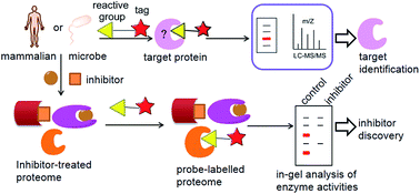
Serine hydrolases (SHs) are involved in a wide range of physiological and pathological processes. Conventional proteomic or genomic methods can only provide an indirect assessment of the functional state of this enzyme in cells and tissues. The lack of effective small-molecule probes or pharmacological tools for the functional characterization of SHs has hindered our understanding of this class of enzymes and its myriad functions and regulation modes. Activity-based protein profiling (ABPP) has emerged as a powerful chemical proteomic method for broad profiling of functional states of enzymes in native biological systems. Herein, we will describe how ABPP has been used to identify and characterize SHs and their inhibitors that are important in physiological and pathological processes of mammals and microbes. Moreover, an integrated workflow for functional mapping of SHs is also depicted.


1. Introduction

Serine hydrolases (SHs) represent one of the largest and most diverse enzyme classes in eukaryotic and prokaryotic proteomes, generally including lipases, esterases, thioesterases, peptidases, proteases and amidases.1–3 In mammals, SHs constitute ∼1% of all proteins and play important roles in a wide range of physiological and pathological processes, including cancer,4,5 inflammation,6,7 neuronal signaling,8 digestion9,10 and blood clotting.11 SHs are also found to perform vital functions in viruses, bacteria and fungi, where they are closely associated with drug resistance, virulence and pathogen life cycle.1 As a widespread group of proteolytic enzymes, SHs are crucial to numerous physiological processes and widely influence microbial pathogenicity. The extensive biological significance of SHs has stimulated many researchers to develop inhibitors for this class of enzymes to be used as both chemical probes to study enzyme function and potentially therapeutic drugs.

In accordance with their biological importance and clinical relevance, SHs are targeted by drugs to treat a variety of diseases such as diabetes,12 obesity,13 Alzheimer's disease,14 bacterial15 and viral infections.16 Selective chemical inhibitors have served as valuable probes for the functional annotation of SHs. Many structurally diverse SH inhibitors have been exploited, which include compounds with reversible and irreversible mechanisms of action. To date, multiple specific chemotypes have been developed for selective SH inhibitors such as lactones/lactams,17,18 carbamates19–21 and ureas,2,22 that inactivate SHs by covalent modification of the conserved serine nucleophile. In some cases, structural features have been introduced into the inhibitors to tailor their selectivity for individual SHs.2,21,23

Despite these advances, the majority of eukaryotic and prokaryotic SHs still lack selective and in vivo-active inhibitors for their functional characterization in terms of their endogenous substrates and cellular functions. The systematic functional characterization of SHs is also limited due to the lack of effective small-molecule probes or pharmacological tools. Moreover, many SHs are regulated by post-translational events, meaning that conventional proteomic24,25 or genomic methods26,27 used to measure protein expression levels can only provide an indirect assessment of their activity changes. These challenges require new proteomic technologies that can facilitate assignment of protein function on a global scale in complex biological systems.

To address this gap, activity-based protein profiling (ABPP), pioneered by Cravatt and Bogyo, has emerged as a powerful chemical proteomic method for broadly profiling of functional states of enzymes in native biological systems.28–30 This chemo-proteomic strategy stands out as a useful method in the application of identifying selective and potent SH inhibitors, including those that are uncharacterized in terms of their substrates and products. Recently, several excellent reviews have extensively summarized the field of ABPP and its implementation in SH superfamily.1,3,29–31 In this review, we will briefly introduce the application of ABPP in the discovery and functional characterization of SHs in mammalian. Specially, we will describe how activity-based probes (ABPs) have been used to successfully identify and characterize SHs inhibitors involved in mammalian physiological and pathological processes. Also, we will describe how ABPP has been used for the characterization of virulence-associated SHs and host immune responses relative to microbial pathogenesis.

2. Serine hydrolase activities profiling by ABPP

2.1. The mammalian serine hydrolases

There are roughly 240 predicted SHs in the mammalian proteome characterized by the presence of an active site serine that is used for the hydrolysis of substrates, with one-half characterized as metabolic serine hydrolases and the other half as serine proteases/peptidases.32 Despite the widely varied structures and sequences, SHs share a common catalytic mechanism. They use a conserved mechanism involving a base-activated serine nucleophile to hydrolyze amide, ester and thioester bonds in small molecules, peptides, or proteins (Fig. 1).31–33 Most serine proteases commonly have chymotrypsin-like or subtilisin-like folds and primarily use a catalytic triad (Ser-His-Asp) to cleave peptide bonds in proteins.34 Serine proteases are typically produced as inactive precursors (zymogens) and therefore are difficult to assay in heterologous expression systems.
image file: c6ra20006k-f1.tif
Fig. 1 Catalytic mechanism of SHs. A base-activated serine nucleophile attacks the substrate carbonyl group to form a covalent intermediate and release the first reaction product. A water molecule then cleaves the resulting acyl-enzyme intermediate to release the second reaction product and regenerate the active enzyme.

The metabolic serine hydrolases (mSHs) are composed of a range of lipases, peptidases, esterases, thioesterases and amidases. The nucleophilicity of the active site serine of mSHs is rendered by the presence of a catalytic dyad (e.g., Ser-Lys or Ser-Asp) or triad (e.g., Ser-His-Asp or Ser-Ser-Lys).35,36 The hydrolysis is completed by the activation of a conserved serine residue, which attacks the carbonyl group of the substrate. The resulting acyl-enzyme intermediate at the active site serine is then cleaved by a water molecule, thus releasing the free serine to its active state (Fig. 1).3,28 The greatly enhanced nucleophilicity of the catalytic serine makes it susceptible to be covalently modified by many types of electrophiles, including fluorophosphonates (FPs) and aryl phosphonates, sulfonyl fluorides, carbamates and ureas.2,21,29

To date, several members of the mSHs have been well characterized, such as monoacylglycerol lipase (MAGL), fatty acid amide hydrolase (FAAH), diacylglycerol lipases (DAGL), KIAA1363 and protein phosphatase methylesterase-1 (PME-1). However, selective inhibitors are still lacking for most mSHs, which further complicates their functional characterization.

2.2. The serine hydrolases involved in microbe

ABPP also served as a powerful strategy for dissecting the function of microbial systems relevant to human health, bioenergy production and the environment.37 In recent years, ABPP has been used to study microbial pathogenesis, and enables the characterization of host immune responses and several virulence-associated enzymes.38 The successful application of ABPP in interrogating host-pathogen interactions has led to the discovery of novel host factors involved in viral pathogenesis.39–41 Recently, ABPP has been used to investigate host–virus interactions and decipher the molecular pathogenesis of virus infections, such as Hepatitis C virus (HCV) infection.41 Monitoring the altered SH activities during viral infection will be useful for discovering novel targets for diagnostic and therapeutic application.

ABPs also have been used to profile host responses to bacterial infection. In many cases, the pathological effects of host response to bacterial infection are closely associated with disease pathology. Hydrolases and post-translational regulation events are often involved in these processes, making ABPs useful tools for studying host defense mechanisms and bacterial pathogenesis.38 FP probes have been used for this purpose during Mycobacterium tuberculosis (Mtb) and Vibrio cholera infection. Also FP probes have been used to measure the proteome responses of Aspergillus fumigatus to Human Serum.42,43

Additionally, caseinolytic protein protease (ClpP)44,45 and rhomboid proteases GlpG46 represent a major virulence regulator and intramembrane proteolytic serine proteases in microbe, respectively. The important pathological and physiological roles of these serine proteases in microbe make them prime targets by ABPs. By far, microbial ABPP studies are still in the infancy, but great promise remains in research to uncover microbial bioenergy conversion processes and microbial pathogens function.37,47 Specific examples will be described in Section 4.

3. Application of ABPP in mammalian SHs

3.1. ABPP for serine hydrolases discovery

Activity-based protein profiling (ABPP), utilizes active site-directed covalent probes to profile the functional state of enzymes and enable direct detection and affinity purification of target enzymes in complex biological samples, such as cell lysates, intact cells and even whole organisms.28,48,49 An activity-based chemical probe typically contains at least two key elements (Fig. 2A): a reactive group for binding and covalently labelling the active sites of enzymes that share conserved mechanistic or structural features, and a reporter tag for promoting a simultaneous read out enzyme activities. The reporter can be a fluorophore for depicting enzyme activities, or a biotin for enrichment, identification and quantification of activities with mass spectrometry-based proteomics (Fig. 2B). Additionally, the reporter can be substituted by a bio-orthogonal chemical handle, such as an alkyne. Then the probe-labelled enzymes are detected by subsequent click chemistry conjugation.50,51
image file: c6ra20006k-f2.tif
Fig. 2 Activity-based protein profiling (ABPP). (A) Schematic representation of an activity-based probe. (B) In gel-based ABPP, native proteomes are reacted with the probe and probe-labeled proteins are visualized by SDS-PAGE and fluorescent scanning (fluorophore probes, top). MS-based ABPP facilitates the identification and quantification of enzyme activities by avidin enrichment, on-bead digestion and Multidimensional Protein Identification Technology (MudPIT) (biotinylated probe, bottom). Blue star represents biotin. (C) Covalent modification of the active-site serine nucleophile of SHs by a FP probe and typical structures of fluorophosphonate probes. Rh represents rhodamine.

ABPP has been widely applied in various biological systems, including target identification,52,53 high-throughput screening (HTS),54 the discovery of deregulated enzyme activities in disease states,5,55 the development of selective enzyme inhibitors,2,19 the characterization of enzyme active sites56,57 and localization of enzyme activity in cells58 and in vivo.59 The original and still most common application of ABPP is for “target discovery” in biological systems. A typical target discovery experiment would allow a comparison and analysis of two or more proteomes to discovery enzymes that show diverse activity. ABPP has been widely used to study the SH superfamily by using the serine hydrolase-directed ABPs fluorophosphonate-rhodamine (FP-Rh) and FP-biotin that covalently phosphorylate the active site serine of nearly all SHs (Fig. 2C).48,60 The FP probes have been widely used in ABPP experiments since they covalently react with most SHs in a potent and irreversible way, while showing minimal cross-reactivity with other hydrolases, including cysteine-, aspartyl- and metallo-hydrolases.28,61

Specially, FP probes have proven to be useful for identifying mSH activities that are dysregulated in cancer cells. For example, Jessani and colleagues utilized FP probes to quantitatively profile enzyme activities in human cancer cells and primary breast tumours, which resulted in the identification of a cluster of proteases, lipases and esterases that distinguished cancer lines based on tissue of origin. These invasiveness associated enzymes included a membrane-associated mSH, KIAA1363, for which no previous association to cancer had been made.62 Using FP probes, the KIAA1363 activity was found to be highly expressed in aggressive human cancer cells and primary tumours (Table 1).62–64 Importantly, knock down of KIAA1363 by RNA interference (RNAi) has disrupted ether lipid metabolism in cancer cells and impaired cell migration and tumor growth in vivo. Further research on functional proteomics indicates that this enzyme is critical in metabolizing an ether lipid signaling network that bridged platelet-activating factor and lysophosphatidic acid.64 Whilst Nomura and colleagues used FP probes to identify KIAA1363 as a principal enzyme for metabolizing low levels of organophosphorus nerve toxins in rodent brains, indicating that this enzyme may play an important role in detoxification of organophosphorus nerve poisons.65

Table 1 Fluorophosphonate and fluorophosphate probes and their applications
Probe Structure Applications
Fluorophosphonate probes image file: c6ra20006k-u1.tif Identified highly expressed KIAA1363[thin space (1/6-em)]62–64 and MAGL5 activities in aggressive human cancer cells and primary tumours
Identified dramatically upregulated uPA and tPA activities in secreted proteomes of aggressive human breast cancer55,68
Identified RBBP9 as a tumor-associated metabolic serine hydrolase and displayed increased activity in pancreatic carcinomas69
Fluorophosphate probes image file: c6ra20006k-u2.tif Identified ATX implicated in tumor progression and inflammation72
image file: c6ra20006k-u3.tif Revealed decreased activity of ESD, which predicts the presence of high-grade lung adenocarcinomas75


Similar to KIAA1363, monoacylglycerol lipase (MAGL), which regulates a free fatty acid network that includes many oncogenic signaling lipids, was also shown to be highly expressed in aggressive human cancer cells and primary tumours by FP probes (Table 1).5,66 The elevated MAGL activity in aggressive cancer cells was confirmed by using the substrate C20[thin space (1/6-em)]:[thin space (1/6-em)]4 MAG and MAG hydrolysis was dramatically reduced when treated with a potent and selective MAGL inhibitor (JZL184[thin space (1/6-em)]20). Nomura and colleagues demonstrated that highly elevated MAG hydrolysis is a prominent feature of both aggressive human cancer cell lines and primary tumors that is mostly the result of up-regulation of MAGL activity. MAGL was also found to be a principal regulator of free fatty acid (FFA) levels in aggressive cancer cells, which is not generally the case in normal cells. The authors went on to demonstrate that overexpression of MAGL in non-aggressive cancer cells increases their pathogenicity, whereas pharmacological or RNA interference disruption of this enzyme impairs cancer aggressiveness.5,67 This work is a representative example of ABPs in application of identifying candidate enzymes potentially involved in aggressive cancers, indicating activity differences may be not detected by the expression level. These results highlight the capability of ABPP in discovering previously uncharacterized enzymes with unknown functions as potential targets.

FP probes have also been used to identify the dramatically upregulated urokinase plasminogen activator (uPA) and tissue plasminogen activator (tPA), as highly secreted enzymes in aggressive human breast cancer line MDA-MB-231 (Table 1).55 On the other hand, although it's already known that urokinase activity is subject to a host of posttranslational mechanism, no direct correlation between mRNA levels and urokinase activity could be found through mRNA levels analysis, indicating that mRNA levels do not represent an accurate measure of enzyme activity as provided by ABPP. Moreover, the FP probes were used to compare serine hydrolase activities in high and low intravasating variants of the human fibrosarcoma HT1080 cell line. The uPA activity was discovered to be elevated in the high intravasating variant, indicating that active uPA promotes tumor cell intravasation and may be a key step in tumor progression.68 This study demonstrates that uPA plays a key role in the process of intravasation, providing strong evidence of ABPP in identifying candidate enzymes involved in regulating intravasation. Using the SH directed probes, Shields and colleagues have determined that the retinoblastoma-binding protein 9 (RBBP9) is a tumor-associated metabolic SH and displayed increased activity in pancreatic carcinomas (Table 1). The enzyme promotes anchorage-independent growth and pancreatic carcinogenesis through overcoming TGF-β mediated antiproliferative signaling by reducing Smad2/3 phosphorylation levels.69

In addition to fluorophosphonate probes, fluorophosphate probes have also been successfully developed to probe serine hydrolase activities in complex biological systems. Cavalli and colleagues designed the first ABP to target autotaxin (ATX), a secreted glycoprotein involved in the hydrolysis of lysophosphatidylcholine (LPC) into the lipid mediator lysophosphatidic acid (LPA),70,71 which has been implicated in tumor progression and inflammation (Table 1).72 This probe specifically labels recombinant ATX and its isoforms in an activity-dependent manner, indicating that the use of an ABP can be an effective tool to monitor ATX levels in plasma. Therefore, ATX-ABPs will function as a potentially diagnostic tool for such tumor by monitoring the level of ATX in body fluids.

Another example of the fluorophosphates probes in the discovery of SH activities is given by Wiedl and colleagues. S-Formylglutathione hydrolase, also known as esterase D (ESD), which was originally involved in the detoxification of formaldehyde,73,74 has not been previously associated with non-small cell lung cancer. With an isopropyl fluorophosphate probe, the authors revealed that a decreased activity of ESD significantly predicted the presence of high-grade lung adenocarcinomas.75 No difference in ESD transcript levels or protein abundance was found when comparing normal and tumor tissue, which highlights the importance of using ABPP for activity quantification. These findings highlight the value of ABPP for the discovery of novel SH activities that depict the origin and pathogenic state of cancer cells. It is more important that ABPP shows great promise for application of ABPs to the identification of novel biomarkers for human disease. In the future, detection of enzyme activity using ABPs may serve as an effective diagnostic method for early detection of aggressive cancers.

3.2. ABPP for SH inhibitors discovery

Developing inhibitors for enzymes typically requires knowledge of their substrates for assay configuration. This criterion has historically hindered the inhibitor discovery for large enzyme classes, such as the SHs, that possess many uncharacterized members. Fortunately, this challenge has been addressed by ABPP in recent years. Competitive ABPP typically involves the treatment of cell or proteomes with small molecule inhibitors, followed by incubation with a fluorescent probe, separation of proteins by SDS–PAGE and quantification of the fluorescence intensity of labelled enzymes relative to a control proteome (known as gel-based competitive ABPP) (Fig. 3A).76 Competitive ABPP was subsequently coupled with multidimensional protein identification technology (MudPIT) to greatly improve the depth of proteome coverage achievable in ABPP experiments.63 Because activity-based probes bind to the active site of their enzyme targets, they can form the basis for a competitive platform for enzyme inhibitors screening.77,78
image file: c6ra20006k-f3.tif
Fig. 3 Competitive ABPP platforms. (A) In competitive ABPP, inhibitors compete with activity-based probes for enzyme targets. Enzyme inhibition is read out by a loss of fluorescence intensity (for fluorophore-conjugated probes) or by a loss of spectral counts by MS signals (for biotinylated probes). (B) Fluopol ABPP is a HTS version of competitive ABPP conducted with pure or recombinant protein. Fluorescence polarization is high if the inhibitor is inactive and low if the inhibitor is active.

Comparing with traditional substrates assay, competitive ABPP has several advantages. First, inhibitors can be directly screened against many enzymes in parallel in native proteomes, cells, or animal models, thereby allowing concurrent optimization of inhibitor potency and selectivity in a single experiment. In addition, inhibitors can also be developed for the uncharacterized enzymes that lack known substrates.19,64 Competitive ABPP has become a primary assay for screening directed libraries of inhibitors and for optimizing their selectivity against SHs.

Using competitive ABPP, multiple selective inhibitors have been found to play important roles in the functional characterization of SHs. Especially, in some cases, they have been developed to be drugs to treat diseases such as diabetes,12 obesity,79 Alzheimer's disease,14 bacterial17 and viral80 infections. Competitive ABPP platforms have proven to be successful in generating small-molecule SH inhibitors using the fluorophosphonate ABP. Here, we review how competitive ABPP is being used to develop inhibitors for SHs.

3.2.1. Fatty acid amide hydrolase (FAAH) inhibitors. FAAH has served as a new therapeutic target for the treatment of a range of clinical disorders including pain, inflammation, and sleep disorders.81 The discovery of selective FAAH inhibitors has become vital for studying the physiological roles of FAAH in animal models82 and humans.83,84 Using competitive ABPP, multiple novel and attractive FAAH inhibitors have been discovered and tested with selectivity to elucidate the role of FAAH as the primary hydrolase for the endogenous cannabinoid system (Table 2).22,85 A competitive ABPP study in mouse brain membrane proteome with FP-rhodamine probe has identified multiple α-ketoheterocycle analogues as reversible FAAH inhibitors.76 In this systematic and extensive study, oxazoles were found to be more potent than thiazoles, imidazoles and the corresponding oxazolines. Like the trifluoromethyl ketones, these α-ketoheterocycle inhibitors reversibly form an enzyme-stabilized hemiketal between the active-site Ser241 and the electrophilic carbonyl.86
Table 2 Representative SH targets and their inhibitors
Target Inhibitor Structure Ref.
FAAH LY2183240 image file: c6ra20006k-u4.tif 87
PF-750 image file: c6ra20006k-u5.tif 88
PF-3845 image file: c6ra20006k-u6.tif 22
PF-04457845 image file: c6ra20006k-u7.tif 89 and 90
MAGL JZL184 image file: c6ra20006k-u8.tif 20
KML29 image file: c6ra20006k-u9.tif 91
JW651 image file: c6ra20006k-u10.tif 92
MJN110 image file: c6ra20006k-u11.tif 93
JJKK-048 image file: c6ra20006k-u12.tif 94
DAGL-α LEI104 image file: c6ra20006k-u13.tif 95
LEI-106 image file: c6ra20006k-u14.tif 96
LEI105 image file: c6ra20006k-u15.tif 97
DAGL-β KT109 image file: c6ra20006k-u16.tif 98
KT172 image file: c6ra20006k-u17.tif 98
KIAA1363 AS115 image file: c6ra20006k-u18.tif 64
JW480 image file: c6ra20006k-u19.tif 99
APEH AA74-1 image file: c6ra20006k-u20.tif 2
PME-1 ABL127 image file: c6ra20006k-u21.tif 100


In addition to the reversible inhibitors, LY2183240 (Table 2) was reported to inhibit FAAH by an irreversible mechanism involving carbamylation of the Ser241.87 This compound was found to block the FP-Rh labeling of FAAH in a concentration-dependent manner and proved to be a remarkably potent inactivator of FAAH (IC50 = 13 nM), suggesting that it might serve as a useful pharmacological tool for studying this enzyme. However, competitive ABPP studies also revealed that LY2183240 inhibited several other brain SHs both in vitro and in vivo with IC50 values in the low nanomolar range. A high-throughput screen of the Pfizer chemical library conducted by Ahn and colleagues has discovered PF-750, a novel piperidine/piperazine urea-containing FAAH inhibitor, which covalently inactivate FAAH via carbamylation of the active-site serine nucleophile.88 Considering the stability of the urea functional group, this irreversible covalent mechanism was rather surprising. Despite the covalent mechanism, competitive ABPP revealed that PF-750 showed no discernible activity against other SHs in vivo at concentrations up to 30 mg kg−1. A possible mechanism that could help explain this remarkable selectivity is a specific binding-induced conformational change of the piperidine/piperazine urea in the FAAH active site, which renders the reactivity of the urea similar to an amide. These findings highlighted the value of chemoproteomic methods like competitive ABPP that can be used to assess compound selectivity broadly across an entire enzyme class.

Based on the PF-750 structure, the authors speculated the extended length would further enhance contacts with the acyl chain-binding channel and may lead to higher potency. Consistent with this premise, PF-3845 was found to have 20-times higher potency for FAAH compared with PF-750 as a result of more extensive van der Waals interactions between the inhibitor 4-trifluoromethyl-2-pyridyl group and the hydrophobic acyl chain-binding pocket of FAAH.22 Competitive ABPP studies confirmed that PF-3845 selectively inhibits FAAH in vivo, designating this agent as a valuable pharmacological tool to study FAAH-regulated endocannabinoid pathways. As expected, PF-3845 was also found to be covalently attached to the catalytic Ser241 nucleophile of FAAH through a carbamate linkage.

Further chemical modification of PF-3845 focused on optimizing biaryl ether moiety, examining the SAR of the 3-aminopyridyl portion and understanding the importance of the pyridyl nitrogen. All these efforts have resulted in the discovery of PF-04457845, which was identified as a highly efficacious FAAH inhibitor by carbamylating the serine nucleophile.89,90 Though there are multiple irreversible inhibitors on the market, the pharmaceutical industry prefers non-covalent, and reversible inhibitors majorly because the irreversible inhibitors lack specificity for a single protein target. However, this problem can be directly solved by competitive ABPP, PF-04457845 was found to be extremely selective for FAAH relative to other SHs. Such a high degree of selectivity has been established for irreversible inhibitors, their distinctive advantages over reversible inhibitors can be exploited, particularly the fact that their protein targets are often inhibited at very low doses in vivo.

3.2.2. Monoacylglycerol lipase (MAGL) inhibitors. Monoacylglycerol lipase (MAGL) and fatty acid amide hydrolase (FAAH) are two enzymes from the mSH family, which can degrade the endocannabinoids 2-arachidonoylglycerol (2-AG) and anandamide (AEA), respectively. Although several MAGL inhibitors have been developed,101–103 high selectivity and in vivo MAGL inhibitors are still lacking. The first selective MAGL inhibitor JZL184 developed by Long and colleagues has greatly accelerated our understanding of the physiological roles of MAGL. As assessed by competitive ABPP, JZL184 was identified as a potent, selective and in vivo MAGL inhibitor and displayed >100-fold selectivity toward this enzyme over FAAH and most other SHs expressed in the brain (Table 2).20 JZL184 is a piperidine carbamate that was shown to inactivate MAGL by covalent carbamylation of the enzyme's serine nucleophile.104,105 Inhibition of MAGL activity reduces 2-AG hydrolysis activity by ∼85% in mouse brain membranes and results in a dramatic elevation in brain 2-AG levels, indicating that MAGL is the primary enzyme involved in degradation of 2-AG in vivo.20 Although JZL184 has been identified as a useful tool to characterize the function of MAGL in vivo, it displays partial cross-reactivity with FAAH when used at high doses.20,104 New MAGL inhibitor based on an O-hexafluoroisopropyl carbamate scaffold was later discovered to show completely selectivity toward MAGL against other SHs. Using ABPP, an O-hexafluoroisopropyl analog of JZL184, KML29, was shown to potently and selectively inhibit MAGL in vitro and in vivo with minimal cross-reactivity toward other SHs, including FAAH and carboxylesterase.91 JZL184 displayed limited efficacy toward rat MAGL both in vitro and in vivo, whereas KML29 showed nearly complete MAGL blockade. The bioisosteric nature of hexafluoroisopropyl leaving group with the 2-AG substrate leads us to speculate that SH inhibitor selectivity may be better achieved by developing inhibitors bearing reactive groups resembling the structures of natural substrates. A subsequent study has demonstrated that, JW651, an analog of KML29, also selectively inhibited MAGL with an IC50 of 38 nM and did not exhibit cross-reactivity with other brain SHs with the exception of ABHD6.92

Despite these advances, the pursuit of other classes of MAGL inhibitors is still necessary, especially compounds that exhibit superior cross-species in vivo activity. Niphakis and colleagues identified MJN110 as a highly potent, selective and in vivo active carbamate inhibitor of MAGL. Comparing with KML29, MJN110 exhibited superior potency and comparable selectivity in both acute and chronic dosing regimens.93 In addition, by systematically exploring the impact of the characteristics of the leaving group on MAGL inhibitor potency, a piperidine triazole urea analogue JJKK-048, was identified as a super potent inhibitor of MAGL (IC50 < 0.4 nM). Competitive ABPP studies in mouse brain membrane proteome using TAMRA-FP probe indicated that MAGL was the only detectable target JJKK-048 at concentrations up to 10−7 M.94 Together with JZL184, JW651, KML29, MJN110 and JJKK-048, these findings indicate that a pharmacological toolkit consisting of potent and selective MAGL inhibitors with varying chemical scaffolds is becoming available. This toolkit can be used in further studies to explore the consequences of pharmacological MAGL inhibition in experimental models of cancer, neurodegeneration, and metabolic disease. Collectively, these MAGL inhibitors have been invaluable in elucidating the role of MAGL in (patho)physiological processes including pain sensation,106,107 inflammation,7 memory,108 anxiety109 and cancer pathogenesis.5

3.2.3. Diacylglycerol lipases (DAGLs) inhibitors. DAGLs are important metabolic SHs in mammalian cells that integrate multiple lipid signaling pathways, including diacylglycerol,110 endocannabinoid111 and eicosanoid112 networks. The biosynthesis of 2-AG relies on two integral membrane enzymes, diacylglycerol lipase-α and -β (DAGL-α and -β), to synthesize 2-AG from hydrolysis of diacylglycerols.113 Previous genetic disruption experiments with knockout mice have revealed that both DAGL-α and DAGL-β are responsible for 2-AG production in vivo, where the relative contribution made by each enzyme depends on tissue type.114,115 Until now, understanding the role of DAGLs in mammalian physiology and pathophysiology has been hindered by lack of efficient inhibitors that are not only specific to DAGLs over other SHs, but also are specific to only one isoform. To illustrate the influence of DAGLs on 2-AG metabolism and cell and animal physiology, selective and in vivo-active DAGL inhibitors would be of value (Table 2).

The first specific and in vivo active DAGL-β inhibitors were reported, based on the triazole urea scaffold.98 Using competitive ABPP, Hsu and colleagues screened recombinantly expressed DAGL enzymes in HEK293T cells against a library of 1,2,3-triazole urea inhibitors and discovered that compound KT109 and KT172, potently inhibited DAGL-β with an IC50 of 82 and 71 nM, respectively. KT109 and KT172 potently inhibited DAGL-β with nearly 60-fold selectivity over DAGL-α. Due to the low abundance, DAGL-β activity was difficult to be detected by using the general probe FP-rhodamine. This problem has been addressed by creating a tailored triazole urea ABP, HT-01, which showed preferential reactivity with DAGL-β over most other SHs. HT-01 was used to detect DAGL-β in various cell and tissue types and confirmed that KT109 and KT172 could inhibit DAGL-β activity in cancer cells and mouse macrophages. In my opinion, the development of DAGL-β inhibitors has nicely showed that ABPP can provide tailored assays to evaluate target engagement for inhibitors of low-abundance enzymes. Further study by Hsu and colleagues has developed ‘‘clickable’’ analogs of KT172 to confirm the selectivity of this inhibitor for DAGL-β and ABHD6 by using click-chemistry-ABPP, indicating this scaffold may serve as a useful tool for developing potent and selective DAGL-β inhibitors.116

Selective DAGL-α inhibitors are also needed to understand the physiological role of 2-AG and may serve as potential drug candidates to treat obesity and neurodegenerative diseases.7,117 Baggelaar and colleagues reported the first DAGL-α sensitive ABP, MB064, a bodipy tagged β-lactone probe based on the nonselective DAGL-α inhibitor tetrahydrolipstatin (THL; also known as Orlistat).95 Using MB064, they identified LEI104, which belongs to the class of α-ketoheterocycles, as the first reversible inhibitor for DAGL-α (Table 2). It is anticipated that the α-ketoheterocycle class will be of significance in dissecting 2-AG and AEA-mediated cannabinoid CB1 signaling and developing selective DAGL-α inhibitors, due to their excellent physicochemical properties, high selectivity, suitable membrane permeability and reversible mechanism. However, the activity of LEI104 on DAGL-β was not investigated, and it is likely that there is cross-reactivity due to the high homology between DAGL-α and DAGL-β.

Moreover, MB064 probe has been used to confirm that the glycine sulfonamides analogue, LEI-106, was a potent DAGL-α inhibitor with an IC50 of 124 nM.96 ABPP assays also revealed that LEI-106 is a potent ABHD6 inhibitor. To improve the potency of LEI104, the authors used a molecular dynamics simulation analysis of the binding pose of LEI104 in DAGL-α to discover that an additional hydrophobic pocket close to the catalytic site was not occupied by LEI104. Introduction of a phenyl substituent at the 6-position of the oxazolopyridine may help probe this pocket with improved potency/or selectivity.97 As a result, LEI105 was discovered to be a potent inhibitor with 10-fold potency against LEI104. Competitive ABPP has identified α-ketoheterocycle LEI105 as a potent, highly selective and reversible dual DAGL-α/DAGL-β inhibitor. Reduction of the α-keto group resulted in a ∼150 fold decrease in activity, thereby indicating that a reversible, enzyme–inhibitor hemiketal adduct was formed between the α-carbonyl and the active site serine.

3.2.4. KIAA1363 inhibitors. The inhibitor discovery by competitive ABPP has been used to probe the functions of uncharacterized enzymes, which then led to insights into their pathophysiological roles. Previous studies indicated the activity of KIAA1363 is highly elevated in both aggressive human cancer cell lines62 and primary tumours.63 However, due to a lack of pharmacological tools to study this enzyme, attempts to further characterize KIAA1363 function in cancer have been hindered. Competitive ABPP screening with a library of candidate inhibitors identified a set of trifluoromethyl ketone (TFMK) inhibitors that showed activity against KIAA1363 in mouse brain proteomes.76 By replacing the reversibly binding TFMK group with a carbamate, Carbamate AS115, which inactivates SHs via a covalent mechanism, has been developed (Table 2).64 The follow-up competitive ABPP profiling indicated that AS115 potently and selectively inhibited KIAA1363 in cancer cells, displaying an IC50 value of 150 nM, while it showed little influence on other SHs activities (IC50 value > 10 μmol). LC-MS analysis of lipophilic metabolites from AS115 treated cancer cells revealed that KIA1363 regulated a class of monoalkylglycerol ether (MAGE) lipids. Additional biochemical experiments identified KIAA1363 as the principal 2-acetyl MAGE hydrolase in cancer cells.

Further optimization of this scaffold resulted in the generation a potent, selective and in vivo active KIAA1363 inhibitor, JW480, which inhibited mouse brain KIAA1363 with exceptional potency and showed negligible cross-reactivity with hormone-sensitive lipase (HSL), acetylcholinesterase (AChE), or other mouse brain SHs.99,118 Treated with JW480, cancer cells showed dramatically reductions in MAGEs in vitro, as well as impairments in migratory and invasive activity. Recently, KIAA1363 inhibitors have been used to reveal that this enzyme regulates platelet and megakaryocyte activation.119 A selective activity-based imaging probe that enables subcellular localization of KIAA1363 activity in cancer cells has also been developed.58

3.2.5. Other SH inhibitors. Though acylpeptide hydrolase (APEH) is considered to serve as a key regulator of N-terminally acetylated proteins in T cells,120 very few endogenous substrates have been identified for APEH. Adibekian and colleagues selected the pyrrolidine- and morpholine-based scaffolds as a starting point for synthetizing a library of 1,2,3-triazole ureas. The authors speculated that the selectivity of individual SHs would be enhanced by introducing substituents into the triazole group. As a result, a series of triazole ureas were synthesized by an efficient click-chemistry approach. In this two-step strategy, substituted alkynes were reacted with in situ-formed azidomethanol to yield 4-substituted triazoles, which were then carbamoylated to give triazole urea products, typically as a 3[thin space (1/6-em)]:[thin space (1/6-em)]1 mixture of N2- and N1-carbamoylated regioisomers. The N2-carbamoyl triazole was then separated and purified by silica gel chromatography and used for subsequent experiments, resulting in a ∼20-member library of 4-aryl- and 4-alkyl triazole derivatives.

Using competitive ABPP, the 1,2,3-triazole urea AA74-1 was identified as an ultrapotent, selective, and in vivo active inhibitors of APEH in mice (Table 2), suggesting that it is suitable for probing the functional characterization of SH targets in biological systems. In addition, AA39-2 and AA44-2 was also found to inhibit platelet activating factor acetylhydrolase 2, and uncharacterized hydrolase ABHD11, respectively.2 Similar to the mechanism of carbamate scaffolds,21,32 1,2,3-triazole ureas inhibit SHs through a covalent, irreversible mechanism involving carbamoylation of the catalytic serine nucleophile.2 These results indicate 1,2,3-triazole ureas can serve as a useful pharmacologically tool for the discovery of potent and selective SH inhibitors involving in eukaryotic and prokaryotic proteomes.

3.3. Fluopol-ABPP for the HTS of SH inhibitors

Competitive ABPP has traditionally been limited by low throughput because it relies on SDS-PAGE or mass spectrometry assays and can only be used to screen a few hundred compounds.121 A high-throughput fluorescence polarization technology for ABPP-based screening (fluopol-ABPP) has recently been introduced to address the limitation (Fig. 3B).54 Fluopol-ABPP monitored the interaction between an activity-based probe and an enzyme by fluorescence polarization and proved applicable to high-throughput screening (HTS) in multi-well format. For instance, by screening 20[thin space (1/6-em)]000 small molecules in 384-well plates, the bioactive alkaloid emetine was identified as a selective inhibitor of the uncharacterized RBBP9, a cancer-associated enzyme.54

Protein phosphatase methylesterase-1 (PME-1), a regulator of protein phosphatase 2A, has been linked to cancer,122,123 and Alzheimer's disease.124 Bachovchin and colleagues has recently used fluopol-ABPP to screen the NIH 300[thin space (1/6-em)]000+ compound library against PME-1, resulting in the discovery of ABL127 (Table 2), a highly potent and selective class of aza-β-lactam (ABL) inhibitors of PME-1 in human cancer cells and mice. ABL127 covalently inhibits PME-1 and decreased the levels of demethylated protein phosphatase 2A. These results demonstrate that ABL127 could serve as a versatile pharmacological probe for probing PME-1 function in living systems.100 More generally, fluopol-ABPP platform has been successfully used to screen other enzymes inhibitors, including protein arginine deiminases,125 arginine methyltransferases126 and glutathione S-transferases.127

As demonstrated above, competitive ABPP has served as a powerful tool for discovering SH inhibitors and for optimizing their selectivity, thus expanding our knowledge of SHs in (patho)physiology. Selective inhibitors developed by competitive ABPP have facilitated the functional characterization of SHs in many biological processes, including neurodegeneration, cancer metabolism and signaling, immunology and infectious disease.

4. ABPP for interrogating SHs involved in microbe

4.1. SHs involved in virus infection

Hepatitis C virus (HCV) is known to induce changes in lipid metabolism128,129 and causes the formation of endoplasmic reticulum (ER)-derived membranous webs where HCV replicates.130 Pezacki and colleagues used FP probe to profile the activity of SHs during HCV replication and discovered carboxylesterase 1 (CES1) expression and activity upregulated in HCV-infected hepatoma cells (Table 3). Knockdown of CES1 with siRNA decreased HCV replication, while the upregulation of CES1 increased HCV propagation. These results indicate that CES1 plays an important role in HCV propagation during host–virus interactions.131
Table 3 Represent activity-based probes and their microbial applications
Probe target Structure Applications
CES1 image file: c6ra20006k-u22.tif FP-Rh to identify the upregulated CES1 expression and activity in HCV-infected hepatoma cells131
FASN image file: c6ra20006k-u23.tif Orlistat-based probe to discover that HCV infection upregulates fatty acid synthase (FASN) expression in Huh7 human hepatoma cells132
IvaP image file: c6ra20006k-u24.tif FP-biotin to discover active IvaP in Vibrio cholerae infected rabbits and in human choleric stool133
ClpP image file: c6ra20006k-u25.tif β-Lactone probes to rapidly and selectively label active ClpP as low as 1.3 μM in S. aureus and completely abolish hemolytic and proteolytic activities134
image file: c6ra20006k-u26.tif VLP probe to label two isoforms of ClpP protease from L. monocytogenes135
image file: c6ra20006k-u27.tif Phenyl ester probe to selectively and potently label ClpP in pathogenic bacteria136
GlpG image file: c6ra20006k-u28.tif FP-Rh to effectively examine the activity of rhomboid proteases that are present in crude lipid extracts isolated from E. coli46 and to be considered as universal ABP for labeling rhomboids137
image file: c6ra20006k-u29.tif Isocoumarin probe to distinguish between active and inactive rhomboids due to covalent, reversible binding of the active-site serine in E. coli138


Since the lipogenic enzymes are important in HCV replication cycle, the group of Pezacki attempted to uncover the possible modulation of the activity of FASN by HCV infection. In a follow-up study, they discovered that HCV infection upregulated fatty acid synthase (FASN) expression at both transcriptional and protein levels in Huh7 human hepatoma cells by using an orlistat-based probe (Table 3).132 It was also indicated that the increase in FASN abundance during HCV replication resulted in an increase in the overall FASN catalytic activity.

4.2. SHs involved in bacterial infection

SHs were reported to have central functions on all levels of Mycobacterium tuberculosis (Mtb) physiology, including persistence. Their wide influence on microbial pathogenicity, such as Mtb metabolism, makes them prime drug targets. Rapid replication of Mtb cells in vitro cultures can't accurately represent the spectrum of growth phases found in chronically infected human tuberculosis patients. To explore the opposite end of the spectrum of metabolic states, the FP-probe was used to label proteins from hypoxic non-replicating cultures and compared to the labeled proteins from rapidly replicating cells.139 In a latest study, Ortega and colleagues systematically investigated SH activity in replicating and non-replicating Mtb by using a click-chemistry-enabled FP probe and identified 27 SHs with previously unknown functions.140 The authors showed that SH activity exhibited large changes during the transition from replication to non-replication. Moreover, several SHs were found to be active during non-replication, providing new drug targets in persistent infection.

Recently, another application in bacterial infection was given by Hatzios and colleagues. The authors used FP probes to discover that four pathogen-secreted serine proteases were consistently active in Vibrio cholera-infected rabbits (Table 3).133 One of these proteases, VC0157, was also active in human choleric stool and influenced the activities of other bacterial and host SHs during infection. These results further highlight the ability of ABPP in studying host-pathogen interaction.

4.3. SHs involved in fungi infection

Aspergillus fumigatus (A. fumigatus) is one of the most prevalent airborne fungal pathogens and has caused various forms of disease in immunocompromised host lung.141–143 Wright and colleagues discovered that exposure of fungal pathogen to host serum altered physiological functions throughout numerous metabolic processes by using FP probe coupled with quantitative proteomic analysis.42 A follow-up study by comparing A. fumigatus with two highly related but rarely pathogenic fungi has identified that multiple processes including actin organization and assembly, transport, and fatty acid, cell membrane, and cell wall synthesis were induced by human serum in A. fumigatus.43 Therefore, ABPP can serve as a powerful tool to understand the fungi's pathogenesis mechanism and discover differences in abundance of ABP-reactive proteins under infection relevant conditions.

4.4. ABPP for ClpP in microbe

SHs represent a widespread group of proteolytic enzymes that are crucial to numerous physiological processes and have been implicated in microbial virulence mechanisms. Caseinolytic protein protease (ClpP) has previously been identified as a major virulence regulator in many bacterial pathogens and represents a central bacterial degradation machinery. Moreover, it has been found to be essential for cellular stress response, cell homeostasis and bacterial virulence.144–146 A genetic ClpP knockout study in Staphylococcus aureus (S. aureus) and Listeria monocytogenes (L. monocytogenes) revealed a significantly reduced virulence in a murine abscess model, making ClpP a suitable target for alternative anti-infective treatment.44,144,145

Previous ABPP studies have identified β-lactone probes as the first selective inhibitors of the highly conserved serine protease ClpP.18 β-Lactones are reported to react covalently with the catalytic Ser98, which results in ring opening and blockage of the active site and provides a catalytically inactive β-hydroxyacyl–enzyme complex (Table 3).147 Böttcher and colleagues utilized ABPP to identify functionalized β-lactones as selective and specific irreversible inhibitors of ClpP in S. aureus and corresponding methicillin-resistant (MRSA) strains. More importantly, β-lactones were discovered to completely abolish hemolytic and proteolytic activities in S. aureus.18,134 Subsequently, competitive ABPP was used to develop a refined inhibitor that inhibited ClpP activity with a four- to five-fold improved potency in S. aureus cultures. Additionally, the production of several virulence factors like enterotoxins and toxic shock syndrome toxin 1 (TSST-1) by MRSA and clinical strains were shown to be dramatically reduced, indicating the expression of these toxins is regulated by ClpP.148 Similar experiments were also performed in the intracellular pathogen L. monocytogenes cultures to demonstrate that β-lactone probes also displayed high selectivity for ClpP.149

To improve the probe specificity against ClpP, Zeiler and colleagues used an alkyne probe VLP based on the bicyclic β-lactone vibralactone to label two isoforms of ClpP protease from L. monocytogenes. The VLP, which was used in E. coli coexpression studies, has showed that ClpP1 was activated by hetero-oligomerization with ClpP2 (Table 3).135 In order to expand the repertoire of ClpP inhibitors, an unbiased high-throughput screen (HTS) of more than 137[thin space (1/6-em)]000 compounds was performed to identify five phenyl ester compounds as highly potent, irreversible ClpP inhibitors in pathogenic bacteria.136 To date, only β-lactones and phenyl esters have been reported as specific ClpP inhibitors in whole proteome studies. ABPP-guided medicinal chemistry studies have led to the discovery of more potent and selective inhibitors in models of bacterial virulence, which have contributed to the investigation of ClpP's functional roles in bacteria and the catalytic mechanism on a molecular level.

4.5. ABPP for rhomboid proteases GlpG in microbe

Rhomboid proteases are a family of intermembrane serine proteases and have been implicated in a variety of biochemical processes, including cell signaling,150 apoptosis151 and activation of signal translocation in bacteria.152 Due to the difficulty involved in purifying rhomboid proteases, the development of small-molecule tools, such as inhibitors and activity-based probes (ABPs), has been relatively slow.

Sherratt and colleagues reported the first rhomboid ABP FP-Rh to probe the activity state of the E. coli rhomboid GlpG and identified that N-terminal sequence is critical for maintaining a catalytically competent state for the E. coli GlpG (Table 3).46 The FP-Rh was used to develop an inhibition assay based on fluorescence polarization (fluopol). Using this method, β-lactones were identified as a novel scaffold of covalent rhomboid inhibitors.153 Isocoumarin scaffold was discovered to be another new class of rhomboid inhibitors developed by Vosyka and colleagues.138 Both the alkynylated isocoumarins and fluorophore-conjugated isocoumarin were identified as useful ABPs to covalently label the rhomboid active site in micelles, lysates and in vivo of E. coli (Table 3).

Recently, FP-Rh probe was applied to label rhomboid proteases in liposomes. Wolf and colleagues discovered that the inhibitor profiles of rhomboids in micelles and liposomes were similar, indicating that micelle-solubilized rhomboids are a model system for inhibitor screening.154 Additionally, the same group also showed that FP-Rh could be considered as a universal ABP for labeling rhomboids from three different phylogenetic domains (Table 3).137

5. Conclusion

As discussed in this review, the chemoproteomic approach ABPP has greatly enhanced our understanding of the role of SHs in complex physiological and pathological processes, implying that detection of enzyme activity using ABPs may serve as an effective diagnostic method for human disease. ABPP can provide information on the relative activity state of SHs in their native environment, thereby directing researchers to specific biological systems in which the enzymes may play a key role. Also we have attempted to highlight the value of ABPP for the discovery of potent and selective SH inhibitors even for previously uncharacterized SHs, which has advanced our understanding of enzyme functions in cell. Selective inhibitors developed by competitive ABPP have facilitated the functional characterization of SHs in many biological processes. Notably, with the advent of fluopol-ABPP, inhibitor discovery and optimization can now be carried out in a high-throughput manner, allowing enzyme targets to be screened against more than 100[thin space (1/6-em)]000 compounds. To date, microbial ABPP studies are only in their infancy, but have been demonstrated great promise in dissecting SH functions involved in virulence regulation and host-pathogen responses. The persistent development of ABPP in microbial research will facilitate our understanding of protein annotation, protein activity characterization, signal transduction and regulatory mechanisms in microbial systems.

Acknowledgements

We wish to thank the financial supports from the Key Technologies R&D Program (2014BAD23B01), National Natural Science Foundation of China (21372052), the Research Project of Chinese Ministry of Education (213033A, 20135201110005).

References

  1. D. A. Bachovchin and B. F. Cravatt, Nat. Rev. Drug Discovery, 2012, 11, 52–68 CrossRef CAS PubMed.
  2. A. Adibekian, B. R. Martin, C. Wang, K. L. Hsu, D. A. Bachovchin, S. Niessen, H. Hoover and B. F. Cravatt, Nat. Chem. Biol., 2011, 7, 469–478 CrossRef CAS PubMed.
  3. J. Z. Long and B. F. Cravatt, Chem. Rev., 2011, 111, 6022–6063 CrossRef CAS PubMed.
  4. J. A. Menendez and R. Lupu, Nat. Rev. Cancer, 2007, 7, 763–777 CrossRef CAS PubMed.
  5. D. K. Nomura, J. Z. Long, S. Niessen, H. S. Hoover, S. W. Ng and B. F. Cravatt, Cell, 2010, 140, 49–61 CrossRef CAS PubMed.
  6. J. V. Bonventre, Z. Huang, M. R. Taheri, E. O'Leary, E. Li, M. A. Moskowitz and A. Sapirstein, Nature, 1997, 390, 622–625 CrossRef CAS PubMed.
  7. D. K. Nomura, B. E. Morrison, J. L. Blankman, J. Z. Long, S. G. Kinsey, M. C. G. Marcondes, A. M. Ward, Y. K. Hahn, A. H. Lichtman, B. Conti and B. F. Cravatt, Science, 2011, 334, 809–813 CrossRef CAS PubMed.
  8. R. M. Lane, S. G. Potkin and A. Enz, Int. J. Neuropsychopharmacol., 2006, 9, 101–124 CrossRef CAS PubMed.
  9. M. Merkel, R. H. Eckel and I. J. Goldberg, J. Lipid Res., 2002, 43, 1997–2006 CrossRef CAS.
  10. D. C. Whitcomb and M. E. Lowe, Dig. Dis. Sci., 2007, 52, 1–17 CrossRef CAS PubMed.
  11. E. W. Davie and O. D. Ratnoff, Science, 1964, 145, 1310–1312 CAS.
  12. N. A. Thornberry and A. E. Weber, Curr. Top. Med. Chem., 2007, 7, 557–568 CrossRef CAS PubMed.
  13. S. Henness and C. M. Perry, Drugs, 2006, 66, 1625–1656 CrossRef CAS PubMed.
  14. M. Racchi, M. Mazzucchelli, E. Porrello, C. Lanni and S. Govoni, Pharmacol. Res., 2004, 50, 441–451 CrossRef CAS PubMed.
  15. I. Staub and S. A. Sieber, J. Am. Chem. Soc., 2008, 130, 13400–13409 CrossRef CAS PubMed.
  16. K. M. Marks and I. M. Jacobson, Antiviral Ther., 2012, 17, 1119–1131 CrossRef CAS PubMed.
  17. A. F. Kluge and R. C. Petter, Curr. Opin. Chem. Biol., 2010, 14, 421–427 CrossRef CAS PubMed.
  18. T. Böttcher and S. A. Sieber, Angew. Chem., Int. Ed., 2008, 47, 4600–4603 CrossRef PubMed.
  19. W. Li, J. L. Blankman and B. F. Cravatt, J. Am. Chem. Soc., 2007, 129, 9594–9595 CrossRef CAS PubMed.
  20. J. Z. Long, W. Li, L. Booker, J. J. Burston, S. G. Kinsey, J. E. Schlosburg, F. J. Pavon, A. M. Serrano, D. E. Selley, L. H. Parsons, A. H. Lichtman and B. F. Cravatt, Nat. Chem. Biol., 2009, 5, 37–44 CrossRef CAS PubMed.
  21. A. B. Cognetta III, M. J. Niphakis, H. C. Lee, M. L. Martini, J. J. Hulce and B. F. Cravatt, Chem. Biol., 2015, 22, 928–937 CrossRef PubMed.
  22. K. Ahn, D. S. Johnson, M. Mileni, D. Beidler, J. Z. Long, M. K. McKinney, E. Weerapana, N. Sadagopan, M. Liimatta, S. E. Smith, S. Lazerwith, C. Stiff, S. Kamtekar, K. Bhattacharya, Y. Zhang, S. Swaney, K. Van Becelaere, R. C. Stevens and B. F. Cravatt, Chem. Biol., 2009, 16, 411–420 CrossRef CAS PubMed.
  23. A. M. Zuhl, J. T. Mohr, D. A. Bachovchin, S. Niessen, K. L. Hsu, J. M. Berlin, M. Dochnahl, M. P. Lopez-Alberca, G. C. Fu and B. F. Cravatt, J. Am. Chem. Soc., 2012, 134, 5068–5071 CrossRef CAS PubMed.
  24. B. Domon and R. Aebersold, Science, 2006, 312, 212–217 CrossRef CAS PubMed.
  25. B. F. Cravatt, G. M. Simon and J. R. Yates III, Nature, 2007, 450, 991–1000 CrossRef CAS PubMed.
  26. T. R. Golub, D. K. Slonim, P. Tamayo, C. Huard, M. Gaasenbeek, J. P. Mesirov, H. Coller, M. L. Loh, J. R. Downing, M. A. Caligiuri, C. D. Bloomfield and E. S. Lander, Science, 1999, 286, 531–537 CrossRef CAS PubMed.
  27. P. O. Brown and D. Botstein, Nat. Genet., 1999, 21, 33–37 CrossRef CAS PubMed.
  28. M. J. Evans and B. F. Cravatt, Chem. Rev., 2006, 106, 3279–3301 CrossRef CAS PubMed.
  29. B. F. Cravatt, A. T. Wright and J. W. Kozarich, Annu. Rev. Biochem., 2008, 77, 383–414 CrossRef CAS PubMed.
  30. M. J. Niphakis and B. F. Cravatt, Annu. Rev. Biochem., 2014, 83, 341–377 CrossRef CAS PubMed.
  31. G. M. Simon and B. F. Cravatt, J. Biol. Chem., 2010, 285, 11051–11055 CrossRef CAS PubMed.
  32. M. K. Ross and R. Wang, Chem. Biol., 2015, 22, 808–809 CrossRef CAS PubMed.
  33. R. E. Moellering and B. F. Cravatt, Chem. Biol., 2012, 19, 11–22 CrossRef CAS PubMed.
  34. J. J. Perona and C. S. Craik, Protein Sci., 1995, 4, 337–360 CrossRef CAS PubMed.
  35. G. Dodson and A. Wlodawer, Trends Biochem. Sci., 1998, 23, 347–352 CrossRef CAS PubMed.
  36. M. P. Patricelli, M. A. Lovato and B. F. Cravatt, Biochemistry, 1999, 38, 9804–9812 CrossRef CAS PubMed.
  37. N. C. Sadler and A. T. Wright, Curr. Opin. Chem. Biol., 2015, 24, 139–144 CrossRef CAS PubMed.
  38. A. W. Puri and M. Bogyo, Biochemistry, 2013, 52, 5985–5996 CrossRef CAS PubMed.
  39. H. Ovaa, B. M. Kessler, U. Rolen, P. J. Galardy, H. L. Ploegh and M. G. Masucci, Proc. Natl. Acad. Sci. U. S. A., 2004, 101, 2253–2258 CrossRef CAS.
  40. U. Rolén, V. Kobzeva, N. Gasparjan, H. Ovaa, G. Winberg, F. Kisseljov and M. G. Masucci, Mol. Carcinog., 2006, 45, 260–269 CrossRef PubMed.
  41. D. R. Blais, N. Nasheri, C. S. McKay, M. C. Legault and J. P. Pezacki, Trends Biotechnol., 2012, 30, 89–99 CrossRef CAS PubMed.
  42. S. D. Wiedner, K. E. Burnum, L. M. Pederson, L. N. Anderson, S. Fortuin, L. M. Chauvigné-Hines, A. K. Shukla, C. Ansong, E. A. Panisko, R. D. Smith and A. T. Wright, J. Biol. Chem., 2012, 287, 33447–33459 CrossRef CAS PubMed.
  43. S. D. Wiedner, C. Ansong, B. J. Webb-Robertson, L. M. Pederson, S. Fortuin, B. A. Hofstad, A. K. Shukla, E. A. Panisko, R. D. Smith and A. T. Wright, Mol. Cell. Proteomics, 2013, 12, 1791–1805 CAS.
  44. H. Brötz-Oesterhelt and P. Sass, Int. J. Med. Microbiol., 2014, 304, 23–30 CrossRef PubMed.
  45. M. Dahmen, M. T. Vielberg, M. Groll and S. A. Sieber, Angew. Chem., Int. Ed., 2015, 54, 3598–3602 CrossRef CAS PubMed.
  46. A. R. Sherratt, D. R. Blais, H. Ghasriani, J. P. Pezacki and N. K. Goto, Biochemistry, 2012, 51, 7794–7803 CrossRef CAS PubMed.
  47. Y. Liu, J. K. Fredrickson, N. C. Sadler, P. Nandhikonda, R. D. Smith and A. T. Wright, Biotechnol. Biofuels, 2015, 8, 156 CrossRef PubMed.
  48. Y. Liu, M. P. Patricelli and B. F. Cravatt, Proc. Natl. Acad. Sci. U. S. A., 1999, 96, 14694–14699 CrossRef CAS.
  49. D. Hunerdosse and D. K. Nomura, Curr. Opin. Biotechnol., 2014, 28, 116–126 CrossRef CAS PubMed.
  50. A. E. Speers, G. C. Adam and B. F. Cravatt, J. Am. Chem. Soc., 2003, 125, 4686–4687 CrossRef CAS PubMed.
  51. A. E. Speers and B. F. Cravatt, Chem. Biol., 2004, 11, 535–546 CrossRef CAS PubMed.
  52. S. Arastu-Kapur, E. L. Ponder, U. P. Fonovic, S. Yeoh, F. Yuan, M. Fonovic, M. Grainger, C. I. Phillips, J. C. Powers and M. Bogyo, Nat. Chem. Biol., 2008, 4, 203–213 CrossRef CAS PubMed.
  53. C. I. Hall, M. L. Reese, E. Weerapana, M. A. Child, P. W. Bowyer, V. E. Albrow, J. D. Haraldsen, M. R. Phillips, E. D. Sandoval, G. E. Ward, B. F. Cravatt, J. C. Boothroyd and M. Bogyo, Proc. Natl. Acad. Sci. U. S. A., 2011, 108, 10568–10573 CrossRef CAS PubMed.
  54. D. A. Bachovchin, S. J. Brown, H. Rosen and B. F. Cravatt, Nat. Biotechnol., 2009, 27, 387–394 CrossRef CAS PubMed.
  55. N. Jessani, M. Humphrey, W. H. McDonald, S. Niessen, K. Masuda, B. Gangadharan, J. R. Yates III, B. M. Mueller and B. F. Cravatt, Proc. Natl. Acad. Sci. U. S. A., 2004, 101, 13756–13761 CrossRef CAS PubMed.
  56. G. C. Adam, J. Burbaum, J. W. Kozarich, M. P. Patricelli and B. F. Cravatt, J. Am. Chem. Soc., 2004, 126, 1363–1368 CrossRef CAS PubMed.
  57. E. Zeiler, A. List, F. Alte, M. Gersch, R. Wachtel, M. Porebac, M. Dragc, M. Grollb and S. A. Sieber, Proc. Natl. Acad. Sci. U. S. A., 2013, 110, 11302–11307 CrossRef CAS PubMed.
  58. J. W. Chang, R. E. Moellering and B. F. Cravatt, Angew. Chem., Int. Ed., 2012, 51, 966–970 CrossRef CAS PubMed.
  59. L. E. Edgington, A. B. Berger, G. Blum, V. E. Albrow, M. G. Paulick, N. Lineberry and M. Bogyo, Nat. Med., 2009, 15, 967–973 CrossRef CAS PubMed.
  60. D. Medina-Cleghorn and D. K. Nomura, Chem. Biol., 2014, 21, 1171–1184 CrossRef CAS PubMed.
  61. L. A. R. Carvalho, E. F. P. Ruivo, S. D. Lucas and R. Moreira, Med. Chem. Commun., 2015, 6, 536–546 RSC.
  62. N. Jessani, Y. Liu, M. Humphrey and B. F. Cravatt, Proc. Natl. Acad. Sci. U. S. A., 2002, 99, 10335–10340 CrossRef CAS PubMed.
  63. N. Jessani, S. Niessen, B. Q. Wei, M. Nicolau, M. Humphrey, Y. Ji, W. Han, D. Y. Noh, J. R. Yates III, S. S. Jeffrey and B. F. Cravatt, Nat. Methods, 2005, 2, 691–697 CrossRef CAS PubMed.
  64. K. P. Chiang, S. Niessen, A. Saghatelian and B. F. Cravatt, Chem. Biol., 2006, 13, 1041–1050 CrossRef CAS PubMed.
  65. D. K. Nomura, D. Leung, K. P. Chiang, G. B. Quistad, B. F. Cravatt and J. E. Casida, Proc. Natl. Acad. Sci. U. S. A., 2005, 102, 6195–6200 CrossRef CAS PubMed.
  66. D. K. Nomura, M. M. Dix and B. F. Cravatt, Nat. Rev. Cancer, 2010, 10, 630–638 CrossRef CAS PubMed.
  67. D. K. Nomura, D. P. Lombardi, J. W. Chang, S. Niessen, A. M. Ward, J. Z. Long, H. H. Hoover and B. F. Cravatt, Chem. Biol., 2011, 18, 846–856 CrossRef CAS PubMed.
  68. M. A. Madsen, E. I. Deryugina, S. Niessen, B. F. Cravatt and J. P. Quigley, J. Biol. Chem., 2006, 281, 15997–16005 CrossRef CAS PubMed.
  69. D. J. Shields, S. Niessen, E. A. Murphy, A. Mielgo, J. S. Desgrosellier, S. K. Lau, L. A. Barnes, J. Lesperance, M. Bouvet, D. Tarin, B. F. Cravatt and D. A. Cheresh, Proc. Natl. Acad. Sci. U. S. A., 2010, 107, 2189–2194 CrossRef CAS PubMed.
  70. A. Tokumura, E. Majima, Y. Kariya, K. Tominaga, K. Kogure, K. Yasuda and K. Fukuzawa, J. Biol. Chem., 2002, 277, 39436–39442 CrossRef CAS PubMed.
  71. M. Umezu-Goto, Y. Kishi, A. Taira, K. Hama, N. Dohmae, K. Takio, T. Yamori, G. B. Mills, K. Inoue, J. Aoki and H. Arai, J. Cell Biol., 2002, 158, 227–233 CrossRef CAS PubMed.
  72. S. Cavalli, A. J. Houben, H. M. Albers, E. W. van Tilburg, A. de Ru, J. Aoki, P. van Veelen, W. H. Moolenaar and H. Ovaa, ChemBioChem, 2010, 11, 2311–2317 CrossRef CAS PubMed.
  73. D. Wu, Y. Li, G. Song, D. Zhang, N. Shaw and Z. J. Liu, FASEB J., 2009, 23, 1441–1446 CrossRef CAS PubMed.
  74. M. Harms, J. Ras, W. N. Reijnders, R. J. van Spanning and A. H. Stouthamer, J. Bacteriol., 1996, 178, 6296–6299 CrossRef PubMed.
  75. T. Wiedl, S. Arni, B. Roschitzki, J. Grossmann, S. Collaud, A. Soltermann, S. Hillinger, R. Aebersold and W. Weder, J. Proteomics, 2011, 74, 1884–1894 CrossRef CAS PubMed.
  76. D. Leung, C. Hardouin, D. L. Boger and B. F. Cravatt, Nat. Biotechnol., 2003, 21, 687–691 CrossRef CAS PubMed.
  77. D. C. Greenbaum, W. D. Arnold, F. Lu, L. Hayrapetian, A. Baruch, J. Krumrine, S. Toba, K. Chehade, D. Bromme, I. D. Kuntz and M. Bogyo, Chem. Biol., 2002, 9, 1085–1094 CrossRef CAS PubMed.
  78. J. L. Counihan, B. Ford and D. K. Nomura, Curr. Opin. Chem. Biol., 2016, 30, 68–76 CrossRef CAS PubMed.
  79. R. H. Nelson and J. M. Miles, Expert Opin. Pharmacother., 2005, 6, 2483–2491 CrossRef CAS PubMed.
  80. J. Vermehren and C. Sarrazin, Eur. J. Med. Res., 2011, 16, 303–314 CrossRef PubMed.
  81. K. Otrubova, C. Ezzili and D. L. Boger, Bioorg. Med. Chem. Lett., 2011, 21, 4674–4685 CrossRef CAS PubMed.
  82. S. Kathuria, S. Gaetani, D. Fegley, F. Valino, A. Duranti, A. Tontini, M. Mor, G. Tarzia, G. La Rana, A. Calignano, A. Giustino, M. Tattoli, M. Palmery, V. Cuomo and D. Piomelli, Nat. Med., 2003, 9, 76–81 CrossRef CAS PubMed.
  83. G. L. Li, H. Winter, R. Arends, G. W. Jay, V. Le, T. Young and J. P. Huggins, Br. J. Clin. Pharmacol., 2012, 73, 706–716 CrossRef CAS PubMed.
  84. T. Bisogno and M. Maccarrone, Expert Opin. Drug Discovery, 2013, 8, 509–522 CrossRef CAS PubMed.
  85. J. Z. Long, M. LaCava, X. Jin and B. F. Cravatt, J. Lipid Res., 2011, 52, 337–344 CrossRef CAS PubMed.
  86. K. Ahn, D. S. Johnson and B. F. Cravatt, Expert Opin. Drug Discovery, 2009, 4, 763–784 CrossRef CAS PubMed.
  87. J. P. Alexander and B. F. Cravatt, J. Am. Chem. Soc., 2006, 128, 9699–9704 CrossRef CAS PubMed.
  88. K. Ahn, D. S. Johnson, L. R. Fitzgerald, M. Liimatta, A. Arendse, T. Stevenson, E. T. Lund, R. A. Nugent, T. K. Nomanbhoy, J. P. Alexander and B. F. Cravatt, Biochemistry, 2007, 46, 13019–13030 CrossRef CAS PubMed.
  89. D. S. Johnson, C. Stiff, S. E. Lazerwith, S. R. Kesten, L. K. Fay, M. Morris, D. Beidler, M. B. Liimatta, S. E. Smith, D. T. Dudley, N. Sadagopan, S. N. Bhattachar, S. J. Kesten, T. K. Nomanbhoy, B. F. Cravatt and K. Ahn, ACS Med. Chem. Lett., 2011, 2, 91–96 CrossRef CAS PubMed.
  90. K. Ahn, S. E. Smith, M. B. Liimatta, D. Beidler, N. Sadagopan, D. T. Dudley, T. Young, P. Wren, Y. Zhang, S. Swaney, K. Van Becelaere, J. L. Blankman, D. K. Nomura, S. N. Bhattachar, C. Stiff, T. K. Nomanbhoy, E. Weerapana, D. S. Johnson and B. F. Cravatt, J. Pharmacol. Exp. Ther., 2011, 338, 114–124 CrossRef CAS PubMed.
  91. J. W. Chang, M. J. Niphakis, K. M. Lum, A. B. Cognetta 3rd, C. Wang, M. L. Matthews, S. Niessen, M. W. Buczynski, L. H. Parsons and B. F. Cravatt, Chem. Biol., 2012, 19, 579–588 CrossRef CAS PubMed.
  92. J. W. Chang, A. B. Cognetta III, M. J. Niphakis and B. F. Cravatt, ACS Chem. Biol., 2013, 8, 1590–1599 CrossRef CAS PubMed.
  93. M. J. Niphakis, A. B. Cognetta III, J. W. Chang, M. W. Buczynski, L. H. Parsons, F. Byrne, J. J. Burston, V. Chapman and B. F. Cravatt, ACS Chem. Neurosci., 2013, 4, 1322–1332 CrossRef CAS PubMed.
  94. N. Aaltonen, J. R. Savinainen, C. R. Ribas, J. Ronkko, A. Kuusisto, J. Korhonen, D. Navia-Paldanius, J. Hayrinen, P. Takabe, H. Kasnanen, T. Pantsar, T. Laitinen, M. Lehtonen, S. Pasonen-Seppanen, A. Poso, T. Nevalainen and J. T. Laitinen, Chem. Biol., 2013, 20, 379–390 CrossRef CAS PubMed.
  95. M. P. Baggelaar, F. J. Janssen, A. C. van Esbroeck, H. den Dulk, M. Allara, S. Hoogendoorn, R. McGuire, B. I. Florea, N. Meeuwenoord, H. van den Elst, G. A. van der Marel, J. Brouwer, V. Di Marzo, H. S. Overkleeft and M. van der Stelt, Angew. Chem., Int. Ed., 2013, 52, 12081–12085 CrossRef CAS PubMed.
  96. F. J. Janssen, H. Deng, M. P. Baggelaar, M. Allara, T. van der Wel, H. den Dulk, A. Ligresti, A. C. van Esbroeck, R. McGuire, V. Di Marzo, H. S. Overkleeft and M. van der Stelt, J. Med. Chem., 2014, 57, 6610–6622 CrossRef CAS PubMed.
  97. M. P. Baggelaar, P. J. Chameau, V. Kantae, J. Hummel, K. L. Hsu, F. Janssen, T. van der Wel, M. Soethoudt, H. Deng, H. den Dulk, M. Allara, B. I. Florea, V. Di Marzo, W. J. Wadman, C. G. Kruse, H. S. Overkleeft, T. Hankemeier, T. R. Werkman, B. F. Cravatt and M. van der Stelt, J. Am. Chem. Soc., 2015, 137, 8851–8857 CrossRef CAS PubMed.
  98. K. L. Hsu, K. Tsuboi, A. Adibekian, H. Pugh, K. Masuda and B. F. Cravatt, Nat. Chem. Biol., 2012, 8, 999–1007 CrossRef CAS PubMed.
  99. J. W. Chang, D. K. Nomura and B. F. Cravatt, Chem. Biol., 2011, 18, 476–484 CrossRef CAS PubMed.
  100. D. A. Bachovchin, J. T. Mohr, A. E. Speers, C. Wang, J. M. Berlinb, T. P. Spicerc, V. Fernandez-Vegac, P. Chasec, P. S. Hodderc, S. C. Schürerc, D. K. Nomuraa, H. Rosena, G. C. Fub and B. F. Cravatt, Proc. Natl. Acad. Sci. U. S. A., 2011, 108, 6811–6816 CrossRef CAS PubMed.
  101. A. G. Hohmann, R. L. Suplita, N. M. Bolton, M. H. Neely, D. Fegley, R. Mangieri, J. F. Krey, J. M. Walker, P. V. Holmes, J. D. Crystal, A. Duranti, A. Tontini, M. Mor, G. Tarzia and D. Piomelli, Nature, 2005, 435, 1108–1112 CrossRef CAS PubMed.
  102. S. M. Saario, O. M. Salo, T. Nevalainen, A. Poso, J. T. Laitinen, T. Jarvinen and R. Niemi, Chem. Biol., 2005, 12, 649–656 CrossRef CAS PubMed.
  103. A. R. King, A. Duranti, A. Tontini, S. Rivara, A. Rosengarth, J. R. Clapper, G. Astarita, J. A. Geaga, H. Luecke, M. Mor, G. Tarzia and D. Piomelli, Chem. Biol., 2007, 14, 1357–1365 CrossRef CAS PubMed.
  104. J. Z. Long, D. K. Nomura and B. F. Cravatt, Chem. Biol., 2009, 16, 744–753 CrossRef CAS PubMed.
  105. J. Z. Long, X. Jin, A. Adibekian, W. Li and B. F. Cravatt, J. Med. Chem., 2010, 53, 1830–1842 CrossRef CAS PubMed.
  106. I. A. Khasabova, A. Chandiramani, C. Harding-Rose, D. A. Simone and V. S. Seybold, Pharmacol. Res., 2011, 64, 60–67 CrossRef CAS PubMed.
  107. B. M. Ignatowska-Jankowska, S. Ghosh, M. S. Crowe, S. G. Kinsey, M. J. Niphakis, R. A. Abdullah, Q. Tao, S. T. O'Neal, D. M. Walentiny, J. L. Wiley, B. F. Cravatt and A. H. Lichtman, Br. J. Pharmacol., 2014, 171, 1392–1407 CrossRef CAS PubMed.
  108. A. Busquets-Garcia, E. Puighermanal, A. Pastor, R. de la Torre, R. Maldonado and A. Ozaita, Biol. Psychiatry, 2011, 70, 479–486 CrossRef CAS PubMed.
  109. S. G. Kinsey, S. T. O'Neal, J. Z. Long, B. F. Cravatt and A. H. Lichtman, Pharmacol., Biochem. Behav., 2011, 98, 21–27 CrossRef CAS PubMed.
  110. S. Carrasco and I. Merida, Trends Biochem. Sci., 2007, 32, 27–36 CrossRef CAS PubMed.
  111. V. Chevaleyre, K. A. Takahashi and P. E. Castillo, Annu. Rev. Neurosci., 2006, 29, 37–76 CrossRef CAS PubMed.
  112. C. A. Rouzer and L. J. Marnett, Chem. Rev., 2011, 111, 5899–5921 CrossRef CAS PubMed.
  113. R. A. Kohnz and D. K. Nomura, Chem. Soc. Rev., 2014, 43, 6859–6869 RSC.
  114. Y. Gao, D. V. Vasilyev, M. B. Goncalves, F. V. Howell, C. Hobbs, M. Reisenberg, R. Shen, M. Y. Zhang, B. W. Strassle, P. Lu, L. Mark, M. J. Piesla, K. Deng, E. V. Kouranova, R. H. Ring, G. T. Whiteside, B. Bates, F. S. Walsh, G. Williams, M. N. Pangalos, T. A. Samad and P. Doherty, J. Neurosci., 2010, 30, 2017–2024 CrossRef CAS PubMed.
  115. A. Tanimura, M. Yamazaki, Y. Hashimotodani, M. Uchigashima, S. Kawata, M. Abe, Y. Kita, K. Hashimoto, T. Shimizu, M. Watanabe, K. Sakimura and M. Kano, Neuron, 2010, 65, 320–327 CrossRef CAS PubMed.
  116. K. L. Hsu, K. Tsuboi, J. W. Chang, L. R. Whitby, A. E. Speers, H. Pugh and B. F. Cravatt, J. Med. Chem., 2013, 56, 8270–8279 CrossRef CAS PubMed.
  117. V. Di Marzo, Nat. Neurosci., 2011, 14, 9–15 CrossRef CAS PubMed.
  118. R. Serwa and E. W. Tate, Chem. Biol., 2011, 18, 407–409 CrossRef CAS PubMed.
  119. S. P. Holly, J. W. Chang, W. Li, S. Niessen, R. M. Phillips, R. Piatt, J. L. Black, M. C. Smith, Y. Boulaftali, A. S. Weyrich, W. Bergmeier, B. F. Cravatt and L. V. Parise, Chem. Biol., 2013, 20, 1125–1134 CrossRef CAS PubMed.
  120. J. Perrier, A. Durand, T. Giardina and A. Puigserver, Biochimie, 2005, 87, 673–685 CrossRef CAS PubMed.
  121. S. L. Schreiber, J. D. Kotz, M. Li, J. Aube, C. P. Austin, J. C. Reed, H. Rosen, E. L. White, L. A. Sklar, C. W. Lindsley, B. R. Alexander, J. A. Bittker, P. A. Clemons, A. de Souza, M. A. Foley, M. Palmer, A. F. Shamji, M. J. Wawer, O. McManus, M. Wu, B. Zou, H. Yu, J. E. Golden, F. J. Schoenen, A. Simeonov, A. Jadhav, M. R. Jackson, A. B. Pinkerton, T. D. Chung, P. R. Griffin, B. F. Cravatt, P. S. Hodder, W. R. Roush, E. Roberts, D. H. Chung, C. B. Jonsson, J. W. Noah, W. E. Severson, S. Ananthan, B. Edwards, T. I. Oprea, P. J. Conn, C. R. Hopkins, M. R. Wood, S. R. Stauffer and K. A. Emmitte, Cell, 2015, 161, 1252–1265 CrossRef CAS PubMed.
  122. P. Puustinen, M. R. Junttila, S. Vanhatupa, A. A. Sablina, M. E. Hector, K. Teittinen, O. Raheem, K. Ketola, S. Lin, J. Kast, H. Haapasalo, W. C. Hahn and J. Westermarck, Cancer Res., 2009, 69, 2870–2877 CrossRef CAS PubMed.
  123. E. Wandzioch, M. Pusey, A. Werda, S. Bail, A. Bhaskar, M. Nestor, J. J. Yang and L. M. Rice, Cancer Res., 2014, 74, 4295–4305 CrossRef CAS PubMed.
  124. J. M. Sontag, V. Nunbhakdi-Craig, M. Mitterhuber, E. Ogris and E. Sontag, J. Neurochem., 2010, 115, 1455–1465 CrossRef CAS PubMed.
  125. B. Knuckley, J. E. Jones, D. A. Bachovchin, J. Slack, C. P. Causey, S. J. Brown, H. Rosen, B. F. Cravatt and P. R. Thompson, Chem. Commun., 2010, 46, 7175–7177 RSC.
  126. M. B. Dillon, D. A. Bachovchin, S. J. Brown, M. G. Finn, H. Rosen, B. F. Cravatt and K. A. Mowen, ACS Chem. Biol., 2012, 7, 1198–1204 CrossRef CAS PubMed.
  127. K. Tsuboi, D. A. Bachovchin, A. E. Speers, T. P. Spicer, V. Fernandez-Vega, P. Hodder, H. Rosen and B. F. Cravatt, J. Am. Chem. Soc., 2011, 133, 16605–16616 CrossRef CAS PubMed.
  128. S. B. Kapadia and F. V. Chisari, Proc. Natl. Acad. Sci. U. S. A., 2005, 102, 2561–2566 CrossRef CAS PubMed.
  129. J. P. Pezacki, R. Singaravelu and R. K. Lyn, Mol. BioSyst., 2010, 6, 1131–1142 RSC.
  130. D. Moradpour, F. Penin and C. M. Rice, Nat. Rev. Microbiol., 2007, 5, 453–463 CrossRef CAS PubMed.
  131. D. R. Blais, R. K. Lyn, M. A. Joyce, Y. Rouleau, R. Steenbergen, N. Barsby, L. F. Zhu, A. F. Pegoraro, A. Stolow, D. L. Tyrrell and J. P. Pezacki, J. Biol. Chem., 2010, 285, 25602–25612 CrossRef CAS PubMed.
  132. N. Nasheri, M. Joyce, Y. Rouleau, P. Yang, S. Yao, D. L. Tyrrell and J. P. Pezacki, Chem. Biol., 2013, 20, 570–582 CrossRef CAS PubMed.
  133. S. K. Hatzios, S. Abel, J. Martell, T. Hubbard, J. Sasabe, D. Munera, L. Clark, D. A. Bachovchin, F. Qadri, E. T. Ryan, B. M. Davis, E. Weerapana and M. K. Waldor, Nat. Chem. Biol., 2016, 12, 268–274 CrossRef CAS PubMed.
  134. T. Böttcher and S. A. Sieber, J. Am. Chem. Soc., 2008, 130, 14400–14401 CrossRef PubMed.
  135. E. Zeiler, N. Braun, T. Böttcher, A. Kastenmuller, S. Weinkauf and S. A. Sieber, Angew. Chem., Int. Ed., 2011, 50, 11001–11004 CrossRef CAS PubMed.
  136. M. W. Hackl, M. Lakemeyer, M. Dahmen, M. Glaser, A. Pahl, K. Lorenz-Baath, T. Menzel, S. Sievers, T. Böttcher, I. Antes, H. Waldmann and S. A. Sieber, J. Am. Chem. Soc., 2015, 137, 8475–8483 CrossRef CAS PubMed.
  137. E. V. Wolf, A. Zeissler and S. H. Verhelst, ACS Chem. Biol., 2015, 10, 2325–2333 CrossRef CAS PubMed.
  138. O. Vosyka, K. R. Vinothkumar, E. V. Wolf, A. J. Brouwer, R. M. J. Liskamp and S. H. L. Verhelst, Proc. Natl. Acad. Sci. U. S. A., 2013, 110, 2472–2477 CrossRef CAS PubMed.
  139. H. I. Boshoff, Cell Chem. Biol., 2016, 23, 209–211 CrossRef CAS PubMed.
  140. C. Ortega, L. N. Anderson, A. Frando, N. C. Sadler, R. W. Brown, R. D. Smith, A. T. Wright and C. Grundner, Cell Chem. Biol., 2016, 23, 290–298 CrossRef CAS PubMed.
  141. J. P. Latgé, Clin. Microbiol. Rev., 1999, 12, 310–350 Search PubMed.
  142. T. R. T. Dagenais and N. P. Keller, Clin. Microbiol. Rev., 2009, 22, 447–465 CrossRef CAS PubMed.
  143. N. M. Moloney, R. A. Owens and S. Doyle, Expert Rev. Proteomics, 2016, 13, 635–649 CrossRef CAS PubMed.
  144. D. Frees, S. N. Qazi, P. J. Hill and H. Ingmer, Mol. Microbiol., 2003, 48, 1565–1578 CrossRef CAS PubMed.
  145. O. Gaillot, E. Pellegrini, S. Bregenholt, S. Nair and P. Berche, Mol. Microbiol., 2000, 35, 1286–1294 CrossRef CAS PubMed.
  146. D. Frees, J. H. Andersen, L. Hemmingsen, K. Koskenniemi, K. T. Baek, M. K. Muhammed, D. D. Gudeta, T. A. Nyman, A. Sukura, P. Varmanen and K. Savijoki, J. Proteome Res., 2012, 11, 95–108 CrossRef CAS PubMed.
  147. M. Gersch, F. Gut, V. Korotkov, J. Lehmann, T. Böttcher, M. Rusch, C. Hedberg, H. Waldmann, G. Klebe and S. A. Sieber, Angew. Chem., Int. Ed., 2013, 52, 3009–3014 CrossRef CAS PubMed.
  148. T. Böttcher and S. A. Sieber, ChemBioChem, 2009, 10, 663–666 CrossRef PubMed.
  149. T. Böttcher and S. A. Sieber, ChemMedChem, 2009, 4, 1260–1263 CrossRef PubMed.
  150. C. Adrain, K. Strisovsky, M. Zettl, L. Hu, M. K. Lemberg and M. Freeman, EMBO Rep., 2011, 12, 421–427 CrossRef CAS PubMed.
  151. S. Cipolat, T. Rudka, D. Hartmann, V. Costa, L. Serneels, K. Craessaerts, K. Metzger, C. Frezza, W. Annaert, L. D'Adamio, C. Derks, T. Dejaegere, L. Pellegrini, R. D'Hooge, L. Scorrano and B. De Strooper, Cell, 2006, 126, 163–175 CrossRef CAS PubMed.
  152. L. G. Stevenson, K. Strisovsky, K. M. Clemmer, S. Bhatt, M. Freeman and P. N. Rather, Proc. Natl. Acad. Sci. U. S. A., 2007, 104, 1003–1008 CrossRef CAS PubMed.
  153. E. V. Wolf, A. Zeissler, O. Vosyka, E. Zeiler, S. A. Sieber and S. H. L. Verhelst, PLoS One, 2013, 8, e72307 CAS.
  154. E. V. Wolf, M. Seybold, R. Hadravova, K. Strisovsky and S. H. Verhelst, ChemBioChem, 2015, 16, 1616–1621 CrossRef CAS PubMed.

This journal is © The Royal Society of Chemistry 2016
Click here to see how this site uses Cookies. View our privacy policy here.