A numerical study of the photothermal behaviour of near-infrared plasmonic colloids

Kai Liua, Xiaozheng Xueb and Edward P. Furlani*ab
aDept. of Electrical Engineering, University at Buffalo SUNY, NY 14260, USA. E-mail: efurlani@buffalo.edu
bDept. of Chemical and Biological Engineering, University at Buffalo SUNY, NY 14260, USA

Received 2nd August 2016 , Accepted 18th October 2016

First published on 18th October 2016


Abstract

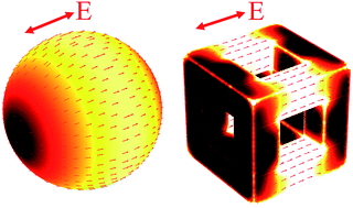
We study the photothermal properties of near-infrared absorbing colloidal plasmonic nanoparticles that are of interest for theranostic applications: SiO2@Au core–shell particles and Au nanocages. Three-dimensional (3D) optical and thermodynamic computational models are developed to explore and compare the optical and thermal response of these particles. The analysis demonstrates that plasmon-enhanced photothermal heating efficiency is a complex function of interrelated factors including the gold content of a particle and the degree to which it supports field-induced current to promote Joule heating. The thermal analysis elucidates fundamental mechanisms that govern the transient temperature distribution and heat dissipation produced by the particles. The modeling approach broadly applies to plasmonic nanostructures with arbitrary shapes, sizes and material constituents and is well suited for the rational design of novel plasmon-assisted photothermal applications.


1 Introduction

Colloidal plasmonic nanoparticles are increasingly used for a broad and diverse range of applications in fields that span biomedicine to analytical chemistry.1,2 Many such applications exploit the unique plasmonic behavior of the particles, i.e. the intense optical absorption and scattering of incident light, and local field enhancement that occur at localized surface plasmon resonance (LSPR). Moreover, the LSPR wavelength can be tuned within the ultraviolet (UV) to near-infrared (NIR) spectrum by controlling the dimensions, constituents and morphology of a particle during synthesis. This is especially important for in vivo applications as it enables the use of incident laser light within the NIR biological window (650–1300 nm), which provides the deepest penetration into tissue3 compared to all other optical wavelengths.

The focus of this work is on the photothermal behavior of colloidal plasmonic nanoparticles used for in vivo theranostic applications. For such applications, a laser is used to illuminate and heat the particles at their LSPR wavelength. This process is exploited in two emerging applications: thermally modulated drug delivery4 and photothermal cancer therapy.5,6 Photothermal modulated drug delivery has been demonstrated using plasmonic core–shell particles (37 nm gold sulfide core within a 4 nm gold shell) embedded within a thermally reversible polymer matrix.4 A pulsed laser is used to heat the particles at their LSPR wavelength, which stimulates thermal release of “model drugs”, such as methylene blue or ovalbumin, from the nanoshell-composite hydrogels. The rate of drug release can be modulated using periodic irradiation, i.e., drug release increases when the laser is activated and returns to a minimum level when the laser is off. The ability to modulate drug delivery in this way can potentially enable a clinician to tailor the release profile of therapeutic agents to match physiologic requirements of the patient. With regard to photothermal cancer therapy, plasmonic particles have been used in various modalities. One such method involves hyperthermia wherein Au nanoparticles are introduced into malignant tissue and heated using a pulsed laser to destroy the tissue by raising the local temperature 3–6 °C. More recently, a different therapeutic modality has emerged in which Au nanoparticles are functionalized and uptaken by cancer cells and then pulsed with sufficient laser intensity to create bubbles within the cells that destroy the cells by rupturing the cell membrane.7 Lapotko et al. have demonstrated this technique for imaging and diagnosis as well as therapy at the cellular level.8,9 They used laser-pulsed spherical (30 nm) gold nanoparticles to rupture K562 and human lymphoid leukemia cell membranes with minimal collateral damage to neighboring healthy cells.8 Lukianova et al. also exploited plasmonic-generated nanobubbles for intracellular drug delivery.10 Gold nanoparticles were used as drug carriers, and when irradiated with a pulsed laser, the nanobubbles that formed caused intracellular dispersal of the drug, which reduced the drug dosage and treatment time while increasing effectiveness.10

Most plasmon-enhanced photothermal applications in vivo utilize biocompatible Au-based nanoparticles with LSPR wavelengths in the NIR window. In this work, we investigate two such nanostructures: core@Au-shell11 and Au nanocage12 particles as shown in Fig. 1. The core–shell particles consist of a silica (SiO2) core with a radius Rc and a gold shell with a thickness ts as shown in Fig. 1a. The Au nanocages are cubic with twelve frame elements in the form of square Au nanowires (Fig. 1b). The nanocage geometry is defined by its length L, which defines the size of the cube, the width W that defines the cross-sectional area of the nanowire, and the aspect ratio R = L/W. This structure is also referred to as a nanoframe.12 These particles are attractive for bioapplications as they can be synthesized in a controllable fashion using bottom-up chemical methods, which enables the tuning of their LSPR absorption within the NIR window as discussed above. In this paper, we combine 3D photonic and thermodynamic computational models to study and compare the plasmonic and photothermal behavior of these particles. We find that the LSPR-enhanced photothermal heating efficiency is a complex function of interrelated factors, including the gold content of a particle and the degree to which it supports an induced current to promote Joule heating. The modeling approach can be used to screen particle designs (shape, size and material constituents) to achieve optimum photothermal performance.


image file: c6ra19566k-f1.tif
Fig. 1 Schematics of NIR plasmonic colloids: (a) SiO2@Au core–shell particles and (b) Au nanocages.

2 Photonic analysis

We study the optical properties of the nanostructures using the finite element (FE)-based Radio Frequency (RF) solver in the COMSOL Multiphysics program (http://www.comsol.com). A typical computational domain (CD) is shown in the inset of Fig. 2a. An incident plane wave is launched from the top of the domain and propagates downward towards the bottom. Perfectly matched layers (PMLs) are imposed at the top and bottom of the CD as shown to eliminate backscatter from these boundaries. A source current is used to generate the field as described in the literature.13,14 The incident light is p-polarized at normal incidence with the E field along the x-axis. More details about CD can be found in the ESI. The optical constants of all materials involved in the models are extracted from the literature.15–18 We account for the fact that the metallic materials (e.g. Au shell) can be thinner than the mean free path of free electrons (∼42 nm) and define a dielectric function of gold based on electron-surface scattering. The details of the material properties can also be found in the ESI.
image file: c6ra19566k-f2.tif
Fig. 2 Optical properties of the SiO2@Au nanostructure with Rc = 27.3 nm and ts = 3.7 nm vs. variation of the domain period along x-axis and y-axis: (a) Px and Py change simultaneously, i.e. P = Px = Py, (b) Px changes and with Py = 2000 nm, and (c) Py changes with Px = 2000 nm. The inset in (a) is the computational domain showing the polarization and propagation direction of the incident field.

It is important to note that we employ symmetry boundary conditions (BCs) perpendicular to E and H. Thus, our computational model mimics the response of an infinite 2D array of coplanar identical nanoparticles with center-to-center x and y lattice spacings, Px and Py respectively. In our preliminary analysis below, we determine values of Px and Py that are large enough so that the field contributions from particles outside the CD are negligible, i.e. so that the analysis accurately reflects the optical response of a single isolated colloidal particle.19 For the purpose of analysis, we fix the total particle volume at Vp = (50 nm)3 = 1.25 × 10⁵ nm3 (i.e., the volume fraction is the same for all colloids) and choose the LSPR wavelength to be λ = 800 nm, which is located within the NIR biological window3 and aligned with one of the most popularly used laser lines, i.e. 808 nm. Based on Mie theory, we find that Rc = 27.3 nm and ts = 3.7 nm render LSPR at 800 nm. More details about Mie theory can be found in the ESI. We now analyze the same particle using the computational model and determine the CD spatial period (P = Px = Py) that reproduces the single particle Mie theory spectrum. In the computational model, we compute the power absorbed by the particle Qabs (W) and then use this to compute the cross section σabs = Qabs/Ilaser where Ilaser (W m−2) is the incident irradiance. The absorption spectra for the period P ranging 100 nm to 2000 nm are shown in Fig. 2a. From this analysis, we find that σabs changes significantly until P ≥ 1000 nm, there is relatively little change in the LSPR wavelength beyond this value. We find that at P = 2000 nm the predicted absorption spectrum is in excellent agreement with Mie theory, as shown in Fig. S1. Thus, at this period the effects of particle coupling are negligible. Based on this observation, we choose the spatial period for the CD to be P = Px = Py = 2000 nm for all of our optical analysis, i.e. for both particles.

It is instructive to investigate the impact of Px and Py separately on particle coupling, i.e. the impact of different array spacing along the x and y directions, respectively. To this end, we first vary Px (in the direction of polarization) from 100 nm to 2000 nm, while fixing the domain size along the y-axis at Py = 2000 nm. As Px increases from 100 nm to 200 nm, the LSPR wavelength blue-shifts from 840 nm to 800 nm, as shown in Fig. 2b, implying a strong LSPR dependence on interparticle coupling between the particles aligned in the direction of polarization. As the distance between neighboring particles increases form 100 nm to 200 nm, their dipole–dipole interaction becomes weaker and the restoring force of the oscillating electrons within a particle increases, which results in a higher oscillation frequency, i.e. a blueshift of LSPR from 840 nm to 800 nm. When Px is in the range of 200–1500 nm, the LSPR wavelength remains constant at 800 nm, but the absorption cross section changes, which indicates particle coupling. However, for Px ≥ 1500 nm there is a negligible change in both the LSPR wavelength and peak absorption, which implies that the particle in the CD is essentially uncoupled from the 2D array.

We perform a similar analysis to explore the coupling between particles aligned along the y-axis, perpendicular to the polarization. We compute the absorption spectrum for Py ranging from 100 nm to 2000 nm, while Px = 2000 nm is fixed. The LSPR wavelength red-shifts from 780 nm to 790 nm as Py increases from 100 nm to 200 nm as shown in Fig. 2c. For Py in the range of 200–1500 nm, the LSPR wavelength is essentially constant at 800 nm, but the absorption amplitude changes, which indicates the existence of particle coupling. When Py ≥ 1500 nm, the coupling is negligible and the optical response is that of an isolated nanoparticle. Additionally, it should be noted that the LSPR wavelength shifts most abruptly as Px or Py increases from 100 nm to 200 nm. This effect can be understood by considering the near field nature of plasmonic resonance and especially inter-particle coupling. The subwavelength variation of particle spacing (i.e. 100–200 nm) is expected to have a more significant impact on plasmonic resonance than larger separation distances. On the other hand, the diffractive coupling between plasmonic particles in a periodic array can range between destructive to constructive interference. Thus, not only the intensity but also the relative phase of the inter-particle coupling will determine the amplitude of the absorption cross section.

Our results demonstrate that the LSPR of SiO2@Au core–shell and Au nanocage particles with identical volumes can be readily tuned to the same NIR wavelength by controlling their dimensions. As previously noted, this NIR tunability is especially attractive for biomedical applications.20,21 As shown in Fig. 3a, an Au nanocage with dimensions of L = 50 nm and W = 13.4 nm possesses the same volume (i.e. Vp = (50 nm)3) and LSPR wavelength (i.e. 800 nm) as an SiO2@Au core–shell particle with dimensions of Rc = 27.3 nm and ts = 3.7 nm. The results reveal comparable absorption cross-sections of the core–shell and nanocage particles that are σabs = 3.63 × 10⁻¹⁴ and 3.59 × 10⁻¹⁴ m2, respectively. It is worth mentioning that despite the comparable optical absorption in two colloids, the Au nanocage has a unique advantage over the core–shell particle in that it exhibits more abundant E field hot spots on its surface and throughout its interior (Fig. S2). On the other hand, it has been demonstrated in our previous work14 that the optical absorption of the Au nanocage is insensitive to changes in its spatial orientation with respect to the incident polarization, which is an important consideration for solution-based colloidal applications. All these advantages can be leveraged for theranostic applications via enabling strongly enhanced biotracking and imaging.22,23


image file: c6ra19566k-f3.tif
Fig. 3 Photothermal profiles of SiO2@Au core shell (Rc = 27.3 nm and ts = 3.7 nm) and Au nanocage (L = 50 nm and W = 13.4 nm) particles with an LSPR wavelength of 800 nm: (a) absorption cross sections of two colloidal particles. (b) and (c) Spatial profiles of the thermal power density of (b) SiO2@Au core–shell particle and (c) Au nanoframe. Small red arrows show the current density on the outer Au surfaces of two nanoparticles.

3 Thermodynamic analysis

We used 3D computational thermodynamics to investigate the transient thermal behaviour and heat transfer of the core–shell and nanocage structures. For consistency, we analysed particles with the same volume and dimensions as in the previous analysis, i.e. the SiO2@Au particle with Rc = 27.3 nm and ts = 3.7 nm, and the nanocage with L = 50 nm and W = 13.4 nm. Recall that these particles have comparable absorption cross-sections despite the substantial difference in their gold content, i.e. approximately a factor of 2. Specifically the SiO2@Au particle contains much less gold (39[thin space (1/6-em)]541 nm3 or 0.764 fg) than the nanocage (69[thin space (1/6-em)]238 nm3 or 1.337 fg). The incident light induces currents within the gold that dissipate energy via Joule heating. The absorption cross-section depends on the magnitude and spatial extent of the LSPR-induced currents, which in turn depend on the amount and importantly, the geometry (configuration) of the constituent gold. The current density and thermal power density (W nm−3) distribution due to Joule heating within the two particles is shown in Fig. 3b and c. The double-headed red arrows in these figures show the direction of polarization. The incident light propagates along the negative direction of z-axis. The small single-headed red arrows represent the current density in the constituent gold. It is important to note that the gold shell in the SiO2@Au particle provides a continuous path for current to flow from one end of its induced dipole structure to the other. Consequently, it exhibits more uniform thermal dissipation over a relatively large volume of its Au shell. This is in contrast to the nanocage wherein only four nanowires, out of the twelve that form the nanoframe, carry significant current and produce heat. These four are aligned with the field polarization. The other eight nanowires in the frame are not aligned and have a negligible impact on optical absorption and heating when the particle has this orientation. Thus, even though the core–shell particle contains less gold than the nanocage, this difference is offset because a higher percentage of the gold in the former contributes to heating, which renders the overall photothermal conversion efficiency of the two particles comparable. An additional factor that can influence absorption includes the special optical cavity configuration of the core–shell particle, which can result in a stronger LSPR resonance than in the nanocage with its open interior region.24,25

We perform 3D thermodynamic analysis of these structures using coupled heat transfer and RF modules of the Comsol program. The absorbed optical power density (heat generated per unit volume of gold) Qh (W m−3) is computed in the stationary (time-harmonic) field solution of the RF model and acts as a heat source in the thermal model. The temperature T throughout the computational domain satisfies the equation,

 
image file: c6ra19566k-t1.tif(1)
where Q (W m−3) is the thermal energy generated per unit volume, i.e. Q = Qh inside the gold and Q = 0 in all other (loss less) materials. Here, ρ is the density, Cp is the specific heat capacity and k is the thermal conductivity of the material. In our analysis, the temperature dependent thermal conductivity, heat capacity and material density of H2O, Au and SiO2 were taken from the literature.26,27

During photothermal therapy, a temperature within a range of 40–49 °C (ref. 28) is required to ensure a therapeutic effect.29 In order to reach this temperature, we choose an incident irradiance of 62.5 kW cm−2 with the linear polarization along x-axis. This level of irradiance is appropriate for a single isolated particle, i.e. reflecting a very low concentration. However, in many photothermal applications, higher concentrations of plasmonic nanoparticles are used, which gives rise to a cooperative interparticle heating that reduces the irradiance required to effect therapy, as discussed in the literature.14,30 Specifically, the heat generated per unit volume in the carrier fluid increases with the particle concentration and this results in a more rapid rise in global fluid temperature for a given irradiance. The thermodynamic model is based on the same computational domain as the photonic analysis except that a thermal insulation condition is imposed on all boundaries. The initial temperature is set to 20 °C.

Fig. 4a shows essentially the same change in the environmental temperature Tenv for the core–shell (blue curve) and nanocage (red curve). Tenv is calculated by averaging the temperature over a spherical surface of radius of 70 nm that is centered with respect to and surrounds the particles. The results indicate that Tenv rises quickly from 20 °C to 43 °C within 50 ns which is consistent with typical laser pulse durations in photothermal therapy applications. Fig. 4b and c show the spatial profiles of the temperature for the SiO2@Au core–shell at t = 10 and 50 ns, respectively. The results imply a rapid exchange of thermal energy between the core–shell particle and the surrounding H2O environment, causing a continuous rise of environmental temperature during 50 ns.


image file: c6ra19566k-f4.tif
Fig. 4 Transient temperature distribution of 62 nm SiO2@Au nanoparticles (Rc = 27.3 nm, ts = 3.7 nm): (a) time-dependent average temperature in proximity to the particles: (b) and (c) temperature distribution due to the core–shell particle at (b) t = 10 ns and (c) 50 ns.

Next, we compare the thermal response of the SiO2@Au and nanocage particles. Since the gold content of the core–shell particle is approximately half that of the nanocage, one might expect that the temperature rise of the former will be essentially twice as fast (due to a lower thermal mass) if the same energy is absorbed within the particles. However, as shown in Fig. 5a, the temperature rise in the Au shell (blue curve) is only slightly higher than that of the nanocage. To explain this, we plot the residual thermal power that heats the gold QAu (W) in the inset of Fig. 5a. Specifically, QAu represents the difference between the optical power absorbed by gold minus the power it dissipates to all surrounding materials. For example, in the SiO2@Au particle thermal energy is dissipated through the inner and outer surface of the gold shell to the SiO2 and H2O, respectively. The data in the inset in Fig. 5a indicates that initially (0–10 ns) the nanocage consumes approximately 70–80% more thermal energy than the Au shell. However, as seen in the main figure, the temperature rise of the gold TAu during this time is essentially the same for both particles. This is due to two factors. First, as noted above the nanocage contains more gold and therefore has a larger thermal mass than the core–shell particle. Thus it needs to absorb more energy to achieve the same temperature. Second, the surface area of the hollow Au nanocage (16[thin space (1/6-em)]745 nm2) is much larger than that of the solid core–shell particle (12[thin space (1/6-em)]070 nm2), and therefore provides faster heat dissipation to the fluid. The combination of these effects results in a comparable heating of surrounding fluid with a difference in the surface (metal) temperature of the two particles.


image file: c6ra19566k-f5.tif
Fig. 5 Thermodynamic profiles in the SiO2@Au (62 nm) and nanocage (50 nm) particles: (a) time-dependent average temperature in the gold material of the core–shell and nanocage. (b) Thermal energy Qc dissipated from the nanoparticles to the surrounding H2O. “Outward” and “Inward” denote the thermal energy dissipated from the outer and inner surface of the Au shell of SiO2@Au particle, respectively. (c)–(f) The plot of the temperature gradient inside (c) and (d) the core–shell and (e) and (f) nanocage at t = 1 ns and 20 ns. Note: all the legends have the same temperature range ΔT = 20 °C in order to highlight the thermal behaviour inside the particles. The local temperature in the black regions is either equal to or below the lower limit of the legend.

Fig. 5b shows the time-dependent conductive heat power Qc dissipated from the nanoparticles to the H2O by the nanocage (blue curve), and by the SiO2@Au particle at the gold–water interface (“Outward”, black curve with diamond markers) and gold–SiO2 interface (“Inward”, black curve with triangular markers). The total power dissipated by the SiO2@Au particle (outward plus inward) is also plotted as the red curve. The “Inward” curve in Fig. 5b indicates that ∼5% of the absorbed power will heat the SiO2 core within t = 2 ns, because of the temperature gradient between the shell and the core, as shown in Fig. 5c. In approaching the steady-state, the temperature becomes more uniform throughout the particle, resulting in a negligible heat exchange between the shell and the core, as shown in Fig. 5d. With regard to the Au nanocage, Fig. 5e, shows there is a more pronounced temperature gradient in its interior, which is due to the lower thermal conductivity of the H2O. However, in the steady-state, a uniform temperature exists in the Au frame elements and the H2O interior, as shown in Fig. 5f. Additionally, Fig. 5b also reveals that in the steady-state, almost all of the energy absorbed by the SiO2@Au and nanocage particles is dissipated in the surrounding H2O. The total thermal energy dissipated to the environment is essentially the same for two nanoparticles, as shown in Fig. 5b.

4 Conclusions

We have used 3D computational models to compare the photothermal behavior of SiO2@Au core–shell and Au nanocage nanostructures. We have found that the SiO2@Au and nanocage particles have comparable photothermal conversion efficiencies even though, in our study, the former has significantly less (approximately 1/2) gold content than the latter. Specifically, we show that the heating efficiency of the particles is a complex function of multiple factors, not only the amount of gold a particle contains, but importantly, how the gold is configured, i.e. the degree to which it supports LSPR-enhanced current to promote Joule heating. In this regard, the symmetry of the particle is also important. In colloidal applications, the particles are randomly oriented in a carrier fluid. The optical absorption of highly symmetric particles is less sensitive to their orientation with respect to the incident field polarization and therefore provide more efficient photothermal heating. The surface to volume ratio (i.e. effective surface area) is another important factor that governs the dissipation of thermal energy to the surrounding environment.

Finally, the combined 3D photonic and thermodynamic computational approach applied here provides insight into fundamental mechanisms that govern the plasmonic and thermal behavior of colloidal nanoparticles. It can be used to screen particle designs (shape, size and material constituents) to achieve optimum photothermal performance. As such, it is well suited for the rational design of novel photothermal applications.

Acknowledgements

We acknowledge financial support from the U.S. National Science Foundation, through Award CBET-1337860.

References

  1. R. Sardar, A. M. Funston, P. Mulvaney and R. W. Murray, Langmuir, 2009, 25, 13840–13851 CrossRef CAS PubMed.
  2. X. Yang, M. Yang, B. Pang, M. Vara and Y. Xia, Chem. Rev., 2015, 115, 10410–10488 CrossRef CAS PubMed.
  3. Y. G. Sun and Y. N. Xia, Nano Lett., 2003, 3, 1569–1572 CrossRef CAS.
  4. S. R. Sershen, S. L. Westcott, N. J. Halas and J. L. West, J. Biomed. Mater. Res., 2000, 51, 293–298 CrossRef CAS PubMed.
  5. E. B. Dickerson, E. C. Dreaden, X. Huang, I. H. El-Sayed, H. Chu, S. Pushpanketh, J. F. McDonald and M. A. El-Sayed, Cancer Lett., 2008, 269, 57–66 CrossRef CAS PubMed.
  6. L. R. Hirsch, R. J. Stafford, J. A. Bankson, S. R. Sershen, B. Rivera, R. E. Price, J. D. Hazle, N. J. Halas and J. L. West, Proc. Natl. Acad. Sci. U. S. A., 2003, 100, 13549–13554 CrossRef CAS PubMed.
  7. E. P. Furlani, I. H. Karampelas and Q. Xie, Lab Chip, 2012, 12, 3707–3719 RSC.
  8. D. Lapotko, E. Lukianova, M. Potapnev, O. Aleinikova and A. Oraevsky, Cancer Lett., 2006, 239, 36–45 CrossRef CAS PubMed.
  9. D. O. Lapotko, E. Lukianova and A. A. Oraevsky, Lasers Surg. Med., 2006, 38, 631–642 CrossRef PubMed.
  10. E. Y. Lukianova-Hleb, A. Belyanin, S. Kashinath, X. Wu and D. O. Lapotko, Biomaterials, 2012, 33, 1821–1826 CrossRef CAS PubMed.
  11. L. Gomez, M. Arruebo, V. Sebastian, L. Gutierrez and J. Santamaria, J. Mater. Chem., 2012, 22, 21420–21425 RSC.
  12. M. A. Mahmoud and M. A. El-Sayed, J. Am. Chem. Soc., 2010, 132, 12704–12710 CrossRef CAS PubMed.
  13. F. Alali, Y. H. Kim, A. Baev and E. P. Furlani, ACS Photonics, 2014, 1, 507–515 CrossRef CAS.
  14. I. H. Karampelas, K. Liu, F. Alali and E. P. Furlani, J. Phys. Chem. C, 2016, 120, 7256–7264 CAS.
  15. P. G. Etchegoin, E. C. Le Ru and M. Meyer, J. Chem. Phys., 2007, 127, 189901 CrossRef.
  16. P. B. Johnson and R. W. Christy, Phys. Rev. B: Solid State, 1972, 6, 4370–4379 CrossRef CAS.
  17. P. Tuersun and X. Han, Appl. Opt., 2013, 52, 1325–1329 CrossRef CAS PubMed.
  18. C. F. Bohren and D. R. Huffman, Absorption and Scattering of Light by Small Particles, Wiley, New York, 1983 Search PubMed.
  19. A. I. Henry, J. M. Bingham, E. Ringe, L. D. Marks, G. C. Schatz and R. P. van Duyne, J. Phys. Chem. C, 2011, 115, 9291–9305 CAS.
  20. C. Ayala-Orozco, J. G. Liu, M. W. Knight, Y. Wang, J. K. Day, P. Nordlander and N. J. Halas, Nano Lett., 2014, 14, 2926–2933 CrossRef CAS PubMed.
  21. W. F. Paxton, S. L. Kleinman, A. N. Basuray, J. F. Stoddart and R. P. van Duyne, J. Phys. Chem. Lett., 2011, 2, 1145–1149 CrossRef CAS PubMed.
  22. S. E. Skrabalak, J. Chen, L. Au, X. Lu, X. Li and Y. Xia, Adv. Mater., 2007, 19, 3177–3184 CrossRef CAS PubMed.
  23. S. Khatua, P. M. R. Paulo, H. F. Yuan, A. Gupta, P. Zijlstra and M. Orrit, ACS Nano, 2014, 8, 4440–4449 CrossRef CAS PubMed.
  24. Y. Pu, R. Grange, C. L. Hsieh and D. Psaltis, Phys. Rev. Lett., 2010, 104, 207402 CrossRef PubMed.
  25. J. J. Penninkhof, L. A. Sweatlock, A. Moroz, H. A. Atwater, A. van Blaaderen and A. Polman, J. Appl. Phys., 2008, 103, 123105 CrossRef.
  26. M. W. Chase Jr, J. Phys. Chem. Ref. Data, Monogr., 1998, 9, 1–1951 Search PubMed.
  27. D. Skelskey and J. van den Sype, J. Appl. Phys., 1970, 41, 4750 CrossRef CAS.
  28. N. Frazier, R. Robinson, A. Ray and H. Ghandehari, Mol. Pharm., 2015, 12, 1605–1614 CrossRef CAS PubMed.
  29. X. Huang and M. A. El-Sayed, J. Adv. Res., 2010, 1, 13–28 CrossRef.
  30. M. F. Tsai, S. H. G. Chang, F. Y. Cheng, V. Shanmugam, Y. S. Cheng, C. H. Su and C. S. Yeh, ACS Nano, 2013, 7, 5330–5342 CrossRef CAS PubMed.

Footnote

Electronic supplementary information (ESI) available. See DOI: 10.1039/c6ra19566k

This journal is © The Royal Society of Chemistry 2016
Click here to see how this site uses Cookies. View our privacy policy here.