DOI:
10.1039/C6RA23718E
(Paper)
RSC Adv., 2016,
6, 100676-100682
Microstructure and corrosion behavior of hafnium coatings on AZ91D magnesium alloys by magnetron sputtering
Received
23rd September 2016
, Accepted 17th October 2016
First published on 17th October 2016
Abstract
Hf coatings are fabricated on the AZ91D Mg alloys by magnetron sputtering with bias voltage ranges from 0 to −125 V. Both potentiodynamic polarization test and neutral salt spray test reveal that the Hf coating deposited at −100 V exhibits the best protective performance. It possesses the lowest corrosion current density of 1.032 μA cm−2 and the highest protection rate of 6, respectively. This perfect anticorrosion property is due to the dense structure and low porosity induced by applying the appropriate bias voltage. Various types of corrosion sites after corrosion tests are examined in detail. The results indicate that the coating failure is strongly dependent on the coating defects and the random phase distribution in the substrate.
1. Introduction
Mg and its alloys are prone to corrosion due to the high electrochemical activity,1,2 although they are promising materials in electronics and aerospace industries. Surface treatment, has been validated as a necessary and effective step to improve the corrosion as well as wear resistance.3–6 Considering the background of environmental protection, physical vapor deposition (PVD) as a ‘green’ method to fabricate functional coatings on Mg alloys has become a research hotspot in recent years.7,8 Moreover, the high-quality of coatings fabricated by PVD is unmatched by the conventional surface treatment methods. Depending on the feature of the material selection diversity, ceramic9 and metallic10–13 protective coatings via this technique have been fabricated on Mg alloy with improved surface resistance.
Actually, although wear resistance was significantly improved, these above coatings exhibit unsatisfactory anticorrosion performances. The above coatings act as the cathode in the galvanic cell with a much noble potential and consequently accelerate the deterioration process of Mg alloy (−2.37 V vs. normal hydrogen electrode). In previous research, the authors found that hafnium coating with the excellent anticorrosion property is a promising material to protect Mg alloys.14 However, for PVD coating, the growth defects such as pore or droplet produced in the deposition process provide deleterious role in corrosion process. The presence of porosity in the PVD coating should be given a careful consideration.15
The negative bias voltage as an important parameter has a significant effect on the microstructure of PVD coatings.16 The change of the microstructure and anti-corrosion property produced by the negative bias voltage has been reported.17 However, there are opposite results of different coatings with the similar bias voltage.17,18 For the PVD Hf coating, it is necessary to study the correlation between the bias voltage and its anti-corrosion property. Moreover, the corrosion behaviors of various PVD-coated Mg alloys have been studied, but rare corrosion mechanism was considering the phase distribution of the substrate.9,10,12,13,19 Song has illustrated that the corrosion formed of Mg alloy strongly depended on composition and microstructure.3 Therefore, in this study, Hf coatings with substrate negative bias varying from 0 to −125 V were deposited on AZ91D Mg alloy surface by magnetron sputtering. The changes of the microstructure and corrosion performance produced by the substrate bias were studied. The coating failure behaviors were comprehensively investigated after selected the optimization in corrosion tests.
2. Experimental details
2.1 Specimen preparation
Hf coatings, 10 ± 0.1 μm thickness, were deposited on silicon (111) wafers and AZ91D (Al: 9.1 wt%, Mn: 0.033 wt%, Zn: 0.91 wt%, Si: 0.04 wt%, Cu: 0.018 wt%, Ni: 0.0006 wt%, Fe: 0.003, Mg: balance) Mg alloy substrates (10 × 10 mm2) by magnetron sputtering from a metallic Hf target (Ø 76 mm, thickness 5 mm, purity 99.9%). Prior to coating deposition, the AZ91D was grinded with emery paper #3000, #7000 and mirror polished with diamond powder (particle size 2.5 μm). Then these AZ91D substrates coupled with silicon wafers were ultrasonically degreased in acetone and ethanol for 10 and 15 min, respectively. After reaching the base pressure of 6.0 × 10⁻⁴ Pa, all the substrates were cleaned by Hall ion source for 5 min (substrate bias of −100 V and working pressure of 2 × 10⁻² Pa) in pure Ar atmosphere in order to reduce the oxide on the substrate surfaces. The Hf target was pre-sputtered for 15 min to remove the surface oxides and other contaminants. The working distance between the substrate and the target was fixed at 100 mm. The working pressure and the Ar flux were maintained at 0.30 Pa and 60 sccm, respectively. The Hf target DC power (Advanced Energy, U.S.) was adjusted to 250 W and the deposition temperature was set at 300 °C. Substrate negative bias was chosen as the controlling parameter varying from 0 to −125 V.
2.2 Characterizations and corrosion tests
The crystal structure and preferred orientation of Hf coatings were investigated by Panalytical X'pert PRO X-ray diffraction (XRD, Philips, Netherlands) using Cu Kα radiation as the X-ray source (λ = 0.154 nm). The Hf-coated specimens (10 × 10 mm2) were operated by the θ–2θ diffraction mode at 35 kV and 15 mA. Surface and cross-sectional morphologies of coatings were observed by scanning electron microscope (SEM, ZEISS SIGMA, Germany). The corrosion performance of specimens in 3.5 wt% sodium chloride aqueous solution was conducted by potentiodynamic polarization test in an electrolyte cell consisted of a working electrode, a counter electrode (platinum chip of 1.0 cm2) and a reference electrode (saturated calomel electrode) via CHI660E electrochemical workstation operated at room temperature. The coated and bare AZ91D substrates were set as the working electrodes. Then the polarization test was carried out at a scan rate of 1 mV s−1. Three Hf-coated samples with the same deposition parameter were used to investigate their corrosion behaviors. Then the average was taken as the final result. Neutral salt spray test was performed to evaluate the long-term corrosive protection function of Hf coating to AZ91D substrates following the standard method ASTM B117. A 5 wt% of sodium chloride solution was used and the temperature of the salt spray chamber was set at 35 ± 2 °C. The salt spray test was lasted for 24 h. Then each specimen was assigned a protection rating following the ASTM B537-70 standard. The corrosion observation was carried out by SEM coupled with Oxford Link energy-dispersive X-ray spectroscopy (EDS) microanalysis hardware after the corrosion tests in order to determine the failure mechanisms of coatings.
3. Results and discussion
3.1 Phase structure and morphology
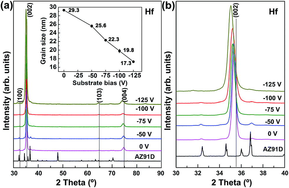
Fig. 1 shows the X-ray patterns of Hf coatings deposited on Si (111) with respect to varying the negative bias voltages from 0 to −125 V. It can be seen that all the diffractograms with (100), (002), (103) and (004) peaks revealed the hcp structured α-Hf (JCPDS no. 05-0670). The densely packed plane Hf (002) was the preferred growth orientation. Moreover, the relative intensity of (103) peak increases with increasing the negative bias while that of (100) peak presents a reverse trend to (103) peak. Both the thermodynamic and kinetic theories have been proposed to explain the transition of the growth orientation of PVD coatings. The surface energy has a dominant contribution to the total free energy minimization for the growth of PVD coatings and the growth orientation corresponds to the plane with the lowest surface energy.20
 |
| | Fig. 1 Normal XRD patterns and grain size of the Hf coatings with respect to substrate bias: (a) the full spectrum and grain size, (b) maximum peak of (002). | |
As shown in Fig. 1b, the diffraction width of the preferred orientation (002) is increased with increasing the negative bias.
Qi et al.21 have demonstrated that this broadening behavior is the result of the finer grain size and the increased microstrain of coating. The relationship of grain size and negative bias can be described by the re-nucleation mechanism based on Abadias et al.22 Applying substrate bias provides more ion bombardment-induced surface nano-defects on substrate surface. The elevated generation of surface nano-defects with increasing the negative bias leads to an increased number of nucleation sites that resulting in a finer grain size. The average grain sizes of the Hf coatings were estimated by the well-known Scherrer formula. As shown in Fig. 1a, the grain size decreases from 29.4 nm to 17.2 nm corresponding to increasing substrate bias from 0 to −125 V. Generally, PVD coatings produced by magnetron sputtering exhibit the pronounced columnar structure. The columnar structure mainly consisted of many micro-defects which attributed to the substrate surface irregularities. The finer grain size of coating suggesting more nucleation sites were produced on the surface irregularities. Hence, the growth of the droplets would be blocked or even healed. In addition, the diffraction peaks of Hf coatings shift to lower angles with increasing the negative bias that reveals the generation of residual stress. This physical effect associated with negative bias exhibited in XRD also has been reported to be presented in hard coatings.16 The bombarding particle energy is proportional to the applied negative bias that causes an increased nucleation sites in the as-deposited coatings and equivalently higher degree of lattice distortion.
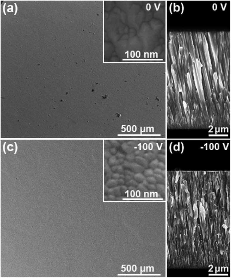
Fig. 2 shows the surface and cross-sectional SEM morphologies of Hf coatings deposited at 0 V (a and b) and −100 V (c and d), respectively. It is clearly revealed that the defects were significantly reduced with increased the substrate bias. The surface morphologies of Hf coatings illustrate a granular microstructure. The size of granular grains decreases with increasing substrate bias which is well agreement with the above analysis in XRD. The roughness of Hf coating (−100 V) surface was much smoother than that of coating without applying substrate bias. The evolution of the surface can be ascribed to high ion bombardment enhanced by substrate bias. Firstly, the large surface particles such as contamination or dust can be filtered by the increase of ion bombardment and electrical repulsion. Secondly, the coating growing surface can also be melted and flattened by the excessive energy which produced by an increasing number of ions bombardments. Moreover, a larger quantity of Ar+ ion bombardments induced by the increased bias voltage would also produce a positive effect on etching the asperities. These effects indicate that improving substrate bias offers a favorable condition for the fabrication of high quality coatings.
 |
| | Fig. 2 Surface and cross-sectional SEM morphologies of the Hf coatings deposited at substrate bias of 0 (a and b) and −100 V (c and d). | |
In addition, the Hf coatings exhibit typically columnar structure in cross-section, with each grain boundary extending across the entire thickness. Since the ratio of deposition temperature Ts (573 K) over the Hf melting point Tm (2500 K) is ∼0.229, the cross-sectional morphology of Hf coatings is identified as zone T structure.23 In this Ts/Tm range, adatom surface diffusion significantly results in local epitaxial growth on individual grains and the formation of the pronounced columnar structure. Being consistent with the evaluation of surface granular size, the columnar grain size clearly decreased with increasing substrate bias from 0 to −100 V. Consequently, a compact and dense microstructure is observed in Fig. 2d.
3.2 Corrosion performance and microstructure correlation
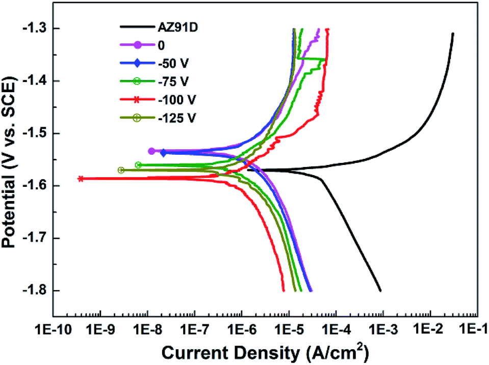
3.2.1 Potentiodynamic polarization test. Fig. 3 shows the potentiodynamic polarization curves of the AZ91D substrate and the Hf-coated specimens in a 3.5 wt% sodium chloride aqueous solution at room temperature. The corrosion potential (Ecorr) and corrosion current density (icorr) were determined using Tafel extrapolation method. The polarization resistance (Rp) values of specimens were calculated with Stern–Geary function. The relative parameters are listed in Table 1. As shown in Fig. 3, the Ecorr of the AZ91D is −1.57 V (vs. SCE) and it revealed the highest icorr corresponding to 162.7 μA cm−2. For the coated specimens, the Ecorr are close or lower to/than that of the bare AZ91D in this condition. Based on the theory of mixed potential,10,14 the similar Ecorr reveals the galvanic corrosion between coating and substrate has been induced by the through-thickness defects such as pore or droplet. The hydrogen bubble was appeared on the coatings' surface during the polarization test. The icorr of Hf-coated specimens are 2.764 μA cm−2 for 0 V, 2.143 μA cm−2 for −50 V, 1.893 μA cm−2 for −75 V, 1.032 μA cm−2 for −100 V and 2.26 μA cm−2 for −125 V exhibit a much lower than that of the uncoated AZ91D about two order of magnitude. The slight higher icorr of these Hf-coated specimens than before14 was mainly due to the rough surface of AZ91D substrate in this study (polished with diamond powder of 2.5 μm). It can be also noted that the improvement corrosion resistance of coatings was proportional to the applied negative bias within a certain range. The slight degraded in protective function of Hf coating with −125 V compared to that of −100 V is probably due to the increased residual stress observed in Fig. 1b. The high residual stress exited in PVD coating led to incomplete surface. The slight peeling and micro-crack were observed on the Hf coating (−125 V) surface after removed from the deposition chamber (shown in Fig. 4).
 |
| | Fig. 3 The electrochemical polarization curves of the AZ91D Mg alloy substrate and the Hf coated specimens in a 3 wt% NaCl aqueous solution at room temperature. | |
Table 1 Potentiodynamic polarization measurement data of coated and uncoated specimens in 3.5% NaCl solution
| Specimen |
Ecorr (VSCE) |
icorr (μA cm−2) |
Rp (Ω cm2) |
| AZ91D |
−1.570 |
162.7 |
188.7 |
| 0 V |
−1.533 |
2.764 |
14![[thin space (1/6-em)]](https://www.rsc.org/images/entities/char_2009.gif) 855.5 855.5 |
| −50 V |
−1.537 |
2.143 |
17125.7 |
| −75 V |
−1.560 |
1.893 |
17383.5 |
| −100 V |
−1.586 |
1.032 |
19100.8 |
| −125 V |
−1.570 |
2.260 |
18317.2 |
 |
| | Fig. 4 The micro-crack observed on Hf coating deposited at substrate bias of −125 V: (a) micro-crack, (b) enlarged view of the red frame in (a). | |
For PVD coatings, the coating defects such as pore or droplets usually originated from the substrate surface irregularities or the deposition conditions. These defects are prone to provide the pathways for corrosion medium to penetrate the physical barrier. Hence, the porosity value is a direct reflection to the coatings' corrosion resistance. The relationship between porosity and bias voltage was calculated by the following equation with the parameters Rps and Rp, which were deduced from the potentiodynamic polarization curves:24
| |
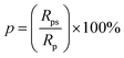 | (1) |
where
p is the total coating porosity,
Rps is the polarization resistance of the uncoated substrates and
Rp is the polarization resistance of the coated substrate (
Table 1). The packing factor related to the porosity as a quantitative measurement of the coating density is determined herein by
eqn (2):
24| |
 | (2) |
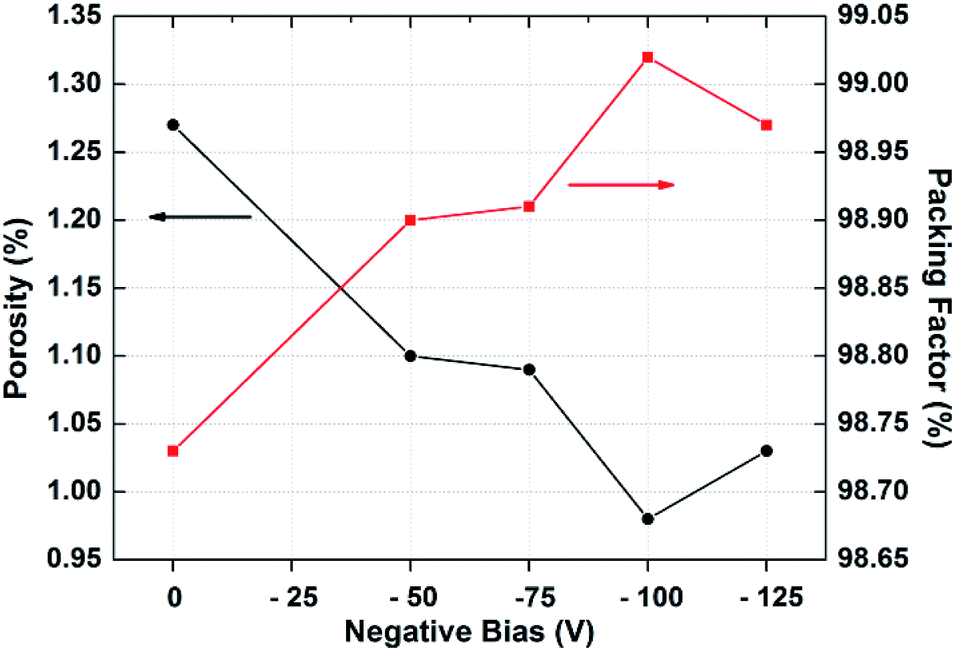
As shown in Fig. 5 and Table 2, all of Hf coatings illustrate the low porosity and high density values. The porosity of Hf coatings at the deposited negative bias from 0 to −125 V exhibits a converse trend with the packing factor. As revealed in above Fig. 1 and 2, the enhanced ion bombardment by applying the appropriate substrate bias provide a positive effect on improving the coating quality such as grain refinement and density improvement. The lower porosity and higher density of the coating reduce the pathway for corrosion media thus decrease the probability of corrosion.
 |
| | Fig. 5 Coating porosity and packing factor as a function of increased negative bias from 0 to −125 V. | |
Table 2 Correlations of porosity and packing factor as a function of bias voltage
| Specimen |
Porosity (%) |
Packing factor (%) |
| 0 V |
1.27 |
98.73 |
| −50 V |
1.1 |
98.9 |
| −75 V |
1.09 |
98.91 |
| −100 V |
0.98 |
99.02 |
| −125 V |
1.03 |
98.97 |
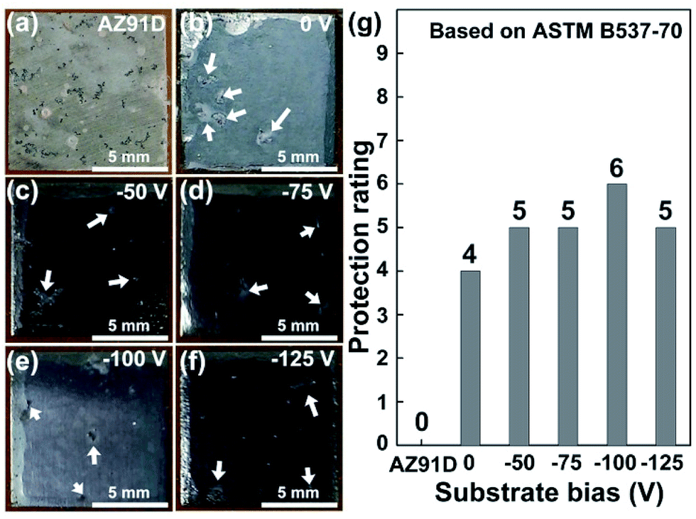
3.2.2 Neutral salt spray test. Electrochemical corrosion test as reported by Fenker et al.25 was not sufficient to evaluate the practical anti-corrosion property of coatings due to the limitation of testing condition. Hoche et al. validated that even micro-defects in PVD coatings can cause serious anodic dissolution of Mg within a few minutes exposed to salt spray test.26 Herein, the neutral salt spray test was further performed to investigate the practical protection of Hf coatings to Mg alloy. Fig. 6a–f show the optical images of the AZ91D substrate and the Hf-coated specimens after the salt spray test for 24 h. The surface of the AZ91D Mg alloy has been corroded severely as a result of the galvanic corrosion between α and β phase.27 In contrast, significant improvement of the corrosion resistance of Mg alloy has achieved by depositing Hf coatings. The pitting corrosion (marked by arrows) found on the surface of Hf coating deposited at substrate bias of −100 V is much less than that of the rest specimens. Fig. 6g shows the protection ratings of the bare and coated specimens. The protection rating of Hf coating first increases from 4 to 6 with increasing the substrate negative bias from 0 to −100 V then it is followed by a decrease to 5 with further increasing the substrate bias to −125 V. This result is in well agreement with that of the electrochemical measurements.
 |
| | Fig. 6 Optical graphs of the AZ91D Mg alloy and coated specimens after the salt spray tests for 24 h (a–f) and the corresponding protection ratings of the specimen faces (g). | |
In general, the kinetic energy of sputtered ions towards the coating is increased with the substrate negative bias increases, results in the increase of the compactness of coating. As indicated in Fig. 1b, the finer and denser microstructures are obtained with increasing the substrate bias. A compact structure exhibits a better ability of physical barrier on corrosion medium for PVD coatings. Moreover, porosity reduction by varying substrate bias helps to reduce the sites of pitting corrosion formation. As shown in Fig. 2a and c, it is clearly observed that the defects of coating with −100 V was significantly reduced compared with the coating of 0 V. Nevertheless, though positive effect on improving corrosion resistance was achieved but negative influence such as residual stress produced by over negative bias should not be ignored.
3.3 Coating failure analysis and simulation
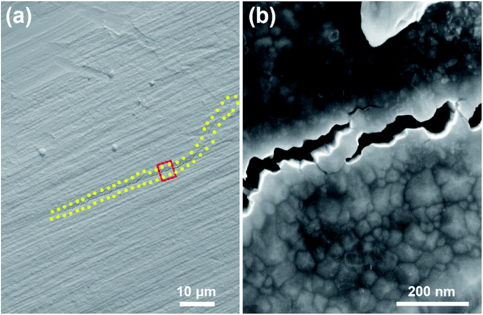
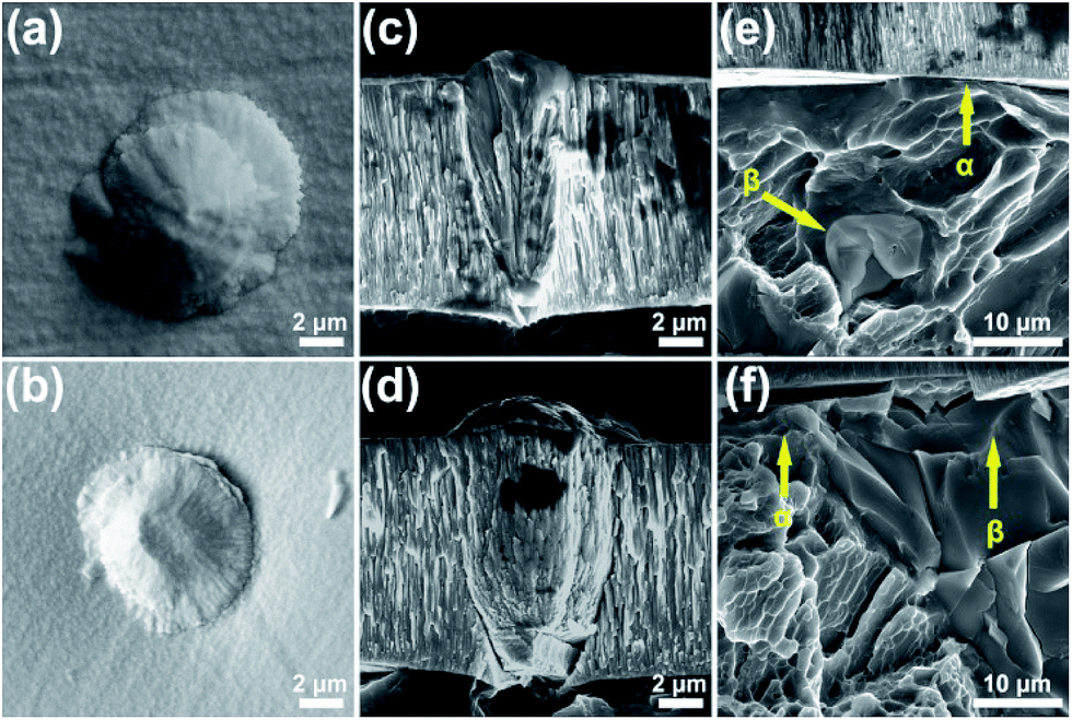
Though Hf coating (−100 V) significantly improved the corrosion resistance of AZ91D Mg alloy, however the coating still has a certain number porosity which resulted in the pitting corrosion after 24 salt spray test. The following is introduced to investigate the corrosion process with the example of Hf (−100 V) coated Mg alloy. Fig. 7a–d show the surface and cross-sectional morphologies of Hf-coated Mg alloy with different through-thickness droplet defects before corrosion test. The defects appear on the coating surface exhibit a cell-shaped structure distinguished from the rest of coating. The cross-sectional morphologies indicate that the cone defects are originated from the substrate. Moreover, the region of defects with clear voids shows a less dense structure that was mainly due to the shadow effect during the deposition process. This less dense structure of defects allows the corrosive media to penetrate the coating to substrate and should be responsible for the porosity revealed in Fig. 6. Fig. 7e and f show the cross-sectional morphologies of AZ91D Mg alloy under the coating. The random distribution of α and β phase suggests that the area where can be contacted with the defects is highly uncertain.
 |
| | Fig. 7 The SEM images of the Hf coated specimens: (a and b) surface view of droplet defects, (c and d) cross-sectional view of through-thickness droplet defects, and (e and f) cross-sectional view of the AZ91D. | |
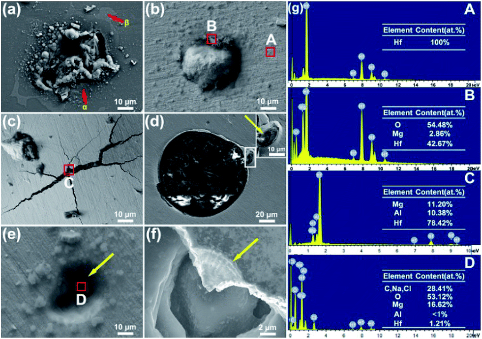
Fig. 8 displays the corrosion characteristics of the bare AZ91D and Hf-coated specimens after 24 salt spray tests. The matrix α phase in Mg alloy always acts as an anode compared to the β phase and is preferentially corroded due to its lower corrosion potential.28 As shown in Fig. 8a, the β phase keeps intact as the barrier to inhibit the extension of corroded area. There is a clear evidence of the preferential corrosion occurred at the defect as indicated in Fig. 8b and g. Corrosion products were appeared on the region of defect whilst the rest was still possessed the protective function. It belongs to a developing corrosion pit in the initial state. However, two different types of developed pitting corrosion are observed in Fig. 8c and e, respectively. The first corrosion type is characterized by a significant expanded micro-crack. There are no defects have been found on the crack suggesting the corrosion is occurred from the inside out. Interestingly, a defect accompanied with corrosion product is observed in a few micrometers away from the micro-crack. According to the analysis in the Fig. 7e and f, it can be speculated that the site of the substrate beneath the defect is β phase, which possessed a more stable chemical property than α phase.28 Therefore, the corrosive media across the β phase and induce the dissolution of the α matrix phase from a distance. Different from the acid autocatalytic effect in local corrosion of other metals, corrosion of Mg exhibits a strange “alkalization effect” behavior even in acid solution.29 Plenty of OH− and Mg2+ were strapped underneath the coating due to their slow diffusion speed. Consequently, the solid Mg(OH)2 eventually formed. The molar volume of Mg(OH)2 about twice as much for that of Mg. As revealed by the EDS of zone C, the increased Mg(OH)2 leads to the increasing volume dilatation of substrate and results in a visible macro-crack in the coating surface. With the deepening of corrosion degree, the coating is failure (shown in Fig. 8d). Fig. 8e and f show the second type of the developed corrosion type. The droplet has came out and left a residual crater (marked by the yellow arrow) in the coatings. The EDS of zone D reveals that the corrosion products are poured out of the pore. Then, the corrosion products are re-dissolved to the salt solution when the coating stripes away (Fig. 8f).
 |
| | Fig. 8 Corrosion morphologies of the AZ91D and Hf coated specimens after salt spray test: (a) AZ91D, (b–f) different corrosion types of the Hf coated specimen, (g) EDS spectra of regions (A)–(D). | |
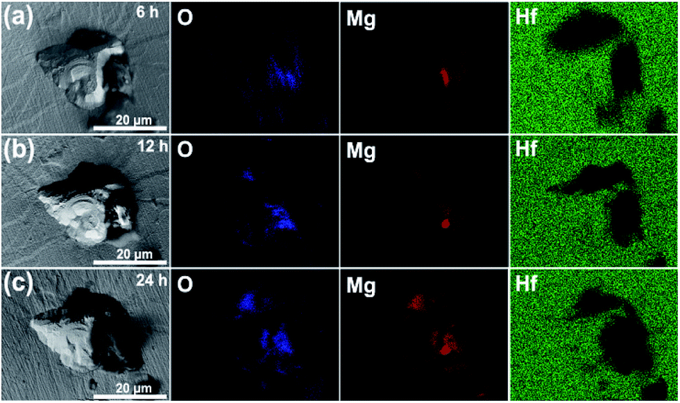
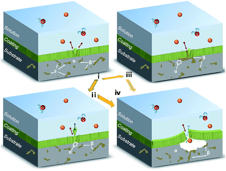
Fig. 9 and 10 show the surface and cross-sectional corrosion process of the Hf coated Mg alloy with varying the salt spray time. In the first stage, as shown in Fig. 9a, the region with droplets in coatings suffers the preferred risk of attacking media when exposed to the corrosion environment. These under-dense structures provide direct passes for corrosive media such as chloride ions and water molecules. Thus Mg alloy begin to be corroded under the driving force of corrosion thermodynamic of the difference potential between coating and substrate. A random phase distribution of α and β in substrate significantly affects the second stage in the coating/substrate interface, which resulted in two different corrosion behaviors. In the first case, β phase of Mg alloy was contacted with the defect. Song et al.1 have confirmed that the β phase was more stable in the salt solution and exhibited more inert to corrosion. Therefore, the corrosive media did not cause corrosion below the defect immediately. As indicated in Fig. 8c, the severed corrosion with a crack was occurred on the distance from the defect. That is to say, the β phase might serve as a barrier to reduce the diffuse speed of corrosive media and extant the diffuse path. However, the β phase also acts as the galvanic cathode and accelerate the corrosion rate of the α matrix eventually resulted in coating failure (as see in Fig. 8a). The contradictory effect of β phase on corrosion has been introduced in detail.1 In the second case, the defect grows on the matrix α phase. In general, the weak bonding between the defect and the surrounding coating resulted from the shadowing effect in the depositing process led to an easy detachment from the coating. On the other hand, the defect flakes off inversely resulted in an accelerating dissolution of Mg alloy. Consequently, the large piece of coating was pilling off from the substrate with naked micro-crack. Fig. 11 shows a comprehensive schematic composes of the above simplification that the pitting corrosion of droplet defect is concurrent with phase distribution of Mg alloy.
 |
| | Fig. 9 Plan views of the droplet after (a) 6 h, (b) 12 h, (c) 24 h salt spray test and corresponding EDS mapping of O, Mg and Hf elements. | |
 |
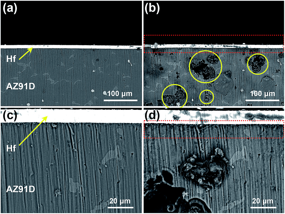
| | Fig. 10 In-suit cross-sectional SEM-BSD images of the Hf coated Mg alloy after (a and c) 0 h, (b and d) 6 h salt spray test. | |
 |
| | Fig. 11 Schematic illustration of the corrosion mechanisms of the Hf coated AZ91D in corrosive condition. | |
It is worth noting that the galvanic corrosion between Hf and Mg alloy is negligible. As indicated in Fig. 10, the pitting corrosion was not consolidated on the interface (marked by red frame) between the coating/substrate after 6 h salt spray test. Inversely, the micro-galvanic corrosion between α and β phase in the Mg alloy (marked by yellow rings) substrate was more severe than the macro-galvanic corrosion between coating and substrate. It is an interesting result for PVD coated-Mg systems whose application usually impeded by the large potential difference between coating and Mg alloy. Therefore, further studies are required to focus on reducing the growth defects or post treatment of Hf coating in order to realize the industrial application of Mg alloys.
4. Conclusion
In conclusions, anticorrosion Hf coatings have been deposited on the AZ91D Mg alloy surfaces by magnetron sputtering varying the negative bias from 0 to −125 V. The Hf coatings present finer microstructure and increased residual stress with increasing the substrate negative bias. The corrosion tests indicate that the Hf coating deposited at substrate bias of −100 V provides the best protection for AZ91D. The porosity of coatings was decreased due to the benefits of denser structure and fewer defects with increasing the bias voltage within a certain range. The droplet defects in the Hf coating were evidenced to be the preferred corrosion sites when exposed to the aggressive environment. Moreover, the micro-galvanic corrosion between α and β phase in substrate determined the whole corrosion process.
Acknowledgements
The authors would like to thank the National Natural Science Foundation of China (51372212 & 20573086).
References
- G. Song and A. Atrens, Adv. Eng. Mater., 2003, 5, 837 CrossRef CAS.
- K. U. Kainer, Magnesium alloys and technologies, John Wiley & Sons, 2006 Search PubMed.
- G. Song, Adv. Eng. Mater., 2005, 7, 563 CrossRef CAS.
- Y. Chen, S. Zhao, M. Chen, W. Zhang, J. Mao, Y. Zhao, M. F. Maitz, N. Huang and G. Wan, Corros. Sci., 2015, 96, 67 CrossRef CAS.
- H. Zhao, S. Cai, Z. T. Ding, M. Zhang, Y. Li and G. H. Xu, RSC Adv., 2015, 5, 24586 RSC.
- L. Y. Cui, R. C. Zeng, S. Q. Li, F. Zhang and E. H. Han, RSC Adv., 2016, 6, 63107 RSC.
- B. Navinšek, P. Panjan and I. Milošev, Surf. Coat. Technol., 1999, 116, 476 CrossRef.
- J. Gray and B. Luan, J. Alloys Compd., 2002, 336, 88 CrossRef CAS.
- F. Hollstein, R. Wiedemann and J. Scholz, Surf. Coat. Technol., 2003, 162, 261 CrossRef CAS.
- B. L. Yu and J. Y. Uan, Scr. Mater., 2006, 54, 1253 CrossRef CAS.
- G. Wu, X. Zeng and G. Yuan, Mater. Lett., 2008, 62, 4325 CrossRef CAS.
- J. Senf and E. Broszeit, Adv. Eng. Mater., 1999, 1, 133 CrossRef CAS.
- G. Wu, Mater. Lett., 2007, 61, 3815 CrossRef CAS.
- D. Zhang, B. Wei, Z. Wu, Z. Qi and Z. Wang, Surf. Coat. Technol., 2016, 303, 94 CrossRef CAS.
- H. Hoche, S. Groß and M. Oechsner, Surf. Coat. Technol., 2014, 259, 102 CrossRef CAS.
- J. Caicedo, C. Amaya, L. Yate, W. Aperador, G. Zambrano, M. Gómez, J. Alvarado-Rivera, J. Muñoz-Saldaña and P. Prieto, Appl. Surf. Sci., 2010, 256, 2876 CrossRef CAS.
- Y. Lv, L. Ji, X. Liu, H. Li, H. Zhou and J. Chen, Appl. Surf. Sci., 2012, 258, 3864 CrossRef CAS.
- J. C. Caicedo, C. Amaya, L. Yate, W. Aperador, G. Zambrano, M. E. Gómez, J. Alvarado-Rivera, J. Muñoz-Saldaña and P. Prieto, Appl. Surf. Sci., 2010, 256, 2876 CrossRef CAS.
- G. Wu, K. Ding, X. Zeng, X. Wang and S. Yao, Scr. Mater., 2009, 61, 269 CrossRef CAS.
- D. McKenzie and M. Bilek, J. Appl. Phys., 1999, 86, 230 CrossRef CAS.
- Z. Qi, P. Sun, F. Zhu, Z. Wang, D. Peng and C. Wu, Surf. Coat. Technol., 2011, 205, 3692 CrossRef CAS.
- G. Abadias, Y. Tse, P. Guérin and V. Pelosin, J. Appl. Phys., 2006, 99, 113519 CrossRef.
- J. A. Thornton, J. Vac. Sci. Technol., A, 1986, 4, 3059 CAS.
- S. H. Ahn, J. H. Lee, J. G. Kim and J. G. Han, Surf. Coat. Technol., 2004, 177–178, 638 CrossRef CAS.
- M. Fenker, M. Balzer and H. Kappl, Surf. Coat. Technol., 2014, 257, 182 CrossRef CAS.
- H. Hoche, S. Groß, T. Troßmann, J. Schmidt and M. Oechsner, Surf. Coat. Technol., 2013, 228, S336 CrossRef CAS.
- G. L. Song and A. Atrens, Adv. Eng. Mater., 1999, 11 CrossRef CAS.
- R. Ambat, N. N. Aung and W. Zhou, Corros. Sci., 2000, 42, 1433 CrossRef CAS.
- G. L. Song and M. Liu, Corros. Sci., 2011, 53, 3500 CrossRef CAS.
|
| This journal is © The Royal Society of Chemistry 2016 |
Click here to see how this site uses Cookies. View our privacy policy here.