
Three-dimensionally microporous and highly biocompatible bacterial cellulose–gelatin composite scaffolds for tissue engineering applications†
Shaukat Khana,
Mazhar Ul-Islamab,
Muhammad Ikramc,
Muhammad Wajid Ullaha,
Muhammad Israra,
Fazli Subhanc,
Yeji Kima,
Jae Hyun Janga,
Sik Yoonc and
Joong Kon Park*a
aDepartment of Chemical Engineering, Kyungpook National University, Daegu, Republic of Korea. E-mail: parkjk@knu.ac.kr; Fax: +82 539506615; Tel: +82 539505621
bDepartment of Chemical Engineering, College of Engineering, Dhofar University, Salalah, Sultanate of Oman
cDepartment of Anatomy, Pusan National University School of Medicine, Yangsan, Gyeongsangnam-do, Korea
First published on 16th November 2016
Abstract
In the current study, highly porous and biocompatible regenerated bacterial cellulose–gelatin (rBC–G) composite scaffolds were fabricated for tissue engineering applications. The scaffolds were prepared from porogen added composite solution of BC–G using a casting and leaching approach. The structural characterization of the scaffolds was carried out through field emission scanning electron microscopy (FE-SEM), Fourier transform infrared spectroscopy (FT-IR), and X-ray photoelectron spectroscopy (XPS). FE-SEM images showed the presence and interconnectivity of pores, while FT-IR and XPS spectra confirmed the composite chemistry of the scaffolds. The observed high porosity and rapid swelling of the scaffolds ensure their nutrient exchange ability during practical applications. In vitro biological tests showed that animal fibroblast cells (NIH 3T3) adhered to and proliferated well on the rBC–G composite scaffolds. Cell penetration assessed by Confocal microscopy indicated up to 200 μm infiltration after 7 days of incubation, suggesting the suitability of the scaffolds for three-dimensional cell culture. The enhanced expression of metalloproteases (MMPs) showed that prolonged cell incubation can lead to extracellular matrix (ECM) production inside the 3D rBC–G scaffolds. These results demonstrated that our 3D rBC–G composite scaffolds are candidates for future biomedical applications, including tissue regeneration.
Introduction
Recent advances in biomedical research have revealed that cell behaviors, such as differentiation1 and tissue regeneration,2 are influenced by the extracellular microenvironment. In particular, the three-dimensional (3D) microenvironment mimics in vivo conditions and enables in vitro tissue regeneration.3 Tissue engineering applies a properly designed combination of functional cells and a 3D scaffold material. The scaffold material is necessary for cell anchorage, growth, and differentiation to provide the biological and physical microenvironment necessary for tissue growth, like the extracellular matrix (ECM).4 The biocompatibility and physico-mechanical properties of the scaffold are critical factors that must be considered in order to produce the appropriate microenvironment and enable specific tissue morphogenesis.5 Scientists have shown immense interest in the development of fabrication techniques and materials to create such 3D microenvironments. The typical materials tested for this purpose include polymeric scaffolds.6 Hydrogel materials, including collagen and fibrinogen, resemble natural ECM, but possess random, sub-micron pores, limiting cell adhesion control and architecture.7 Polymeric scaffolds provide architecture, but the fabrication techniques often result in non-uniform pore sizes and shapes.8 Therefore, it is necessary to construct a well-defined scaffold with a uniform microenvironment to tailor in vitro tissue regeneration for advanced biomedical applications.The natural and synthetic polymers used for scaffold fabrication include gelatin, chitosan, cellulose, polycaprolactone, and polyglycolic acid.9 Among biopolymers, cellulose is the most abundant and important. In particular, bacterial cellulose (BC) produced by acetic acid bacteria is the purest form of cellulose, lacking biogenic impurities, such as lignin and hemicelluloses.10,11 BC offers a unique combination of excellent physico-mechanical and structural properties with a micro-fibrous structure, microporosity, biocompatibility, moldability, and high water-holding capacity.12,13 Owing to these properties, BC is a candidate for advanced biomedical fields. BC has already been tested in biomedical applications, such as for vascular grafts, bone regeneration and cartilage replacement, and as tissue engineering scaffolds.14 Recently, BC has been successfully applied to tissue regeneration research.15
Researchers have augmented BC to match the materials that is intended to replace or regenerate.16 For example, BC soaked in hydroxyapatite yields a scaffold for bone tissue regeneration.17 Similarly, BC nanocomposites with poly (vinyl alcohol) show improved mechanical properties and have been tested for applications in cardiovascular implants.18 Recent studies have focused on BC surface modifications to improve its biocompatibility. Tailoring the microporosity of BC networks for increased cell infiltration is one such effort. For example, patterned BC has been fabricated by the in situ production of BC on honeycomb-patterned agarose film surfaces.19 Similarly, highly porous BC scaffolds have been fabricated by placing starch and paraffin microparticles in BC harvesting media.20 However, the aforementioned efforts have focused on enhancements in either the biocompatibility or microporosity of BC. For practical applications of BC in biomedical fields, it is desirable to consider the simultaneous enhancement of biocompatibility and microporosity.
3D microporosity is one of the primary requirements for the scaffold material to be applied in tissue engineering. Various techniques have been applied to tailor the microporosity of 3D scaffolds, including high-pressure processing,21 phase separation,22 particulate leaching,23 gyration, and electrospinning.24 However, each of these methods has limitations in terms of uniform porosity, interconnectivity, and applicability to different materials. For example, gyration and electrospinning involve polymer solutions at ambient temperature and are not useful to fabricate a true 3D scaffold. Other methods lead to irregular pores and poor interconnectivity, and it is difficult to control the shape and size of scaffolds. The particulate leaching method is widely used to fabricate biopolymer scaffolds.14 This method involves the incorporation of porogen particles into a polymer solution, followed by casting and leaching in a suitable solvent. It is easy, inexpensive, and can be applied to large number of soluble polymers. However, limitations of this method include the irregular pore size, poor interconnectivity, and the application of toxic solvents. Additionally, the complete removal of porogens from the scaffolds is not an easy task and results in residual particles.25
Herein, we report a composite solution casting and particulate leaching scaffold fabrication method that is easy, inexpensive, ensures the complete removal of porogens, and is tunable to maximize biocompatibility and microporosity. This research considers the biocompatibility and microporosity of BC simultaneously via control of the BC to gelatin (G) ratio and the polymer to porogen ratio. The fabricated 3D rBC–G composite scaffolds demonstrated better cell adhesion and proliferation properties indicating their future prospects in tissue regeneration applications.
Materials and methods
Materials
Dulbecco's minimum essential medium (MEM), penicillin, streptomycin, phosphate-buffered saline (PBS), glucose, sodium hydroxide (NaOH), succinic acid, acetic acid, glutaraldehyde, fetal bovine serum (FBS) and bovine serum albumin (BSA) were purchased from Sigma-Aldrich (St. Louis, MO, USA). Fluorescein isothiocyanate (FITC)–phalloidin was purchased from Promega (Madison, WI, USA) and 4,6-diamidino-2-phenylindole (DAPI) was purchased from Vector Laboratories (Burlingame, CA, USA). Murine NIH 3T3 fibroblasts (ATCC no. CRL-1658) were obtained from the American Type Culture Collection (ATCC, Rockville, MD, USA). The reagents were used without any additional processing.Preparation of BC sheets and fabrication of 3D rBC and 3D rBC–G scaffolds
Gluconacetobacter hansenii PJK (KCTC 10505BP) was cultured using a chemically defined medium containing glucose (10 g L−1), yeast extract (10 g L−1), peptone (7 g L−1), acetic acid (1.5 mL L−1) and succinic acid (0.2 g L−1), and the BC sheets were prepared as previously reported by our group.14 The solution casting method was used to fabricate 3D rBC and 3D rBC–G scaffolds using sodium chloride (NaCl) crystals as porogens.14 To fabricate 3D rBC scaffolds, 98 g of N-methyl N-morpholine oxide monohydrate (NMMO·H2O) was taken in a beaker and 2% (v/w) DI water was added to it. The NMMO/water system was then heated to 80 °C on a hot plate. After liquefaction in 3 h, 4 g of BC was added to it and stirred overnight at 600 rpm for complete dissolution. Sieved NaCl crystals (12 g) with diameter ranging from 400 μm to 500 μm were added to the BC solution (BC to NaCl ratio, 1![[thin space (1/6-em)]](https://www.rsc.org/images/entities/char_2009.gif) :3) and stirred for 1 min for uniform mixing. To fabricate 3D rBC–G composite scaffolds, 2 g of gelatin was dissolved in NMMO/water mixture through overnight stirring, followed by the addition of 2 g of BC to the gelatin solution (BC to gelatin ratio, 1:1) and again stirred overnight for BC dissolution. The same NaCl crystals were added as porogens and stirred for 1 min leading to a uniform mixture (polymer to NaCl ratio, 1:3). The mixtures were casted into molds and allowed to solidify at room temperature. After complete solidification in 30 min, the scaffolds were incubated in water bath at 10 °C for salt leaching and solvent removal for 3 days with the supply of fresh water after each 12 h. The scaffolds were then freeze dried before further investigations.
:3) and stirred for 1 min for uniform mixing. To fabricate 3D rBC–G composite scaffolds, 2 g of gelatin was dissolved in NMMO/water mixture through overnight stirring, followed by the addition of 2 g of BC to the gelatin solution (BC to gelatin ratio, 1:1) and again stirred overnight for BC dissolution. The same NaCl crystals were added as porogens and stirred for 1 min leading to a uniform mixture (polymer to NaCl ratio, 1:3). The mixtures were casted into molds and allowed to solidify at room temperature. After complete solidification in 30 min, the scaffolds were incubated in water bath at 10 °C for salt leaching and solvent removal for 3 days with the supply of fresh water after each 12 h. The scaffolds were then freeze dried before further investigations.
Characterization
Fourier transform infrared (FT-IR) spectra of the scaffolds were recorded using a Spectrum GX &AutoImage. An FT-IR spectrophotometer was used [spectral range: 4000–400 cm−1; beam splitter: Ge coated on KBr; detector: deuterated triglycine sulfate; resolution: 0.25 cm−1 (step selectable); PerkinElmer, Melville, NY, USA]. The samples were mixed with KBr and formed into pellets prior to FT-IR analysis (IR grade; Merck, Darmstadt, Germany). FE-SEM analysis of the scaffolds was carried out through Hitachi S-4800 (Tokyo, Japan) and Horiba EDX-350 (Tokyo, Japan). The samples were fixed on a brass holder and coated with osmium tetroxide using a VD HPC-ISW osmium coater (Tokyo, Japan) prior to FE-SEM observations. The fabricated scaffolds were also characterized using XPS performed through a PHI Quantera SXM (ULVAC-PHI, Inc., Chigasaki, Japan) instrument having AlK X-ray source at 15 K and 25 W. The Ar ion gun acceleration was performed at 2 kV, with a sputter rate of 5 nm min−1 for an SiO2 standard. The emission angle was kept at 45°. Photographs of the scaffolds were obtained using a digital camera (Model 5050; Olympus, Tokyo, Japan).Porosity determination and swelling studies
A liquid displacement method was applied for the determination of % porosity of the 3D scaffolds.26 Briefly, scaffolds of known weight W (diameter = 1 cm and height = 0.5 cm) were soaked in degassed n-hexane of known volume (V1). The combined volume of hexane and the immersed scaffold was recorded as V2. After 1 h, the scaffold was removed and the volume of the remaining hexane was measured as V3. The % porosity was calculated using the following formula:
The swelling ratio (SR) of rBC and rBC–G composite scaffolds was determined as reported previously.14 Briefly, a preweighed dry scaffold was soaked in water for a defined length of time at ambient temperature and reweighed. The SR of the respective samples was determined using the following formula:
| SR = (ww − wd)/wd × 100 |
In vitro biocompatibility test
NIH 3T3 were cultured in DMEM containing FBS (10%), streptomycin (100 U mL−1), and penicillin (100 U mL−1) in 5% CO2 at 37 °C. For in vitro biocompatibility determination, the scaffolds were cut into round pieces (20 mm diameter) and sterilized under ultraviolet light. The samples were then incubated in cell growth medium for 12 h. The NIH 3T3 cells were then seeded on the scaffolds at a density of 1.8 × 10⁴ cells per scaffold. Cell adhesion and proliferation were assessed by an FE-SEM analysis. After 3 days of cell incubation, the scaffolds were washed three times with PBS followed by cell fixation with 2.5% glutaraldehyde for 15 min. The scaffolds were again washed with PBS and allowed to dry at ambient temperature. Prior to FE-SEM observations, the samples were fixed on a brass holder and coated with osmium tetroxide using a VD HPC-ISW osmium coater (Tokyo, Japan).Cell infiltration into scaffolds
Cell infiltration into the scaffolds was monitored using a Confocal laser scanning microscope (Olympus FV1000-IX81).14 Briefly, NIH 3T3 cells were seeded on the scaffolds (20 mm diameter) at a density of 1.7 × 10⁴ cells per scaffold, cultured for one week, washed with PBS, fixed with 2.5% glutaraldehyde, and again washed with PBS. Permeabilization was then performed with 0.1% Triton X-100 in PBS for 5 min, followed by washing with PBS. The cells were then incubated in 1% BSA for 1 h at room temperature. After the removal of excess BSA, the samples were incubated with FITC–phalloidin (diluted 1:100) at ambient temperature for 1 h and washed with PBS. The samples were then mounted on glass slides using Vectashield® containing DAPI. The cell fluorescence was determined using a Confocal laser scanning microscope with a 40× oil-immersion objective for image acquisition. The excitation wavelength for FITC was fixed at 488 nm, while the emission wavelength was set at 500–540 nm. The excitation and emission wavelengths for DAPI were fixed at 358 nm and 461 nm, respectively. The 3D reconstruction image of the scaffolds was recorded and stacks of Confocal images were collected for scaffolds with a step size of 10 μm for 3D rBC and 20 μm for 3D rBC–G between adjacent optical planes. These stacks of images were used to generate a 3D animation sequence using 3D projections.
RNA extraction and reverse transcription polymerase chain reaction (RT-PCR) for cell activity determination
The protocol for total RNA extraction was adopted from the RNeasy Mini Kit (Qiagen, Valencia, CA, USA). Briefly, the 3D rBC and rBC–G scaffolds were cut into round pieces (20 mm diameter) and sterilized under UV light. NIH 3T3 cells were cultured on the scaffold at a density of 1.76 × 10⁴ cells per scaffold for 7 days. The scaffolds were then transferred to a mortar containing liquid nitrogen, and ground to a fine powder using a pestle. The scaffolds containing cells were lysed with RLT buffer, incubated on ice, homogenized by vortexing, and supplemented with 2 mL of 70% ethanol. The subsequent RNA purification steps were performed according to the manufacturer's instructions. RNA quantity and quality were assessed using a NanoDrop 2000 (Thermo Scientific, Waltham, MA, USA). First-strand cDNA was reverse transcribed from 2 μg of total RNA in 25 μL of solution using a Promega kit according to manufacturer's instructions. Briefly, the reaction was conducted in 20 μL of buffer containing 0.5 μg of oligo (dT) 12–18 primer (Promega), 50 mM Tris–HCl (pH 8.3), 75 mM KCl, 3 mM MgCl2, 40 mM dithiothreitol, 0.5 mM deoxynucleotide triphosphate mixture (Promega),10 U RNase inhibitor (Promega), and 200 U moloney murine leukemia virus (MMLV) reverse transcriptase (Promega). After incubation at 37 °C for 60 min, the reaction was stopped via heating at 70 °C for 5 min. The cDNA was used as a template for PCR amplification with gene-specific primers. Amplification primer pairs were selected (Table 1) to examine the mRNA expression of the marker genes MMP-2 and MMP-9 and the expression of the house-keeping gene glyceraldehyde-3-phosphatase dehydrogenase (GAPDH) was monitored as a control for RNA loading. PCR amplification of the cDNA was performed in an automated thermal cycler (PC 320; Astec, Osaka, Japan) in a final volume of 25 μL containing 100 ng of cDNA solution, 5 μL of 5 × GOTaq buffer, 3 μL of 10 mM dNTP, 0.5 μL of 10 pmol of each primer, and 0.25 μL of GoTaq DNA polymerase, cycled 30–35 times at 94 °C for 30 s, 60 °C for 30 s, and a final extension of 5 min at 72 °C. After PCR, the amplified products were analyzed by electrophoresis in a 2% agarose gel and visualized using ethidium bromide staining under ultraviolet light. Band intensities of the PCR products were measured using ImageJ. Data are expressed as ratios of each mRNA normalized to GAPDH mRNA expression levels amplified from the same cDNA sample to correct for errors in spectrophotometric RNA quantification or pipetting.| Symbol | Gene name | Accession number | Primer sequences | Size |
|---|---|---|---|---|
| GAPDH | Glyceraldehyde-3-phosphate dehydrogenase | NM_001289726.1 | F.P 5′-TGGAGA AACCTG CCAAGTATG-3′, R.P 5′-TTGTCATACCAGGAA ATGAGC-3′ | 202 bp |
| MMP-2 | Matrix metallopeptidase 2 | NM_008610.2 | F.P 5′-CACCATCGCCCATCATCAAGT-3′, R.P 5′-TGGATTCGAGAAAAGCGCAGCGG-3′ | 400 bp |
| MMP-9 | Matrix metallopeptidase 9 | NM_013599.3 | F.P 5′-ACTCACACGACATCTTCCAG-3′, R.P 5′-AGAAGGAGCCCTAGTTCAAG-3′ | 176 bp |
Cell viability
Cell viability on the fabricated rBC and rBC–G scaffolds was determined using a 3-dimethylthiazol-2,5-diphenyltetrazolium bromide (MTT) colorimetric assay.10 Briefly, the scaffolds were cut into round pieces (20 mm diameter) and sterilized under UV light. NIH 3T3 were cultured on the scaffold at 1.3 × 10⁴ cells per scaffold for 1, 3, 5, and 7 days. After completion of the incubation period, the scaffolds were rinsed with PBS and MTT solution (0.5 mg mL−1) was added. The mixture was incubated at 37 °C for 4 h. The excess MTT solution was discarded from the scaffolds and DMSO (200 μL per scaffold) was added, followed by incubation at 37 °C for 30 min to dissolve formazan crystals. A microplate reader was used to record the optical density of the formazan solution at 540 nm. The cells cultured on the 3D rBC scaffold served as a control.Statistical analysis
All quantitative data are expressed as means ± standard deviation. Statistical comparisons were performed using one-way ANOVA implemented in SPSS 13.0 for Windows (SPSS Inc., Chicago, IL, USA). P-Values of less than 0.05 were considered statistically significant.Results and discussion
Fabrication and structural analysis of the 3D rBC and 3D rBC–G scaffolds
The microporous 3D rBC and 3D rBC–G scaffolds were prepared by a solution casting and particulate leaching method as explained in experimental section. The structural homogeneity of scaffolds is affected by the BC and gelatin concentrations, BC to gelatin ratio, porogen diameter, and polymer to porogen ratio. These parameters were assessed and the optimal values were observed as follows: BC and gelatin concentration, 2%; BC to gelatin ratio, 1:1; porogen diameter, 400–500 μm; and polymer to porogen ratio 1:3.
Fig. 1a shows camera images of the 3D scaffolds. The images clearly show the surface pores in the fabricated scaffolds. The 3D rBC scaffolds appear white owing to the pure cellulose composition, while the 3D rBC–G scaffolds look yellowish owing to the presence of gelatin, indicating the uniformity of the composite scaffold. The microporosity of the scaffolds was further confirmed through SEM analysis. Fig. 1b shows the surface and cross-sectional morphology of the 3D scaffolds. The surface images of the 3D rBC and 3D rBC–G scaffolds show the homogeneous macroporous structures. Additionally, the SEM observation shows the pore interconnectivity (indicated by the arrows in the cross-sectional images of the scaffolds) and similar internal morphologies of the scaffolds. However, the surface morphology of the pore walls was quite different between the two scaffolds. The cross-sections of the 3D rBC–G scaffolds indicate presence of nanofibers between the pore walls while no such nanofibers were observed in the 3D rBC scaffolds. The SEM analysis showed the successful fabrication of the microporous scaffolds with high interconnectivity. The porosity and interconnectivity of scaffolds are essential for cell proliferation, migration, and the transport of nutrients and oxygen during tissue regeneration.27
 | ||
| Fig. 1 (a) Camera pictures of the synthesized 3D scaffolds, scale bar represents 1 cm, (b) SEM analysis of the scaffolds. The green arrows points towards the windows connecting two adjacent pores confirming the pore interconnectivity. | ||
Structural characterization, porosity and swelling of the 3D scaffolds
Fig. 2a shows the FT-IR spectra of 3D rBC, gelatin and 3D rBC–G composite scaffolds. The characteristic peaks for BC were easily assigned as follows: 3330 cm−1 to O–H stretching, 2900 cm−1 to C–H stretching of CH and CH2 functional groups, 1630 cm−1 to bending vibration of residual water, 1355 cm−1 to symmetric bending of CH2, and the bands at 1170–1000 cm−1 indicate the O–H and C–H stretching and C–O–C bending vibrations.28 For gelatin, the absorption band at 3410 cm−1 was assigned to N–H stretching, 1640 cm−1 to C![[double bond, length as m-dash]](https://www.rsc.org/images/entities/char_e001.gif) O, 1550 cm−1 to C–N stretching, and 1247 cm−1 to N–H bending vibration.29 The FT-IR spectra of the 3D rBC–G composite scaffold showed all the characteristic bands for both BC and gelatin, confirming the uniformity of the composite scaffolds. Compared to pure gelatin, the characteristic bands of gelatin at 1640 cm−1 and 3410 cm−1 shifted to higher wavelengths in the 3D rBC–G composite scaffolds, indicating strong intermolecular hydrogen bonding between gelatin and BC.29
O, 1550 cm−1 to C–N stretching, and 1247 cm−1 to N–H bending vibration.29 The FT-IR spectra of the 3D rBC–G composite scaffold showed all the characteristic bands for both BC and gelatin, confirming the uniformity of the composite scaffolds. Compared to pure gelatin, the characteristic bands of gelatin at 1640 cm−1 and 3410 cm−1 shifted to higher wavelengths in the 3D rBC–G composite scaffolds, indicating strong intermolecular hydrogen bonding between gelatin and BC.29
 | ||
| Fig. 2 FT-IR (a), XPS spectra of the scaffolds (b) and swelling properties of the 3D rBC and 3D rBC–G scaffolds incubated in DI water (c). | ||
An XPS analysis (Fig. 2b) was carried out to determine the elemental composition of the fabricated 3D rBC and 3D rBC–G composite scaffolds. The atomic percentage of nitrogen was higher in the 3D rBC–G scaffold and the oxygen and carbon atomic percentages were lower compared to the 3D rBC scaffolds. In the rBC scaffolds, carbon and oxygen were recorded as 59.76% and 40.23%, respectively, but no nitrogen was detected. However, the nitrogen and carbon percentage increased to 9.4% and 64.1% respectively, while oxygen decreased to 26.5%, in the 3D rBC–G scaffold. The O 1s and C 1s spectra of 3D rBC and the O 1s, C 1s, and N 1s spectra of the 3D rBC–G scaffolds are shown in Fig. 2b. The O 1s and C 1s peaks of gelatin in the 3D rBC–G scaffolds overlapped with the same peaks from 3D rBC.12 The increase in the atomic percentage of nitrogen confirms the successful fabrication of the 3D rBC–G composite scaffolds.
A porosity evaluation using the volume displacement method showed that the synthesized 3D rBC and 3D rBC–G composite scaffolds have average porosities of approximately 68.67 ± 4.73% and 70.15 ± 2.2%, respectively. No statistically significant differences in porosity were observed between the 3D rBC and 3D rBC–G composite scaffolds. These results are concordant with the results of the SEM analysis and confirmed the uniform porosity and interconnectivity of the micropores in the 3D rBC and 3D rBC–G composite scaffolds fabricated by solution casting and salt leaching.14
The percent swelling results for 3D rBC and 3D rBC–G composite scaffolds for various water contact times are depicted in Fig. 2c. The SR of the 3D rBC scaffold increased rapidly to 912% in the initial 10 min and reached saturation at 1436% in only 30 min. This is due to the large 3D porous structure of the 3D rBC scaffold, allowing water to penetrate the scaffold very quickly. However, the blending of 50% gelatin with BC in the 3D rBC–G composite scaffolds resulted in increased swelling to 1646% in 10 min and saturation at 2205% in 30 min, which is promising for enhanced cell adhesion, growth, and proliferation. Gelatin contains numerous polar hydrophilic groups, such as –COOH, –NH2, and –OH, which contribute to the increased swelling ability observed for 3D rBC–G scaffolds. This could explain the high SR observed for the 3D rBC–G composite scaffolds. These results are in complete agreement with those of previous reports.30 A high water-holding capacity of a scaffold is favorable for cell adhesion and growth. Water absorption facilitates nutrient transport from the scaffold to cells in the culture system during tissue regeneration.
In vitro biocompatibility of the 3D scaffolds
The biocompatibility of the 3D rBC and 3D rBC–G composite scaffolds was investigated against NIH 3T3 cells in vitro to evaluate their potential applications in tissue engineering. Fig. 3 shows SEM images of the scaffolds after cell adhesion and proliferation for 3 days. The images show good cell adhesion inside the pores on both the 3D rBC and 3D rBC–G composite scaffolds. Incubation period of 3 days resulted in cell growth and proliferation, leading to interconnections via filopodia formation in both the samples. However, the denser filopodia network on the 3D rBC–G scaffolds suggests better cell adhesion and proliferation on these samples compared to the 3D rBC scaffolds. Gelatin is a highly biocompatible polymer and is often employed as a biocompatibility enhancer in tissue regeneration scaffolds. For example, gelatin–silica composite scaffolds have been fabricated and tested for in vitro bone regeneration properties.31 Gelatin has also been employed to enhance the biocompatibility of alginate and alginate–hydroxyapatite scaffolds.29 The denser filopodia network on our 3D rBC–G scaffolds showed their enhanced biocompatibility upon the addition of gelatin compared to pure 3D rBC scaffolds. | ||
| Fig. 3 SEM images showing cell adhesion and proliferation on the 3D rBC (a) and 3D rBC–G scaffolds (b). The arrows indicate the interconnected network of cell filopodia after 3 days of incubation. | ||
ECM synthesis and organization are affected by the microenvironment of growing NIH 3T3; accordingly, it was important to observe the morphology of cultured cells on the 3D scaffolds. Typically, NIH 3T3 cells spread like a fried egg on a 2D surface, but form elongated filopodia in the 3D scaffolds. While most cells attained a spindle shape via the formation of long filopodia extending through the pores (stretchers), some cells spread on the scaffold walls only (stickers).32 Previous studies have shown that NIH 3T3 cells cultured on non-porous 2D surfaces and scaffolds with very large pore sizes are characterized by more stickers than stretchers.32 Our results clearly show extended filopodia formation through the pores (indicated by red arrows in Fig. 3) on the 3D scaffolds, which not only confirms the suitability of the pore size of the scaffolds for cell proliferation, but also suggests a mechanism for improved ECM synthesis in these scaffolds.
Cell infiltration into the 3D scaffolds
Confocal microscopy was used to determine the cell morphology and infiltration into the 3D scaffolds. Fig. 4a shows the differential interference contrast (DIC), DAPI, F-actin, and DAPI/F-actin merged images of the stained NIH 3T3 cells after 7 days of incubation. These images indicate good cell growth and proliferation on the scaffolds. However, increased cell growth and proliferation was observed on the 3D rBC–G scaffolds compared to the 3D rBC scaffolds (Fig. 4a). Cell infiltration was determined by optical slicing in the Z-direction from the top to bottom of the scaffolds, allowing us to observe the penetration of cells into the 3-D scaffolds. Fig. 4b contains the Z-stack images from top to bottom for 3D rBC and 3D rBC–G scaffolds, respectively. And, Fig. 4c shows the reconstructed 3-D projection image of the scaffolds. These results clearly demonstrate the successful growth, proliferation, and infiltration of cells up to 100 μm in 3D rBC and 200 μm in the 3D rBC–G scaffolds, indicating the enhanced biocompatibility of 3D rBC–G scaffolds and also their suitability for tissue regeneration applications.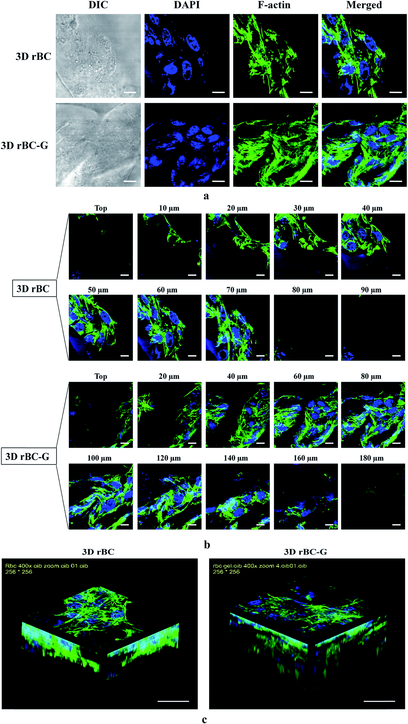 | ||
| Fig. 4 Confocal images of DAPI and F-actin stained NIH 3T3 cultured for one week on 3-D rBC and 3-D rBC–G scaffolds (a). Z-Stack images obtained from surface to bottom of the scaffold for 3D rBC and 3D rBC–G are shown (b). The scale bar in (a) and (b) represents 10 μm. Reconstructed 3-D projection image of the scaffolds (c). The scale bar in (c) represents 100 μm. | ||
Synthesis and organization of ECM in the 3D scaffolds
NIH 3T3 activity was compared for the 3D rBC and 3D rBC–G scaffold cultures by quantifying the mRNA expression of MMP-2 and MMP-9, which are involved in extracellular matrix production via differentiation of NIH 3T3. We examined matrix metalloproteases (MMPs) after 7 days of cell culture to determine the effect of scaffold composition on specific cell functions. The most important function of NIH 3T3 cells is ECM synthesis; therefore, we focused on MMPs involved in ECM organization. MMP-2 and MMP-9 are important differentiation markers and the expression levels of these genes were examined by PCR (Fig. 5a). The densitometry results in Fig. 5b shows quantitative enhancement of the transcription of MMP-9 in 3D rBC–G (6.12) compared to 3D rBC scaffolds (1.02), suggesting that the 3D rBC–G scaffold provides a better microenvironment for cell differentiation. | ||
| Fig. 5 RT-PCR analysis (a) and densitometry profiles (b) showing the expression of MMP-2 and MMP-9 genes in the 3D scaffolds after 7 days of cell incubation. | ||
NIH 3T3 regulate the synthesis and remodeling of the ECM to maintain homeostasis in the connective tissue. MMPs are involved in the degradation of ECM components during the organization of the actin cytoskeleton. In addition, MMPs regulate non-matrix substrates and matrix- and surface-bound growth regulators via the activation of other MMPs, cytokines, and chemokines.33 In this study, both 3D rBC and rBC–G scaffolds exhibited MMP-2 and MMP-9 activity. However, the genes were significantly up regulated in the 3D rBC–G scaffolds compared to the 3D rBC. This effect was attributed to the enhanced biocompatibility of the 3D rBC–G scaffolds compared to the 3D rBC scaffolds. These results indicated that 3D rBC–G scaffolds better regulated function of NIH 3T3 via ECM synthesis and organization compared to the 3D rBC scaffolds.
Cell viability
An MTT assay was used to examine cell proliferation on the 3D rBC and 3D rBC–G composite scaffolds. The results are presented in Fig. 6. The cell numbers increased on both the 3D rBC and 3D rBC–G scaffolds as the incubation time increased. No significant difference in cell proliferation was observed on first day of incubation, although similar 3D space was available for growth and proliferation in the 3D rBC and 3D rBC–G scaffolds. However, the difference got increased with incubation time of 3, 5 and 7 days and augmented higher compatibility and non-toxicity of the 3D rBC–G scaffold. These results clearly show the improved biocompatibility in the 3D rBC–G composite scaffolds owing to gelatin incorporation. Our results are in complete agreement with those of previous reports showing better cell proliferation for scaffolds when gelatin was incorporated into alginate.29 The alginate/gelatin scaffolds demonstrated good cell adhesion and proliferation when tested against human bone marrow-derived mesenchymal stem cells (hBMSC). The cells also showed uniform distribution and excellent infiltration into the scaffolds over 21 days of incubation. It was concluded that the constructed alginate/gelatin scaffolds are potential candidate materials for bone regeneration.29 A major challenge in tissue engineering is the survival and growth of cells inside the scaffold. Cells are typically attached to the surface and fail to migrate inside the scaffold owing to the poor interconnectivity of the pores and limited nutrient and oxygen supply in the central regions. Our SEM and porosity results confirmed the porous nature of the scaffolds while the high swelling behavior augment the successful transfer of nutrients and oxygen leading to the possible migration of cells into the internal matrix of the scaffolds. These results were also supported by the Confocal analysis. The results of the MTT assay are completely concordant with the in vitro biocompatibility tests for the 3D scaffolds and verify our scaffold fabrication and biocompatibility enhancement technique.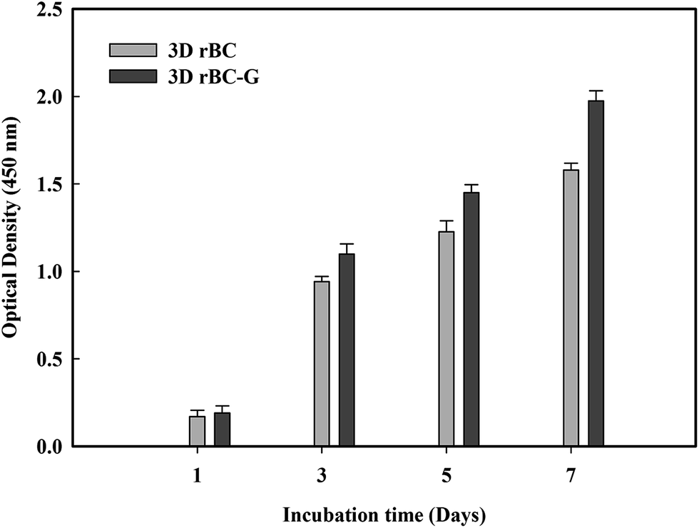 | ||
| Fig. 6 Cell viability assay performed after 1, 3, 5 and 7 days of cell adhesion. Mean values are shown ± SD (P < 0.05). | ||
Conclusion
3D rBC–G composite scaffolds, with enhanced biocompatibility, were fabricated using a solution casting and porogen leaching method. The scaffolds were characterized by a porous geometry and high absorbing capabilities that facilitate their biomedical applications. Animal cells adhered to the surfaces and infiltrated the scaffolds. Moreover, an RT-PCR analysis showed successful ECM synthesis via activity of NIH 3T3. The high biocompatibility toward animal cells and ECM synthesis inside the scaffolds makes them strong candidates for future tissue engineering applications.Acknowledgements
This research was supported by the Basic Science Research Program through the National Research Foundation (NRF) funded by the Ministry of Education, Science and Technology, Korea (NRF-2014-R1A1A2055756) and by the BK21 plus (2014–2019), Korea (21A.2013-1800001).References
- M. Ul-Islam, N. Shah, J. H. Ha and J. K. Park, Korean J. Chem. Eng., 2011, 28, 1736–1743 CrossRef CAS.
- C. M. Nelson and M. J. Bissell, Annu. Rev. Cell Dev. Biol., 2006, 22, 287 CrossRef CAS PubMed.
- L. G. Griffith and M. A. Swartz, Nat. Rev. Mol. Cell Biol., 2006, 7, 211–224 CrossRef CAS PubMed.
- V. P. Shastri, Adv. Mater., 2009, 21, 3246–3254 CrossRef CAS PubMed.
- J. Lee, M. J. Cuddihy and N. A. Kotov, Tissue Eng., Part B, 2008, 14, 61–86 CrossRef CAS PubMed.
- R. Zhang and P. X. Ma, J. Biomed. Mater. Res., 1999, 44, 446–455 CrossRef CAS PubMed.
- A. Barbetta, A. Gumiero, R. Pecci, R. Bedini and M. Dentini, Biomacromolecules, 2009, 10, 3188–3192 CrossRef CAS PubMed.
- A. Guiseppi-Elie, Biomaterials, 2010, 31, 2701–2716 CrossRef CAS PubMed.
- S. Jebahi, H. Oudadesse, G. B. Saleh, M. Saoudi, S. Mesadhi, T. Rebai, H. Keskes, A. El Feki and H. El Feki, Korean J. Chem. Eng., 2014, 31, 1616–1623 CrossRef CAS.
- S. Khan, M. Ul-Islam, W. A. Khattak, M. W. Ullah and J. K. Park, Cellulose, 2015, 22, 565–579 CrossRef CAS.
- M. W. Ullah, M. Ul-Islam, S. Khan, Y. Kim and J. K. Park, Carbohydr. Polym., 2015, 132, 286–294 CrossRef CAS PubMed.
- S. Khan, M. Ul-Islam, W. A. Khattak, M. W. Ullah and J. K. Park, Carbohydr. Polym., 2015, 127, 86–93 CrossRef CAS PubMed.
- C. Seo, H. W. Lee, A. Suresh, J. W. Yang, J. K. Jung and Y.-C. Kim, Korean J. Chem. Eng., 2014, 31, 1433–1437 CrossRef CAS.
- S. Khan, M. Ul-Islam, M. W. Ullah, M. Ikram, F. Subhan, Y. Kim, J. H. Jang, S. Yoon and J. K. Park, RSC Adv., 2015, 5, 84565–84573 RSC.
- L.-Y. Zhu, X.-Q. Yan, H.-M. Zhang, S.-J. Yao and L. Jiang, Korean J. Chem. Eng., 2015, 32, 369–372 CrossRef CAS.
- M. Fiayyaz, K. M. Zia, M. Zuber, T. Jamil, M. K. Khosa and M. A. Jamal, Korean J. Chem. Eng., 2014, 31, 644–649 CrossRef CAS.
- A. F. Leitão, S. Gupta, J. P. Silva, I. Reviakine and M. Gama, Colloids Surf., B, 2013, 111, 493–502 CrossRef PubMed.
- D. Ciechańska, Fibres Text. East. Eur., 2004, 12, 48 Search PubMed.
- H. Bäckdahl, M. Esguerra, D. Delbro, B. Risberg and P. Gatenholm, J. Tissue Eng. Regener. Med., 2008, 2, 320–330 CrossRef PubMed.
- J. J. Yoon and T. G. Park, J. Biomed. Mater. Res., 2001, 55, 401–408 CrossRef CAS PubMed.
- D. J. Mooney, D. F. Baldwin, N. P. Suh, J. P. Vacanti and R. Langer, Biomaterials, 1996, 17, 1417–1422 CrossRef CAS PubMed.
- P. X. Ma and R. Zhang, J. Biomed. Mater. Res., 1999, 46, 60–72 CrossRef CAS PubMed.
- C. J. Liao, C. F. Chen, J. H. Chen, S. F. Chiang, Y. J. Lin and K. Y. Chang, J. Biomed. Mater. Res., 2002, 59, 676–681 CrossRef CAS PubMed.
- J. A. Matthews, G. E. Wnek, D. G. Simpson and G. L. Bowlin, Biomacromolecules, 2002, 3, 232–238 CrossRef CAS PubMed.
- D. W. Hutmacher, Biomaterials, 2000, 21, 2529–2543 CrossRef CAS PubMed.
- F. Palumbo, C. Fiorica, G. Pitarresi, S. Agnello and G. Giammona, RSC Adv., 2015, 5, 61440–61448 RSC.
- F. Wang, J.-H. Jeon, S. Park, C.-D. Kee, S.-J. Kim and I.-K. Oh, Soft Matter, 2016, 12, 246–254 RSC.
- M. W. Ullah, M. Ul-Islam, S. Khan, Y. Kim and J. K. Park, Carbohydr. Polym., 2016, 136, 908–916 CrossRef CAS PubMed.
- Y. Luo, A. Lode, A. R. Akkineni and M. Gelinsky, RSC Adv., 2015, 5, 43480–43488 RSC.
- M. Moffa, A. Polini, A. G. Sciancalepore, L. Persano, E. Mele, L. G. Passione, G. Potente and D. Pisignano, Soft Matter, 2013, 9, 5529–5539 RSC.
- B. Lei, K.-H. Shin, D.-Y. Noh, I.-H. Jo, Y.-H. Koh, W.-Y. Choi and H.-E. Kim, J. Mater. Chem., 2012, 22, 14133–14140 RSC.
- J.-Y. Lin, W.-J. Lin, W.-H. Hong, W.-C. Hung, S. H. Nowotarski, S. M. Gouveia, I. Cristo and K.-H. Lin, Soft Matter, 2011, 7, 10010–10016 RSC.
- M. A. Karsdal, L. Larsen, M. T. Engsig, H. Lou, M. Ferreras, A. Lochter, J.-M. Delaissé and N. T. Foged, J. Biol. Chem., 2002, 277, 44061–44067 CrossRef CAS PubMed.
Footnote |
| † Electronic supplementary information (ESI) available. See DOI: 10.1039/c6ra18847h |
| This journal is © The Royal Society of Chemistry 2016 |