Alternative role of cisplatin in DNA damage – theoretical studies on the influence of excess electrons on the cisplatin–DNA complex

Shuang Zhao and Ru-bo Zhang*
School of Chemistry, Beijing Institute of Technology, South Street No. 5, Zhongguancun, Haidian District 100081, Beijing, China. E-mail: zhangrubo@bit.edu.cn

Received 14th July 2016 , Accepted 17th August 2016

First published on 17th August 2016


Abstract

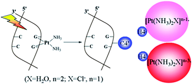
Reliable DFT calculations were used to gain insights into the effects of excess electrons on the cisplatin–DNA complex in a water solution. One electron injection is enough to break the two Pt–N7 bonds, which is driven by the rare symmetrical in-plane bending vibration. The dissociated [Pt(NH3)2]+˙ group from the cisplatin–DNA complex could combine with H2O in the surroundings to form a reactive species, which can abstract the most solvent accessible H4′ of the sugar with a barrier of ca. 17.5 kcal mol−1. Upon influence of the multiple electrons addition, the H4′-abstraction reaction by the stable radical anion is feasible with a lower barrier and is exothermic. Thereby, they have high efficiency for DNA damage. The synergic effect between the metal and the ligand is highlighted due to failure of the isolated [Pt(NH3)2]+˙ and [Pt(NH3)2]˙ to abstract H4′ of sugar because the overlap between the SOMO (on Pt) and the C4′H4′ anti-bonding orbital is zero. In the present studies, an alternate role of cisplatin in DNA damage was discovered, which strongly confirmed that the cisplatin–DNA complex is more vulnerable to attack from low-energy electrons.


Introduction

Cisplatin is one of the most widely used drugs for treating cancer.1 The related mechanisms have been extensively studied.2–10 It is well-known that cisplatin must undergo a slow hydrolysis process to replace one or two chlorides by water molecules to form reactive intermediates. Afterwards, predominantly intra-crosslinked adducts are formed through the cisplatin intermediates binding to the N7 sites of the two consecutive guanines in DNA such that the cross-link product can bend and unwind the DNA duplex, efficiently suppressing DNA transcription and ultimately leading to cell death.

Single- and double-strand DNA breakage (SSB and DSB) induced by the low energy electrons (LEE) have been widely studied.11–17 The C3′–O3′ and N-glycosidic bonds are more vulnerable to attack from LEE.18–21 In many clinical trials, improved local tumor control was achieved when both radiation and the chemotherapeutic agent (such as cisplatin) were synchronously used,22–24 which has been attributed to a superadditive effect on tumor regression. Another proposal is that the new reactive intermediates could be created from the primary radiation of the cisplatin–DNA complex and causes the additional DNA damage.25 Compared to pure DNA, Kobayashi et al. reported that the cisplatin–DNA complex was more inclined to suffering from the substantial strand-break damage enhancement under the concomitant X-ray irradiation.26 Recently, bombarding the solid films composed of the cisplatin–DNA complexes with different energy electrons was performed by Sanche and Zheng et al.27 The significant promotion of the strand breakage was found in the low-energy electron region. The effective rate constants of both the phosphodiester and glycosidic bond cleavages were enhanced to be 1.8- and 1.9-fold larger for the cisplatin–DNA complexes compared to the corresponding unmodified DNA under the X-ray radiation.28 Thus, the cisplatin–DNA complexes could be thought to be more effective to trigger radiosensitization by the secondary LEE in concomitant chemoradiation therapy.27,29,30

Since cisplatin, the classic anti-cancer drug, has been used for a long time in therapy, the very significant and fascinating experimental results promote one to think about why the cisplatin–DNA complex is more sensitive to electrons than the related pure DNA, and what role cisplatin plays in the cisplatin–DNA complex attacked by the excess electrons. Are there some reactive cisplatin species contributing to the enhanced DNA damage? Hence, we attempted to use the reliable DFT method with the aim of exploring the influence of one and more electrons on stability of the cisplatin–DNA complex at the electronic and atomic level. Whether it is possible for the biological molecules to capture more than one electron is strongly determined by the electron affinities (EA) of the molecular complex and the vertical electron detachment energy (VDE) of the corresponding anion. We discovered that attachment of only one electron can transform the planar four-coordinated [Pt(NH3)2GG]0 into the three-coordinated single cross-link [Pt(NH3)2G + G]˙ species, which could abstract H2′ in the solution with a 20.3 kcal mol−1 barrier. In addition, a more significant and competitive product is the free [Pt(NH3)2]+˙, which originates from the bond scission of Pt–N7b in [Pt(NH3)2G + G]˙. The reaction needs to override quite a low or even negative barrier and is exothermic, driven by the symmetrically in-plane bending vibration of the [NH3–Pt–NH3]+˙ group. The resulting free [Pt(NH3)2]+˙, together with surrounding H2O as a ligand is highly reactive towards C–H abstraction, which is the crucial step of DNA strand scission. The more reactive [Pt(NH3)2H2O]˙ could be produced through injection of more electrons into the [Pt(NH3)2H2O]+˙ + DNA system. Highly reactive species could be obtained, which also caused enhancement of DNA damage when cisplatin–DNA complex was under radiation.

Computational model and methods

The calculation model consisted of two continuously stacking guanine (G) bases, one neighboring cytosine (C) base at the 3′ terminal, the lateral sugar–phosphate backbone, and cisplatin without two Cl ions, as displayed in Scheme 1. The labeling of some important atoms is given in the conventional definition. G3 and G5 represent the two guanine bases, which are localized at 3′ and 5′ terminals of the model DNA fragment, respectively. The abovementioned cisplatin–GGC fragment was based on the structure of 5′-d(CCTCAGGCCTCC)-3′ dodecamer duplex, selected from PDB entry 2NPW.31 The neutralized cisplatin–GGC fragment was also studied through protonation of the phosphate group, which is also often used for nucleic acid studies.32–34
image file: c6ra17919c-s1.tif
Scheme 1 The significant labels in the cisplatin–GGC complex.

All the calculations were carried out in Gaussian 09 series.35 The geometries were optimized in a water solution using the Stuttgart–Dresden pseudopotential (SDD) and its accompanying basis set36 for the Pt atom, and 6-31G(d) basis set for other atoms. M06 meta hybrid GGA functional37 was used throughout the calculations. The M06 functional performed the best for metal atom excitations, transition metal bonding, and organometallic and inorganometallic thermochemistry.37 Moreover, M06 is suitable for cisplatin–DNA complex since the complex contains important contributions from electrostatic as well as dispersion interactions.38 The conductor-like polarizable continuum model (CPCM)39,40 was employed to simulate the solvent environment. Frequency calculations were performed at the same level to confirm that the optimized geometry corresponds to a stationary point and to extract the zero-point vibrational energies (ZPE). If necessary, intrinsic reaction coordinate (IRC) calculations were used to determine which minima were connected by a particular transition state. Subsequent single point energies were obtained at the same functional with 6-31+G(d,p) and 6-31++G(d,p) basis sets.

Results and discussion

Structures of the neutral [cisplatin–GGC]0 and its single-electron adducts [cisplatin–GGC]˙

The optimized geometries of the neutral [cisplatin–GGC]0 and its single-electron adduct [cisplatin–GGC]˙ in a water solution are given in the ESI (Table S1). The distances of Pt–N7a and Pt–N7b bonds were 2.077 and 2.069 Å in the solution, respectively. In 5′-d(CCTCAGGCCTCC)-3′ dodecamer duplex,31 they were 2.009 and 2.013 Å, respectively. When one electron was added into the [cisplatin–GGC]0 complex, the Pt–N7a distance was extended up to 3.820 Å in the solution, showing that the Pt–N7a bond could be broken with no barrier. However, the Pt–N7b distance was changed by only ca. 0.03 Å. The lowest unoccupied molecular orbital (LUMO) of [cisplatin–GGC]0 and the singly occupied molecular orbital (SOMO) of [cisplatin–GGC]˙ in the solution is shown in Fig. 1. The LUMO of [cisplatin–GGC]0 was mainly populated in the cisplatin and had the distinct repulsive σ* character in the solution. The SOMO of [cisplatin–GGC]˙ was revealed to be a non-coordinated d orbital, which results from one excess electron captured by LUMO of [cisplatin–GGC]0. These results show that the excess electron mainly occupied the anti-bonding orbital of Pt–N7a, which resulted in transformation of the primary double crosslink complex into the single crosslink complex.
image file: c6ra17919c-f1.tif
Fig. 1 The lowest unoccupied molecular orbital (LUMO) in [cisplatin–GGC]0 and singly occupied molecular orbital (SOMO) in [cisplatin–DNA]˙ in a water solution.

The NPA charge difference between [cisplatin–GGC]0 and [cisplatin–GGC]˙ provides further insight into the overall electronic effects brought by the excess electron. Table 1 summarizes the charge distributions among the bases, riboses, phosphates and Pt(NH3)2 groups for the neutral [cisplatin–GGC]0 and its single-electron adduct. As shown in Table 1, the charge difference on the Pt(NH3)2 group was −0.55e in a water solution. On G5, it was −0.29e in a water solution. These results show that the excess electron was localized mainly on the Pt(NH3)2 group and guanines in the [cisplatin–GGC]˙.

Table 1 NPA charge distributions of the neutral and radical anion for the cisplatin–GGC adduct
Components In water solution
Neutral Anion Δa
a NPA charge difference between analogous neutral and anionic species.b Definition shown in Scheme 1.c Ribose 1, 2 and 3 represent the sugar moieties connected with G5, G3 and cytosine, respectively.
Pt(NH3)2 1.32 0.77 −0.55
G5b 0.02 −0.27 −0.29
G3b 0.01 −0.12 −0.13
Cytosine −0.27 −0.26 0.01
Ribose 1c 0.61 0.59 −0.02
Ribose 2c 0.92 0.91 −0.01
Ribose 3c 0.62 0.61 −0.01
Phosphates −3.22 −3.23 −0.01


It is very significant to estimate the stability of the [cisplatin–GGC]˙ since it could reflect the possible reactivity of the electron adduct. Hence, we also calculated the vertical electron affinity (VEA) and adiabatic electron affinity (AEA) of the [cisplatin–GGC]0 complex, as well as the vertical electron detachment energy (VDE) of the [cisplatin–GGC]˙ adduct. These values are presented in Table 2.

Table 2 Electron affinitiesa (in eV) of [cisplatin–GGC]0 at the M06/SDD, 6-31++G(d,p) level in a water solution
[Cisplatin–GGC]0 → [cisplatin–GGC]˙ VEA AEA VDE
a Their definitions can be found in ref. 41.
In water solution 2.18 3.76 4.66


The VEA predicted for [cisplatin–GGC]0 is 2.18 eV in a water solution, which indicate that the [cisplatin–GGC]0 is a good electron acceptor. The VDE was also calculated to assess the stability of the radical anions, which is most readily observed from the important anion photo detachment experiments.42,43 As shown in Table 2, the VDE of [cisplatin–GGC]˙ was 4.7 eV in a water solution. Consequently, the [cisplatin–GGC]˙ radical anion could exist in the solution.

The possible reactions occurring in [cisplatin–GGC]˙

As mentioned above, the first electron attachment induces a spontaneous scission of the Pt–N7a bond to form the non-coordinated d-orbital on Pt. Therefore, one of the possible reactions of the [cisplatin–GGC]˙ includes H atom abstraction by Pt. As depicted in Fig. 2, in the solution, only H2′ from the sugar of the G5-containing deoxynucleotide is available to the radical-centered Pt and its abstraction is an endothermic reaction with a barrier of 20.3 kcal mol−1. Another parallel reaction includes the scission of the Pt–N7b bond with a negative barrier of −1.9 kcal mol−1. It is exothermic by 0.8 kcal mol−1 and forms [Pt(NH3)2]+˙. Interestingly, the low barrier corresponds to the rarely symmetrically in-plane bending vibration of NH3–Pt–NH3 group rather than the classic Pt–N7b bond stretch vibration, which is ascertained through the calculations of both the harmonic frequencies and the IRC calculations (as shown in Fig. S1 of the ESI). In order to eliminate the influence of the negative charge on the phosphate anion group, the protons were used to neutralize the two phosphate groups.
image file: c6ra17919c-f2.tif
Fig. 2 Stationary structures and the selected geometrical parameters along the H abstraction and Pt–N7b bond scission reactions of [cisplatin–GGC]˙ in a water solution (bond length in Å and energies in kcal mol−1).

The same calculations were performed and show that the reaction barriers of the H2′ abstraction and the Pt–N7b bond scission were 20.2 and 7.4 kcal mol−1, respectively. The structures are shown in Fig. S2 of the ESI. The present calculation results indicate that the activation and reaction energies of H2′ abstraction were kept essentially constant whether the phosphate groups were protonated or not. However, when the phosphate groups were neutralized, the Pt–N7b bond scission could override slightly small positive barrier with endothermic heat of 1.8 kcal mol−1.

From [cisplatin–GGC]0 to [cisplatin–GGC]˙ in an aqueous solution, Fig. 3 shows that the torsion angles of the backbone involving the sugar and phosphate had no drastic change. However, for TS1, the torsion angles β and χ-1 were found to change drastically, showing that the G5 and the local backbone of DNA were varied in a rotation manner. For the product P1, these torsion angles were similar to those in TS1.


image file: c6ra17919c-f3.tif
Fig. 3 The curve variation of the backbone torsion angles (δ-1, ε-1, ζ-1, α, β, γ and δ) and glycosyl angles (χ-1) along the H-abstraction reaction in the water solution. The δ-1 torsion angle is defined as C5′G5–C4′G5–C3′G5–O3′, ε -1 as C4′G5–C3′G5–O3′–P, ζ-1 as C3′G5–O3′–P–O5′, α as O3′–P–O5′–C5′G3, β as P–O5′–C5′G3–C4′G3, γ as O5′–C5′G3–C4′G3–C3′G3, δ as C5′G3–C4′G3–C3′G3–O3′,and χ-1 as O4′–C1′–N1–C4 in G5, which are shown in Fig. S3 of the ESI.

Our present theoretical results are consistent with the experiments by Sanche and co-workers,44 in which they found that an electron of 1.6–3.6 eV was enough to simultaneously break the two Pt–N7 bonds in the cisPt–DNA complex. According to our calculations, one electron attachment first triggers the spontaneous Pt–N7a bond scission, and then the Pt–N7b bond is ruptured with a low or even negative barrier, which is driven by the symmetrically in-plane bending vibration of NH3–Pt–NH3 group. Hence, the present theoretical and experimental results drew the same conclusion that one electron is enough to break the two Pt–N7 bonds. It is noteworthy that the previous reports by Lu et al.45,46 and Illenberger et al.47 pointed out that one electron attachment to cisplatin resulted in two Pt–Cl bond cleavages, thereby forming [Pt(NH3)2]+˙, which is regarded as the reactive intermediate for formation of cisplatin–DNA adducts through bonding to the N7 sites of guanine and adenine.

Based on the aforementioned results, the Pt–N7b bond breakage results in formation of the intermediates P2, which include the [Pt(NH3)2]+˙ fragment with the N–Pt–N bond angle of 174.0°. The aqueous association energy between the free 3′GGC5′ and the isolated [Pt(NH3)2]+˙ fragments was −39.0 kcal mol−1. The negative association energy in an aqueous solution signifies the [Pt(NH3)2]+˙ species freely shifting to other sites, where some possible reactions are induced by the [Pt(NH3)2]+˙ and its derivatives.

H abstraction on the sugar by the reduced Pt-containing species

The electron-injected DNA adducts led to the strand scission and breakage of the N-glycosidic bond of DNA, which were thoroughly investigated by the experiments and the theoretical calculations.48,49 The most facile rupture of the N-glycosidic bond in DNA occurred in guanine nucleotide, whose activation energy was estimated to be 10.0 kcal mol−1.49 If the effective rate constants for the electron-induced N-glycosidic and phosphodiester bond cleavages in cisplatin–DNA complex were 1.8 and 1.9 folder than those in pure DNA,28 it could be inferred that the secondary electron may not be the only active species to cause DNA damage. Thus, the contribution of the formed Pt(NH3)2+ to the DNA damage needs to be explored.

Hence, the [Pt(NH3)2]+˙ species, in addition to itself, can possibly exist in an associated manner with surrounding H2O. The complex could approach the deoxyribose of DNA in the major groove. It is generally accepted that the H4′ on the sugar is the most solvent accessible site in B-form DNA.50 3′dMP was selected as a model compound (seen in Fig. 4) to investigate the C–H abstraction reaction. The relative activation barriers and reaction energies are shown in Table 3.


image file: c6ra17919c-f4.tif
Fig. 4 M06/SDD, 6-31++G(d,p) free energy profile (in kcal mol−1) for H4′ abstraction from nucleotide by (a) cis-[Pt(NH3)2H2O]+˙, (b) trans-[Pt(NH3)2H2O]+˙, (c) cis-[Pt(NH3)2H2O]˙ and (d) trans-[Pt(NH3)2H2O]˙ in water solution.
Table 3 ZPE-corrected activation barriers (ΔE) and reaction energies (ΔE) for H4′-abstraction reactions at the M06/SDD, 6-31++G(d,p) level, in kcal mol−1
Complexes ΔEE) AOOLa SOMO (occupancy)
a AOOL represents the orbital overlap between the SOMO of the complex and the C4′H4′ anti-bonding orbital.
[Pt(NH3)2]+˙ – containing species
3′dMP + [Pt(NH3)2]+˙ N/A 0 dyz (1.447)
3′dMP + cis-[Pt(NH3)2Cl]˙ 18.3 (10.6) −0.011 dz2 (1.597)
3′dMP + trans-[Pt(NH3)2Cl]˙ 17.4 (17.7) 0.011 dyz (1.654)
3′dMP + cis-[Pt(NH3)2H2O]+˙ 17.6 (7.4) 0.032 dz2 (1.466)
3′dMP + trans-[Pt(NH3)2H2O]+˙ 17.5 (18.0) −0.009 dyz (1.649)
[thin space (1/6-em)]
[Pt(NH3)2]˙ – containing species
3′dMP + [Pt(NH3)2]˙ N/A 0 dyz (1.744)
3′dMP + cis-[Pt(NH3)2Cl]2−˙ 11.4 (−0.2) 0.002 dxy (1.779)
3′dMP + trans-[Pt(NH3)2Cl]2−˙ 27.8 (9.5) −0.045 dyz (1.633)
3′dMP + cis-[Pt(NH3)2H2O]˙ 15.5 (−7.8) 0.076 dyz (1.716)
3′dMP + trans-[Pt(NH3)2H2O]˙ 26.1 (10.7) −0.035 dyz (1.719)


As shown in Table 3, the H4′-abstraction barriers of [Pt(NH3)2H2O]+˙ was ca. 17.5 kcal mol−1. These results show that the small Pt-containing radical cation could be one of the candidates for DNA damage. An interesting fact is that the charge of the ligand plays a trivial role on the H4′-abstraction barrier. For instance, replacement of H2O with Cl ion as the ligand to Pt could not obviously influence the H4′-abstraction barrier, as shown in Table 3. Both the cis- and trans-[Pt(NH3)2H2O]+˙ had almost the same efficiency to H4′ abstraction. Moreover, these reactions were endothermic and the reaction energies of cis-[Pt(NH3)2H2O]+˙ were clearly lower by 10.6 kcal mol−1 than those corresponding to the trans-counterpart. The trans effect was very effective for the present radical reaction since the H-abstraction products from the anti-Pt-containing species are very unstable. Thus, only cis-species are the valid candidates for DNA damage. Unexpectedly, H4′ cannot be abstracted by the isolated [Pt(NH3)2]+˙ fragment, showing an interesting fact that the synergetic effect of both the ligand and [Pt(NH3)2]+˙ is the driving force for H4′ abstraction. Further NBO calculations showed that the SOMOs of the complexes were assigned to dyz for the trans-complexes and dz2 for the cis-complexes. The orbital overlap (AOOL) between the SOMOs of the complexes and the image file: c6ra17919c-t1.tif is also presented in Table 3. The AOOL was observed to be zero for the complex 3′dMP + [Pt(NH3)2]+˙, whereas the other AOOLs could not be ignored. This could be used to rationalize why the isolated [Pt(NH3)2]+˙ would not abstract H4′ on the sugar.

As discussed above, the [cisplatin–GGC]˙ adduct with the unpaired electron mainly localized on Pt can be dissociated into [3′GGC5′]2− and [Pt(NH3)2]+˙ fragments. Addition of more electrons into the [Pt(NH3)2]+˙ + [3′GGC5′]2− system in the solution could occur in the concrete experiments, which was determined by both the number of the secondary electrons and the capacity of capturing electrons by the small Pt-containing species. Extension of other new reactive species for DNA damage would be worthy to be attempted.29 Hence, under high-energy radiation, a large number of secondary electrons could be accumulated and the DNA and the [Pt(NH3)2H2O]+˙ could also capture more than one electron, which strongly depends on the fragment molecular electron affinities. As shown in Fig. S4 of the ESI, both the two- and the three-electron adducts of [Pt(NH3)2H2O]2+ had positive VEA, AEA and VDE values, calculated at the M06/SDD, 6-31++G(d,p) level in bulk water. The calculation results further ascertained that the Pt-containing anion species could actually exist in a water solution. The radical adducts could be also formed through the single-electron reduction of Pt atom in the NH3–H2O solution. [Pt(NH3)2]˙ was once deduced as the significant reactive species in recent experiments.47 However, the role is still unclear to date. Thus, in addition to the aforementioned [Pt(NH3)2H2O]+˙ as the potentially reactive species, one question arises about how the possible [Pt(NH3)2H2O]˙ radical anion performs when it is used to attack the sugar.

According to both Table 3 and Fig. 4, when the [Pt(NH3)2H2O]˙ species attack the sugar, smaller H4′-abstraction barriers were observed. They were estimated to be 26.1 kcal mol−1 for the trans-conformer and 15.5 kcal mol−1 for the cis-conformer, which is slightly lower than those of the aforementioned cases with the [Pt(NH3)2H2O]+˙ as the reactive species. More importantly, the reaction was exothermic, which is also dramatically different from the cases with the reactions of the [Pt(NH3)2H2O]+˙ reactive species participation. Hence, the H-abstraction efficiency by the [Pt(NH3)2H2O]˙ depends on the obvious stereoselectivity. Again, the isolated [Pt(NH3)2]˙ anion could not abstract the H4′ at all since the AOOL is zero, as seen in Table 3. Thus, the novel small [Pt(NH3)2H2O]˙ is presently also proposed as the reactive species towards the C–H abstraction reaction, which strongly contributes to DNA damage.

In order to depict the change of the electronic structures during the reaction paths labeled by (a) and (c) in Fig. 4, both the unpaired spin density and NPA charges of the H4′ on each point along the H4′-abstraction reaction path are shown in Fig. 5. The reaction of DNA model with the [Pt(NH3)2H2O]+˙ or [Pt(NH3)2H2O]˙ leads to migration of free radical site from Pt to C4′ atom. For Fig. 5(a), starting from the [Pt(NH3)2H2O]+˙–DNA model reactant complex, the charge of H4′ gradually decreased down to zero and the spin population of the H4′ was very small for the entire transition process. For Fig. 5(b) with the [Pt(NH3)2H2O]˙ species, the spin population of the H4′ was kept close to zero in the process while its charges were changed from ca. 0.2e to −0.3e of the product, which implies that the anion character of H4′ could be very important for this reaction path.


image file: c6ra17919c-f5.tif
Fig. 5 The unpaired spin density and NPA charges of the H4′, C4′ group (the phosphate backbone except H4′) and Pt group (the center atom Pt and its ligands NH3 and H2O) on each point along the H4′-abstraction reaction path in (a) cis-[Pt(NH3)2H2O]+˙ and (b) cis-[Pt(NH3)2H2O]˙.

Note that Chen et al.51 recently studied the reaction of the neutral [Pt(NH3)2Cl]˙ with DNA model in a H-abstraction manner in order to understand how the reactive radical lead to DNA damage. The neutral [Pt(NH3)2Cl]˙ was formed through capture of one electron by the cisplatin + water cluster, which was once proposed by Lu et al. in their cell experiments.23,45,46 According to our calculations, the [Pt(NH3)2H2O]+˙ reactive species was also derived from addition of one electron into the cisplatin–DNA complex. It has almost the same reactivity with [Pt(NH3)2Cl]˙ studied by Chen et al. More importantly, we discovered that the further reduced species, [Pt(NH3)2H2O]˙ shows more reactivity towards C–H abstraction and much attention has been focused on the microscopic mechanism of how [Pt(NH3)2H2O]+˙ and [Pt(NH3)2H2O]˙ perform DNA damage in our studies.

Conclusions

In this study, reliable DFT calculations are used to gain insights into influence of excess electrons on the cisplatin–DNA model complex in a water solution. The potentially reactive species towards DNA damage were explored and the relevant mechanisms are suggested in detail.

The present investigations indicate that [cisplatin–GGC]0 is a good electron acceptor. One electron addition can lead to primary rupture of the Pt–N7a bond. The following Pt–N7b scission reaction is more competitive and its barrier is even negative in a solution, which is driven by the rarely symmetrical in-plane bending vibration rather than the common Pt–N7b stretch vibration. Thus, both the [Pt(NH3)2]+˙ and the DNA model fragments could be separated through only one-electron addition in the solution.

The dissociated [Pt(NH3)2]+˙ group could combine with H2O in the surroundings to form some Pt-based reactive species such as [Pt(NH3)2H2O]+˙, which can abstract the most solvent accessible H4′ on the sugar with a barrier of ca. 17.5 kcal mol−1. Upon influence of the multiple electrons considered in the concrete experiments, H4′-abstraction reaction by the stable [Pt(NH3)2H2O]˙ was found to have lower barrier of 15.5 kcal mol−1 and was exothermic. Thus, the [Pt(NH3)2H2O]˙ is more reactive than the [Pt(NH3)2H2O]+˙ towards H atom abstraction, and therefore has higher efficiency for DNA damage. The isolated [Pt(NH3)2]+˙ and [Pt(NH3)2]˙ would not abstract H4′ on the sugar because of the overlap between the SOMO (on Pt) and the C4′H4′ anti-bonding orbital was zero. Compared with the previous findings about the excess electron influence on DNA damage, the present studies contribute to discover the alternative role of cisplatin in DNA damage and strongly confirm that the DNA damage is enhanced when the transient and reduced Pt-containing species exist.

Notes and references

  1. Y. Jung and S. J. Lippard, Chem. Rev., 2007, 107, 1387–1407 CrossRef CAS PubMed.
  2. P. Carloni, M. Sprik and W. Andreoni, J. Phys. Chem. B, 2000, 104, 823–835 CrossRef CAS.
  3. A. Melchior, M. Tolazzi, J. M. Martínez, R. R. Pappalardo and E. S. Marcos, J. Chem. Theory Comput., 2015, 11, 1735–1744 CrossRef CAS PubMed.
  4. L. A. Truflandier, K. Sutter and J. Autschbach, Inorg. Chem., 2011, 50, 1723–1732 CrossRef CAS PubMed.
  5. L. A. Truflandier and J. Autschbach, J. Am. Chem. Soc., 2010, 132, 3472–3483 CrossRef CAS PubMed.
  6. J. V. Burda, M. Zeizinger and J. Leszczynski, J. Comput. Chem., 2005, 26, 907–914 CrossRef CAS PubMed.
  7. J. K. C. Lau and B. Ensing, Phys. Chem. Chem. Phys., 2010, 12, 10348–10355 RSC.
  8. A. Melchior, E. S. Marcos, R. R. Pappalardo and J. M. Martinez, Theor. Chem. Acc., 2011, 128, 627–638 CrossRef CAS.
  9. J. P. Ceron-Carrasco, D. Jacquemin and E. Cauet, Phys. Chem. Chem. Phys., 2012, 14, 12457–12464 RSC.
  10. M. E. Alberto, V. Butera and N. Russo, Inorg. Chem., 2011, 50, 6965–6971 CrossRef CAS PubMed.
  11. B. Boudaiffa, P. Cloutier, D. Hunting, M. A. Huels and L. Sanche, Science, 2000, 287, 1658–1660 CrossRef CAS PubMed.
  12. J. Nguyena, Y. H. Ma, T. Luo, R. G. Bristow, D. A. Jaffray and Q. B. Lu, Proc. Natl. Acad. Sci. U. S. A., 2011, 108, 11778–11783 CrossRef PubMed.
  13. J. Kopyra, Phys. Chem. Chem. Phys., 2012, 14, 8287–8289 RSC.
  14. H. Y. Chen, P. Y. Yang, H. F. Chen, C. L. Kao and L. W. Liao, J. Phys. Chem. B, 2014, 118, 11137–11144 CrossRef CAS PubMed.
  15. J. D. Gu, J. Wang and J. Leszczynski, Phys. Chem. Chem. Phys., 2016, 18, 13657–13665 RSC.
  16. A. Frances-Monerris, J. Segarra-Marti, M. Merchan and D. Roca-Sanjuan, J. Chem. Phys., 2015, 143, 215101 CrossRef PubMed.
  17. R. B. Zhang and L. A. Eriksson, Chem.–Eur. J., 2009, 15, 2394–2402 CrossRef CAS PubMed.
  18. X. Pan and L. Sanche, Chem. Phys. Lett., 2006, 421, 404–408 CrossRef CAS.
  19. Y. Zheng, P. Cloutier, D. J. Hunting, J. R. Wagner and L. Sanche, J. Am. Chem. Soc., 2004, 126, 1002–1003 CrossRef CAS PubMed.
  20. J. Berdys, I. Anusiewicz, P. Skurski and J. Simons, J. Am. Chem. Soc., 2004, 126, 6441–6447 CrossRef CAS PubMed.
  21. J. Berdys, I. Anusiewicz and J. Simons, J. Phys. Chem. A, 2004, 108, 2999–3005 CrossRef CAS.
  22. P. J. Eifel, Nat. Clin. Pract. Oncol., 2006, 5, 248–255 CrossRef PubMed.
  23. T. Luo, J. Q. Yu, J. Nguyen, C. R. Wang, R. G. Bristow, D. A. Jaffray, X. Z. Zhou, K. P. Luand and Q. B. Lu, Proc. Natl. Acad. Sci. U. S. A., 2012, 109, 10175–10180 CrossRef CAS PubMed.
  24. B. Behmand, P. Cloutier, S. Girouard, J. R. Wagner, L. Sanche and D. J. Hunting, J. Phys. Chem. B, 2013, 117, 15994–15999 CrossRef CAS PubMed.
  25. L. Dewit, Int. J. Radiat. Oncol., Biol., Phys., 1987, 13, 403–426 CrossRef CAS.
  26. K. Kobayashi, H. Frohlich, N. Usami, K. Takakura and C. Le Sech, Radiat. Res., 2002, 157, 32–37 CrossRef CAS PubMed.
  27. Y. Zheng, D. J. Hunting, P. Ayotte and L. Sanche, Phys. Rev. Lett., 2008, 100, 198101 CrossRef PubMed.
  28. F. X. Xiao, X. L. Luo, X. Z. Fu and Y. Zheng, J. Phys. Chem. B, 2013, 117, 4893–4900 CrossRef CAS PubMed.
  29. M. Rezaee, E. Alizadeh, P. Cloutier, D. Hunting and L. Sanche, ChemMedChem, 2014, 9, 1145–1149 CrossRef CAS PubMed.
  30. M. Rezaee, D. Hunting and L. Sanche, Int. J. Radiat. Oncol., Biol., Phys., 2013, 87, 847–853 CrossRef CAS PubMed.
  31. Y. B. Wu, D. Bhattacharyya, C. L. King, I. Baskerville-Abraham, S. H. Huh, G. Boysen, J. A. Swenberg, B. Temple, S. L. Campbell and S. G. Chaney, Biochemistry, 2007, 46, 6477–6487 CrossRef CAS PubMed.
  32. V. Labet, C. Morell, A. Grand, J. Cadet, P. Cimino and V. Barone, Org. Biomol. Chem., 2008, 6, 3300–3305 CAS.
  33. C. Dupont, C. Patel, J. L. Ravanat and E. Dumont, Org. Biomol. Chem., 2013, 11, 3038–3045 CAS.
  34. S. Zhao, R. B. Zhang and Z. S. Li, Phys. Chem. Chem. Phys., 2016, 18, 23763–23768 RSC.
  35. M. J. Frisch, G. W. Trucks, H. B. Schlegel, G. E. Scuseria, M. A. Robb, J. R. Cheeseman, G. Scalmani, V. Barone, B. Mennucci, G. A. Petersson, H. Nakatsuji, M. Caricato, X. Li, H. P. Hratchian, A. F. Izmaylov, J. Bloino, G. Zheng, J. L. Sonnenberg, M. Hada, M. Ehara, K. Toyota, R. Fukuda, J. Hasegawa, M. Ishida, T. Nakajima, Y. Honda, O. Kitao, H. Nakai, T. Vreven, J. A. Montgomery Jr, J. E. Peralta, F. Ogliaro, M. Bearpark, J. J. Heyd, E. Brothers, K. N. Kudin, V. N. Staroverov, R. Kobayashi, J. Normand, K. Raghavachari, A. Rendell, J. C. Burant, S. S. Iyengar, J. Tomasi, M. Cossi, N. Rega, J. M. Millam, M. Klene, J. E. Knox, J. B. Cross, V. Bakken, C. Adamo, J. Jaramillo, R. Gomperts, R. E. Stratmann, O. Yazyev, A. J. Austin, R. Cammi, C. Pomelli, J. W. Ochterski, R. L. Martin, K. Morokuma, V. G. Zakrzewski, G. A. Voth, P. Salvador, J. J. Dannenberg, S. Dapprich, A. D. Daniels, O. Farkas, J. B. Foresman, J. V. Ortiz, J. Cioslowski and D. J. Fox, Gaussian, 09, Revision A.02, Gaussian, Inc., Wallingford CT, 2009 Search PubMed.
  36. J. V. Burda, M. Zeizinger, J. Sponer and J. Leszczynski, J. Chem. Phys., 2000, 113, 2224–2232 CrossRef CAS.
  37. Y. Zhao and D. G. Truhlar, Theor. Chem. Acc., 2008, 120, 215–241 CrossRef CAS.
  38. M. Walker, A. J. A. Harvey, A. Sen and C. E. H. Dessent, J. Phys. Chem. A, 2013, 117, 12590–12600 CrossRef CAS PubMed.
  39. E. Cances, B. Mennucci and J. Tomasi, J. Chem. Phys., 1997, 107, 3032–3041 CrossRef CAS.
  40. M. Cossi, V. Barone, B. Mennucci and J. Tomasi, Chem. Phys. Lett., 1998, 286, 253–260 CrossRef CAS.
  41. J. C. Rienstra-Kiracofe, G. S. Tschumper and H. F. Schaefer III, Chem. Rev., 2002, 102, 231–282 CrossRef CAS PubMed.
  42. S. T. Stokes, X. Li, A. Grubisic, Y. J. Ko and K. H. Bowen, J. Chem. Phys., 2007, 127, 084321 CrossRef PubMed.
  43. D. Radisic, K. H. Bowen, I. Dabkowska, P. Storoniak, J. Rak and M. Gutowski, J. Am. Chem. Soc., 2005, 127, 6443–6450 CrossRef CAS PubMed.
  44. Q. H. Bao, Y. F. Chen, Y. Zheng and L. Sanche, J. Phys. Chem. C, 2014, 118, 15516–15524 CAS.
  45. Q. B. Lu, S. Kalantari and C. R. Wang, Mol. Pharm., 2007, 4, 624–628 CrossRef CAS PubMed.
  46. Q. B. Lu, J. Med. Chem., 2007, 50, 2601–2604 CrossRef CAS PubMed.
  47. J. Kopyra, C. Koenig-Lehmann, I. Bald and E. Illenberger, Angew. Chem., Int. Ed., 2009, 48, 7904–7907 CrossRef CAS PubMed.
  48. J. Simons, Acc. Chem. Res., 2006, 39, 772–779 CrossRef CAS PubMed.
  49. J. D. Gu, J. Leszczynski and H. F. Schaefer III, Chem. Rev., 2012, 112, 5603–5640 CrossRef CAS PubMed.
  50. L. Pardo, R. Osman, J. Banfelder, A. P. Mazurek and H. Weinstein, Free Radical Res. Commun., 1991, 12–13, 461–463 CrossRef CAS PubMed.
  51. H. Y. Chen, H. F. Chen, C. L. Kao, P. Y. Yang and S. C. N. Hsu, Phys. Chem. Chem. Phys., 2014, 16, 19290–19297 RSC.

Footnote

Electronic supplementary information (ESI) available: All the coordinates of optimized structures, the detail information of the torsion and glycosyl angles of the reactant, transition state and product belong the H abstraction in water solution, and the electron affinities of the Pt-containing species. See DOI: 10.1039/c6ra17919c

This journal is © The Royal Society of Chemistry 2016
Click here to see how this site uses Cookies. View our privacy policy here.