DOI:
10.1039/C6RA17309H
(Paper)
RSC Adv., 2016,
6, 79533-79541
Visible light assisted electro–photo synergistic catalysis of heterostructured Pd–Ag NPs/graphene for methanol oxidation
Received
6th July 2016
, Accepted 15th August 2016
First published on 16th August 2016
Abstract
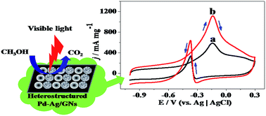
In this work, a facile method was used to synthesize the heterostructured bimetallic catalyst Pd–Ag/graphene (simplified as Pd–Ag/GNs). In order to achieve the heterostructured Pd–Ag nanoparticles (NPs), Ag2O was firstly produced on GNs from AgOH under UV irradiation, then the heterogeneous phase between the precursor Pd2+ and Ag2O contributes to the formation of the highly dispersed heterostructured Pd–Ag NPs with the average diameter of 5 nm on GNs after a synchronous reduction process. Electro-catalytic performances of Pd–Ag/GNs were investigated by cyclic voltammetry (CV), chronoamperometry, COad stripping voltammetry, and electrochemical impedance spectroscopy (EIS). It is shown that Pd–Ag/GNs has a much higher catalytic activity (595 mA mg−1) and stability for methanol oxidation reaction (MOR) and improved tolerance of CO compared with the counterpart Pd/GNs and the commercial 20% Pd/C catalyst (Pd/C-SA). And under visible light irradiation, the catalytic activity and stability of Pd–Ag/GNs are remarkably enhanced. Especially, its mass activity is 1128 mA mg−1 which is 1.9 times higher than that without light irradiation. The significantly improved performance benefits from the efficient electro–photo synergistic catalysis for MOR under visible light irradiation, which is in favor of harvesting sunlight to produce clean energy.
1. Introduction
Direct methanol fuel cells (DMFCs) are attracting much more attention in clean energy technology, due to their high energy density, high efficiency and cleanliness, and easy storage and transportation of liquid methanol at ambient conditions.1–3 Although Pt (Pd)-based catalysts exhibit valuable electro-catalytic activity for methanol oxidation in DMFCs, the expense of Pt (Pd), the poor tolerance toward CO-like intermediate species poisoning and the sluggish electro-catalytic process of methanol oxidation have hampered the scale-up of their use in applications.4–8 Therefore, research has been conducted to solve these problems by combining Pt (Pd)-based catalysts with metal oxides.6,9,10 And it is found that the involvement of metal oxides in catalysts result in higher catalytic activities and better tolerance of CO during methanol oxidation reaction (MOR). Especially, some photo-responsive metal oxides such as TiO2,11,12 SnO2,13 ZnO,14 WO3,15 Cu2O16 etc. were introduced into traditional Pt (Pd)-based catalysts to boost MOR, due to the synergistic effects of photo- and electro-catalysis under external light irradiation. Zhang et al.17 and Wang et al.18 presented the enhanced electro-catalytic performance of Pt–TiO2 based catalysts for MOR under external light illumination. Zhai et al.19 reported that Pt nanoflowers deposited on graphene modified TiO2 nanotube arrays, and the obtained ternary composite was employed as an electro–photo catalyst for MOR in alkaline medium. Li et al.20 reported synthesis of Pt (Pd)/ZnO/graphene composites and its efficient electro–photo synergistic catalysis toward methanol oxidation in the presence of light irradiation.
However, the low electric conductivity of metal oxides is one of the major obstacles to prevent their wide application in electro–photo synergistic catalysis,11,21,22 which can affect the electron transfer efficiency during MOR. Furthermore, metal oxides are easy to dissolve out in electrolyte in the long term catalytic process, leading to the low stability of electro–photo synergistic effect of this kind of catalysts. So it is very desirable to design Pt (Pd)-based bimetallic catalysts for electro–photo synergistic catalysis, due to the good electric conductivity and high stability of pure metallic catalysts.
Among various bimetallic catalysts, Pd–Ag materials are very fascinating.23–26 Pd is more abundant and cheaper than Pt. And Ag is proposed to effectively reduce the poisoning of CO-like species on catalysts' surface and increase the electro-catalytic performance.23,25 Wang et al. have reported that Pd–Ag alloy catalysts exhibited higher activity, enhanced CO tolerance and stability in alkaline direct alcohol fuel cell anodic reaction.27,28 But up to date, rare efforts were focused on Pd–Ag catalysts to explore their potential electro–photo synergistic catalysis for MOR. To the best of our knowledge, only Lin et al. have presented silicon-based Pt–Ag nano-forests catalyst for MOR under external light irradiation.29 However, in this case, electro–photo synergistic catalysis effect was poor, and the methanol oxidation current was boosted by only 6.4% under external light irradiation. So it is very essential to construct novel Pd–Ag nanostructure for achieving a better electro–photo synergistic catalysis effect under external light irradiation.
In the present work, a novel heterostructured Pd–Ag bimetallic catalyst was synthesized with graphene as a support material. The intermediates specie Ag2O plays a key role in the formation of heterostructured bimetallic Pd–Ag nanoparticles (NPs) and results in their high dispersion on the surface of graphene. Pd–Ag NPs/graphene (simplified as Pd–Ag/GNs) not only exhibits higher electro-catalytic performance, but also presents a significant electro–photo synergistic effect under visible light irradiation for MOR. For the first time, efficient electro–photo catalysis has been successfully realized on Pd-based bimetallic catalysts, providing a promising strategy to dispose of lower electric conductivity of noble metal–metal oxide electro–photo catalysis system.
2. Experimental
2.1. Materials
Natural graphite powder (−325 mesh, 99.9995%) was purchased from Alfa Aesar. The commercially available 20% Pd/C catalyst (simplified as Pd/C-SA) and Nafion solution(R) 117 (5%) were purchased from Sigma-Aldrich. Pd(NO3)2 aqueous solution (40%), AgNO3 (99%), sodium hydroxide, potassium permanganate, potassium peroxydisulfate, phosphorus pentoxide, concentrated sulfuric acid (98%), concentrated hydrochloric acid (36%), hydrogen peroxide (30%), formic acid, anhydrous alcohol and anhydrous methanol were all analytical reagents and purchased from Sinopharm Chemical Reagent Co., Ltd. (Shanghai, China).
2.2. Synthesis of Pd–Ag/GNs
Graphite oxide (GO) was synthesized from natural graphite powder by a modified Hummers' method.30–32 And it was purified by dialyzing in deionized water for two week with BIOSHARP membrane (MW: 8000–1400). The overall synthetic route of Pd–Ag/GNs is as depicted in Scheme 1, and its detailed procedure is as follows: 50 mg GO were dispersed into 50 mL deionised water by sonication for 90 min, and 22.5 mg AgNO3 were added. The pH was adjusted to 9.0 by dilute sodium hydroxide solution. Then the mixture was sonicated for 60 min, and irradiated under UV light for 2 h. During irradiation, AgOH was converted to Ag2O on GNs.33 After that, 3 mL of Pd(NO3)2 (1 g/100 mL) were added, and the mixed solution stirred for 30 min. The atomic ratio of Pd
:
Ag for the feeding solution was 1
:
1. The mixture was reduced using 5 mL HCOOH within 72 h. Subsequently, the product was purified by repeated centrifugation (10
000 rpm, 20 min) and deionised water washing cycles. Finally, the black precipitate was lyophilized. Pd/GNs and Ag/GNs were synthesized by the similar procedure without the addition of AgNO3 or Pd(NO3)2. The actual Pd and Ag contents were determined by ICP-AES. The Pd contents for Pd–Ag/GNs and Pd/GNs were 19.11% and 22.16%, respectively. And the Ag contents for Pd–Ag/GNs and Ag/GNs were 20.75% and 23.96% respectively.
 |
| Scheme 1 Synthetic route of heterostructured Pd–Ag/GNs composite. | |
2.3. Characterization
An X'pert Pro X-ray diffractometer (PANalytical, NED) was used to determine X-ray diffraction (XRD) patterns of as-synthesized samples, and Cu-Kalpha radiation source was used, scanning between 5° and 90° at a rate of 5°/min−1. X-ray photoelectron spectroscopy (XPS) was performed on Thermo Scientific VG ESCALAB 250 electron spectrometer. An Al-Kalpha radiation source (1486.6 eV) was used, and the binding energy was referenced to C 1s peak at 284.8 eV. Shirley-type background corrections and Gaussian–Lorentzian peak shapes of 80 ± 10% and 20 ± 10% were used to fit C 1s spectra with the aid of the software “XPSPEAK (Version 4.1)”. Raman spectra were conducted on a Renishaw INVIA Reflex Raman spectrometer with a 514.5 nm diode laser excitation on a 300 lines per mm grating. Transmission electron microscopy (TEM), high-angle annular dark-field scanning transmission electron microscopy (HAADF-STEM) and the element mapping analysis were used for observing the morphologies of samples on a FEI TECNAI G2 electron microscope. Inductively coupled plasma-atomic emission spectrometer (ICP-AES, ICAP6300, Thermo Scientific) was used to determine the actual Pd and Ag contents, and aqua regia was used to dissolve the samples.
2.4. Electrochemical measurements
Electrochemical measurements were carried out at 25 °C in a typical three electrode cell, using CHI 650E electrochemical workstation (ChenHua Instrument, China). Platinum column and Ag|AgCl (saturated KCl) electrode was used as the counter electrode and the reference electrode, respectively. The working electrode was glassy carbon electrode (GCE, 3.0 mm in diameter) pretreated by polishing and rinsing step. Then it was coated with catalysts' suspension prepared by sonicating 5 mg sample in 1 mL ethanol. After drying, 5.0 mL 0.5% Nafion diluted solution was dropped to fix the samples. Cyclic voltammetric (CV) curves for MOR were conducted in 1.0 M KOH solution containing 1.0 M CH3OH, at 100 mV s−1. CO stripping voltammetry was performed by oxidizing preadsorbed CO on the electrode surface, and its detailed procedure is as follows: firstly, 1.0 M KOH solution was purged with N2 for 30 min, then CO was bubbled for 30 min to allow the complete adsorption of CO onto the electrode surface while the potential was kept at 0.1 V. After that, excess CO in electrolyte was purged out with N2 for another 30 min. Electrochemical impedance spectroscopy (EIS) was conducted in 1.0 M KOH solution containing 1.0 M CH3OH under −0.3 V. Typical Nyquist plots were collected in the frequency range between 100 kHz and 0.1 Hz. Chronoamperometric curves were carried on for 2000 s in 1.0 M KOH solution containing 1.0 M CH3OH, at −0.15 V.
2.5. Photo-electrochemical measurements
A quartz window was reserved at the bottom of the electrochemical cell, facing toward the working electrode surface. A 300 W xenon lamp (CEL-HXUV300, Aulight, China) was utilized to provide visible light via tuning the optical filters. The irradiation light penetrates through the quartz window and illuminates the electrode surface. The lamp emits visible light with the wavelength beyond 420 nm. The distance between the GCE and the lamp is 30 cm, and the irradiation intensity is 0.3 mW cm2.
3. Results and discussion
3.1. Characterization
Fig. 1 displays the XRD patterns of Pd/GNs and Pd–Ag/GNs. As shown in curve b of Fig. 1, the four characteristic peaks located at 40.2°, 46.8°, 68.3°, and 82.3° can be observed for Pd/GNs, corresponding to (111), (200), (220), and (311) planes of the face centered cubic (fcc) structure for metallic Pd, which confirms that the formation of Pd(0) NPs after the reduction by HCOOH.34,35 However, the corresponding diffraction peaks is apparently different when Ag was introduced into Pd–Ag/GNs composite (curve c), which is broader and looks like shoulder peaks. In contrast with the peaks for Pd(0) (curve b) & Ag(0) (curve a), it is interesting to found that the four broad peaks are consisting of the diffraction peaks for fcc structure of Pd(0) and Ag(0), respectively, suggesting the formation of heterostructured Pd(0) and Ag(0) in Pd–Ag/GNs composite.23,36 This is also supported by the ICDD PDF cards of Pd (No. 46-1043) & Ag (No. 04-0783) in Fig. 1.34,37 And according to ICDD PDF No. 41-1104, the diffraction peaks of representative (111) and (220) planes for Ag2O should be located at 32.8° and 54.9°, respectively.38 However, there are no corresponding peaks to be observed, suggesting that the intermediate Ag2O was all transformed into Ag after the reduction by formic acid.
 |
| Fig. 1 XRD patterns of Ag/GNs (a), Pd/GNs (b) and Pd–Ag/GNs (c). | |
XPS spectra analysis is employed to characterize the chemical states of different elements in the composites above. As shown in Fig. 2A, the binding energies of Pd 3d for Pd/GNs (curve a) and Pd–Ag/GNs (curve b) are 335.74 & 341.01 eV and 335.62 & 340.89 eV, respectively, corresponding to palladium in the zero-valent state,25,39 which further verifies the formation of Pd(0) NPs in Pd/GNs and Pd–Ag/GNs composites. And the binding energy of Pd 3d for Pd–Ag/GNs presents a slightly negative shift. As depicted in Fig. 2B, the binding energy of Ag 3d5/2 and Ag 3d3/2 for Pd–Ag/GNs is located at 368.37 eV and 374.32 eV, respectively, in line with that of monometallic Ag.34,40 And the peaks related to Ag2O was not observed in Fig. 2B, further suggesting that intermediate Ag2O during synthesis process was completely transformed into Ag after reduction.39 Correspondingly, the binding energy of Ag 3d for Pd–Ag/GNs shifts positively in comparison with the reported value.34,39 The shift of Pd 3d and Ag 3d should be ascribed to the strong electronic interactions between Pd and Ag, indicating the obvious electron transfer between them.25,40 And the changes in the electronic structure affect the d-band center of the Pd, which would further affect the electro-catalytic performance of Pd.23,40
 |
| Fig. 2 Pd 3d XPS profile (A) Pd/GNs (curve a) and Pd–Ag/GNs (curve b); (B) Ag 3d XPS profile of Pd–Ag/GNs; C 1s XPS profile of Pd/GNs (C) and Pd–Ag/GNs (D). | |
The structure change of GNs after reduction can be observed in C 1s XPS profile, as presented in Fig. 2C and D. The C 1s XPS spectra of Pd/GNs and Pd–Ag/GNs can be deconvoluted into three peaks using the software “XPSPEAK (Version 4.1)”. It is found that the dominant peak at approximately 284.6 eV is ascribed to sp2-hybridized C–C bonds, and the other two peaks at about 285.8 and 288.1 eV are assigned to C–O (epoxy and alkoxy) and O–C
O bonds, respectively.34,35,41 The prominent intensity of sp2 hybridized C–C bonds and significantly decreasing intensity of oxygen-containing carbon confirm the reduction of GO into GNs in the Pd/GNs and Pd–Ag/GNs with HCOOH as a reducing agent.41,42 It is interesting to find that the percentage of C–C bonds for Pd–Ag/GNs is 71.12%, which is much higher than that for Pd/GNs (54.64%). This indicates that the introduction of Ag into Pd–Ag/GNs is of great benefit to repairing the sp2 hybridized network of graphene, which also reflects in Raman spectra. As shown in Fig. 3, two characteristic peaks of GNs are observed, namely D band at 1317 cm−1 and G band at 1603 cm−1 in the Raman spectra of Pd/GNs and Pd–Ag/GNs. The D peak for GNs is due to a defect-induced breathing mode of sp2 carbon rings, and the G peak is ascribed to the E2g vibration mode of sp2 bonded carbon atoms.43 The intensity ratio (ID/IG) of D and G bands can be considered as a measure of disorder degree and average size of the sp2 domains, increasing with the amount of disorder for graphitic materials and vanishing for completely defect-free graphite.20,44,45 The ID/IG value of Pd–Ag/GNs can be calculated to be 1.508 which is lower than that of Pd/GNs (1.654), meaning that GNs in Pd–Ag/GNs has fewer defects and contains more sp2 hybridized C–C bonds.
 |
| Fig. 3 Raman spectra of Pd/GNs (curve a) and Pd–Ag/GNs (curve b). | |
The morphologies of Pd–Ag/GNs and Pd/GNs are characterized by TEM and HRTEM. As shown in Fig. 4a and b, it can be found that highly dispersed Pd and Ag NPs with relatively uniform size distribution are deposited on the surface of GNs. By determining the diameter of 100 randomly selected particles from Fig. 4b, it is evaluated statistically that the average diameter for Pd and Ag NPs is 5 nm, as presented in the inset of Fig. 4b. Fig. 4c exhibits the detailed crystal lattice of Pd and Ag for Pd–Ag/GNs. The lattice fringe with an interplanar spacing of 0.228 nm is assigned to (111) planes of face centered cubic (fcc) Pd for Pd–Ag/GNs.16,40 And the lattice fringe with an interplanar spacing of 0.235 nm corresponds to the (111) planes of fcc Ag.37,40 It is easy to find that Pd and Ag NPs are separately deposited on GNs, instead of being formed into Pd–Ag alloy, which is in good agreement with XRD analysis. Furthermore, the selected-area electron diffraction (SAED) pattern of Pd–Ag/GNs displays distinct diffraction rings related to fcc crystalline planes (Fig. 4f), confirming the high crystallinity of Pd (Ag) NPs for Pd–Ag/GNs. HAADF-STEM and elemental mapping of Pd and Ag were used to further investigate the distribution of Pd and Ag NPs on GNs. As shown in Fig. 5a, Pd and Ag NPs are evenly dispersed on the surface of GNs and their morphologies are in line with those in TEM images. And as indicated in the elemental mapping of Pd and Ag (Fig. 5b and c), it is found that Pd and Ag NPs is in good co-existence and intimate contact. For comparison, TEM images of Pd/GNs are shown in Fig. 4d and e. It is found that Pd NPs with the average diameter of 6.5 nm are deposited on the surface of GNs with less uniform distribution.
 |
| Fig. 4 TEM images of Pd–Ag/GNs (a and b) and Pd/GNs (d and e); HRTEM of Pd–Ag/GNs (c); SAED of Pd–Ag/GNs (f); the particle size distributions of Pd–Ag/GNs (the inset of b) and Pd/GNs (the inset of e). | |
 |
| Fig. 5 HAADF-STEM of Pd–Ag/GNs (a); elemental mapping of Pd (b) and Ag (c) for Pd–Ag/GNs. | |
Generally, the simultaneous reduction of Ag and Pd results in the formation of Pd–Ag bimetallic alloy.25,46,47 However, heterostructured bimetallic Ag and Pd NPs are obtained though the simultaneous reduction in this work, which is attributed to the unique synthesis strategy of Pd–Ag/GNs. As shown in Scheme 1, owing to being oxygenated with hydroxyl and epoxide functional groups, graphene oxide sheets possess negative charge which provides even sites for trapping positive Ag+.48 Ag+ is transformed into AgOH when pH was tuned to 9, then AgOH was converted to Ag2O on the surface of GNs under UV irradiation.33,49 After the addition of Pd(NO3)2, Pd and Ag are deposited onto GNs via synchronous reduction of Pd(NO3)2, Ag2O and GO with HCOOH as a reducing agent. During reduction process, solid Ag2O can further provide anchoring sites for palladium ions or particles, in favor of the dispersion of Pd, and the heterogeneous phase between precursor Pd2+ (liquid) and Ag2O (solid) effectively prevents Pd and Ag from forming alloy, resulting in heterostructured bimetallic Pd–Ag NPs on GNs.
3.2. Electro-catalysis
Fig. 6A displays the cyclic voltammetry (CV) curves of Pd/C-SA, Pd/GNs and Pd–Ag/GNs in 1.0 M KOH. All curves show the similar electrochemical features of palladium. The peaks in the range between −1.0 and −0.5 V correspond to hydrogen ad/desorption on Pd surface and the obvious reduction peak of palladium oxide at around −0.4 V is observed on the backward scanning.23,24,26,35 No typical redox couple of Ag is observed in CV curves of Pd–Ag/GNs in 1.0 M KOH, indicating that Ag NPs is not the key component for electro-catalytic property towards methanol oxidation. As for the Pd-based catalysts, it is imprecise to calculate the electrochemical surface areas (ECSA) from the integrated charge of hydrogen ad/desorption peaks because of the absorption of hydrogen in bulk for Pd.24,50 Hence, ECSA of Pd-based catalysts should be measured through the reduction peak of palladium oxide, according to the equation ECSA = Q/(405 mC cm−2 × Pd loading), by assuming that only monolayer of PdO covers on surface and the charge produced from reduction of the monolayer PdO is 405 mC cm−2.24,35,50 It is calculated that ECSA of Pd–Ag/GNs is 72.6 m2 g−1 from curve c in Fig. 6A, which is higher than that of Pd/GNs (60.2 m2 g−1) and Pd/C-SA (25.7 m2 g−1). The value is also higher than that of other recently reported Pd based catalysts or Pd–Ag bimetallic catalysts.16,24,35,50 The highly dispersion of Pd NPs with small size (about 5 nm in TEM analysis) for Pd–Ag/GNs should be responsible for the higher ECSA value, which is very important for improving the practical performance of DMFCs.
 |
| Fig. 6 (A) CV curves in 1.0 M KOH: Pd/C-SA (a), Pd/GNs (b) and Pd–Ag/GNs (c), scan rate: 100 mV s−1; (B) CV curves in 1.0 M KOH + 1.0 M CH3OH: Pd/C-SA (a), Pd/GNs (b), Pd–Ag/GNs (c), scan rate: 100 mV s−1. | |
To investigate the electro-catalytic activities of Pd/C-SA, Pd/GNs and Pd–Ag/GNs for MOR, CV curves in 1.0 M KOH solution containing 1.0 M CH3OH were conducted, as presented in Fig. 6B. Two characteristic oxidation peaks for all curves can be observed: the forward anodic peak of methanol electro-oxidation and the backward scanning peak of the intermediates oxidation. The mass activity (forward peak current density) of Pd–Ag/GNs is 595 mA mg−1 which is higher than that of its counterparts Pd/GNs (442 mA mg−1) and Pd/C-SA (398 mA mg−1). It is also found that the peak potential value for MOR on Pd–Ag/GNs is −0.14 V, which shifts to a lower value than that of Pd/GNs (−0.10 V) and Pd/C-SA (−0.11 V). A lower peak potential suggests more ease oxidation of methanol, contributing to a superior electro-catalytic performance of Pd–Ag/GNs.
Above observations suggest that the introduction of Ag has improved the electro-catalytic activity of Pd nanoparticles for MOR. These enhancements can be attributed to the following reasons: firstly, the introduction of Ag into Pd–Ag/GNs composite results in fewer defects and more sp2-hybridized C–C bonds domains in GNs (as analyzed in C 1s XPS profile and Raman spectra), which is in favor of improving the electronic conductivity of Pd–Ag/GNs composite.35 This can be further supported by EIS spectra analysis of Pd–Ag/GNs, Pd/GNs, and Pd/C-SA, respectively. As shown in Fig. 7A, there exists a semicircle at a higher frequency region on all three electrodes. The semicircle diameters at high frequency have been considered as a measure of charge transfer resistance between molecules in electrolyte and composites at electro-catalytic interface.51,52 Charge transfer resistance is a main parameter to evaluate the inherent speed of the charge-transfer step of an electrode reaction.37 The semicircle diameter of Pd–Ag/GNs (curve c) is much smaller than Pd/GNs (curve b) and Pd/C-SA (curve a), indicating that Pd–Ag/GNs electrode (curve c) has lower charge transfer resistance and its electron-transfer kinetics for MOR are much better facilitated.16,37 Secondly, the intimate contact between Pd and Ag NPs leads to a strong interaction between them as analyzed in Pd 3d XPS profile, which induces the shift of d-band center of Pd.26,35 The shift causes the adsorption ability of the OH anion onto the Pd to become stronger.26,53 Then increasing the adsorption of OHads onto the catalyst surface may facilitate the release of free active catalytic sites and lead to a faster MOR with better intermediates tolerance.23,26,50 Thirdly, the introduction of Ag in the Ag–Pd bimetallic catalyst allows for water activation at a lower potential as compared to that over Pd mono metallic catalyst. The activated water is dissociated to create OHad species and make easily donate O-species to CO to form CO2 during the electro-oxidation of CO.26,50 The improved tolerance of CO can be confirmed by CO stripping voltammetric curves in 1.0 M KOH at 100 mV s−1. As shown in Fig. 7B, the peak potential value for CO oxidation on Pd–Ag/GNs (curve c) is −0.15 V which is lower than that of Pd/GNs (curve b, −0.13 V) and Pd/C-SA (curve a, −0.12 V), indicating more ease oxidation of CO on the surface of Pd–Ag/GNs. So better anti CO-like species poisoning for Pd–Ag/GNs is achieved.
 |
| Fig. 7 (A) Nyquist plots of EIS in 1.0 M KOH + 1.0 M CH3OH: Pd/C-SA (a), Pd/GNs (b) and Pd–Ag/GNs (c), at −0.3 V; (B) CO stripping voltammetric curves in 1.0 M KOH: Pd/C-SA (a), Pd/GNs (b) and Pd–Ag/GNs (c), scan rate 100 mV s−1. | |
3.3. Electro–photo synergistic catalysis
The effect of visible light irradiation on MOR for Pd–Ag/GNs, Pd/GNs and Pd/C-SA was investigated. As shown in Fig. 8A, Pd–Ag/GNs composite exhibits drastically increasing mass activity with the value of 1128 mA mg−1 (curve b in Fig. 8A) under visible light irradiation, which is 1.9 times higher than that without light irradiation (595 mA mg−1). It is worth mentioning that the mass activity and its increasing rate (about 90%) are also significantly higher than that of our reported Pd–Cu2O (ZnO)–GNs catalysts under external light irradiation.16,20 The reasonable interpretation may be that the added photo-responsive Ag possesses better electric conductivity than its counterparts Cu2O or ZnO, resulting in improved electron-transfer kinetics for MOR under light irradiation. However, only slight increase of mass activity for Pd/GNs and Pd/C-SA is observed under visible light irradiation (Fig. 8B and C). The remarkable improvement of catalytic performance for Pd–Ag/GNs apparently stems from the synergistic interaction of photo- and electro-catalysis for MOR.11,12 It is worthy to note that this is the first example that efficient electro–photo synergistic catalysis has been successfully realized on Pd bimetallic catalysts, which is promising to effectively overcome the relatively low electric conductivity of Pd–metal oxide electro–photo catalysts.
 |
| Fig. 8 CV curves under light irradiation in 1.0 M KOH + 1.0 M CH3OH, scan rate 100 mV s−1: Pd–Ag/GNs (A), Pd/GNs (B) and Pd/C-SA (C), curve a: without irradiation, curve b: after visible irradiation for 30 min. | |
To further discuss the synergistic effect between the electro-catalytic and photo-catalytic properties of Pd–Ag/GNs for MOR, chronoamperometric curves under visible light irradiation and without irradiation are conducted for comparison. As displayed in Fig. 9, the current densities of Pd–Ag/GNs, Pd/GNs and Pd/C-SA decay quickly during the initial minute, which mainly arises from the unavoidable formation of Pd oxides and CO-like intermediate species poisoning on the electrode surface during the early stage of MOR.18,52 Following, the current densities diminish slowly and reach at a quasi-stationary value after 2000 s. Without irradiation, Pd–Ag/GNs (curve c) exhibits a slightly slower current density declining rate than Pd/C-SA (curve a) and Pd/GNs (curve b), which indicates that Pd–Ag/GNs possesses better electro-catalytic stability and improved tolerance of CO for MOR due to the existence of Ag species. Especially, under visible light irradiation, it is interesting to find that both initial and quasi-stationary current density for Pd–Ag/GNs increases drastically (curve d in Fig. 9). It can be calculated that the quasi-stationary current density of Pd–Ag/GNs in the presence of visible irradiation is about 3-fold higher than that without irradiation. And the current density decay rate of Pd–Ag/GNs under visible irradiation is much lower than that of Pd–Ag/GNs (curve c), Pd/GNs (curve b) and Pd/C-SA (curve a) without irradiation. In contrast, almost no change in chronoamperometric tests under visible light irradiation is observed for the Pd/GNs and Pd/C-SA (not presented in Fig. 9 for clarity). The significantly lower decay rate of Pd–Ag/GNs under visible irradiation clearly indicates the positive effect of visible light irradiation on the catalytic stability.
 |
| Fig. 9 Chronoamperometric curves in 1.0 M KOH + 1.0 M CH3OH: Pd/C-SA (a), Pd/GNs (b), Pd–Ag/GNs (c), Pd–Ag/GNs under the durative visible irradiation (d). | |
The reproducibility of Pd–Ag/GNs catalyst response to visible light irradiation is confirmed by polarizing the electrode at −0.15 V in repeated on–off cycles under visible light irradiation. It can be seen that the electrode displays a quite sensitive current response to the visible light irradiation (Fig. 10). When visible light is on, the current density increases correspondingly and keeps essential stability; on the contrary, when the light is off, the current density falls back instantly. This phenomenon clearly indicates that the catalytic performance of Pd–Ag/GNs for MOR is improved by visible light irradiation and can be sustained for cycles.12,18 Ag has high free electron mobility and exhibits molecular-like excited-state properties with well-defined absorption and emission features, indicating that Ag is photoresponsive and it can act as electron-donors.54,55 When the external light irradiates on metallic Ag, photo-generated electrons (e−) can produce photo-current, contributing to extra increase current in addition to that deriving from methanol electro-oxidation at Pd active sites.13,14 And GNs in Pd–Ag/GNs can act as efficient electron acceptors and carriers to extract photo-generated electrons from Ag and transfer to Pd particles, resulting in limited self photo-reduction process and more easy recovery of partially oxidized Pd active sites.11,16
 |
| Fig. 10 Chronoamperometric curves of Pd–Ag/GNs in 1.0 M KOH + 1.0 M CH3OH: without irradiation (a), under intermittent visible irradiation (b). | |
It should be noted that Ag NPs in Pd–Ag/GNs play the key role in the visible light assisted synergistic effects of photo- and electro-catalysis.29,56 As shown in HAADF-STEM and elemental mapping, Pd and Ag nanoparticles are in good co-existence and intimate contact, which is beneficial for localized surface plasmon resonance effect (LSPR).56,57 LSPR boosts the catalytic capability of Pd in the hot spot region and extends the spectral response of Pd–Ag/GNs.29,58 So the strong absorption of visible light can be produced to realize the well synergistic effects of photo- and electro-catalysis under visible light irradiation. And the heterostructured pattern may be helpful for the photo-response of Ag, due to that its photoactive property is fully retained in Pd–Ag/GNs. Furthermore, it is reported that the introduction of the graphene nanosheets in catalysts' nanostructure could promote the utilization of the visible-light spectra.19 GNs not only improve the dispersion of Pd NPs and the charge transfer performance, but also promotes the visible light absorption and the electron emission on Pd–Ag/GNs catalyst.18
4. Conclusion
In summary, the heterostructured Pd–Ag/GNs electro–photo catalyst was synthesized by a facile method, in which Ag2O was undergone as intermediate species and HCOOH was used as a reducing agent. The formation of Ag2O during synthesis process results in the heterogeneous phase between Pd2+ and Ag2O, which effectively prevents Pd and Ag from forming alloy and makes heterostructured Pd and Ag NPs in good co-existence and intimate contact. The good electron transfer capacity, the shift of d-band center of Pd and the anti-CO poisoning role of the second metal Ag contribute to a much higher electro-catalytic activity and better stability. Especially, under visible light irradiation, the catalytic activity and stability of Pd–Ag/GNs are remarkably improved due to synergistic effects of photo- and electro-catalysis. The localized surface plasmon resonance effect on Pd–Ag/GNs contributes to efficient visible light assisted electro–photo synergistic catalysis, which provides a new paradigm for harvesting sunlight to develop novel catalysts for DMFCs anodic application.
Acknowledgements
This work was financially supported by the National Natural Science Foundation of China (No. 21171037), Research Foundation of the Education Department of Fujian Province (No. JA13082 & No. JB13010).
Reference
- X. Cui, Y. Zhu, Z. Hua, J. Feng, Z. Liu, L. Chen and J. Shi, Energy Environ. Sci., 2015, 8, 1261–1266 CAS.
- H. Yan, C. Tian, L. Sun, B. Wang, L. Wang, J. Yin, A. Wu and H. Fu, Energy Environ. Sci., 2014, 7, 1939–1949 CAS.
- J. Zhu, M. Xiao, X. Zhao, K. Li, C. Liu and W. Xing, Chem. Commun., 2014, 50, 12201–12203 RSC.
- Y. Kim, Y. Noh, E. J. Lim, S. Lee, S. M. Choi and W. B. Kim, J. Mater. Chem. A, 2014, 2, 6976–6986 CAS.
- X.-L. Sui, Z.-B. Wang, C.-Z. Li, J.-J. Zhang, L. Zhao, D.-M. Gu and S. Gu, J. Mater. Chem. A, 2015, 3, 840–846 CAS.
- Y. Y. Chu, J. Cao, Z. Dai and X. Y. Tan, J. Mater. Chem. A, 2014, 2, 4038–4044 CAS.
- H. Huang, G. Ye, S. Yang, H. Fei, C. S. Tiwary, Y. Gong, R. Vajtai, J. M. Tour, X. Wang and P. M. Ajayan, J. Mater. Chem. A, 2015, 3, 19696–19701 CAS.
- A. Dutta and J. Ouyang, Appl. Catal., B, 2014, 158, 119–128 CrossRef.
- W. Zhuang, L. He, J. Zhu, R. An, X. Wu, L. Mu, X. Lu, L. Lu, X. Liu and H. Ying, Int. J. Hydrogen Energy, 2015, 40, 3679–3688 CrossRef CAS.
- H. Huang, Y. Liu, Q. Gao, W. Ruan, X. Lin and X. Li, ACS Appl. Mater. Interfaces, 2014, 6, 10258–10264 CAS.
- L. Ye, Z. Li, L. Zhang, F. Lei and S. Lin, J. Colloid Interface Sci., 2014, 433, 156–162 CrossRef CAS PubMed.
- W. Li, Y. Bai, F. Li, C. Liu, K.-Y. Chan, X. Feng and X. Lu, J. Mater. Chem., 2012, 22, 4025–4031 RSC.
- F. Lei, Z. Li, L. Ye, Y. Wang and S. Lin, Int. J. Hydrogen Energy, 2016, 41, 255–264 CrossRef CAS.
- C.-Y. Su, Y.-C. Hsueh, C.-C. Kei, C.-T. Lin and T.-P. Perng, J. Phys. Chem. C, 2013, 117, 11610–11618 CAS.
- J. Georgieva, S. Sotiropoulos, E. Valova, S. Armyanov and N. Karanasios, J. Electroanal. Chem., 2014, 727, 135–140 CrossRef CAS.
- L. Ye, Z. Li, X. Zhang, F. Lei and S. Lin, J. Mater. Chem. A, 2014, 2, 21010–21019 CAS.
- H. Zhang, W. Zhou, Y. Du, P. Yang, C. Wang and J. Xu, Int. J. Hydrogen Energy, 2010, 35, 13290–13297 CrossRef CAS.
- C. Wang, F. Jiang, R. Yue, H. Wang and Y. Du, J. Solid State Electrochem., 2014, 18, 515–522 CrossRef CAS.
- C. Zhai, M. Zhu, D. Bin, H. Wang, Y. Du, C. Wang and P. Yang, ACS Appl. Mater. Interfaces, 2014, 6, 17753–17761 CAS.
- Z. Li, L. Ye, F. Lei, Y. Wang, S. Xu and S. Lin, Electrochim. Acta, 2016, 188, 450–460 CrossRef CAS.
- B. Y. Xia, B. Wang, H. B. Wu, Z. Liu, X. Wang and X. W. Lou, J. Mater. Chem., 2012, 22, 16499–16505 RSC.
- X. Wang, X. Li, D. Liu, S. Song and H. Zhang, Chem. Commun., 2012, 48, 2885–2887 RSC.
- Y. Yang, W. Wang, Y. Liu, F. Wang, Z. Zhang and Z. Lei, Int. J. Hydrogen Energy, 2015, 40, 2225–2230 CrossRef CAS.
- G. Li, L. Jiang, Q. Jiang, S. Wang and G. Sun, Electrochim. Acta, 2011, 56, 7703–7711 CrossRef CAS.
- S. Fu, C. Zhu, D. Du and Y. Lin, ACS Appl. Mater. Interfaces, 2015, 7, 13842–13848 CAS.
- L. Xu, Z. Luo, Z. Fan, S. Yu, J. Chen, Y. Liao and C. Xue, Chem.–Eur. J., 2015, 21, 8691–8695 CrossRef CAS PubMed.
- S. T. Nguyen, H. M. Law, H. T. Nguyen, N. Kristian, S. Wang, S. H. Chan and X. Wang, Appl. Catal., B, 2009, 91, 507–515 CrossRef CAS.
- S. T. Nguyen, Y. Yang and X. Wang, Appl. Catal., B, 2012, 113–114, 261–270 CrossRef CAS.
- C.-T. Lin, M.-H. Shiao, M.-N. Chang, N. Chu, Y.-W. Chen, Y.-H. Peng, B.-H. Liao, H. J. Huang, C.-N. Hsiao and F.-G. Tseng, Nanoscale Res. Lett., 2015, 10, 1–8 CrossRef PubMed.
- Q. Zeng, J. Cheng, L. Tang, X. Liu, Y. Liu, J. Li and J. Jiang, Adv. Funct. Mater., 2010, 20, 3366–3372 CrossRef CAS.
- N. I. Kovtyukhova, P. J. Ollivier, B. R. Martin, T. E. Mallouk, S. A. Chizhik, E. V. Buzaneva and A. D. Gorchinskiy, Chem. Mater., 1999, 11, 771–778 CrossRef CAS.
- W. S. Hummers and R. E. Offeman, J. Am. Chem. Soc., 1958, 80, 1339 CrossRef CAS.
- D. Sarkar, C. K. Ghosh, S. Mukherjee and K. K. Chattopadhyay, ACS Appl. Mater. Interfaces, 2013, 5, 331–337 CAS.
- M. Liu, Y. Lu and W. Chen, Adv. Funct. Mater., 2013, 23, 1289–1296 CrossRef CAS.
- L. Zhang, H. Wang, X. Li, F. Xia, Y. Liu, X. Xu, J. Gao and F. Xing, Electrochim. Acta, 2015, 172, 42–51 CrossRef CAS.
- S. Ma, J. Xue, Y. Zhou and Z. Zhang, J. Mater. Chem. A, 2014, 2, 7272–7280 CAS.
- Y. Lu and W. Chen, J. Phys. Chem. C, 2010, 114, 21190–21200 CAS.
- G. Wang, X. Ma, B. Huang, H. Cheng, Z. Wang, J. Zhan, X. Qin, X. Zhang and Y. Dai, J. Mater. Chem., 2012, 22, 21189–21194 RSC.
- C.-L. Lee, H.-P. Chiou, C.-M. Syu, C.-R. Liu, C.-C. Yang and C.-C. Syu, Int. J. Hydrogen Energy, 2011, 36, 12706–12714 CrossRef CAS.
- J. Huang, S. Vongehr, S. Tang and X. Meng, J. Phys. Chem. C, 2010, 114, 15005–15010 CAS.
- Z. Li, X. Huang, X. Zhang, L. Zhang and S. Lin, J. Mater. Chem., 2012, 22, 23602–23607 RSC.
- D. S. Yu and L. M. Dai, J. Phys. Chem. Lett., 2010, 1, 467–470 CrossRef CAS.
- Z. Li, F. Lei, L. Ye, X. Zhang and S. Lin, J. Nanopart. Res., 2015, 17, 192–202 CrossRef.
- M. Zhang, J. Xie, Q. Sun, Z. Yan, M. Chen and J. Jing, Int. J. Hydrogen Energy, 2013, 38, 16402–16409 CrossRef CAS.
- X. Zhong, Z. Wang, Y. Huang, Y. Yu, Q. Feng and Q. Li, Int. J. Hydrogen Energy, 2014, 39, 15920–15927 CrossRef CAS.
- A. Safavi, H. Kazemi, S. Momeni, M. Tohidi and P. K. Mehrin, Int. J. Hydrogen Energy, 2013, 38, 3380–3386 CrossRef CAS.
- C.-L. Lee, H.-P. Chiou, K.-C. Chang and C.-H. Huang, Int. J. Hydrogen Energy, 2011, 36, 2759–2764 CrossRef CAS.
- C. Chen, L. Wang, Y. Liu, Z. Chen, D. Pan, Z. Li, Z. Jiao, P. Hu, C.-H. Shek, C. M. L. Wu, J. K. L. Lai and M. Wu, Langmuir, 2013, 29, 4111–4118 CrossRef CAS PubMed.
- Z. Ji, X. Shen, J. Yang, Y. Xu, G. Zhu and K. Chen, Eur. J. Inorg. Chem., 2013, 2013, 6119–6125 CrossRef CAS.
- K. Kakaei and M. Dorraji, Electrochim. Acta, 2014, 143, 207–215 CrossRef CAS.
- Y. Feng, N. Feng, Y. Wei and G. Zhang, RSC Adv., 2014, 4, 7933–7943 RSC.
- Z. Li, L. Zhang, X. Huang, L. Ye and S. Lin, Electrochim. Acta, 2014, 121, 215–222 CrossRef CAS.
- Y. Wang, Z. M. Sheng, H. Yang, S. P. Jiang and C. M. Li, Int. J. Hydrogen Energy, 2010, 35, 10087–10093 CrossRef CAS.
- M. J. Kale, T. Avanesian and P. Christopher, ACS Catal., 2014, 4, 116–128 CrossRef CAS.
- W. Hou and S. B. Cronin, Adv. Funct. Mater., 2013, 23, 1612–1619 CrossRef CAS.
- S. Wang, H. Qian, Y. Hu, W. Dai, Y. Zhong, J. Chen and X. Hu, Dalton Trans., 2013, 42, 1122–1128 RSC.
- H. Eom, J.-Y. Jung, Y. Shin, S. Kim, J.-H. Choi, E. Lee, J.-H. Jeong and I. Park, Nanoscale, 2014, 6, 226–234 RSC.
- L. Zhang, L. Chen, L. Chen and G. Zhu, RSC Adv., 2014, 4, 54463–54468 RSC.
Footnote |
† Indicates equal contributors. |
|
This journal is © The Royal Society of Chemistry 2016 |
Click here to see how this site uses Cookies. View our privacy policy here.