Shell thickness effects on reconfiguration of NiOcore–Ptshell anodic catalysts in a high current density direct methanol fuel cell

Tsan-Yao Chen*ab, Po-Chun Huanga, Yen-Fa Liaoc, Yu-Ting Liud, Tsung-Kuang Yeha and Tsang-Lang Lin*a
aDepartment of Engineering and System Science, National Tsing Hua University, Hsinchu 30013, Taiwan. E-mail: chencaeser@gmail.com; tllin@mx.nthu.edu.tw; Tel: +886-3-5715131 ext. 34271
bInstitute of Nuclear Engineering and Science, National Tsing Hua University, Hsinchu 30013, Taiwan
cNational Synchrotron Radiation Research Center, Hsinchu 30013, Taiwan
dDepartment of Soil and Environmental Science, National Chung Hsing University, Taichung 402, Taiwan

Received 2nd July 2016 , Accepted 22nd July 2016

First published on 25th July 2016


Abstract

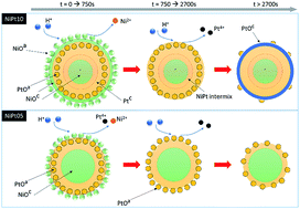
The atomic structure relocation of a NiOcore–Ptshell nanocatalyst (NC) in direct methanol fuel cells for a long-term work cycle is quantitatively investigated by performing both X-ray absorption spectroscopy and electrochemical potential sweeping analyses. We found that Pt atoms in this metal-to-metal oxide junction NC tend to dissolute on reducing the domain size in a long-term DMFC work cycle in the condition of Pt > 30 at%. On the other hand, with Pt < 30%, Pt atoms are rearranged into a thin homoatomic shell at the NiO surface. Such an atomic reconfiguration is controlled by competition between dissolution and relocation of Pt/Ni atoms at varying facets. For the optimum case (Pt < 30%), the current density and the output power density of DMFC are doubled by using NiO–Ptshell NC at the anode as compared to that using a PtRu alloy.


Introduction

The atomic structure stability, fabrication cost, and chemical activity of a nanocatalyst (NC) in methanol oxidation reaction (MOR) are important indexes of the development of reliable and cost-effective direct methanol fuel cells (DMFC) particularly in high current density sectors. These indexes can be manipulated by modifying the surface structure of NC or changing the heteroatomic intermixture.1 In addition to these “intra-particle” structure parameters, build-ups and chemical identity of heterogeneous support also play important roles in electrochemical properties of NC.2 A change in the structure and geometric configuration in atomic scale is believed to be a promising approach for developing chemical durable NC.3 Among existing intra-particle configurations (alloy, cluster-in-cluster, jaunts, onion, try-layer, core–shell, and etc.), the core–shell structure of NC could induce the strongest structure stabilization in electrochemical reactions. Likewise, the crystal junction with core–shell structure has a simple configuration for programming heteroatomic intermix and lattice strain in surface region, thus optimizing the redox activity with minimum noble metal loading in NC.

For electrochemical applications, NC comprising Pt atoms are preferred due to its good performance and reliability in low temperature conditions for state-of-the-art devices.4 The relocation of Pt atoms into homoatomic cluster is an intrinsic manner in thermodynamic nature.5 This behaviour reduces the surface dangling atoms, which is a deciding factor in the stability of NC in chemical devices. In these studies, the stabilization of NC is conducted by incorporating high electronegative atoms (for example, Pt or Au)6 in top of Pd@Co, Ru@Pt, or Cu@Pd NCs.7 These atoms forms strong metallic bonding as rivets tightening the surface structure of NC. To be concrete, when we utilize metal-to-metal oxide junction with high chemical corrosion resistance (specific to Ru and Pd) in the core region, the stability of core–shell NC in low temperature fuel cells is significantly improved.6,8 However, there is a trade-off between fabrication cost and device performance particularly in presence of poor interface coherence in core–shell without proper treatment. Consequently, finding core crystal with relatively acceptable activity and chemical durability is a crucial issue in future fuel cell development.

A correlation between atomic structure evolution and electrochemical properties in a long-term work cycle test is an important index for developing chemical durable NC in DMFC. Our previous studies have shown that the stability of NC in long-term work cycle of DMFC can be optimized by adding monolayers of electrooxidation active atom (Pt) in the top of metal oxide crystallite.9 In this research, we utilized nickel oxide NC as core crystal underneath the Pt and Ni mix-oxide shell crystal. For the optimum case, NiO@Pt NC has double power and current density for a DMFC module as compared to that using PtRu alloy in anode. The effects of shell crystal thickness on the structure durability for current NC in long-term MOR are quantitatively examined through a spectroscopy analysis. Details of the heteroatomic restructure of NC in relation to the thickness of shell crystal are given in the following sections. The structure evolution results of PtRu alloy NC in the same work cycle test in DMFC are compared as a reference.

Experiment

The nanostructure of NC is controlled using the sequential designed polyol reduction.10 To provide proper structure information in explaining the impacts of Pt shell thickness on electrochemical properties of experimental NCs, systematic characterizations from micrometre to electronic regimes are performed. The MOR activity and chemical durability of NC in long-term DMFC work cycle are determined by electrochemical analyses.

Materials

Hexachloride platinum(VI) acid (H2PtCl6·6H2O, 99 at%), nickel(II) chloride (NiCl2, 99 at%), polyvinylpyrrolidone stabilizer (PVP-40, MW ∼42[thin space (1/6-em)]000, 99 wt%), H2SO4 (99.9 wt%), and HNO3 (99.9 wt%) are obtained from Sigma-Aldrich. Ruthenium(III) chloride hydrate (RuCl3·3H2O, 99 at%) is obtained from Strem. The synthesis and surface modification of multi-walled carbon nanotube on carbon fibre (MWCNT) are conducted by a bias assisted microwave plasma enhanced chemical vapour deposition system. The surface modification of MWCNT is performed by two steps of acid treatment followed by ligandization of 2-mercaptoethanol, where details of the experimental procedures and parameters can be referred to our previous works.11

Catalyst preparation, deposition, and DMFC assembly

The NiOcore–Ptshell NC (in a Pt/Ni ratio of 0.5 “NiPt05” with 33 at% of Pt and 1.0 “NiPt10” with 50 at% of Pt) is synthesized by a two-step polyol method with a control of reaction sequences. In the first step, fcc Ni core with an average diameter of ∼2.5 nm are prepared by reducing 10 mM of NiCl2 in EG with the thermal treatments shown in the previous work.12 Second, the Pt shell with different thickness is grown based on a heterogeneous nucleation and growth pathway by reducing Pt anions in top of the Ni core in presence of 10 wt% polyvinylpyrrolidone (PVP) stabilizer in EG at 160 °C for 2 h. In this step, the shell thickness was controlled by adjusting the Pt/Ni ratios. The PtRu alloy NP was grown by reducing the mixture of Pt and Ru anions (Pt = 65 at%) in the presence of PVP stabilizer in EG at 160 °C for 2 h. The dispersion of NC was conducted by immersing the MWCNT/CF electrode into the ethanol solution which contains polymer blended NC (metal content = ∼120 mg mL−3) at room temperature for 15 min and then dried in air for 30 min. Four immersion cycles were conducted for loading comparable metal to that of commercial electrode (∼1.0 mg cm−2). For reinforcing the interface between NC and CNT electrode, the sample was annealed in air at 330 °C for 30 min. The method for installing DMFC single cell is given in the previous works.11 After installation, the catalyst and the proton exchange membrane inside the cell is activated by flowing with 0.5 M of H2SO4 solution for 24 h. The cell is connected to the 850e station with fuel supply prior to the long-term work cycle test.

Nanocatalyst characterizations and DMFC module long-cycle test

The microstructures of NC are illustrated by high-resolution transmission electron microscopy (HRTEM) at an acceleration voltage of 200 kV. The facility (FEI E.O Tecnai F20 G2 Field-Emission TEM) is located at center of electron microscopy, National Sun Yat-sen University, Kaohsiung, Taiwan, R.O.C. Atomic structure of NC are characterized by X-ray absorption spectroscopy at beamlines of BL-07A, BL-17C at NSRRC (Taiwan). For XRD analysis, the incident X-ray wavelength is 0.693 Å (17.89 keV) and the patterns are measured by a 2D area detector (Rayonix MX225) at BL-12B2 at SPring-8 (Japan). The structure parameters are obtained by fitting the diffraction peaks with Lorentz peak functions. Considering to the consistency of crystal growth rate in an absence of protection agents at crystal facets, the FWHM of peak from assigned crystal phase are constrained in a variation less than 5%. The electrochemical surface area (ECSA) and long-term stability of NC in methanol oxidation reaction (MOR) are determined by cycling voltammetry (CV) analysis (CH Instruments Model 600B). The electrochemical cell for CV analysis (with a potential sweeping rate of 20 mV s−1) includes a working electrode, an Ag/AgCl reference electrode, and a 10 mm × 10 mm platinum foil counter electrode. For measuring ECSA, the CV test was conducted in the electrolyte of 0.5 M H2SO4(aq). In long-term MOR, the electrolyte is mixture of 0.5 M methanol and H2SO4(aq). During the DMFC measurements, the cell is heated to 70 °C and is fed with 1.0 M of methanol (MeOH) and H2SO4 solution with a flux of 60 mL h−1 at anode and the 100 sccm of oxygen flow at cathode. The flow liquid and gas are kept at identical temperature to the DMFC cell. For the work cycle test, we repeatedly collect the polarization curves in uniform parameters. There are 400 duty cycles in total for DMFC cells. To minimize the effects of severe hydration on cathode performance, the cycle period is 100 and the cell is baked for 12 hours at 70 °C at the check points (arrows in Fig. 7).

Results and discussion

Effects of Pt shell thickness on crystal structure of NiOcore–Ptshell NC

Results of HRTEM and XRD analysis are performed to determine crystal structure, particle size, and surface morphologies of the experimental NC. Fig. 1 shows the HRTEM images of PtRu (a), PtNi05 (b), and PtNi10 (c) on CNT support. As indicated in Fig. 1a, the PtRu NC are grown in hexagonal shape, showing the typical atomic stacking of fcc crystallite (d(111)_Pt = 2.232 Å) constrained by the stabilization effects of pyrrolidone ligands at the {110} facets.13 The Ru atoms are mostly located at the interfaceted regions (see rectangles in red) with a large extent of roughness at NC in reducing the free surface energy of the system.5,8a For NiPt05, the particles are grown in two groups in the average size (Davg) of 1.5 and 4.1 nm, respectively. In the large particle, the lattice space is 2.241 Å. This value of lattice space is ∼1.3% smaller than that of Pt (111) facet, implying the formation of local PtNi alloying. On the other hand, the lattice space in small particles is 2.087 Å, similar to the lattice space of (200) facet for NiO (2.051 Å), where the chemical state of Ni is complementary confirmed by Ni K-edge XAS analysis (Fig. S4). The lattice expansion (1.71%) implies the slight Pt alloy in Ni oxide NC. The presence of two Davg groups accounted for the dramatic crystal growth rate at Ni facets driven by an initial heterogeneous nucleation and crystal growth (i.e., layer-plus-island Stranski–Krastanov growth mode) and a subsequent layer-by-layer crystal growth at core crystal with full Pt shell coverage (Frank–van der Merve growth mode). This phenomenon invokes a local depletion of Pt anions in the reaction system and thus grows a certain amount of NC with incomplete shell coverage. By increasing Pt/Ni to 1.0 (PtNi10), the Davg for the large particle group increases to 5.1 nm. In comparison to NiPt05, the difference between Davg of two groups is reduced by ∼34% (from 2.6 to 1.5 nm), accounting for the uneven growth rates of Pt allocation at Ni and Pt facets. The lattice space is the same with that of Pt (111) facet, namely 2.262 Å. This indicated the absence of interface confinement in the growth of Pt shell crystal on Ni core surface. The formation of semicoherent interface in twin boundaries (denoted by the yellow dashed line) with large angle (θ = 72°) directly evidences the substantial loss of substrate confinement in the shell crystal growth. Such a feature is further confirmed by the performance of NC in the long-term MOR and the duty cycle of DMFC module as will be discussed later.
image file: c6ra17013g-f1.tif
Fig. 1 High resolution transmission electron microscopy images of (a) PtRu, (b) NiPt05, and (c) NiPt10 supported by multi-walled carbon nanotube.

Fig. 2 compares the XRD patterns with the fitting curves of PtRu (a), NiPt05 (b), and NiPt10 NC (c), where structural parameters are summarized in Table 1. In Fig. 2a, the three peaks A, B, and C at 17.687, 20.225, and 28.978° denote the diffraction lines of (111), (200), and (220) facets of Pt crystallite with the lattice space d(hkl)_Pt (coherent length, D(hkl)_Pt) of 2.256 Å (32.1 Å), 1.955 Å (27.9 Å), and 1.378 Å (36.9 Å), respectively. The lattice constant is 3.907 Å which is slightly compressed (0.736%) from that of standard Pt metal crystal (3.92 Å) due to the heteroatomic intermixture between Ru and Pt atom. The anisotropic growth of NC is noticed between the three facets. This phenomenon can be explained by minimization of surface free energy (γ) in a presence of steric effect by PVP ligand chelation upon the NC growth.13 In crystal with fcc phase, it is expectable to find the preferential growth of (111) due to the lowest γ (1.004 eV) among the three facets. On the other hand, preferential growth of (220) facet is resulted from steric effect of pyrrolidone ligands which substantially reducing γ of the chelated facets.13


image file: c6ra17013g-f2.tif
Fig. 2 XRD patterns of PtRu alloy (a), NiPt05 (b), and NiPt10 (c) NCs. The X-ray wavelength is 0.693 Å (17.89 keV).
Table 1 XRD determined structure parameters of PtRu, NiPt05, and NiPt10 NCs
NC index Δ2θ red 2θ D (Å) d (Å)
PtRu Pt(111) 1.121 17.68 32.1 2.256
Pt (200) 1.289 20.22 27.9 1.955
Pt (220) 0.993 28.98 36.9 1.378
NiPt05 Pt (111) 1.266 17.67 29.6 2.242
NiO (200) 1.693 18.97 22.2 2.091
Pt (200) 1.733 20.18 21.7 1.965
Pt (220) 1.214 28.78 31.5 1.386
NiPt10 Pt (111) 0.655 17.53 57.2 2.261
NiO (200) 1.218 18.79 30.8 2.112
Pt (200) 0.791 20.17 47.6 1.967
Pt (220) 0.729 28.80 52.5 1.385


In XRD pattern of PtNi05 (Fig. 2b), the diffraction peaks of Pt fcc facets (A′, B′, and C′) are shifted to different sites as compared to that of PtRu. Accordingly, d(111)_Pt is compressed by 0.62% (0.014 Å) while the lattice expansion in similar value is found on (200) and (220) facets in comparison to that of PtRu. Such an uneven lattice strain implies the preferential allocation of Ni in Pt (111) facet and the displacement of Pt atoms at (200)/(220) facets when they are allocated at facets with widened interatomic distance between open sites (NiO (200) facet). In this case, the oxidation induced Pt segregation from high index facets could be the reason for the severe Pt corrosion in long-term MOR in subsequent sections. The D(200)_NiO is 22.21 Å which consistently proved the finding of small particle group in Fig. 1b. With segregation and the insufficient supply of Pt anions, a severe anisotropic growth between (111) and (200) facets is found comparing to that on PtRu. In addition, the smeared diffraction peak across (111) and (200) facets shows the typical feature of core–shell type nanocrystal with Pt in shell region.8a

By increasing Pt/Ni to 1.0, the five peaks A*, B*, C*, and D* of PtNi10 (Fig. 2c) refer to diffraction lines of (111), (200), (220) facets for Pt metal, and (200) facet for NiO with the lattice spaces of 2.261, 1.967, 1.385, and 2.000, respectively. Accordingly, the anisotropic lattice strain is absence and the lattice constant of Pt fcc phase is 3.914 Å. Compared to that of NiPt05, the lattice compression is reduced by 0.782% (from 1.329 to 0.547%) which suggesting the lattice relaxation by formation of semicoherent/incoherent interfaces. The coherent lengths for the Pt (111), (200), and (220) facets are 57.284, 47.617, 52.518 Å depicting the reduced anisotropic crystal growth (also explained by HRTEM observation in Fig. 1c). The coherent length of NiO (200) is 30.86 Å and is accounted for the dissolution of metallic Ni into Ni2+ by interacting with Pt4+ (galvanic replacement) followed by the segregation and oxidation of Ni2+ into NiO (which is further confirmed by Ni K-edge XAS analysis in Fig. S4).

XPS spectra of experimental NCs are compared in Fig. 3, where the five emission peaks correspond to signals from Ni(OH)2/NiO(II) 3p (67.5 eV), Pt(0) 4f7/2 (71.2 eV), Pt(0) 4f5/2 (74.4 eV), doublet peaks of Pt(II) (peak A: 4f5/2 at 72.1 eV and peak A* 4f7/2 at 75.6 eV), and doublet peaks of Pt(IV) (peak A*: 4f7/2 at 77.4 eV and corresponding 4f5/2 as a background emission underneath Pt(0) 4f5/2 at 74.4–74.6 eV). The emission peaks are shifted to high binding energy side and the Pt(0) 4f5/2 peak is lifted by ∼5–7% by reducing Pt content to ∼33% (NiPt05). These phenomena are accounted for the backgrounds from elastic photoelectron scattering and the interferences of Ni(II) and Pt(II) oxide emission singles (i.e., the higher extent of Pt oxidation as indicated by the doublet peaks of PtO2 in A and A*) in NiPt05 as compared to NiPt10. In this event, the broadened Ni(II) 3p peak intensity suggests the varying metallic species by intermixture between Ni and Pt atoms in NiPt10 and is attributed to the galvanic replacement between Ni and Pt4+ ions upon growing Pt shell crystal. These characteristics feature the formation of thin (or incomplete) Pt shell with slight Ni oxide/Pt(OH)2 segregation in NC surface and are consistently explained by results of Fig. 4.


image file: c6ra17013g-f3.tif
Fig. 3 XPS spectra of NiPt05 and NiPt10 NCs.

image file: c6ra17013g-f4.tif
Fig. 4 (a) Chronoamperometric curves (measured in a mixture of 0.5 M methanol and H2SO4(aq)) of experimental NC in long-term MOR and CV sweeping curves of (b) PtRu, (c) NiPt05, and (d) NiPt10 (measured in 0.5 M H2SO4(aq) solution and at a potential sweeping rate of 20 mV s−1) before and after long-term MOR.

Effects of shell thickness on surface electrochemical performances of Nicore–Ptshell NC

The results of XRD and HRTEM reveal that the lattice strain is dominated by the extent of heteroatomic intermix upon changing the shell thicknesses on the NiO core crystal. From this understandings, proper control is adopted to the arrangement of active atoms in optimizing the surface activity and noble metal utility of NC.

The electrochemical characterizations provide direct implication for the effects of Pt shell thickness on the structure configurations and the subsequent surface atomic restructure of NC in the long-term methanol oxidation reaction (MOR). The structure durability of NC against MOR is revealed by a serious of half-cell electrochemical analyses including chronoamperometric analysis (CA) on chemical durability test and cyclic voltammetry (CV) analysis on electrochemical surface area (ECSA) test before and after the long-term MOR.

The CA curves of the MWCNT supported PtRu, NiPt05 and NiPt10 NCs for MOR at 0.5 volt (vs. Ag/AgCl) are shown in Fig. 4a. As can be seen, the initial activity for NiPt05 is 0.2 A mg−1 and for NiPt10 is 0.32 A mg−1. These results are ∼70% and ∼170% higher than that of the PtRu (0.117 A mg−1) suggesting the possibility of high output current density of NiO@Pt NCs.

In region A, the MOR activity of PtRu is exponentially decayed by ∼80% in the first 750 s and could be rationalized by the strong chemisorption of hydroxide ligands (which leading to a thick electrostatic double layer (EDL) as indicated by the large current density difference (ΔI) between the forward and backward sweeping curve at a fixed voltage range) at NC. The EDL hindered the subsequent sorption of MeOH molecules at NC surface thus reducing the MOR current and was proved by the electrochemical surface analysis (ECSA) in later sections.14 For PtRu, the Ru atoms tend to intercalate at the near surface region. Compared to NiPt (which comprising weak hydrophilic atoms), the Ru atoms leaves a high density of hydrophilic sites and thus a thicker EDL is expected because of this. Once the competition chemisorption kinetics between MeOH molecule and OH ligand reached a metastable balance (denoted by the arrows O), the reaction activity (current density in region B) is dominated by the atomic structure and the crystal facets. After reaction time passed the O node, a significant current vibration on the AC curve is noticed between arrows O and O*. This feature, which was consistently proved by results of CV sweeping (Fig. 4b) and XAS analysis in later sections, indicating the atomic restructure between Pt and Ru atoms at interfaceted regions of NC. For NiPt, the MOR activities were exponentially decayed by larger than −80% in the first 15 min (region A). Aside from EDL formation, the substantial current decay could be attributed to the dissolution of Ni oxide followed by the Pt restructure at NC surface. These characters were further confirmed by CV spectra for samples with long-term MOR (Fig. 3c and d) measurement. Compared to PtRu, the long-term activity of NiPt05 is improved by 110% suggesting it's higher specific surface sites for MOR reaction.

Fig. 4b–d compared the CV curves of electrodes coated by PtRu, NiPt05, and NiPt10 NC in an aqueous solution of 0.5 M H2SO4, respectively. In general, both CV curves show the typical voltammetric behaviour of hydro-reacted species (Pt) in acidic media and three regions including the under-potential deposition (UPD) of hydrogen in region A, the formation of EDL by the OH ligand chemisorption in region B, and the reduction of chemisorbed oxygen at surface adatoms (at an onset potential of Eoii at forward scan) as well as the reduction of alpha Pt oxide (at a peak potential of Ed at backward scan) in region C were qualitatively elucidated on these CV curves. In these figures, the solid sphere and solid lines denote the CV curves of the two NCs with and without experiencing the long-term MOR; where the corresponding I vs. t curves (collected at 0.5 V (vs. Ag/AgCl) for 2 h) are given in Fig. 4a, respectively. For the NC without MOR, at a potential negative to approximately ∼0.3 V (region A), the reversible proton adsorption (c1, c2) and desorption (a1, a2) responses are clearly presented on the negative and positive sweeps, respectively. The area and features of UPD region determined the ECSA (see below) and the active facets to the specific reactants in the electrolyte.14 For PtRu, the position (Va1 and Va2) and intensity (Ha1 and Ha2) in Fig. 4b for the two characteristic peaks (a1 and a2) depict the applied potential and reactivity for the weakly and strongly adsorbed H+ that dissociate from the close packed and opened facets, respectively. The profiles in UPD region illustrated the responses of the low index facets on Pt fcc crystallite for H+ redox reactions. In this region, the symmetry responses in positive and negative potential sweeps suggested that the PtRu was grown in heteroatomic alloyed crystallite with the ratio of Pt/Ru = 0.45 to 0.55.15 The current difference (ΔI) of PtRu at a fixed applied voltage was determined to be ∼1.3 mA cm−2. This value was quadrupled to that of NiPt05 (ΔI′) and NiPt10 (ΔI*) proving the strong competition of surface reaction sites between OH ligand and MeOH molecules and thus the suppression of current density in long-term MOR test. This phenomenon was complimentary rationalized by the smearing effects in UPD region after long-term MOR (PtRu-a in Fig. 4b). The smearing of UPD features resembled the suppression of H+ redox reaction at Pt fcc facets by the surface passivation by OH that dramatically suppressed the current density of PtRu on AC curve. Such feature depicted the surface segregation/de-alloying of hydrophilic atoms (i.e., Ru in this study) as consistently revealed by local structure analysis (Fig. 5a).6,16 The changes of the CV features are important indication for the surface chemical environments so as to the structure stability and activity of NC for MOR. For PtRu, the increasing currents indicate the formation of Pt hydroxide on positive sweep at the applied potentials higher than ∼0.1 V vs. Ag/AgCl. This potential referred to the onset (Eoi) of Ru assisted OH chemisorption which kept increasing with the applied voltage in the positive sweep. At ∼0.3 V vs. Ag/AgCl, the applied potential is higher than that of OH diffusion barrier at heterogeneous interface. It leads to collisions between chemisorbed OH followed by the formation of Pt oxide and dissociation of H2 at ∼0.9 V vs. Ag/AgCl (the second onset Eoii) in the positive sweep. In the backward sweep, a broad peak at ∼0.5 V vs. Ag/AgCl (Ebd) resembled the reduction current for amorphous metal oxide with complicated conformations (PtOx, RuOx, or PtRuO) on the PtRu.17 This peak was shifted by −0.12 V to Ead at (0.38 V vs. Ag/AgCl) with a slight decrease of current density suggesting the increased energy barrier for the oxide reduction. Such phenomenon explained the decrement of amorphous oxides (which taking lower energy for reduction compared to that of crystallite PtO2) as a result of the surface polishing by protonation corrosion.


image file: c6ra17013g-f5.tif
Fig. 5 Fourier transformed Pt L3-edge EXAFS spectra of PtRu, NiPt05, and NiPt10 NCs w/ and w/o experiencing the long-term work cycle of DMFC.

For NiPt05 and NiPt10, the asymmetry peaks in region A indicated the coexistence of current responses from metallic Pt (a1′, a2′ and c2′) and NiO (c1′) facets. The strong peak c1′ implied to the responses of oxygen reduction from NiO at the applied voltage higher than 1.1 volt vs. Ag/AgCl in the negative sweeping curve. This character disclosed the substantial higher kinetics of oxygen reduction compared to that of hydrogen which were caused by the dissolution of NiO crystal and surface Ni oxide residual at NiPt NC. In region B, the narrowed current intensity differences (ΔI′ and ΔI*) suggesting the suppressed EDL due to the prevailing amorphous metal oxide at NC surface. Compared to that of PtRu, the upshift of OH chemisorption potential (Eoi) complementary with the narrowed EDL revealed the higher energy barrier for OH chemisorption NiPt surface due to the oxide segregation. Consequently, the onset energies for OH diffusion at metal oxide surface and the subsequent oxide formation by inter-OH collisions were substantially reduced (as probed by the downshift of Eoii). In the negative sweep, the reduction potential for Pt oxide was determined to be 0.43 V vs. Ag/AgCl image file: c6ra17013g-t1.tif. Compared to that of PtRu, this value is reduced by 0.08 V suggesting the reducing of energy barrier for metal oxide and is expectable given that the NiO segregation was adjacent to Pt crystal with severe lattice mismatch (i.e., weak substrate confinement effects).3c,5 Significant modifications (with the suppression of intensity and upshift of position on C1′ peak) were found on UPD region indications for the protonation corrosion of Ni oxides on NiPt05 after long-term MOR. In this circumstance, the further upshift (downshift) of Eoi (Eoii) potential and the narrowing of ΔI featured the hydrophobic surface of NC with negative charge dipole exposing to electrolyte. It is important to note that the image file: c6ra17013g-t2.tif and image file: c6ra17013g-t3.tif were determined to be the same position indicating the identical oxide reduction pathways on NC in long-term MOR. It evidenced the strong resistance of protonation corrosion on Pt shell crystal by lattice confinement at interface adjacent to core crystal at NiPt05. Fig. 4d compared the CV sweeping curves of NiPt10 w/ and w/o exposing to MOR for 2 h. Compared to that of NiPt05, NiPt10 possessed the similar features in UPD (region A) and EDL (region B) therefore the surface chemical components for redox reactions of H+ and the OH ligand at NC before long-term MOR. On the other hand, the voltage difference between image file: c6ra17013g-t4.tif and image file: c6ra17013g-t5.tif was further tightened resembling the raising energy barriers with the Pt content at NC surface. The identical position and width of image file: c6ra17013g-t6.tif to image file: c6ra17013g-t7.tif illustrated the same form of Pt oxide at pristine NiPt10 with NiPt05. After long-term MOR, the peak position for oxide reduction was back shifted by 1.0 volt (from image file: c6ra17013g-t8.tif to image file: c6ra17013g-t9.tif) coincide to the increasing energy barrier for Pt oxide decomposition. This indicates the response of oxygen desorption at Pt sites as complementary shown by the symmetry peak X at positive sweeping curve.

The local atomic structure evolution of PtRu, NiPt05, and NiPt10 after long-term MOR were depicted by Fourier transformed extended X-ray absorption fine structure (FT-EXAFS) spectra and the obtained structure parameters were summarized in Table 2. In Fig. 5a, peak A refer to the interferences of metallic Pt–Pt or Pt–Ru bond pairs in PtRu alloy; where the position and intensity refer to the inter-atomic distance and coordination numbers, respectively. As clearly illustrates, the intensity of peak A for PtRu before long-term MOR (PtRu-b) was determined to be 15.4 (a.u.) corresponding to the coordination number for Pt–Pt (CNPt–Pt) and Pt–Ru (CNPt–Ru) bond pair of 8.21 and 0.39, respectively. In this case, the extent of Ru alloy in Pt region (χRu–Pt = CNPt–Ru/(CNPt–Pt + CNPt–Ru)) is determined to be 4.53% coincide to the formation of Ru clusters in corner region of NC surface. After long-term MOR (PtRu-a), the intensity of peak A is increased by a ratio of 7%. Given that the value of χRu–Pt remain unchanged, the increasing CNPt–Pt can be rationalized by removal of oxides (corrosion of Ru oxide and desorption of oxygen chemisorption) and defects (Pt restructure) in NC surface. For pristine NiPt05 (NiPt05-b), the broad radial peak (intensity = 9 a.u.) features the contribution of CNPt–Pt = 6.29 and CNPt–Ni = 0.45 (i.e., χNi–Pt = 6.67%), respectively. After MOR, the peak intensity is increased by 3.0 (a.u.) and the parameters of CNPt–Pt and CNPt–Ni are determined to be 8.31 and 0.31, respectively. In this case the χNi–Pt is determined to be 2.46%. These results illustrate the same pathway for oxygen decomposition and Pt relocation at Ni core surface. For pristine NiPt10, the radial peak intensity denote the CNPt–Pt (CNPt–Ni) values of 8.72 (0.31). This result is reduced by 0.44 (0.10) indicating the dissolution of Pt oxides in NC surface after long-term MOR.

Table 2 XAS determined structure parameters of PtRu, NiPt05, and NiPt10 NCs w/ and w/o experiencing long-term work cycle of DMFC
NC Path CN R χ
PtRu-b Pt–Pt 8.21 2.751 4.53
Pt–Ru 0.39 2.745
PtRu-a Pt–Pt 8.61 2.754 4.33
Pt–Ru 0.39 2.745
NiPt05b Pt–Pt 6.29 2.748 6.67
Pt–Ni 0.45 2.657
NiPt05a Pt–Pt 8.31 2.756 2.46
Pt–Ni 0.21 2.638
NiPt10b Pt–Pt 8.72 2.757 3.43
Pt–Ni 0.31 2.718
NiPt10a Pt–Pt 8.28 2.757 2.47
Pt–Ni 0.21 2.677


The nanostructure of NiPt is further conformed by FT EXAFS function in comparison with NiPt alloy in the same Pt/Ni ratios (Fig. S1) and electrochemical characterizations (Fig. 4b–d). As can be seen, the radial peak of NiPt NC is located at that of metallic Pt. This character coincide to a low intermetallic intermix between metallic Pt and Ni oxide. In addition, the features of radial peak of NiPt05 is completely different from that of NiPt alloy indicating the segregation between Pt and Ni oxides. Further implication is given by local coordination number of Pt with a steric consideration on NC. The average CN of surface (CNsurfaceavg) and all (CNNPavg) atoms with respect to NC curvature are estimated by numerical representation in ESI and are shown in Fig. S2. Accordingly, CNNPavg is decreased from 11.5 to 9.0 in a linear proportion to NC curvature from 0.02 to 0.16 Å−1 (i.e., NC diameter from 10.0 to 1.2 nm). In this region, CNsurfaceavg is decreased from 8.6 to 7.6. Accordingly to results of atomic model fitting (Table 2), the average CN of Pt (CNPt–Pt + CNPt–Ni) in NiPt after long-term MOR is ∼8.7. According to HRTEM, the average size of NiPt05 and NiPt10 is ∼3 and ∼5 nm (i.e., with a curvature of 0.067 and 0.04 Å−1) corresponding to the average CN of 10.5 and 11.2, respectively. These values are substantially higher than that of CNPt–Pt + CNPt–Ni indicating the preferential segregation of Pt in NC shell region. Combining electrochemical characterization results, the structure of experimental NC is composed of a thin Ni oxide coating, a Pt riched shell crystal and a NiO core in a size of 3.0 nm in both NiPt05 and NiPt10 before long-term MOR. After MOR for 7200 s, the surface Ni oxide is removed that exposing the metallic Pt to electrolyte. In this case, the thin Pt layer at NiO possesses a high density of defect sites and could be the reason for the substantially enhanced the DMFC current with NiPt05 anode. The schematic representation for the electrochemical induced restructure of NiPt and PtRu NCs are shown in Scheme 1.


image file: c6ra17013g-s1.tif
Scheme 1 Atomic restructure pathways of Ni10, Ni05, and PtRu NCs in long-term MOR. The captions of (PtOa/NiOa/RuOa), (PtOc, NiOc, Ptc), RuPt, and (Ru-OHads, and Pt-OHads) denote the species of amorphous (Pt oxide, Ni oxide, Ru oxide), crystalline (PtO, NiO, Pt), PtRu alloy, and OH ligand chemisorbed (Ru and Pt), respectively.

The surface composition and nanostructure of NCs directly influences the performance of DMFC. The polarization curves of fuel cells with the experimental NC purged by methanol solution (1 M, 60 mL h−1) and 100 sccm O2 is shown in Fig. 6. To rule out the influences of assembly processes, the anode coated by Pt NC is employed as a reference. As depicted (Fig. 6), the DMFC with Pt NC anode performs an open-circuit voltage (OCV) of 0.491 V and a maximum power density (ηmax) of 17.19 (mW cm−2 mg−1). Compared to results in literatures, the low cell voltage might owing to sever hydration of cathode, where the prevailing H2O molecules form an internal resistance in a constant ohmic voltage loss in a DMFC single cell. The node in polarization curve (denoted by black arrow in Fig. S3) depicts the substantial charge transfer resistance at interfaces between NC, CNT, and chemisorbed methanol molecules. The loss of local contact is caused by homoatomic Pt clustering at a heterogeneous surface and the CO poisoning at Pt NC surface.4 The OCV and ηmax of DMFC with PtRu anode are 0.538 V and 35.61 (mW cm−2 mg−1), respectively. Compared to that of DMFC with Pt NC, these improvements are mainly attributed to the influences of bifunctional and ligand effects. In this case, the absence of node indicates the strong contact between NC and CNT/CF support due to the strong Ru to carbon bonding and the suppressed interparticle sintering. By adopting NiPt05 to the anode, the current density and ηmax of DMFC are improved by ratios of ∼60% (from 280 to 450 mA cm−2 mg−1) and ∼31% (from 35.61 to 46.01 mW cm−2 mg−1), respectively. On the other hand, the reduced OCV by ∼0.045 V (0.486 V) accountable for the poor charge transfer resistance between NiPt and carbon support. For the case of DMFC using NiPt10, the current density and ηmax are dramatically reduced to 150 mA cm−2 mg−1 and 18.10 mW cm−2 mg−1. The obtained DMFC performance and the ECSA of experimental anodes are summarized in Table S1. Accordingly, given that identical Pt loading is adopted in experimental anodes, the DMFC performance is not dominated by ECSA of the catalysts adopted in anode. Consequently, the substantial improvement of DMFC current density illustrates the superior MOR reactivity of NiPt05 among experimental NC in DMFC.


image file: c6ra17013g-f6.tif
Fig. 6 Polarization curves of fuel cells with the experimental NC at anode.

Fig. 7 shows the changes of OCV (a), current density (b), and power density (c) of DMFC with experimental NC in a work cycle test by 400 cycle times. A substantial drop on the OCV is found coincide to the current and power density leap at check points (denoted by arrows at each 100 cycle number). For eliminating the complexity of discussion, only the DMFC cell with optimum performance is discussed here; where results of DMFC cell with PtRu and Pt NC are shown as reference. In the work cycle tracks, the glitches of OCV (Fig. 7a) at each check point coincide to the drop of maximum current density (Fig. 7b) and ηmax (Fig. 7c) of experimental DMFC cell during the work cycle test. These abnormal tracks are induced by severe hydration at the cathodes as a result of the improper dehydration control in gas channel at cathode of experimental DMFC cells. A baking treatment is applied for dehydration for restoring the electrochemical properties of NC in subsequent work cycles. Accordingly, the parameters of all DMFC cells can be restored or even improved to maximum conditions at the 2nd check point (200th work cycles). For the optimum case (NiPt05), the cell current is improved to 580 mA cm−2 mg−1 with the power density of 63 mW cm−2 mg−1. It is important to note that the differences of electrochemical properties between NiPt05 and PtRu kept at the same value in each data point number from 1st to 400th cycles. These features the possibility of using NiPt05 NC as high current and power density anode in DMFC cell.


image file: c6ra17013g-f7.tif
Fig. 7 (a) OCV, (b) current density, and (c) power density of DMFC module with experimental NC at anode in a work cycle test by 400 cycles.

Conclusions

In this study, nanocatalysts in NiOcore–Ptshell structure in monolayers shell thickness are prepared by polyol reduction method with sequence control. The structure relocation is quantitatively determined combining X-ray spectroscopy and electrochemical analyses. Results indicate that this NC is consisted of a NiO core and a thin Pt shell. The rearrangement between Ni oxide and Pt shell atoms in the outmost layer are dominated by competition between the homoatomic clustering of Pt atoms and the heteroatomic bonding of Pt at NiO core in a long-term MOR cycle test. We show that Pt atoms in such unique metal to oxide junction structure tend to dissolute in reducing the domain size in long-term MOR when Pt > 30 at%. On the other hand, Pt atoms rearranged into homoatomic thin shell at NiO surface the when Pt < 30 at%. Such atomic restructure is dominated by the competition between the dissolution and relocation of Pt/Ni atoms at varies facets. Compared to PtRu alloy, the current density and the output power density of DMFC cell are doubled by adopting NiOcore–Ptshell NC (Pt < 30%) at anode.

Acknowledgements

The authors thank the help by staffs of National Synchrotron Radiation Research Center (NSRRC), Hsinchu, Taiwan for helping in various synchrotron-based measurements. T.-Y. Chen acknowledges the funding support from the National Tsing Hua University, Taiwan (N103K30211 and 103N1200K3) and the Ministry of Science and Technology, Taiwan (MOST 103-2112-M-007-022-MY3 and MOST 105-3113-E-006-019-CC2).

Notes and references

  1. T.-Y. Chen, H.-D. Li, G.-W. Lee, P.-C. Huang, P.-W. Yang, Y.-T. Liu, Y.-F. Liao, H.-T. Jeng, D.-S. Lin and T.-L. Lin, Phys. Chem. Chem. Phys., 2015, 17, 15131 RSC.
  2. (a) S.-Y. Huang, C.-M. Chang and C.-T. Yeh, J. Catal., 2006, 241, 400 CrossRef CAS; (b) S.-Y. Huang, C. M. Chang, K. W. Wang and C.-T. Yeh, ChemPhysChem, 2007, 8, 1774 CrossRef CAS PubMed.
  3. (a) B. J. Hwang, L. S. Sarma, J. M. Chen, C. H. Chen, S. C. Shih, G. R. Wang, D. G. Liu, J. F. Lee and M. T. Tang, J. Am. Chem. Soc., 2005, 127, 11140 CrossRef CAS PubMed; (b) K. Sasaki, H. Naohara, Y. Cai, Y. M. Choi, P. Liu, M. B. Vukmirovic, J. X. Wang and R. R. Adzic, Angew. Chem., Int. Ed., 2010, 49, 8602 CrossRef CAS PubMed; (c) Y. Ding, X. Sun, Z. L. Wang and S. Sun, Appl. Phys. Lett., 2012, 100, 111603 CrossRef.
  4. M. P. Hogarth and T. R. Ralph, Platinum Met. Rev., 2002, 46, 117 Search PubMed.
  5. T. Bligaard and J. K. Nørskov, Electrochim. Acta, 2007, 52, 5512 CrossRef CAS.
  6. P. Strasser, S. Koh, T. Anniyev, J. Greeley, K. More, C. Yu, Z. Liu, S. Kaya, D. Nordlund, H. Ogasawara, M. F. Toney and A. Nilsson, Nat. Chem., 2010, 2, 454 CrossRef CAS PubMed.
  7. K. J. Mayrhofer, V. Juhart, K. Hartl, M. Hanzlik and M. Arenz, Angew. Chem., Int. Ed., 2009, 48, 3529 CrossRef CAS PubMed.
  8. (a) S. Alayoglu, A. U. Nilekar, M. Mavrikakis and B. Eichhorn, Nat. Mater., 2008, 7, 333 CrossRef CAS PubMed; (b) T.-Y. Chen, Y.-T. Liu, H. M. Nguyen, L.-J. Fan, C.-Y. Wu, T.-J. M. Luo, C.-H. Lee, Y.-W. Yang, T.-C. Wen and T.-L. Lin, J. Mater. Chem. A, 2013, 1, 5660 RSC.
  9. J.-J. Wang, Y.-T. Liu, I.-L. Chen, Y.-W. Yang, T.-K. Yeh, C.-H. Lee, C.-C. Hu, T.-C. Wen, T.-Y. Chen and T.-L. Lin, J. Phys. Chem. C, 2014, 118, 2253 CAS.
  10. Y. Sun and Y. Xia, Science, 2002, 298, 2176 CrossRef CAS PubMed.
  11. T.-Y. Chen, T.-L. Lin, C.-C. Chen, C.-M. Chen and C.-F. Chen, J. Chin. Chem. Soc., 2009, 56, 1236 CrossRef CAS.
  12. T.-Y. Chen, T.-L. Lin, T.-J. M. Luo, Y. Choi and J.-F. Lee, ChemPhysChem, 2010, 11, 2383 CrossRef CAS PubMed.
  13. J. Chen, T. Herricks and Y. Xia, Angew. Chem., Int. Ed., 2005, 44, 2589 CrossRef CAS PubMed.
  14. (a) C.-C. Hu and K.-Y. Liu, Electrochim. Acta, 1999, 44, 2727 CrossRef CAS; (b) C.-C. Hu and K.-Y. Liu, Electrochim. Acta, 2000, 45, 3063 CrossRef CAS.
  15. T.-Y. Chen, Y.-T. Liu, H.-S. Chen, K.-W. Wang, C.-T. Yang, T.-J. M. Luo, C.-H. Lee and T.-L. Lin, CrystEngComm, 2013, 15, 3932 RSC.
  16. (a) S. Goto, S. Hosoi, R. Arai, S. Tanaka, M. Umeda, M. Yoshimoto and Y. Kudo, J. Phys. Chem. C, 2014, 118, 2634–2640 CrossRef CAS; (b) K. Maruya, R. Yamauchi, T. Narushima, S. Miura and T. Yonezawa, J. Nanosci. Nanotechnol., 2013, 13, 2999 CrossRef CAS PubMed.
  17. J. Jiang and A. Kucernak, J. Electroanal. Chem., 2003, 543, 187 CrossRef CAS.

Footnote

Electronic supplementary information (ESI) available: Fourier transformed EXAFS spectra of NiPt core–shell and NiPt alloy were compared. See DOI: 10.1039/c6ra17013g

This journal is © The Royal Society of Chemistry 2016
Click here to see how this site uses Cookies. View our privacy policy here.